Article

A Genome-Wide Phenotypic Analysis of Saccharomyces cerevisiae’s Adaptive Response and Tolerance to Chitosan in Conditions Relevant for Winemaking

by
Patrícia Lage
1,2,
Bárbara B. Coelho
3,4,
Nuno P. Mira
3,4,* and
Ana Mendes-Ferreira
1,2,*
1
WM&B—Laboratory of Wine Microbiology & Biotechnology, Department of Biology and Environment, University of Trás-os-Montes and Alto Douro, 5000-801 Vila Real, Portugal
2
BioISI—Biosystems & Integrative Sciences Institute, Campo Grande, 1749-016 Lisbon, Portugal
3
iBB—Institute for Bioengineering and Biosciences, Instituto Superior Técnico, Department of Bioengineering, Universidade de Lisboa, 1049-001 Lisbon, Portugal
4
Associate Laboratory i4HB—Institute for Health and Bioeconomy at Instituto Superior Técnico, Universidade de Lisboa, Av. Rovisco Pais, 1049-001 Lisbon, Portugal
*
Authors to whom correspondence should be addressed.
Fermentation 2023, 9(2), 172; https://doi.org/10.3390/fermentation9020172
Submission received: 29 December 2022 / Revised: 6 February 2023 / Accepted: 8 February 2023 / Published: 14 February 2023
(This article belongs to the Special Issue Wine Microbiology)

Abstract

:
In the wine industry, the use of chitosan, a non-toxic biodegradable polysaccharide with antimicrobial properties, has been gaining interest with respect to envisaging the reduction in the use of sulfur dioxide (SO2). Although the mechanisms of toxicity of chitosan against fungal cells have been addressed before, most of the studies undertaken used other sources of chitosan and/or used conditions to solubilize the polymer that were not compatible with winemaking. Herein, the effect of a commercial formulation of chitosan approved for use in winemaking over the growth of the spoilage yeast species Dekkera anomala, Saccharomycodes ludwigii, Zygosaccharomyces bailii, and Pichia anomala was assessed. At the legally allowed concentration of 0.1 g/L, chitosan inhibited the growth of all spoilage yeasts, except for the tested Pichia anomala strains. Interestingly, the highly SO2-tolerant yeasts S. ludwigii and Z. bailii were highly susceptible to chitosan. The growth of commercial Saccharomyces cerevisiae was also impacted by chitosan, in a strain-dependent manner, albeit at higher concentrations. To dissect this differential inhibitory potential and gain further insight into the interaction of chitosan over fungal cells, we explored a chemogenomic analysis to identify all of the S. cerevisiae genes conferring protection against or increasing susceptibility to the commercial formulation of chitosan. Among the genes found to confer protection against chitosan, a high proportion was found to encode proteins required for the assembly and structuring of the cell wall, enzymes involved in the synthesis of plasma membrane lipids, and components of signaling pathways that respond to damages in the plasma membrane (e.g., the Rim101 pathway). The data obtained also suggest that the fungal ribosome and the vacuolar V-ATPase could be directly targeted by chitosan, since the deletion of genes encoding proteins required for the structure and function of these organelles was found to increase tolerance to chitosan. We also demonstrated, for the first time, that the deletion of ITR1, AGP2 and FPS1, encoding plasma membrane transporters, prominently increased the tolerance of S. cerevisiae to chitosan, suggesting that they can serve as carriers for chitosan. Besides providing new insights into the mode of action of chitosan against wine yeasts, this study adds relevant information for its rational use as a substitute/complementary preservative to SO2.

1. Introduction

The occurrence of microbial contamination is a major concern for winemakers because it can result in a profound depreciation of wines, with consequent high economic losses [1,2,3]. To prevent spoilage, winemakers make use of chemical preservatives, of which sulfur dioxide (SO2) stands out as the most commonly used [1,4]. The molecular mechanisms by which SO2 inhibits microbial growth follow that of other weak acids, relying on the lipophilic properties of its undissociated form that prevail in the acidic environment of wine must [5]. Despite its efficacy and wide application, in recent years, there has been increasing awareness of the utilization of SO2 mainly resulting from its adverse effects on human health [6,7]. Consequently, there has been increased pressure in the wine industry to search for alternatives, preferably molecules with an antimicrobial activity identical to SO2 but with a “green” label [1,8,9]. In this context, the use of chitosan, a natural polysaccharide derived from chitin, as an alternative preservative has emerged, supported by good antimicrobial properties against microbial wine spoilage species [10,11], a high biodegradability [10,12,13,14,15,16], lack of described toxic effects in humans [17,18], antioxidant properties [9,19], and potential as a fining agent [9,20]. An aspect of the antimicrobial potential of chitosan that stands out is its capacity to hamper the growth of spoilage yeasts of Dekkera/Brettanomyces spp. [13,15,21,22,23,24], Saccharomycodes ludwigii or Zygosaccharomyces bailii [10,11,25,26] (although a significant strain-to-strain variation has been observed [13,26,27]), while no significant effects are reported in the growth of S. cerevisiae, the species that leads vinification [10,11,13,28].
Although the antimicrobial properties of chitosan have been known for quite some time, the molecular mechanisms underlying it remain elusive, in part due to a high number of factors that have been found to modulate it, which include molecular weight, the degree of acetylation, the pH and the temperature or the ionic strength of the media [15,29,30,31]. In bacteria, chitosan has been shown to perturb the structure of the cell wall and of the plasma membrane, resulting in severe nutrient leakage [32,33]. The inhibition of mRNA and/or protein synthesis and the sequestration of essential trace metals (such as Ni2+, Zn2+, Co2+, Fe2+, Mg2+ and Cu2+) from the environment are other described effects underlying the antibacterial effect of chitosan [13,31,34]. Similarly, chitosan has also been described to affect the structure of the fungal plasma membrane [29,35], to which S. cerevisiae and Neurospora crassa were found to respond to significant alterations in their transcriptomes [36,37]. Under chitosan stress, S. cerevisiae was found to trigger the activation of the Hog1p and Slt1p signaling pathways [37,38], known for their role in response to alterations in osmotic pressure and in damages to cell walls [39].
Although the studies undertaken so far have provided an overall picture of the possible effects of chitosan over fungal cells, it is important to note that they have been undertaken using very different experimental setups and, most importantly, using conditions that are not relevant in the winemaking context. Indeed, although the only sources of chitosan legally accepted are those derived from Aspergillus niger or Agaricus bisporus [40], the studies performed used hydrolyzed chitin retrieved from shellfish or chitosan salts [13,26]. Another important issue comes from the fact that most of the studies undertaken used high pHs [17,41] or acetic acid (typically 1% (v/v)) to solubilize chitosan [11,15,24]. Vinification occurs at an acidic pH, and acetic acid has a well-described toxic effect on yeast cells [42], and its supplementation can thus create confounding effects attributable, for example, to synergies with chitosan. Indeed, chitosan has before been reported to synergistically interact with benzoic acid [43], which, like acetic acid, is also a carboxylic weak acid. More than 30% of the S. cerevisiae genes reported to provide protection to chitosan [38,44] are also required for tolerance to acetic acid [42], suggesting that some of the previously identified determinants of tolerance to chitosan can actually provide protection against the acetic acid used to solubilize it and not directly to chitosan.
To obtain a clearer picture of the interaction of chitosan with fungal cells in an oenologically relevant context, we explored a commercial formulation of chitosan approved for wine application, No Brett Inside® (Lallemand). We examined how this formulation impacted the growth of S. cerevisiae starter strains and also of strains belonging to relevant spoilage species, including those refractory to SO2-based preservation, at pH 3.5 and without adding acetic acid to the medium. We also leveraged the genetic resources available in S. cerevisiae to make a chemogenomic screening that helped us to understand the toxic effects of this commercial formulation of chitosan over this species and establish hypothesis for how it may interact with other fungal cells.

2. Materials and Methods

2.1. Strains and Growth Media

As detailed in Table 1, this work made use of nineteen S. cerevisiae commercial strains used as starters in wine fermentations, one sake S. cerevisiae strain, and the laboratory strain S. cerevisiae BY4741 (genotype MATa, his3Δ1, leu2Δ0, met15Δ0, and ura3Δ0, acquired from the Euroscarf collection). Two Dekkera anomala strains, one Saccharomycodes ludwigii strain, one Zygosaccharomyces bailii strain, and three Pichia anomala strains, all isolated from wine musts, were also used. All the non-Saccharomyces strains were identified through the amplification and subsequent Sanger sequencing of their conserved ribosomal DNA internal transcribed spacer region (ITS) and D1/D2 domain of 26S rDNA. The chemogenomics screening was performed using the haploid mutant S. cerevisiae collection, derived from the BY4741 background, acquired from Euroscarf.
Yeast cells from the different species were maintained in YPD medium, which contains 20 g/L glucose (Merck, Rahway, NJ, USA), 10 g/L peptone (Himedia, Shenzhen, China), 5 g/L yeast extract (Himedia), and 20 g/L agar (Merck). Cells were also cultivated in liquid mineral medium MMB (containing, per liter, 1.7 g YNB without amino acids or ammonium sulfate (Difco Laboratories, Detroit, Michigan), 20 g glucose (Merck), and 2.65 g (NH4)2HPO4, (Merck)). To supplement the auxotrophies of the BY4741 background, the MMB medium was further supplemented with 20 mg/L methionine, 60 mg/L leucine, 20 mg/L histidine, and 20 mg/L uracil, all acquired from Sigma (Barcelona, Spain). Whenever required, the pH of the media was adjusted to 3.5 using HCl as the acidulant. Preparation of MMB solid media was achieved upon supplementation with 2% agarose (Seakem® LE, Lonza, Siena, Italy) of the corresponding liquid medium. After autoclaving, pH of the solid media was confirmed to range between 3.5 and 3.8.

2.2. Susceptibility Assays to Chitosan

The susceptibility of S. cerevisiae and of the spoilage yeast strains to the commercial formulation of chitosan was based on spot assays. For this purpose, a pre-inoculum of the different strains was performed by cultivating them, overnight, in liquid MMB medium (at pH 3.5) at 30 °C with orbital agitation (250 rpm). On the next day, each culture was diluted 1:10 (resulting in suspensions with cellular densities ranging from 104 to 108 cells/mL), and 4 μL of these diluted cell suspensions were spotted onto the surface of the MMB solid medium, either supplemented or not supplemented with 0.1, 0.5, 1.0, 1.5, or 2 g/L of chitosan. Stock solutions of chitosan were prepared, in water, by dissolving the proper amounts of the formulation No Brett Inside® (chitosan extracted from Aspergillus niger with a molecular weight of 10–15 kDa, degree of acetylation below 30%). The stock solution was acidified to pH 3.5 using HCl as the acidulant and then sterilized by autoclaving (at 1 atm and 121 °C for 15 min), after which it was added to the solid medium. After inoculation of the different yeast cell suspensions in solid MMB, either supplemented or not supplemented with chitosan, the plates were incubated at 30 °C for 2 to 3 days, depending on the severity of the growth inhibition.
The susceptibility of the D. anomala, P. anomala, Z. bailii, and S. ludwigii spoilage strains and of three randomly selected wine S. cerevisiae commercial strains (K7, UCD595, and AWRI R2) and of the laboratory strain S. cerevisiae BY4741 to chitosan was also studied by accompanying their growth curve (in 96-multiwell plates) in liquid MMB medium (at pH 3.5), either supplemented or not supplemented with chitosan (0.25, 0.5 or 1 g/L). For this purpose, cells of the different strains were cultivated in an MMB medium (at pH 3.5) until the mid-exponential phase (OD600nm~0.6). At this point, cells were harvested and used to inoculate (at an initial OD600nm of 0.2) the 96-multiwell plates containing the liquid MMB medium, either supplemented or not supplemented with chitosan. Growth was based on the increase in OD600nm and was monitored, hourly, using a plate reader Multiskan Ascent spectrophotometer (Thermo Fisher Scientific Inc., Waltham, MA, USA).

2.3. Phenotypic Screening of the S. cerevisiae Deletion Mutant Collection of Chitosan

To screen the Euroscarf haploid S. cerevisiae mutant collection, an experimental setting previously used by this team to profile growth in the presence of other stressors was used [45]. Briefly, the ~4000 S. cerevisiae mutant strains were cultivated in MMB medium (at pH 3.5), at 30 °C, with orbital agitation (250 rpm), for 16 h, in 96-well plates. After that period, 4 mL of each cellular suspension was spotted, using a 96-pin replica platter, onto the surface of MMB solid medium supplemented or not supplemented with 0.25, 0.50, or 1 g/L chitosan. The plates were afterward incubated at 30 °C for 3 days, after which the growth of the different strains was examined. Five replicates of the wild-type BY4741 strain were included in each plate to ensure the reproducibility of the results among the different plates. Only mutant strains whose growth in unsupplemented MMB medium was identical to the one of the parental strain were considered for downstream analysis. The mutant strains were classified based on their susceptibility to chitosan in three different categories: (i) hyper-susceptible mutants, corresponding to those that did not exhibit growth in the presence of 0.25 g/L of chitosan; (ii) susceptible mutants, corresponding to those that exhibited growth in the presence of 0.25 g/L, but not in the presence of 0.5 or 1 g/L chitosan. Strains showing growth in MMB medium supplemented with 1 g/L chitosan were classified as resistant strains, as the parental strain BY4741 does not grow at such a concentration. Yeast deletion strains previously found to be resistant to chitosan were selected for individual confirmation by spot assays on solid MMB medium (at pH 3.5) supplemented with 1, 1.25, 1.5, or 1.75 g/L of chitosan No Brett Inside®.

3. Results

3.1. Effect of Chitosan on Growth of S. cerevisiae and of Wine Spoilage Yeast Strains

As said above, almost all previous studies that examined the antimicrobial effect of chitosan on fungal cells explored chitosan sources not of fungal origin (the only source allowed in vinification), used high pHs, and/or used acetic acid as the chitosan-solubilizing agent. To overcome these limitations, we designed a more oenologically relevant experimental setup that bypasses the use of acetic acid and focus on the use of commercial formulation of chitosan available for winemakers. We started by assessing, under the established conditions, the inhibitory effects of the chitosan’s commercial formulation in the growth of a set of well-recognized wine spoilage species, including D. anomala, S. ludwigii, Z. bailii and P. anomala. The results obtained are shown in Figure 1. At 0.1 g/L, the legally allowed limit, chitosan fully abolished the growth of all tested strains of D. anomala, Z. bailii, and S. ludwigii, but it had no effect against the P. anomala strains (Figure 1). This inhibitory effect of chitosan over Z. bailii, S. ludwigii, and D. anomala is in line with results reported in other studies that resorted to other sources of chitosan [11,25,26]. The tolerance of P. anomala to chitosan is reported herein for the first time. Notably, the S. ludwigii and Z. bailli strains found to be highly susceptible to chitosan exhibited very high tolerance to SO2, while the chitosan-tolerant P. anomala strains exhibited the opposite growth pattern (Figure 1).
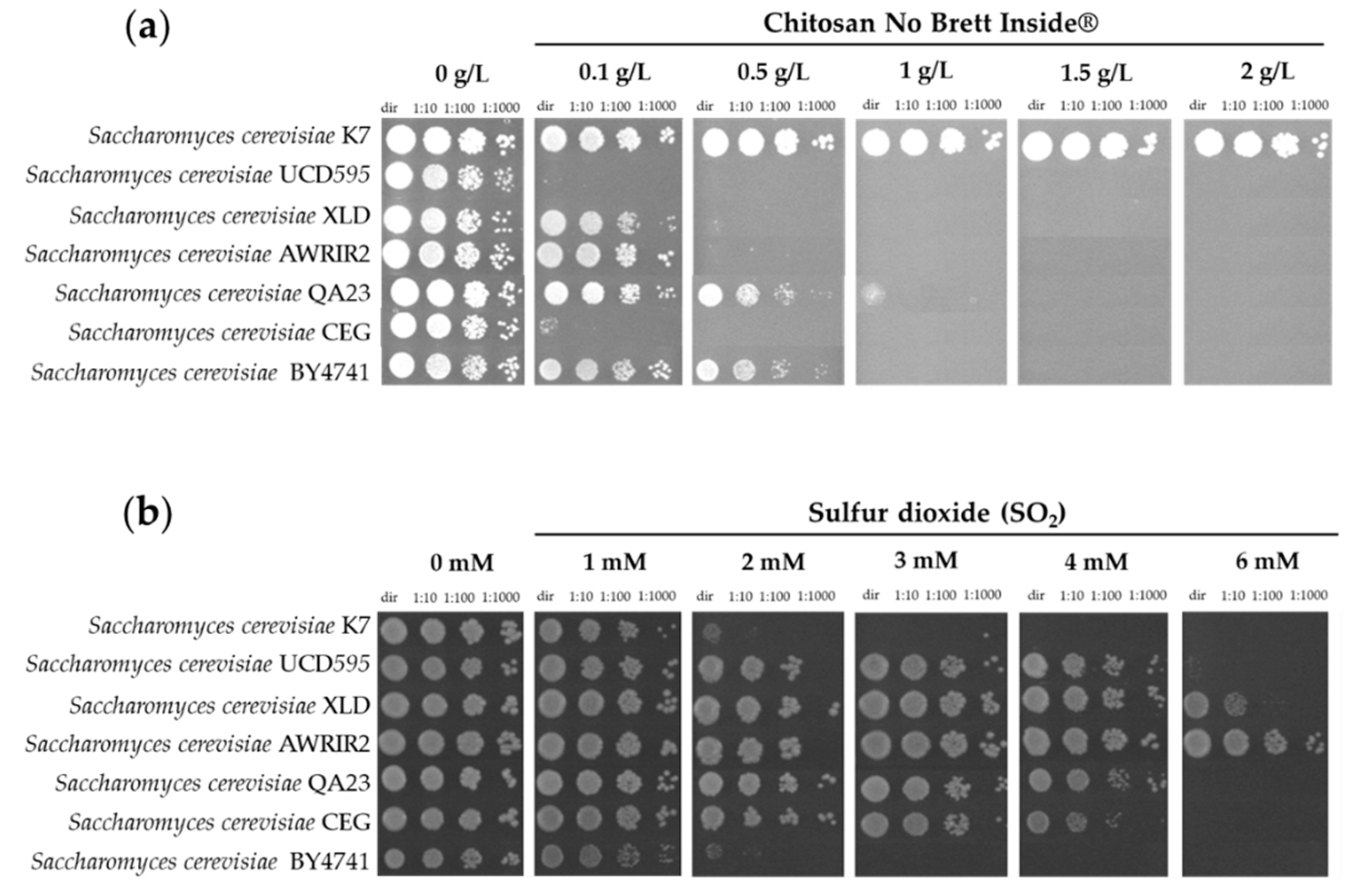
Subsequently, we examined the effect of the commercial formulation of chitosan in the growth of S. cerevisiae strains, including a cohort of those used by winemakers as starters (Figure 2). The results show a considerable degree of variation in tolerance to chitosan with strains K7, EC1118, and BRL97 being very tolerant, while CEG and UCD595 were highly susceptible (Figure 2 and Supplementary Figure S1). This result contrasts, to some extent, with the reported lack of inhibition of chitosan over the growth of S. cerevisiae [10,11,13]. Besides the differences in the experimental setups used by those studies, herein, we also examined a larger cohort of S. cerevisiae, which increased the chances of finding strain-to-strain phenotypic variation. Notably, as previously observed for the spoilage yeasts, the S. cerevisiae strains more tolerant to chitosan did not coincide with those more tolerant to SO2 and vice versa (Figure 2 and Supplementary Figure S2).
To obtain more quantitative data on the impact of chitosan on the growth of S. cerevisiae and of the spoilage yeasts, we selected a cohort of strains and followed their growth in liquid MMB medium, either supplemented or not supplemented with chitosan (0.25, 0.5, or 1.0 g/L) (Supplementary Figure S3). The results obtained coincide with those obtained in the spot assays concerning the differential susceptibility of the strains to chitosan, also clarifying that the most drastic effect of exposure to chitosan is the reduction in the final biomass of the cultures (detectable even in the more tolerant strains), while the impact in growth rate is very mild (Supplementary Figure S3).

3.2. Chemogenomic Analysis of Chitosan-Stressed S. cerevisiae Cells

3.2.1. Overview

Chemogenomic screenings were useful in elucidating the molecular mechanisms of toxicity of different xenobiotics in S. cerevisiae, clarifying not only the main routes by which these molecules are deleterious for the cells but also the corresponding adaptive responses [46,47]. With this in mind, we profiled the Euroscarf haploid S. cerevisiae mutant collection (composed of approximately 5000 mutants individually devoid of all non-essential genes) in the presence of the commercial formulation of chitosan and under our developed experimental setup. To select the best concentration to use in this large-scale phenotypic assay, we performed a preliminary screening in which BY4741 cells (the wild-type strain of the collection) were cultivated in the presence of increasing concentrations of chitosan. Based on these results (shown in Supplementary Figure S4), we decided to undertake the phenotypic screening with three concentrations of the commercial formulation of chitosan: (i) 0.25 g/L, a concentration that mildly reduced the growth rate of BY4741 cells; (ii) 0.5 g/L, a concentration that significantly decreased the final biomass of the BY4741 culture; and (iii) 1 g/L, a concentration that fully abolished the growth of the BY4741 strain (Supplementary Figure S4). Supplementary Figure S5 shows a representative image of the results obtained while phenotyping the mutant collection and that led us to classify the mutant strains into four different categories: (i) mutants not affected by the presence of chitosan; (ii) hyper-susceptible mutants; (iii) susceptible mutants; and (iv) resistant mutants. In Materials and Methods, we detail the criteria that we used for this classification.
Overall, we identified 539 mutants whose growth in the presence of the commercial formulation of chitosan was reduced, compared to the one exhibited by the wild-type strain, with 252 strains being considered hyper-susceptible and 287 being susceptible. A selected set of genes whose deletion resulted in these susceptibility phenotypes is described in Table 2, while the full dataset is available in Supplementary Table S1. Under the conditions used, we could also identify 207 chitosan-resistant mutant strains, which are listed in Supplementary Table S2.
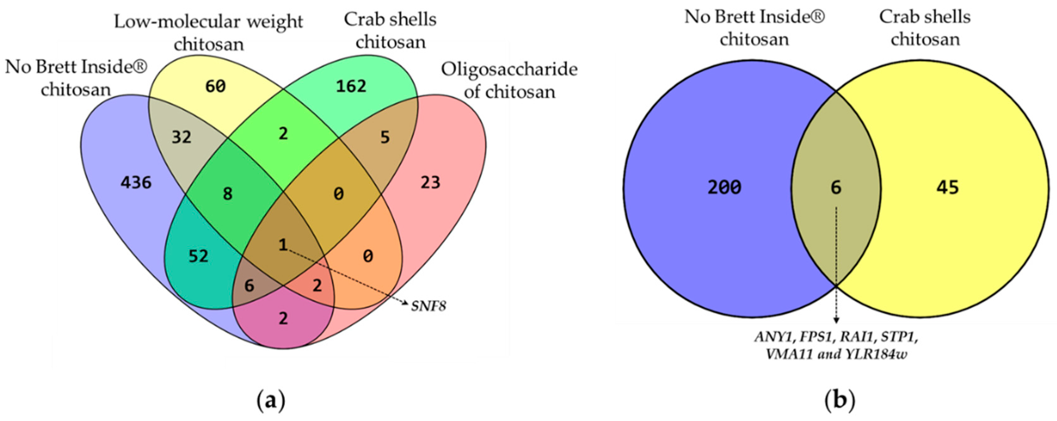
Comparing the results obtained in our phenotypic screening with others previously published revealed that only one mutant strain, Δsnf8, emerged as susceptible in the four studies, while six mutant strains (Δany1, Δfps1, Δrai1, Δstp1, Δvma11, and Δylr184w) were commonly identified as chitosan-resistant in our study and in another (Figure 3 and Supplementary Tables S1 and S2). The identification of resistance phenotypes in this type of phenotypic screening is considered particularly interesting because it has the potential to reveal which processes can be directly targeted by xenobiotics, as performed before with much success [48]. The small overlaps observed in both cohorts of susceptible and resistant strains reflect the impact of the differences in experimental setups (e.g., different pHs, different sources of chitosan) and also in the genetic background of the S. cerevisiae strains (we used BY4741, while others explored the diploid BY4743). The lack of overlap also reinforces the importance of studying tolerance to chitosan in an oenologically relevant context as a generalization of results obtained in other studies seems difficult, if even possible. SNF8 encodes a component of the ESCRT-II complex, involved in ubiquitin-dependent sorting of proteins into the endosome. This gene is part of a large cohort of genes found to confer protection against a wide array of xenobiotics in S. cerevisiae, which are considered multidrug-resistant (MDR) genes [49]. Together with SNF8, we could identify 117 other MDR genes in our cohort of genes conferring protection to chitosan (these are highlighted in grey in Table S1), including genes encoding proteins involved in lipid-based signaling (e.g., IPT1, IRS4, OPI3), intracellular transport (e.g., STP22, VPS25, VPS36), or enzymes required for cell wall biosynthesis (e.g., ROT2, SMI1, MKC7). Although the susceptibility phenotype toward chitosan of mutants devoid of MDR genes was expected, the manipulation of the expression of MDR genes can be used to improve tolerance of S. cerevisiae to stressors, and, in fact, over-expression of ARL1 and BCK2 improved the tolerance of wild-type cells to other sources of chitosan [50].

3.2.2. Functional Distribution of S. cerevisiae Genes Contributing to the Tolerance to Chitosan

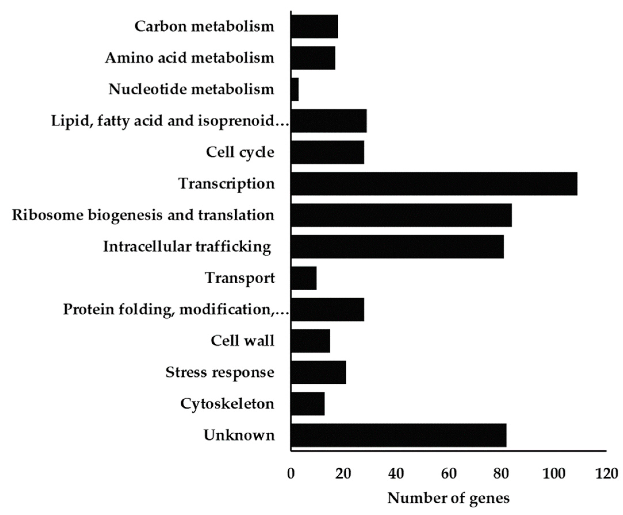
Functional clustering of the genes shown to provide protection to the commercial formulation of chitosan used shows that the highest number of those are involved in “intracellular trafficking”, in “translation”, and in “transcription”, as detailed in Figure 4 and in Table 2.
Many of the genes clustered in the “Intracellular trafficking” class are, like SNF8, MDR genes, and their involvement in chitosan tolerance (and in tolerance to xenobiotics in general) likely reflects the cell’s need to relocate damaged proteins for degradation while targeting newly synthesized proteins to their native locations. The chitosan-protective genes clustered in the “Transcription” functional class include genes required for the basic function of the transcriptional machinery (like ASK10, CDC73, and GAL11, encoding proteins involved in the assembly and catalytic activity of RNA pol II) and enzymes involved in remodeling of histone acetylation and deacetylation levels (e.g., SPT10, SPT21 and the four genes of the sirtuin SIR complex, SIR1, SIR2, SIR3, and SIR4). Under environmental stress, S. cerevisiae cells induce significant modifications in their genomic expression, accompanied by dynamic modifications of the accessibility of transcriptional regulators to chromatin that are largely dictated by histone modifications [51,52]. In line with this fact, exposure to chitosan (even if its origins differ from the one explored herein) leads to prominent alterations in S. cerevisiae’s genomic expression [37], and this is consistent with the identification in our set of chitosan-susceptible strains of mutants lacking genes involved in basic transcription functions or in histone acetylation/deacetylation. Other chitosan-protective genes clustered in the “Transcription” class include the transcription factors Crz1, the effector of the calcineurine signaling pathway; Dal81 and Gln3, involved in response to nitrogen availability; Pho2, Aft1, and Zap1, involved in response to phosphate, iron, and zinc depletion, respectively; and the Rim101p-transcription factor (Table 2 and Supplementary Table S1). Besides Rim101, the remaining members of the Rim101-signaling pathway were also found to provide protection against chitosan (including Rim20, Rim21, Rim13, Rim8, and Rim9, all involved in the cascade that promotes proteolytic activation of Rim101p (Table 2 and Supplementary Table S1)). No previous reports of the involvement of the Rim101p pathway in S. cerevisiae response and tolerance to chitosan have been previously described. Concerning the “chitosan-protection” genes clustered in the “translation” functional class, these were found to encode components of the ribosomal large and small subunits (e.g., RPL13B, RPL1B, RPL21A, RPL31B, RPL38, RPS17B, RPS18A, and RPS18B; see Supplementary Table S1). Strikingly, the deletion of some genes related with the ribosomal apparatus resulted in a resistance phenotype (e.g., RPL12B, RPL16B, RPL19B, RPL34A, or RPL40B) (Supplementary Table S2), suggesting that the effects of chitosan over this cellular process may go beyond a general impact of chitosan over translation.
Consistent with the described effects of chitosan in perturbing the lipid structure of the plasma membrane of fungal cells [37,38], the deletion of several genes encoding enzymes required for synthesis and transport to the plasma membrane of phospholipids (CHO1 and CHO2, OPI3, FPK1, LEM3, SCS2, and CST26), fatty acids (ALE1 and LAS21), and sphingolipids (IPT1, SAC1, SCS7, SUR3, and TSC3) increased the susceptibility of S. cerevisiae cells to chitosan (Table 2 and Supplementary Table S1). Both phospholipids and sphingolipids play an essential role in determining the lipid composition of S. cerevisiae cells’ plasma membrane and, consequently, in determining its architectural and physical properties [53]. Notably, the ABC transporters Pdr5 and Yor1, two multidrug resistance transporters whose role in conferring protection against drugs in S. cerevisiae has been linked to their role in the control of the asymmetric distribution of phospholipids across the plasma membrane [54,55,56,57], were also identified as determinants of yeast tolerance to chitosan (Table 2 and Supplementary Table S1).
Yeast exposure to chitosan has been shown to result in the activation of the Slt2-signalling pathway, known for its role in response to cell wall perturbations, suggesting that chitosan causes damage to this structure [37]. In this context, our phenotypic analysis uncovered several mutants with increased susceptibility to chitosan devoid of genes required for cell wall assembly and function, such as Δbgl2, devoid of the major endo-β-1,3 glucanase; Δfks1, devoid of β-1,3-glucan synthase or Δgas1; and Δgas2, devoid of β-1,3-glucanosyltransferases. Notably, the loss of both Stl2 and Rlm1 did not render chitosan-sensitive strains, suggesting that the observed Stl2 activation [37] could be a response to the acetic acid used to dissolved the chitosan tested as Slt2-phosphorylation was described to occur in response to this organic acid [58].
Chitosan also has recognized metal-chelating capacities [30], and, consistent with this, we found that the deletion of genes encoding iron (AFT1, FET3, and FTR1), potassium (TRK1), or copper (CTR1) transporters increases S. cerevisiae susceptibility to chitosan (Table 2 and Supplementary Table S1).

3.2.3. Functional Distribution of S. cerevisiae Genes Whose Deletion Increases Tolerance to Chitosan

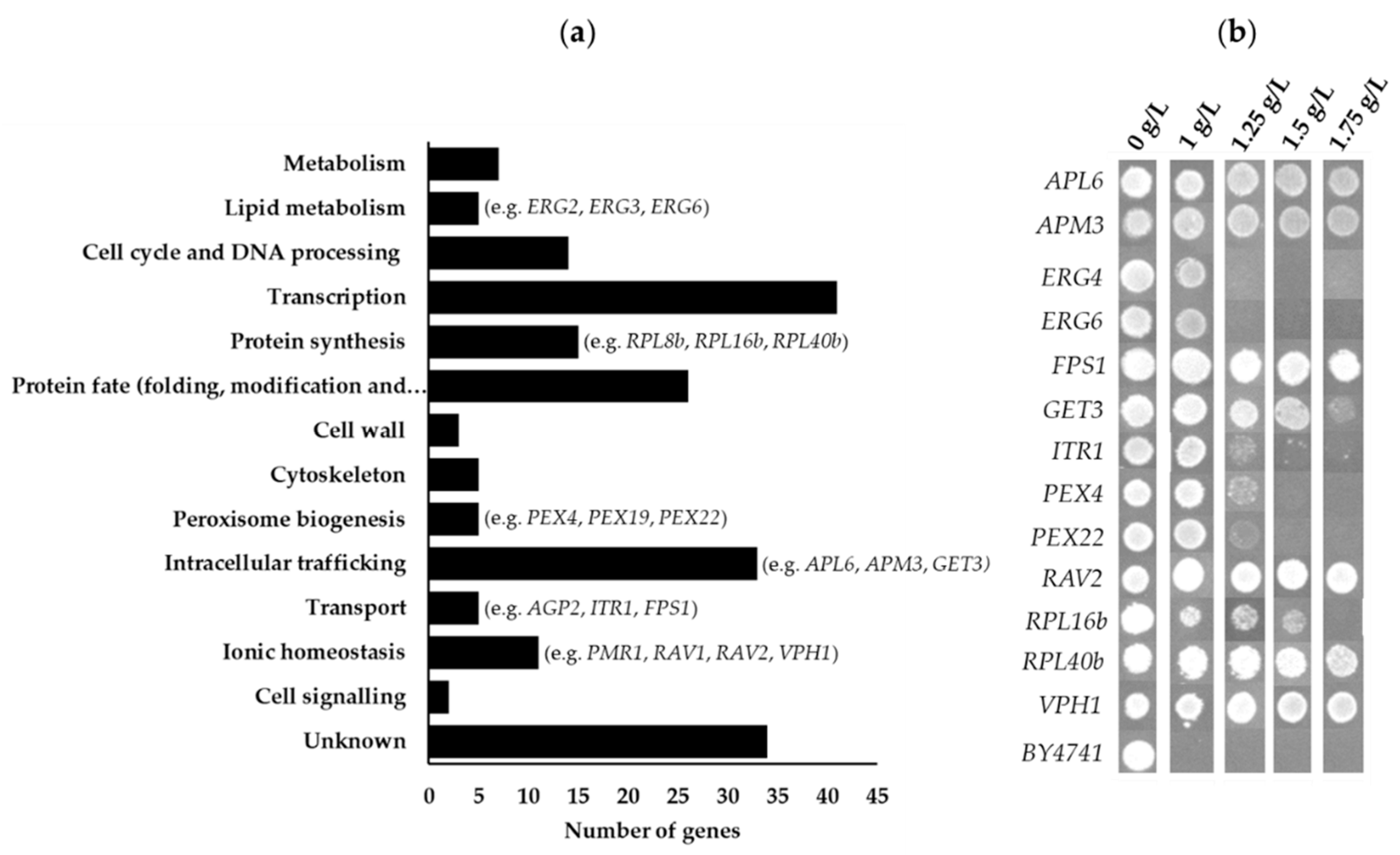
Under the conditions in which we performed the phenotypic screening, we uncovered 207 strains exhibiting growth in the presence of the highest concentration of chitosan tested (1 g/L), unlike cells of the parental strain BY4741, which were only able to grow in the presence of 0.5 g/L (Figure 5 and Supplementary Figure S4). This list of chitosan-resistant strains is detailed in Supplementary Table S2, while in Figure 5, we provide a picture associating these genes with their biological functions. To confirm the “chitosan-resistance” phenotypes, we individually profiled the growth of the resistant mutants in the solid MMB medium (at pH 3.5) supplemented with 1 g/L of chitosan (the highest concentration used to screen the mutant collection), but also with 1.25, 1.5, and 1.75 g/L of chitosan (Figure 5).
The results obtained confirmed the ability of the uncovered resistant mutants to grow in the presence of 1 g/L of the commercial formulation of chitosan, unlike wild-type BY4741 cells (Figure 5 and Supplementary Table S2). Notably, 33 chitosan-resistant mutants (Δany1, Δapl6, Δapm3, Δaps3, Δbrp1, Δbud13, Δcdc50, Δemp65, Δerv14, Δfes1, Δfps1, Δget3, Δglo3, Δhur1, Δist3, Δlea1, Δpkr1, Δpmr1, Δrad27, Δrav1, Δrav2, Δrcy1, Δrpl40b, Δrtt103, Δsnt309, Δted1, Δtef4, Δtop1, Δvma21, Δvph1, ΔYDR203W, ΔYLR338W, and ΔYML095C-A) exhibited growth in the presence of 1.75 g/L chitosan, a concentration that is almost 4 times higher than the one tolerated by the parental strain (Figure 5). The biological function of the genes whose deletion improves S. cerevisiae tolerance to chitosan is scattered (as evidenced by Figure 5 and Supplementary Table S2), ranging from genes involved in peroxisome biogenesis, to those involved in protein synthesis, in protein targeting and folding or serving as solute importers (Figure 5 and Supplementary Table S2).

4. Discussion

In recent years, chitosan attracted attention in the winemaking industry as a possible alternative to SO2; however, the mechanisms underlying the antimicrobial properties of chitosan, especially in the formulations legally allowed for use, remain elusive. In this work, we examined, for the first time, the impact of a commercially used formulation of chitosan in oenologically relevant conditions. Under these conditions, 0.1 g/L of chitosan inhibited growth of the spoilage yeasts D. anomala (which was expected since the formulation is sold by the supplier to prevent the growth of Brettanomyces/Dekkera yeasts), Z. bailii, and S. lugdwigii. In contrast, no effect was observed against P. anomala, even when the concentration of chitosan tested was 20 times higher than the maximum legal dosage. This result compromises the potential use of chitosan during storage to control this oxidative SO2-sensitive spoilage yeast, which is known to form a film on the surface of wines with low SO2 in unfilled containers with consequent negative effects on the quality of wines, imparting an oxidized flavor due to the production of acetaldehyde [4].
Interestingly, the spoilage species Z. bailii and S. ludwigii showed higher susceptibility to chitosan but were tolerant to SO2. The same trend of observation was made for S. cerevisiae, as strains more tolerant to chitosan did not coincide with those more tolerant to SO2. These observations suggest that the biological targets of chitosan do not coincide with those targeted by SO2, an idea further reinforced by the low overlap of genes providing protection against these two stressors, which basically include the MDR genes (see results in Supplementary Figure S6). It is important to emphasize that this comparison was made using results that were obtained from two phenotypic analyses carried out in similar experimental setups (the same phenotypic screening methodology, the same set of strains, and the same media at the same pH). The different susceptibility to SO2 and chitosan found amongst spoilage species strengthens the idea of the rational use of each of the preservatives, either alone or in combination, considering the winemaking stage and the associated prevalence of each targeted spoilage species. A differential susceptibility of yeast species to chitosan has also been previously observed [10,24], and despite several attempts, it has not yet been possible to pinpoint these variations to a specific feature, a task certainly made difficult by the variety of experimental settings explored when approaching the interaction of chitosan with fungal cells. We also cannot rule out the possibility that tolerance to chitosan results from the combined action of a network of functions that, synergistically, cooperate to facilitate tolerance to chitosan. The high number of S. cerevisiae genes identified herein as determining resistance or sensitivity to chitosan, along with the wide distribution of biological functions that these genes have, suggests that, indeed, the process of adaptation and response to chitosan stress in S. cerevisiae is multi-factorial and unlikely to be determined by a single phenotypic trait. In a more applied perspective, the observed phenotypic variation observed towards the commercial formulation of chitosan suggests that winemakers should also consider chitosan tolerance (for which they can consult the results shown herein) in the selection of their starter cultures of S. cerevisiae strains if the use of the preservative prior to alcoholic fermentation is intended.
The protective effect against chitosan played by a variety of genes with biological functions related to the synthesis of phospholipids and sphingolipids in S. cerevisiae is in line with the demonstrated strong interaction of the amino groups of chitosan with negatively charged lipids [37,59]. The role of the Rim101p-pathway in S. cerevisiae tolerance to chitosan uncovered herein further substantiates the interference of chitosan over the plasma membrane lipid structure, as this signaling system was recently shown to become activated in response to perturbations causing asymmetries in this lipid bilayer [60,61,62,63]. However, tolerance to chitosan does not correlate with the levels of phospholipids, sphingolipids, or ergosterol [37,64], showing that the mechanism by which chitosan perturbs the plasma membrane structure goes beyond the mere sequestration of these lipids. Interestingly, we found that the deletion of ERG2, ERG6, and ERG3 genes, encoding three enzymes of the ergosterol biosynthetic pathway, potently enhances S. cerevisiae tolerance to chitosan. However, chitosan tolerance was found not to be correlated with ergosterol levels [59]. Lipidomic analysis of erg mutants showed that these genes’ deletion abolishes the production of ergosterol, but also leads to prominent changes in the balances of other lipid species present in the plasma membrane, including the amount of phospholipids and sphingolipids, with consequences on the biophysical properties of the membrane [65]. It is thus possible that the interaction of chitosan with the plasma membrane may depend on its biophysical properties (for example, its fluidity) and not on the individual level of a certain lipid. In this context, it would be the outcome of the balance of the concentration of the different lipids present, translated into a given biophysical trait, that would determine a higher or lower tolerance of the cells to chitosan. This hypothesis is consistent not only with the differential effect exerted by the expression of genes involved in the metabolism of lipids that we have uncovered herein (in some cases contributing to tolerance, while in others enhancing susceptibility), but also with the establishment of a positive correlation between chitosan tolerance and the saturation degree of the plasma membrane [59], which depends on the proportion of saturated and unsaturated fatty acids and not on their individual concentration.
Even though it seems clear that chitosan interacts with the plasma membrane and with the cell wall, the mechanism by which it enters fungal cells remains unclear since it was shown not to occur by diffusion and depend on energy, which suggests the involvement of a yet unidentified transporter [30,32,37,59]. In this context, the resistance phenotype obtained for the Δfps1 mutant was very interesting since Fps1 encodes an aquaglyceroporin found to mediate the entry of xenobiotics such as arsenic or acetic acid [66,67]. Similarly, Δagp2, a mutant devoid of the low-affinity amino acid permease Agp2, and Δitr1, a mutant devoid of a myo-inositol importer, were also highly resistant to chitosan (Supplementary Table S2). Similar to Fps1, both Agp2 and Itr1 were also found to serve as xenobiotic carriers, with Agp2 promoting the import of the anticancer drug bleomycine [68] and Itr1 of azoles [69]. Further studies will be required to demonstrate clearly and unequivocally whether these promiscuous transporters serve as carriers of chitosan and if their activity can also explain the different degrees of susceptibility of different fungal species.
Another striking observation that emerged from our chemogenomics screening was the implication in tolerance to chitosan of a wide range of genes encoding structural subunits of the ribosome, while the deletion of others confers a resistance phenotype. The need for a ribosomal function to improve tolerance to chitosan is not surprising since, as in response to other stresses, under chitosan stress, the cells need to adjust their proteome accordingly. A reduction in the activity of ribosomal function caused by the deletion of ribosomal components is, thus, expected to reduce the ability of the yeast cells to cope with chitosan stress. However, the fact that the deletion of some components of the large ribosomal subunit confers resistance to chitosan suggests that the role of the ribosome could go beyond the modification in the proteome, with one possibility being a direct binding of chitosan to this structure. Notably, the deletion of ribosomal components of the large ribosomal subunit has been previously found to increase S. cerevisiae tolerance to chitosan obtained from crab shells [38], although the genes they identified in that study do not coincide with those that we have uncovered in our study. Another functionally related set of genes whose deletion increases tolerance to chitosan is those involved in the assembly and function of the vacuolar V-ATPase. Thus far, no studies have addressed the direct interaction of chitosan with the ribosome or with the V-ATPase; however, the results obtained in our work suggest that as an interesting avenue of future research. Indeed, other molecules, such as the anticancer drug imatinib, have been found to directly interact with the yeast V-ATPase [48], and ribosomes are known to be targeted by a number of small molecules that have been explored as putative pharmaceuticals [70].
In this work, we have provided evidence supporting the utilization of a commercial formulation of chitosan as an alternative preservative to SO2 showing a potent inhibitory effect against the spoilage species D. anomala, as well as S. ludwigii and Z. bailii (usually resilient to SO2-based preservation), while affecting the growth of S. cerevisiae strains much less, despite an important strain-to-strain variation that was observed. With the results from the large-scale phenotypic screening, we have compiled a schematic representation of the interaction of chitosan with S. cerevisiae cells (Figure 6), and this knowledge can be further used to understand the tolerance interaction of chitosan with other fungal cells. Three possible carriers of chitosan, Itr1, Agp2, and Fps1, were herein identified based on the potent resistance phenotypes obtained upon their deletion. Resistance phenotypes obtained upon the deletion of multiple genes involved in the assembly of a large ribosomal subunit or for the assembly and function of the vacuolar V-ATPase were also uncovered, suggesting a possible direct interaction of chitosan with these organelles. Overall, the results presented herein pave the way to reducing the utilization of SO2 in winemaking, with positive consequences for the health of more susceptible consumers, but also in the competitiveness of winemaking companies that face a growing market of consumers increasingly fond of products with a green label.

Supplementary Materials

The following supporting information can be downloaded at https://www.mdpi.com/article/10.3390/fermentation9020172/s1: Figure S1: Phenotypic profiling of chitosan No Brett Inside® of a set of commercially available strains for wine fermentation, as revealed by spot assays. Figure S2: Phenotypic profiling to SO2 of a set of commercially available strains for wine fermentation, as revealed by spot assay. Figure S3: Growth curves of three commercial S. cerevisiae strains (K7, UCD595, and AWRIR2) and the laboratory strain BY4741 and four spoilage yeast (Dekkera anomala IGC5152, Pichia anomala UTAD37, Saccharomycodes ludwigii UTAD17, and Zygosaccharomyces bailii UTAD265) in MMB media (at pH 3.5) at different chitosan concentrations (0.25, 0.5 and 1 g/L); Figure S4: Comparison of the susceptibility to chitosan No Brett Inside® of the Saccharomyces cerevisiae BY4741 parental strain with spot assay, at the indicated concentrations of chitosan in MMB (pH 3.5) agarized medium; Figure S5: Illustrative example of deletion strains exhibiting different susceptibility profiles in the chemogenomics screenings carried out. Figure S6: Venn diagram comparing genes that were found to confer resistance to SO2 with those that were found to confer resistance to chitosan No Brett inside®.

Author Contributions

Conceptualization of the study: A.M.-F.; investigation: P.L., B.B.C.; resources, A.M.-F.; data curation: P.L., N.P.M. and A.M.-F.; writing—original draft preparation: P.L., N.P.M. and A.M.-F.; writing—review and editing: P.L., A.M.-F. and N.P.M.; supervision: N.P.M. and A.M.-F.; project administration: A.M.-F.; funding acquisition: A.M.-F. All authors have read and agreed to the published version of the manuscript.

Funding

This research was funded by Fundação para a Ciência e Tecnologia [grant numbers EXPL/AGR-TEC/1823/2013; UIDB/04046/2020, UIDB/04565/2020, LA/P/0140/2020] and by European Regional Development Fund (ERDF) through Norte 2020 research program [grant number INNOVINE&WINE Norte-01-0145-FEDER-000038].

Data Availability Statement

The data obtained in the chemogenomics screening is provided in Supplementary Materials. Any other data will be provided pending contact to corresponding authors.

Conflicts of Interest

The authors declare no conflict of interest.

References

  1. Renouf, V.; Strehaiano, P.; Lonvaud-Funel, A. Effectiveness of Dimethlydicarbonate to Prevent Brettanomyces bruxellensis Growth in Wine. Food Control 2008, 19, 208–216. [Google Scholar] [CrossRef]
  2. Vally, H.; Misso, N.L.A.; Madan, V. Clinical Effects of Sulphite Additives. Clin. Exp. Allergy 2009, 39, 1643–1651. [Google Scholar] [CrossRef]
  3. Costantini, A.; García-Moruno, E.; Moreno-Arribas, M.V. Biochemical Transformations Produced by Malolactic Fermentation. In Wine Chemistry and Biochemistry; Springer: New York, NY, USA, 2009; pp. 27–57. ISBN 9780387741161. [Google Scholar]
  4. Du Toit, M.; Pretorius, I.S. Microbial Spoilage and Preservation of Wine: Using Weapons from Nature’s Own Arsenal-a Review. S. Afr. J. Enol. Vitic. 2000, 21, 74–96. [Google Scholar] [CrossRef]
  5. Corte, L.; Roscini, L.; Zadra, C.; Antonielli, L.; Tancini, B.; Magini, A.; Emiliani, C.; Cardinali, G. Effect of PH on Potassium Metabisulphite Biocidic Activity against Yeast and Human Cell Cultures. Food Chem. 2012, 134, 1327–1336. [Google Scholar] [CrossRef] [PubMed]
  6. Vally, H.; Thompson, P.J. Role of Sulfite Additives in Wine Induced Asthma: Single Dose and Cumulative Dose Studies. Thorax 2001, 56, 763–769. [Google Scholar] [CrossRef]
  7. Irwin, S.V.; Fisher, P.; Graham, E.; Malek, A.; Robidoux, A. Sulfites Inhibit the Growth of Four Species of Beneficial Gut Bacteria at Concentrations Regarded as Safe for Food. PLoS ONE 2017, 12, e0186629. [Google Scholar] [CrossRef]
  8. Divol, B.; Strehaiano, P.; Lonvaud-Funel, A. Effectiveness of Dimethyldicarbonate to Stop Alcoholic Fermentation in Wine. Food Microbiol. 2005, 22, 169–178. [Google Scholar] [CrossRef]
  9. Castro Marín, A.; Colangelo, D.; Lambri, M.; Riponi, C.; Chinnici, F. Relevance and Perspectives of the Use of Chitosan in Winemaking: A Review. Crit. Rev. Food Sci. Nutr. 2021, 61, 3450–3464. [Google Scholar] [CrossRef]
  10. Miot-Sertier, C.; Paulin, M.; Dutilh, L.; Ballestra, P.; Albertin, W.; Masneuf-Pomarède, I.; Coulon, J.; Moine, V.; Vallet-Courbin, A.; Maupeu, J.; et al. Assessment of Chitosan Antimicrobial Effect on Wine Microbes. Int. J. Food Microbiol. 2022, 381, 109907. [Google Scholar] [CrossRef]
  11. Bağder Elmaci, S.; Gülgör, G.; Tokatli, M.; Erten, H.; İşci, A.; Özçelik, F. Effectiveness of Chitosan against Wine-Related Microorganisms. Antonie Van Leeuwenhoek 2015, 107, 675–686. [Google Scholar] [CrossRef]
  12. Aider, M. Chitosan Application for Active Bio-Based Films Production and Potential in the Food Industry: Review. LWT-Food Sci. Technol. 2010, 43, 837–842. [Google Scholar] [CrossRef]
  13. Gómez-Rivas, L.; Escudero-Abarca, B.I.; Aguilar-Uscanga, M.G.; Hayward-Jones, P.M.; Mendoza, P.; Ramírez, M. Selective Antimicrobial Action of Chitosan against Spoilage Yeasts in Mixed Culture Fermentations. J. Ind. Microbiol. Biotechnol. 2004, 31, 16–22. [Google Scholar] [CrossRef] [PubMed]
  14. Portugal, C.; Sáenz, Y.; Rojo-Bezares, B.; Zarazaga, M.; Torres, C.; Cacho, J.; Ruiz-Larrea, F. Brettanomyces Susceptibility to Antimicrobial Agents Used in Winemaking: In Vitro and Practical Approaches. Eur. Food Res. Technol. 2014, 238, 641–652. [Google Scholar] [CrossRef]
  15. Ferreira, D.; Moreira, D.; Costa, E.M.; Silva, S.; Pintado, M.M.; Couto, J.A. The Antimicrobial Action of Chitosan Against the Wine Spoilage Yeast Brettanomyces/Dekkera. J. Chitin Chitosan Sci. 2013, 1, 240–245. [Google Scholar] [CrossRef]
  16. Bornet, A.; Teissedre, P.L. Chitosan, Chitin-Glucan and Chitin Effects on Minerals (Iron, Lead, Cadmium) and Organic (Ochratoxin A) Contaminants in Wines. Eur. Food Res. Technol. 2008, 226, 681–689. [Google Scholar] [CrossRef]
  17. Rabea, E.I.; Badawy, M.E.T.; Stevens, C.V.; Smagghe, G.; Steurbaut, W. Chitosan as Antimicrobial Agent: Applications and Mode of Action. Biomacromolecules 2003, 4, 1457–1465. [Google Scholar] [CrossRef]
  18. Darwesh, O.M.; Sultan, Y.Y.; Seif, M.M.; Marrez, D.A. Bio-Evaluation of Crustacean and Fungal Nano-Chitosan for Applying as Food Ingredient. Toxicol. Rep. 2018, 5, 348–356. [Google Scholar] [CrossRef]
  19. Abd El-Hack, M.E.; El-Saadony, M.T.; Shafi, M.E.; Zabermawi, N.M.; Arif, M.; Batiha, G.E.; Khafaga, A.F.; Abd El-Hakim, Y.M.; Al-Sagheer, A.A. Antimicrobial and Antioxidant Properties of Chitosan and Its Derivatives and Their Applications: A Review. Int. J. Biol. Macromol. 2020, 164, 2726–2744. [Google Scholar] [CrossRef]
  20. Colangelo, D.; Torchio, F.; De Faveri, D.M.; Lambri, M. The Use of Chitosan as Alternative to Bentonite for Wine Fining: Effects on Heat-Stability, Proteins, Organic Acids, Colour, and Volatile Compounds in an Aromatic White Wine. Food Chem. 2018, 264, 301–309. [Google Scholar] [CrossRef]
  21. Nardi, T.; Vagnoli, P.; Minacci, A.; Gautier, S.; Sieczkowski, N. Evaluating the Impact of a Fungal-Origin Chitosan Preparation on Brettanomyces Bruxellensis in the Context of Wine Aging. Wine Stud. 2014, 3, 4574. [Google Scholar] [CrossRef] [Green Version]
  22. Zuehlke, J.M.; Petrova, B.; Edwards, C.G. Advances in the Control of Wine Spoilage by Zygosaccharomyces and Dekkera/Brettanomyces. Annu. Rev. Food Sci. Technol. 2013, 4, 57–78. [Google Scholar] [CrossRef] [PubMed]
  23. Taillandier, P.; Joannis-Cassan, C.; Jentzer, J.B.; Gautier, S.; Sieczkowski, N.; Granes, D.; Brandam, C. Effect of a Fungal Chitosan Preparation on Brettanomyces bruxellensis, a Wine Contaminant. J. Appl. Microbiol. 2015, 118, 123–131. [Google Scholar] [CrossRef] [PubMed]
  24. Petrova, B.; Cartwright, Z.M.; Edwards, C.G. Effectiveness of Chitosan Preparations against Brettanomyces bruxellensis Grown in Culture Media and Red Wines. J. Int. Sci. Vigne Vin 2016, 50, 49–56. [Google Scholar] [CrossRef]
  25. Rhoades, J.; Roller, S. Antimicrobial Actions of Degraded and Native Chitosan against Spoilage Organisms in Laboratory Media and Foods. Appl. Environ. Microbiol. 2000, 66, 80–86. [Google Scholar] [CrossRef] [PubMed]
  26. Roller, S.; Covill, N. The Antifungal Properties of Chitosan in Laboratory Media and Apple Juice. Int. J. Food Microbiol. 1999, 47, 67–77. [Google Scholar] [CrossRef]
  27. Raafat, D.; Fouad, G. Chitosan as an Antimicrobial Compound: Modes of Action and Resistance Mechanisms. Ph.D. Thesis, University of Bonn, Bonn, Germany, 2008. [Google Scholar]
  28. Scansani, S.; Rauhut, D.; Brezina, S.; Semmler, H.; Benito, S. The Impact of Chitosan on the Chemical Composition of Wines Fermented with Schizosaccharomyces pombe and Saccharomyces cerevisiae. Foods 2020, 9, 1423. [Google Scholar] [CrossRef]
  29. Liu, H.; Du, Y.; Wang, X.; Sun, L. Chitosan Kills Bacteria through Cell Membrane Damage. Int. J. Food Microbiol. 2004, 95, 147–155. [Google Scholar] [CrossRef]
  30. Helander, I.M.; Nurmiaho-Lassila, E.L.; Ahvenainen, R.; Rhoades, J.; Roller, S. Chitosan Disrupts the Barrier Properties of the Outer Membrane of Gram-Negative Bacteria. Int. J. Food Microbiol. 2001, 71, 235–244. [Google Scholar] [CrossRef]
  31. Kong, M.; Chen, X.G.; Xing, K.; Park, H.J. Antimicrobial Properties of Chitosan and Mode of Action: A State of the Art Review. Int. J. Food Microbiol. 2010, 144, 51–63. [Google Scholar] [CrossRef]
  32. Palma-Guerrero, J.; Huang, I.C.; Jansson, H.B.; Salinas, J.; Lopez-Llorca, L.V.; Read, N.D. Chitosan Permeabilizes the Plasma Membrane and Kills Cells of Neurospora crassa in an Energy Dependent Manner. Fungal Genet. Biol. 2009, 46, 585–594. [Google Scholar] [CrossRef]
  33. Goy, R.C.; de Britto, D.; Assis, O.B.G. A Review of the Antimicrobial Activity of Chitosan. Polímeros 2009, 19, 241–247. [Google Scholar] [CrossRef]
  34. Hernández, R.B.; Yola, O.R.; Mercê, A.L.R. Chemical Equilibrium in the Complexation of First Transition Series Divalent Cations, Cu2+, Mn2+ and Zn2+, with Chitosan. J. Braz. Chem. Soc. 2007, 18, 1388–1396. [Google Scholar] [CrossRef]
  35. Teixeira, M.C.; Monteiro, P.T.; Guerreiro, J.F.; Gonçalves, J.P.; Mira, N.P.; Dos Santos, S.C.; Cabrito, T.R.; Palma, M.; Costa, C.; Francisco, A.P.; et al. The YEASTRACT Database: An Upgraded Information System for the Analysis of Gene and Genomic Transcription Regulation in Saccharomyces cerevisiae. Nucleic Acids Res. 2014, 34, D446–D451. [Google Scholar] [CrossRef]
  36. Lopez-Moya, F.; Kowbel, D.; Nueda, M.J.; Palma-Guerrero, J.; Glass, N.L.; Lopez-Llorca, L.V. Neurospora crassa Transcriptomics Reveals Oxidative Stress and Plasma Membrane Homeostasis Biology Genes as Key Targets in Response to Chitosan. Mol. BioSyst. 2016, 12, 391–403. [Google Scholar] [CrossRef]
  37. Zakrzewska, A.; Boorsma, A.; Brul, S.; Hellingwerf, K.J.; Klis, F.M. Transcriptional Response of Saccharomyces cerevisiae to the Plasma Membrane-Perturbing Compound Chitosan. Eukaryot. Cell 2005, 4, 703–715. [Google Scholar] [CrossRef] [PubMed]
  38. Zakrzewska, A.; Boorsma, A.; Delneri, D.; Brul, S.; Oliver, S.G.; Klis, F.M. Cellular Processes and Pathways That Protect Saccharomyces cerevisiae Cells against the Plasma Membrane-Perturbing Compound Chitosan. Eukaryot. Cell 2007, 13, 2–9. [Google Scholar] [CrossRef]
  39. Bermejo, C.; Rodríguez, E.; García, R.; Rodríguez-Peña, J.M.; Rodríguez de la Concepción, M.L.; Rivas, C.; Arias, P.; Nombela, C.; Posas, F.; Arroyo, J. The Sequential Activation of the Yeast HOG and SLT2 Pathways Is Required for Cell Survival to Cell Wall Stress. Mol. Biol. Cell 2008, 19, 1113–1124. [Google Scholar] [CrossRef]
  40. Organisation Internationale de la Vigne et du Vin (OIV). International Code of Oenological Practices OIV/OENO, 338A/2009; International Organisation of Vine and Wine: Paris, France, 2009. [Google Scholar]
  41. Lim, S.H.; Hudson, S.M. Synthesis and Antimicrobial Activity of a Water-Soluble Chitosan Derivative with a Fiber-Reactive Group. Carbohydr. Res. 2004, 339, 313–319. [Google Scholar] [CrossRef]
  42. Mira, N.P.; Teixeira, M.C.; Sá-Correia, I. Adaptive Response and Tolerance to Weak Acids in Saccharomyces cerevisiae: A Genome-Wide View. Omi. J. Integr. Biol. 2010, 14, 525–540. [Google Scholar] [CrossRef]
  43. Sagoo, S.K.; Board, R.; Roller, S. Chitosan Potentiates the Antimicrobial Action of Sodium Benzoate on Spoilage Yeasts. Lett. Appl. Microbiol. 2002, 34, 168–172. [Google Scholar] [CrossRef]
  44. Galván Márquez, I.; Akuaku, J.; Cruz, I.; Cheetham, J.; Golshani, A.; Smith, M.L. Disruption of Protein Synthesis as Antifungal Mode of Action by Chitosan. Int. J. Food Microbiol. 2013, 164, 108–112. [Google Scholar] [CrossRef]
  45. Lage, P.; Sampaio-Marques, B.; Ludovico, P.; Mira, N.P.; Mendes-Ferreira, A. Transcriptomic and Chemogenomic Analyses Unveil the Essential Role of Com2-Regulon in Response and Tolerance of Saccharomyces cerevisiae to Stress Induced by Sulfur Dioxide. Microb. Cell 2019, 6, 509–523. [Google Scholar] [CrossRef]
  46. Gaikani, H.; Smith, A.M.; Lee, A.Y.; Giaever, G.; Nislow, C. Systematic Prediction of Antifungal Drug Synergy by Chemogenomic Screening in Saccharomyces cerevisiae. Front. Fungal Biol. 2021, 2, 683414. [Google Scholar] [CrossRef]
  47. Barazandeh, M.; Kriti, D.; Nislow, C.; Giaever, G. The Cellular Response to Drug Perturbation Is Limited: Comparison of Large-Scale Chemogenomic Fitness Signatures. BMC Genom. 2022, 23, 197. [Google Scholar] [CrossRef] [PubMed]
  48. Dos Santos, S.C.; Sá-Correia, I. Genome-Wide Identification of Genes Required for Yeast Growth under Imatinib Stress: Vacuolar H+-Atpase Function Is an Important Target of This Anticancer Drug. Omi. J. Integr. Biol. 2009, 13, 185–198. [Google Scholar] [CrossRef] [PubMed]
  49. Hillenmeyer, M.E.; Fung, E.; Wildenhain, J.; Pierce, S.E.; Hoon, S.; Lee, W.; Proctor, M.; St Onge, R.P.; Tyers, M.; Koller, D.; et al. The Chemical Genomic Portrait of Yeast: Uncovering a Phenotype for All Genes. Science 2008, 320, 362–365. [Google Scholar] [CrossRef]
  50. Jaime, M.D.L.A.; Lopez-Llorca, L.V.; Conesa, A.; Lee, A.Y.; Proctor, M.; Heisler, L.E.; Gebbia, M.; Giaever, G.; Westwood, J.T.; Nislow, C. Identification of Yeast Genes That Confer Resistance to Chitosan Oligosaccharide (COS) Using Chemogenomics. BMC Genom. 2012, 13, 267. [Google Scholar] [CrossRef]
  51. Friedman, N.; Rando, O.J. Epigenomics and the Structure of the Living Genome. Genome Res. 2015, 25, 1482–1490. [Google Scholar] [CrossRef]
  52. Magraner-Pardo, L.; Pelechano, V.; Coloma, M.D.; Tordera, V. Dynamic Remodeling of Histone Modifications in Response to Osmotic Stress in Saccharomyces cerevisiae. BMC Genom. 2014, 15, 247. [Google Scholar] [CrossRef]
  53. De Almeida, R.F.M. A Route to Understanding Yeast Cellular Envelope–Plasma Membrane Lipids Interplaying in Cell Wall Integrity. FEBS J. 2018, 285, 2402–2404. [Google Scholar] [CrossRef] [Green Version]
  54. Kodedová, M.; Sychrová, H. Changes in the Sterol Composition of the Plasma Membrane Affect Membrane Potential, Salt Tolerance and the Activity of Multidrug Resistance Pumps in Saccharomyces cerevisiae. PLoS ONE 2015, 10, e0139306. [Google Scholar] [CrossRef]
  55. Khakhina, S.; Johnson, S.S.; Manoharlal, R.; Russo, S.B.; Blugeon, C.; Lemoine, S.; Sunshine, A.B.; Dunham, M.J.; Cowart, L.A.; Devaux, F.; et al. Control of Plasma Membrane Permeability by ABC Transporters. Eukaryot. Cell 2015, 14, 442–453. [Google Scholar] [CrossRef]
  56. Kaur, R.; Bachhawat, A.K. The Yeast Multidrug Resistance Pump, Pdr5p, Confers Reduced Drug Resistance in Erg Mutants of Saccharomyces Cerevisiae. Microbiology 1999, 145, 809–818. [Google Scholar] [CrossRef]
  57. Decottignies, A.; Grant, A.M.; Nichols, J.W.; De Wet, H.; McIntosh, D.B.; Goffeau, A. ATPase and Multidrug Transport Activities of the Overexpressed Yeast ABC Protein Yor1p. J. Biol. Chem. 1998, 273, 12612–12622. [Google Scholar] [CrossRef]
  58. Mollapour, M.; Shepherd, A.; Piper, P.W. Presence of the Fps1p Aquaglyceroporin Channel Is Essential for Hog1p Activation, but Suppresses Slt2(Mpk1)p Activation, with Acetic Acid Stress of Yeast. Microbiology 2009, 155, 3304–3311. [Google Scholar] [CrossRef]
  59. Palma-Guerrero, J.; Lopez-Jimenez, J.A.; Pérez-Berná, A.J.; Huang, I.C.; Jansson, H.B.; Salinas, J.; Villalaín, J.; Read, N.D.; Lopez-Llorca, L.V. Membrane Fluidity Determines Sensitivity of Filamentous Fungi to Chitosan. Mol. Microbiol. 2010, 75, 1021–1032. [Google Scholar] [CrossRef]
  60. Ikeda, M.; Kihara, A.; Denpoh, A.; Igarashi, Y. The Rim101 Pathway Is Involved in Rsb1 Expression Induced by Altered Lipid Asymmetry. Mol. Biol. Cell 2008, 49, 1922–1931. [Google Scholar] [CrossRef]
  61. Mattiazzi, M.; Jambhekar, A.; Kaferle, P.; DeRisi, J.L.; Križaj, I.; Petrovič, U. Genetic Interactions between a Phospholipase A2 and the Rim101 Pathway Components in S. cerevisiae Reveal a Role for This Pathway in Response to Changes in Membrane Composition and Shape. Mol. Genet. Genom. 2010, 283, 519–530. [Google Scholar] [CrossRef]
  62. Rockenfeller, P.; Smolnig, M.; Diessl, J.; Bashir, M.; Schmiedhofer, V.; Knittelfelder, O.; Ring, J.; Franz, J.; Foessl, I.; Khan, M.J.; et al. Diacylglycerol Triggers Rim101 Pathway-Dependent Necrosis in Yeast: A Model for Lipotoxicity. Cell Death Differ. 2018, 25, 767–783. [Google Scholar] [CrossRef]
  63. Obara, K.; Kihara, A. Signaling Events of the Rim101 Pathway Occur at the Plasma Membrane in a Ubiquitination-Dependent Manner. Mol. Cell. Biol. 2014, 34, 3525–3534. [Google Scholar] [CrossRef] [Green Version]
  64. Mira, N.P.; Palma, M.; Guerreiro, J.F.; Sá-Correia, I. Genome-Wide Identification of Saccharomyces cerevisiae Genes Required for Tolerance to Acetic Acid. Microb. Cell Fact. 2010, 9, 79. [Google Scholar] [CrossRef]
  65. Johnston, E.J.; Moses, T.; Rosser, S.J. The Wide-Ranging Phenotypes of Ergosterol Biosynthesis Mutants, and Implications for Microbial Cell Factories. Yeast 2020, 37, 27–44. [Google Scholar] [CrossRef] [PubMed]
  66. Maciaszczyk-Dziubinska, E.; Migdal, I.; Migocka, M.; Bocer, T.; Wysocki, R. The Yeast Aquaglyceroporin Fps1p Is a Bidirectional Arsenite Channel. FEBS Lett. 2010, 584, 726–732. [Google Scholar] [CrossRef]
  67. Mollapour, M.; Piper, P.W. Hog1 Mitogen-Activated Protein Kinase Phosphorylation Targets the Yeast Fps1 Aquaglyceroporin for Endocytosis, Thereby Rendering Cells Resistant to Acetic Acid. Mol. Cell. Biol. 2007. [Google Scholar] [CrossRef]
  68. Aouida, M.; Pagé, N.; Leduc, A.; Peter, M.; Ramotar, D. A Genome-Wide Screen in Saccharomyces cerevisiae Reveals Altered Transport As a Mechanism of Resistance to the Anticancer Drug Bleomycin. Cancer Res. 2004, 64, 1102–1109. [Google Scholar] [CrossRef] [PubMed]
  69. Dias, A.L.; Faraj, S.A.S.; Calixto, M.D.; Ayumi, V.A.; Pedro, C.A.; Silva, P.G.; Monique, F.; da Cunha, F.H.M.R.; Hollunder, K.A.; Fernandes, S.S.; et al. Yeast Double Transporter Gene Deletion Library for Identification of Xenobiotic Carriers in Low or High Throughput. MBio 2021, 12, e03221-21. [Google Scholar] [CrossRef]
  70. Dmitriev, S.E.; Vladimirov, D.O.; Lashkevich, K.A. A Quick Guide to Small-Molecule Inhibitors of Eukaryotic Protein Synthesis. Biochemistry 2020, 85, 1389–1421. [Google Scholar] [CrossRef] [PubMed]
Figure 1. Spot assay of non-Saccharomyces yeast strains at several concentrations of (a) chitosan and (b) SO2. Ten-fold dilutions were spotted on an MMB plate or YPD (respectively) and incubated for 2 to 3 days, at 30 °C, depending on the severity of the growth inhibition.
Fermentation 09 00172 g001
Figure 2. Spot assay of non-Saccharomyces yeast strains at several concentrations of (a) chitosan and (b) SO2. Ten-fold dilutions were spotted on an MMB plate or YPD (respectively) and incubated for 2 to 3 days.
Fermentation 09 00172 g002
Figure 3. Venn diagrams indicating the number of overlapping genes whose deletion was found to confer a susceptibility phenotype (a) or a resistance phenotype (b) with other previously published studies, using different origins of chitosan, namely low molecular weight (LMW) [38]; chitosan from crab shells [44]; chitosan oligosaccharide (COS) [50] and No Brett Inside® (this study).
Fermentation 09 00172 g003
Figure 4. Functional categorization of genes whose deletion led to increased sensitivity to chitosan No Brett Inside®. Assignment of genes to their functional categories was performed using information from MIPS and SGD databases, further subjected to some manual curation.
Fermentation 09 00172 g004
Figure 5. Functional distribution of genes whose deletion increases tolerance to chitosan (a). Genes were clustered using MIPS and SGD databases; (b) validation of “chitosan-resistance” phenotypes by spot assay on solid MB medium (at pH 3.5) supplemented with 1, 1.25, 1.5 and 1.75 g/L of chitosan No Brett Inside®.
Fermentation 09 00172 g005
Figure 6. Schematic model for the adaptive response of S. cerevisiae to chitosan-induced stress according to genome-wide phenotyping analysis results, as detailed in the text. Abbreviations: phosphatidylcholine (PC), phosphatidylserine (PS), phosphatidylethanolamine (PE).
Fermentation 09 00172 g006
Table 1. Yeast strains used in this study.
SpeciesStrainSource
Saccharomyces cerevisiae strains
Saccharomyces cerevisiaeLalvin T73Lalvin, Proenol, Vila Nova de Gaia, Portugal
Saccharomyces cerevisiaeLalvin EC1118Lalvin, Proenol, Vila Nova de Gaia, Portugal
Saccharomyces cerevisiaeFermivinDSM, Vila Nova de Gaia, Portugal
Saccharomyces cerevisiaeZymaflore VL1Laffort, Proenol, Vila Nova de Gaia, Portugal
Saccharomyces cerevisiaeLalvin QA23Lalvin, Proenol, Vila Nova de Gaia, Portugal
Saccharomyces cerevisiaeUvaferm CEGUVAFERM, Proenol, Vila Nova de Gaia, Portugal
Saccharomyces cerevisiaeVIN13Anchor, Vila Nova de Gaia, Portugal
Saccharomyces cerevisiaeNT116Anchor, Vila Nova de Gaia, Portugal
Saccharomyces cerevisiaeLalvin BM45UVAFERM, Proenol, Vila Nova de Gaia, Portugal
Saccharomyces cerevisiaeLalvin BRL97UVAFERM, Proenol, Vila Nova de Gaia, Portugal
Saccharomyces cerevisiaeFermicru XLDSM, Vila Nova de Gaia, Portugal
Saccharomyces cerevisiaeFermicru LVCBDSM, Vila Nova de Gaia, Portugal
Saccharomyces cerevisiaeXLDDSM, Vila Nova de Gaia, Portugal
Saccharomyces cerevisiaeUCD522Maurivin, Enovitis, Peso da Régua, Portugal
Saccharomyces cerevisiaeUCD595UC Davis collection, California, USA
Saccharomyces cerevisiaeUCD505UC Davis collection, California, USA
Saccharomyces cerevisiaeAWRI796Maurivin, Enovitis, Peso da Régua, Portugal
Saccharomyces cerevisiaeAWRI R2Maurivin, Enovitis, Peso da Régua, Portugal
Saccharomyces cerevisiaeW3Brewing Society of Japan (NRIB)
Saccharomyces cerevisiaeK7Brewing Society of Japan (NRIB)
Saccharomyces cerevisiaeBY4741Euroscarf collection, Frankfurt, Germany
Non-Saccharomyces strains
Dekkera anomalaIGC5153Portuguese Collection of Yeast Cultures
Dekkera anomalaIGC5152Portuguese Collection of Yeast Cultures
Pichia anomalaUTAD37University of Trás-os-Montes and Alto Douro, Vila Real, Portugal
Pichia anomalaUTAD38University of Trás-os-Montes and Alto Douro, Vila Real, Portugal
Pichia anomalaUTAD40University of Trás-os-Montes and Alto Douro, Vila Real, Portugal
Saccharomycodes ludwigiiUTAD17University of Trás-os-Montes and Alto Douro, Vila Real, Portugal
Zygosaccharomyces bailiiUTAD265University of Trás-os-Montes and Alto Douro, Vila Real, Portugal
Table 2. Genes whose deletion increases the susceptibility to chitosan No Brett Inside ® (at pH 3.5).
Gene/ORFFunctionSusceptibility to Chitosan
Cell wall biosynthesis
BGL2Endo-beta-1,3-glucanase, a major protein of the cell wall, involved in cell wall maintenance++
GAS1Beta-1,3-glucanosyltransferase, required for cell wall assembly and also has a role in transcriptional silencing++
GAS21,3-beta-glucanosyltransferase, involved with Gas4p in spore wall assembly+
Lipid metabolism
CHO1Phosphatidylserine synthase, functions in phospholipid biosynthesis++
CHO2Phosphatidylethanolamine methyltransferase (PEMT), catalyzes the first step in the conversion of phosphatidylethanolamine to phosphatidylcholine during the methylation pathway of phosphatidylcholine biosynthesis++
OPI1Transcriptional regulator of a variety of genes; phosphorylation by protein kinase A stimulates Opi1p function in negative regulation of phospholipid biosynthetic genes++
OPI3Methylene-fatty-acyl-phospholipid synthase; catalyzes the last two steps in phosphatidylcholine biosynthesis++
SUR1Mannosylinositol phosphorylceramide (MIPC) synthase catalytic subunit++
SUR4Elongase, involved in fatty acid and sphingolipid biosynthesis; synthesizes very long chain 20-26-carbon fatty acids from C18-CoA primers+
Ribosome biosynthesis
RPS24BProtein component of the small (40S) ribosomal subunit+
RPS26BProtein component of the small (40S) ribosomal subunit++
RPS27AProtein component of the small (40S) ribosomal subunit++
RPS28AProtein component of the small (40S) ribosomal subunit++
RPL31BProtein component of the large (60S) ribosomal subunit++
RPL38Protein component of the large (60S) ribosomal subunit+
Rim101 pathway
RIM101Transcriptional repressor involved in response to pH and in cell wall construction++
RIM13Protein involved in proteolytic activation of Rim101p; part of response to alkaline pH++
RIM20Protein involved in proteolytic activation of Rim101p; part of response to alkaline pH++
RIM21pH sensor molecule, component of the RIM101 pathway; has a role in cell wall construction and alkaline pH response++
RIM8Protein involved in proteolytic activation of Rim101p in response to alkaline pH++
RIM9Protein involved in the proteolytic activation of Rim101p in response to alkaline pH++
Ion transport
FTR1High-affinity iron permease involved in the transport of iron across the plasma membrane+
CTR1High-affinity copper transporter of the plasma membrane; mediates nearly all copper uptake under low copper conditions+
PDR5Plasma membrane ATP-binding cassette (ABC) transporter, multidrug transporter actively regulated by Pdr1p+
YOR1Plasma membrane ATP-binding cassette (ABC) transporter, multidrug transporter mediates export of many different organic anions including oligomycin+
Disclaimer/Publisher’s Note: The statements, opinions and data contained in all publications are solely those of the individual author(s) and contributor(s) and not of MDPI and/or the editor(s). MDPI and/or the editor(s) disclaim responsibility for any injury to people or property resulting from any ideas, methods, instructions or products referred to in the content.