Abstract
Lactic acid bacteria (LAB) and probiotics promise specific health benefits to their host. However, good storage stability is a prerequisite for their functioning and industrial use. This study aimed to evaluate glutathione (GSH) as a potential protective agent for improving microbial stability deteriorated by freeze-drying, freeze-thawing, and cold treatments. In this study, the optimal concentration of glutathione (50% w/w) was 1%, showing effective protection on the viability and stability of various LAB strains (Lactiplantibacillus plantarum MG4229 and MG4296, Lactococcus lactis MG5125, Limosilactobacillus fermentum MG4295, Lacticaseibacillus paracasei MG5012, and Bifidobacterium animalis ssp. lactis MG741). Glutathione-containing protectants considerably improved the viability of all of these strains after freeze-drying compared with non-coated probiotics. Survivability in the gastrointestinal (GI) tract, accelerated stability tests, and adhesion assays on intestinal epithelial cells were performed to determine whether glutathione enhances bacterial stability. Based on morphological observations, protectants containing GSH were coated onto the cell surface, resulting in effective protection against multiple external stress stimuli. The applicability of GSH as a new and effective protective agent can improve the stability and viability of various probiotics with anti-freezing and anti-thawing effects.
1. Introduction
Probiotics are live microorganisms that, when administered in appropriate amounts, provide health benefits to the host [1]. Probiotics have been studied to maintain the balance of intestinal microbes and exert various therapeutic effects against human diseases, such as diarrhea, inflammatory bowel disease (IBD), and cancer [2]. The therapeutic functions of probiotics are expanding from their health benefits to disease prevention. Accordingly, commercial interest in probiotics is increasing in fermented foods and cosmetics, non-chemotherapeutics, and pharmaceuticals [3].
The most common method for preserving probiotic stability is freeze-drying, which avoids water phase transition and oxidation [4]. The process is performed by freezing probiotics at low temperatures, followed by drying them under a vacuum. Cryoprotectants, such as sugar alcohols (for example, xylitol, sorbitol, and mannitol), saccharides (for example, sucrose, trehalose, and fructooligosaccharide), and gel compounds (for example, alginate, xanthan gum, and gellan gum) complex compounds (for example, yeast extract, skimmed milk, and whey) are frequently added to preserve probiotic activity and improve viability during storage upon freeze-drying [5,6,7,8]. However, some protectants may cause undesirable effects such as whey and skimmed milk which might cause adverse effects to hosts due to lactose intolerance and allergies to milk protein, and some are metabolized by Lactobacillus with the production of acids which has a negative effect on their survival rate [9,10]. Therefore, selecting a suitable protectant is crucial to increase the stability of the cell membrane structure, causing an increase in viability.
Previous studies have reported that bacterial membrane structure is associated with the bacterial response to stressful conditions [11]. Thus, exogenous compounds can interact with the membrane lipid bilayer to form a new layer structure that enhances the membrane fluidity and viability of bacteria in a comfortable environment [12]. Among various protectants, glutathione is the most important non-protein thiol compound in plants, animals, fungi, and some bacteria and archaea, known as a new factor for protecting probiotics against oxidative stress, acid stress, and cold stresses [13,14,15]. However, in a study, GSH was found in many of the Gram-negative bacteria but not in most of the Gram-positive bacteria that were examined [16]. Therefore, adding glutathione to probiotic protectants is a strategy for improving the stability of probiotics.
This study aimed to evaluate the protective effects of adding glutathione to cryoprotectants on the survival rate and viability of various probiotic strains after freeze-drying.
2. Materials and Methods
2.1. Microorganisms and Cultivate Preparation
Lactiplantibacillus plantarum MG4229 and MG4296, Lactococcus lactis MG5125, Limosilactobacillus fermentum MG4295, Lacticaseibacillus paracasei MG5012, and Bifidobacterium animalis subsp. lactis MG741 were obtained from Mediogen, Jecheon-si, Republic of Korea. The stock culture was mixed with sterile 25% (v/v) glycerol, aliquoted into cryovials, and stored at −80 °C. The strains were subcultured twice in de Man, Rogosa, and Sharpe (MRS) broth (Difco, Sparks, MD, USA) before use. Activated strains at an inoculum volume of 2% (v/v) were inoculated into MRS broth and incubated at 37 °C for approximately 12 h. B. animalis ssp. lactis MG741 was incubated in a CO2 incubator (Vision Scientific Co., Bucheon, Republic of Korea), and the other strains were incubated in a BOD incubator (HanBaek Science Co., Bucheon, Republic of Korea).
2.2. Preparation of Cryoprotectants and Vacuum Freeze-Drying
In this study, we investigated the cryoprotective effect of 1.0% glutathione (GSH)-containing protectants compared with that of non-coated probiotics. The protectant contained 20% skim milk and 5% sucrose (SK) as positive controls. Protectant solutions were sterilized by autoclaving at 121 °C for 15 min. The cell cultures were harvested at 4000× g and 4 °C for 20 min using an ultra-speed centrifuge (Hanil Science Industrial Co., Daejeon, Republic of Korea). The protectant solutions and harvested cell pellets were mixed at a ratio of 1:2 (w/w). The cell mixture was pre-frozen at −80 °C and freeze-dried (drying at different steps of −30 °C, −20 °C, −10 °C, 0 °C, 10 °C, 20 °C, and 30 °C and vacuum 0.5 mbar) for 24 h using a freeze dryer (Ilshin Biobased Co., Dongducheon, Republic of Korea). Dried powder samples were collected and refrigerated for further experiments.
2.3. Viable Counts and Cell Viability Determination
The viability of the samples was serially diluted in sterilized 0.1% (w/w) buffered peptone water (Oxoid, Hampshire, UK) and spread onto L. plantarum MG4229 and MG4296, L. lactis MG5125, L. fermentum MG4295, and L. paracasei MG5012 on MRS agar plates, and B. animalis ssp. lactis MG741 was spread onto a transgalactosylated oligosaccharide-mupirocin lithium salt agar plate (TOS-MUP, MB cell, Seoul, Republic of Korea). Colony-forming units (CFU) were counted after incubation at 37 °C for 48 h. B. animalis ssp. lactis MG741 was incubated in a CO2 incubator, and the other strains were incubated in a BOD incubator. Counting was carried out with a plate containing 30–300 colonies and converted to log CFU. Cell viability after freeze-drying was calculated using Equation (1).
where N0 and Na are the log CFU values before and after freeze-drying, respectively.
Cell viability (%) = (Na/N0) × 100
2.4. Determination of Simulated Gastrointestinal Tract Tolerance of Freeze-Dried Probiotics
Artificial gastric and intestinal juices were prepared and simulated to determine the viability of the bacterial strains in the human gastrointestinal (GI) tract. The conditions of the simulated GI juice were designed by modifying the method described by Qi et al. [17]. One gram of freeze-dried cells was added to a tube with 9 mL of simulated gastric fluid (SGF, 0.3% pepsin, pH 2.5) and incubated at 37 °C for 2 h. After 2 h of SGF incubation, the pellet was collected using centrifugation and washed twice before adding 9 mL of simulated intestinal fluid (SIF, 1% pancreatin, 1% bile salt, pH 7.4). The tubes were again incubated at 37 °C for 2.5 h. Subsequently, the enumerated cells were determined by dilution and plate assays on MRS and TOS for Lactobacilli and Bifidobacteria, respectively. The plates were cultivated in a BOD and CO2 incubator at 37 °C for 48 h. Survival after freeze-drying was calculated using Equation (2).
2.5. Conditions for the Accelerated Stability Test Using the Different Strains
The freeze-dried products were divided into 25 g portions and sealed after storage in a thermo-hygrostat (Memmert, Büchenbach, Germany) at 40 °C and 70% relative humidity for seven days. Viable cell counts of the freeze-dried products were determined each week. Subsequently, serial dilutions were performed in sterile peptone water, with spreading MRS and TOS agar media in duplicates. The plates were cultivated in a BOD and CO2 incubator at 37 °C for 48 h. Survival after freeze-drying was calculated using Equation (2).
where N0 and Nt were the log CFU at time zero and time t, respectively.
Survival rate (%) = (Nt/N0) × 100
2.6. Animal Cell Culture and Adhesion Assay on Intestinal Epithelial Cells
The HT-29 cells were purchased from the Korea Cell Line Bank (Seoul, Republic of Korea). The HT-29 cells were cultured in Dulbecco’s modified Eagle’s medium (DMEM; Welgene, Gyeongsan, Republic of Korea) at 37 °C in a 5% CO2 incubator. All media used for cell culture contained 10% fetal bovine serum (FBS; Welgene) and 1% penicillin–streptomycin (Gibco BRL, Burlington, Canada). The cells were subcultured to 70–80% confluency.
Adhesion of the strains to HT-29 cells, a colonic epithelial cell line, was evaluated as previously described [18]. The HT-29 cells were seeded at a density of 4 × 10⁵ cells/mL in 12-well plates under 5% CO2 at 37 °C until a monolayer was formed. The probiotic strains (each 1 × 10⁸ CFU/mL) in DMEM without FBS and PS were added to each well and incubated for 2 h. The HT-29 cells were washed twice with PBS to remove non-adherent bacterial cells. The adhesion ratio (%) was calculated by comparing the number of adherent cells to the initial number of viable cells. The number of viable strains was determined by plate counting on MRS agar. The adhesion percentage was calculated using Equation (3).
where Nf is the log number of adhered bacterial cells at the end of the test and N0 is the log number of bacteria added.
Adhesion rate (%) = (Nf/N0) × 100
2.7. Scanning Electron Microscopy (SEM)
The morphological characteristics of the treated cryoprotectant cells were determined using scanning electron microscopy (SEM) (Hitachi TM3000, Tokyo, Japan) at Pusan National University. The samples were analyzed by modifying the method described by Nguyen et al. [19]. The freeze-dried samples were mounted on a stub using double-sided adhesive metallic tape and coated with gold. The beam was operated at an accelerating voltage of 15 kV. The cells were viewed at 1000× and 5000× magnifications.
2.8. Statistical Analysis
Statistical analyses were performed using SPSS software version 21 (IBM Inc., Armonk, NY, USA). The experimental data for the bacterial viability analyses are expressed as the mean ± standard deviation (SD) of at least three independent experiments. Analysis of variance (ANOVA) was used to determine significant differences (p < 0.05) among the treatments.
3. Results and Discussion
3.1. Viability of Bacteria after Freeze-Drying
Viability was determined after the freeze-drying process. A viability comparison was carried out between the non-coated cells, coating with basic protectants containing skim milk and sucrose (SK) and coating with protectants containing GSH. The results show that the survival of B. animalis ssp. lactis MG741, L. plantarum MG4229, MG4296, and L. paracasei MG5012 with 86.52, 77.76, 73.63, and 93.13%, respectively, when coated with protectant-containing GSH were significantly higher than non-coating and coating with the SK protectant. The survival of L. lactis MG5125 and L. fermentum MG4295 were significantly increased compared with the non-coated samples, but there was no significant increase with the SK protectant. The highest survival percentage was of L. plantarum MG4229 and MG4296, with 76.38 and 71.15% growths, respectively, compared with the non-coated sample (Figure 1). As a result, the GSH contained protectant effect efficiency on the viability of probiotics, and different species of probiotic strains have differed in survival under similar freeze-drying conditions explained by various factors related to the cell structure [4,20]. Zang et al. also reported that GSH reduces the cryodamage of cell membranes by protecting K+, Na+ -ATPase and preventing peroxidation of membrane fatty acids [15].
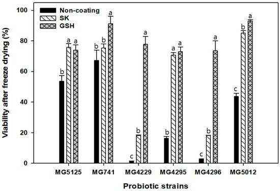
Figure 1.
The viability of probiotics after the freeze-drying process. Different letters on the columns indicate significant differences among means within each strain at p < 0.05 based on Tukey’s post-hoc test.
3.2. Survival of Non-Coated and Coated Bacteria under In Vitro Simulated Gastrointestinal Conditions
The viability of non-coated and coated probiotics with a protectant in the gastrointestinal tract was evaluated. The freeze-dried powders were incubated for 120 and 150 min in simulated gastric and intestinal juices, and the viability was reported as a percentage of log CFU/mL (Table 1). The results showed that their viability was not significantly different between non-coated and coated probiotics with GSH-containing protectants of L. plantarum MG4296 after inclusion in the simulated gastrointestinal tract. However, the viability of GHS-containing protectants in L. lactis MG5125, B. animalis ssp. lactis MG741, L. plantarum MG4229, L. fermentum MG4295, and L. paracasei MG5012 with 77.50, 99.98, 89.13, 98.38, and 98.34%, respectively, were significantly higher than those of non-coated probiotics with 75.92, 91.39, 86.35, 87.13, and 95.12%, respectively. The cell survival rate is reduced because of the low pH at which probiotics pass through the stomach [21,22]. The survival rate of GSH-coated probiotics is higher than that of non-coated probiotics because GSH plays a role in protecting against acid stress in probiotic strains [14].

Table 1.
Survival rates of the selected strains in the simulated gastrointestinal tract.
3.3. Survival of Non-Coated and Coated Bacteria under the Accelerated Stability Test
The storage duration is the principal factor for a probiotic candidate and is affected by increases in temperature. The loss of viability in accelerated storage conditions (40 °C) is higher than that at room temperature or long-term storage conditions [23]. The effect of coated probiotics with protectants on the survival of probiotics after seven days of storage is presented in Table 2. A difference in survival was observed between the non-coated and coated probiotics with GSH-containing protectants. The viability values of the coated L. lactis MG5125, B. animalis ssp. lactis MG741, L. plantarum MG4229, L. fermentum MG4295, L. plantarum MG4296, and L. paracasei MG5012 were higher than those of the uncoated samples (98.85, 97.92, 93.99, 86.82, 90.69, and 93.47%, respectively). A loss of viability in accelerated storage conditions may be explained by the oxidation of membrane lipids and protein denaturation resulting in macromolecular degradation of probiotic cells, which occurs faster at high temperatures.
Table 2.
Survival rates of the non-coated and coated bacteria under the accelerated stability test.
3.4. Adhesion Assay on Intestinal Epithelial Cells
One of the most preferred characteristics of probiotic strains is their adhesion. An adhesion assay on intestinal epithelial cells was conducted to evaluate the differences between the non-coated and coated probiotic strains with GSH-containing protectants. The results showed that the adhesive abilities of L. lactis MG5125, L. plantarum MG4229, and L. paracasei MG5012 coated with GSH were higher than those of non-coated probiotics (67.25, 70.26, and 82.41%, respectively) (Figure 2). The adhesiveness of B. animalis ssp. lactis MG741, L. fermentum MG4295, and L. plantarum MG4296 showed no significant differences between the non-coated and coated probiotics. Previous studies have reported that probiotics that are adhesive to intestinal epithelial cells can inhibit intestinal colonization and the attachment of pathogens [24]. Therefore, coating probiotics with a GSH-containing protectant does not affect the probiotic properties of the studied strains.
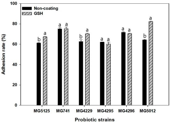
Figure 2.
The adhesion ability of the selected strains to the HT-29 cells. Different letters on the columns indicate significant differences among means within each strain at p < 0.05 based on Tukey’s post-hoc test.
3.5. Scanning Electron Microscopy (SEM)
Scanning electron microscopy was used to observe the non-coated and coated probiotics. The images show that the interval between the cells was irregular in the non-coated sample and a soft structure could be observed (Figure 3a,b), while the cells were covered in the GSH-containing protectant matrix (Figure 3c,d). A previous study also reported that when coated with a protectant, a thin layer of the protective matrix was formed to cover the cells [25,26,27]. Additionally, Kim et al. showed that a collagen peptide-containing protectant prevented membrane disruption and maintained the structural integrity of the cell membrane barrier [26]. Therefore, coating with a protectant can protect probiotics against external stress factors, leading to increased viability after freeze-drying, storage stability, and thermal stability.
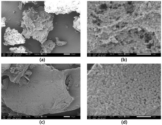
Figure 3.
Scanning electron microscopy images of freeze-dried L. plantarum MG4229: micrograph of the non-coated sample at 1000× magnification (a) and 5000× magnification (b), micrograph of the GSH-coated sample at 1000× magnification (c) and 5000× magnification (d).
4. Conclusions
The GSH-containing protectant increased the viability of all of the studied strains after the freeze-drying process, including L. plantarum MG4229 and MG4296, L. lactis MG5125, L. fermentum MG4295, L. paracasei MG5012, and B. animalis subsp. lactis MG741. The survival ability of many strains was higher with the GSH-coating than with no coating, except L. fermentum MG4295. In the simulated gastrointestinal tract, most of the studied strains showed a significant increase in the survival rate, except for L. plantarum MG4296. All of the GSH-coated strains increased their survival rate after seven days under accelerated conditions. There was no loss of adhesion in most of the studied strains. Furthermore, GSH is now produced on an industrial scale at an affordable price. Based on these results, GSH-containing protectants are a promising technique for increasing the survival of probiotic strains.
Author Contributions
Conceptualization, C.-H.K.; methodology, T.H.N., J.-S.K. and H.-J.K.; formal analysis, T.H.N., J.-S.K. and H.-J.K.; investigation, T.H.N., J.-S.K. and H.-J.K.; data curation, T.H.N. and J.-S.K.; writing—original draft preparation, T.H.N.; writing—review and editing, T.H.N., J.-S.K. and C.-H.K.; visualization, T.H.N.; supervision, C.-H.K.; project administration, C.-H.K. All authors have read and agreed to the published version of the manuscript.
Funding
This research received no external funding.
Institutional Review Board Statement
Not applicable.
Informed Consent Statement
Not applicable.
Data Availability Statement
All data are presented in the paper.
Conflicts of Interest
The authors declare no conflict of interest.
References
- Son, S.J.; Koh, J.H.; Park, M.R.; Ryu, S.; Lee, W.J.; Yun, B.; Lee, J.H.; Oh, S.; Kim, Y. Effect of the Lactobacillus rhamnosus Strain GG and Tagatose as a Synbiotic Combination in a Dextran Sulfate Sodium-Induced Colitis Murine Model. J. Dairy Sci. 2019, 102, 2844–2853. [Google Scholar] [CrossRef] [PubMed]
- Kailasapathy, K. Survival of Free and Encapsulated Probiotic Bacteria and Their Effect on the Sensory Properties of Yoghurt. LWT Food Sci. Technol. 2006, 39, 1221–1227. [Google Scholar] [CrossRef]
- Nami, Y.; Abdullah, N.; Haghshenas, B.; Radiah, D.; Rosli, R.; Khosroushahi, A.Y. Assessment of Probiotic Potential and Anticancer Activity of Newly Isolated Vaginal Bacterium Lactobacillus plantarum 5BL. Microbiol. Immunol. 2014, 58, 492–502. [Google Scholar] [CrossRef] [PubMed]
- Meng, X.C.; Stanton, C.; Fitzgerald, G.F.; Daly, C.; Ross, R.P. Anhydrobiotics: The Challenges of Drying Probiotic Cultures. Food Chem. 2008, 106, 1406–1416. [Google Scholar] [CrossRef]
- Semyonov, D.; Ramon, O.; Kaplun, Z.; Levin-Brener, L.; Gurevich, N.; Shimoni, E. Microencapsulation of Lactobacillus paracasei by Spray Freeze Drying. Food Res. Int. 2010, 43, 193–202. [Google Scholar] [CrossRef]
- Stummer, S.; Toegel, S.; Rabenreither, M.C.; Unger, F.M.; Wirth, M.; Viernstein, H.; Salar-Behzadi, S. Fluidized-Bed Drying as a Feasible Method for Dehydration of Enterococcus faecium M74. J. Food Eng. 2012, 111, 156–165. [Google Scholar] [CrossRef]
- Ta, L.P.; Bujna, E.; Antal, O.; Ladányi, M.; Juhász, R.; Szécsi, A.; Kun, S.; Sudheer, S.; Gupta, V.K.; Nguyen, Q.D. Effects of Various Polysaccharides (Alginate, Carrageenan, Gums, Chitosan) and Their Combination with Prebiotic Saccharides (Resistant Starch, Lactosucrose, Lactulose) on the Encapsulation of Probiotic Bacteria Lactobacillus casei 01 Strain. Int. J. Biol. Macromol. 2021, 183, 1136–1144. [Google Scholar] [CrossRef]
- Khodadadi, P.; Tabandeh, F.; Alemzadeh, I.; Soltani, M. Effects of Sucrose, Skim Milk and Yeast Powder on Survival of Lactobacillus rhamnosus GG Encapsulated with Alginate during One-Week Storage at Room Conditions. Appl. Food Biotechnol. 2022, 9, 251–259. [Google Scholar] [CrossRef]
- Zhen, N.; Zeng, X.; Wang, H.; Yu, J.; Pan, D.; Wu, Z.; Guo, Y. Effects of Heat Shock Treatment on the Survival Rate of Lactobacillus acidophilus after Freeze-Drying. Food Res. Int. 2020, 136, 109507. [Google Scholar] [CrossRef]
- Savedboworn, W.; Teawsomboonkit, K.; Surichay, S.; Riansa-ngawong, W.; Rittisak, S.; Charoen, R.; Phattayakorn, K. Impact of Protectants on the Storage Stability of Freeze-Dried Probiotic Lactobacillus plantarum. Food Sci. Biotechnol. 2019, 28, 795. [Google Scholar] [CrossRef]
- Santivarangkna, C.; Wenning, M.; Foerst, P.; Kulozik, U. Damage of Cell Envelope of Lactobacillus helveticus during Vacuum Drying. J. Appl. Microbiol. 2007, 102, 748–756. [Google Scholar] [CrossRef] [PubMed]
- Cui, S.; Hu, K.; Qian, Z.; Mao, B.; Zhang, Q.; Zhao, J.; Tang, X.; Zhang, H. Improvement of Freeze-Dried Survival of Lactiplantibacillus plantarum Based on Cell Membrane Regulation. Microorganisms 2022, 10, 1985. [Google Scholar] [CrossRef] [PubMed]
- Li, Y.; Hugenholtz, J.; Abee, T.; Molenaar, D. Glutathione Protects Lactococcus Lactis against Oxidative Stress. Appl. Environ. Microbiol. 2003, 69, 5739. [Google Scholar] [CrossRef] [PubMed]
- Zhang, J.; Fu, R.Y.; Hugenholtz, J.; Li, Y.; Chen, J. Glutathione Protects Lactococcus lactis against Acid Stress. Appl. Environ. Microbiol. 2007, 73, 5268–5275. [Google Scholar] [CrossRef]
- Zhang, J.; Du, G.C.; Zhang, Y.; Liao, X.Y.; Wang, M.; Li, Y.; Chen, J. Glutathione Protects Lactobacillus sanfranciscensis against Freeze-Thawing, Freeze-Drying, and Cold Treatment. Appl. Environ. Microbiol. 2010, 76, 2989–2996. [Google Scholar] [CrossRef]
- Fahey, R.C.; Brown, W.C.; Adams, W.B.; Worsham, M.B. Occurrence of Glutathione in Bacteria. J. Bacteriol. 1978, 133, 1126. [Google Scholar] [CrossRef]
- de Menezes, M.F.D.S.C.; da Silva, T.M.; Etchepare, M.D.A.; Fonseca, B.D.S.; Sonza, V.P.; Codevilla, C.F.; Barin, J.S.; da Silva, C.D.B.; de Menezes, C.R. Improvement of the Viability of Probiotics (Lactobacillus acidophilus) by Multilayer Encapsulation. Ciência Rural 2019, 49, 9. [Google Scholar] [CrossRef]
- Lee, J.Y.; Kang, C.-H. Probiotics Alleviate Oxidative Stress in H2O2-Exposed Hepatocytes and t-BHP-Induced C57BL/6 Mice. Microorganisms 2022, 10, 234. [Google Scholar] [CrossRef]
- Nguyen, T.H.; Kim, Y.G.; Kim, J.S.; Jeong, Y.; Park, H.M.; Kim, J.W.; Kim, J.E.; Kim, H.; Paek, N.S.; Kang, C.H. Evaluating the Cryoprotective Encapsulation of the Lactic Acid Bacteria in Simulated Gastrointestinal Conditions. Biotechnol. Bioprocess Eng. 2020, 25, 287–292. [Google Scholar] [CrossRef]
- Carvalho, A.S.; Silva, J.; Ho, P.; Teixeira, P.; Malcata, F.X.; Gibbs, P. Relevant Factors for the Preparation of Freeze-Dried Lactic Acid Bacteria. Int. Dairy J. 2004, 14, 835–847. [Google Scholar] [CrossRef]
- Cook, M.T.; Tzortzis, G.; Charalampopoulos, D.; Khutoryanskiy, V.V. Microencapsulation of Probiotics for Gastrointestinal Delivery. J. Control Release 2012, 162, 56–67. [Google Scholar] [CrossRef]
- da Silva, M.N.; Tagliapietra, B.L.; do Amaral Flores, V.; dos Santos Richards, N.S.P. Pereira dos Santos Richards, N.S. In Vitro Test to Evaluate Survival in the Gastrointestinal Tract of Commercial Probiotics. Curr. Res. Food Sci. 2021, 4, 320–325. [Google Scholar] [CrossRef] [PubMed]
- Makinen, K.; Berger, B.; Bel-Rhlid, R.; Ananta, E. Science and Technology for the Mastership of Probiotic Applications in Food Products. J. Biotechnol. 2012, 162, 356–365. [Google Scholar] [CrossRef] [PubMed]
- Eom, J.S.; Song, J.; Choi, H.S. Protective Effects of a Novel Probiotic Strain of Lactobacillus plantarum JSA22 from Traditional Fermented Soybean Food Against Infection by Salmonella Enterica Serovar Typhimurium. J. Microbiol. Biotechnol. 2015, 25, 479–491. [Google Scholar] [CrossRef] [PubMed]
- Halim, M.; Mohd Mustafa, N.A.; Othman, M.; Wasoh, H.; Kapri, M.R.; Ariff, A.B. Effect of Encapsulant and Cryoprotectant on the Viability of Probiotic pediococcus Acidilactici ATCC 8042 during Freeze-Drying and Exposure to High Acidity, Bile Salts and Heat. LWT 2017, 81, 210–216. [Google Scholar] [CrossRef]
- Kim, S.I.; Kim, J.W.; Kim, K.T.; Kang, C.H. Survivability of Collagen-Peptide Microencapsulated Lactic Acid Bacteria during Storage and Simulated Gastrointestinal Conditions. Fermentation 2021, 7, 177. [Google Scholar] [CrossRef]
- Hongpattarakere, T.; Rattanaubon, P.; Buntin, N. Improvement of Freeze-Dried Lactobacillus plantarum Survival Using Water Extracts and Crude Fibers from Food Crops. Food Bioprocess Technol. 2013, 6, 1885–1896. [Google Scholar] [CrossRef]
Disclaimer/Publisher’s Note: The statements, opinions and data contained in all publications are solely those of the individual author(s) and contributor(s) and not of MDPI and/or the editor(s). MDPI and/or the editor(s) disclaim responsibility for any injury to people or property resulting from any ideas, methods, instructions or products referred to in the content. |
© 2023 by the authors. Licensee MDPI, Basel, Switzerland. This article is an open access article distributed under the terms and conditions of the Creative Commons Attribution (CC BY) license (https://creativecommons.org/licenses/by/4.0/).