Article

Mediator Subunit RhMED15a Regulates Drought Tolerance in Rose

1
Key Laboratory of Quality and Safety Control for Subtropical Fruit and Vegetable, Ministry of Agriculture and Rural Affairs, Collaborative Innovation Center for Efficient and Green Production of Agriculture in Mountainous Areas of Zhejiang Province, College of Horticulture Science, Zhejiang A&F University, Hangzhou 311300, China
2
School of Urban Construction, Zhejiang Shuren University, Hangzhou 310015, China
*
Authors to whom correspondence should be addressed.
These authors contributed equally to this work.
Horticulturae 2024, 10(1), 84; https://doi.org/10.3390/horticulturae10010084
Submission received: 19 December 2023 / Revised: 8 January 2024 / Accepted: 8 January 2024 / Published: 15 January 2024

Abstract

:
Mediator is a multiprotein complex integral to the transcription machinery, mediated by RNA polymerase II. Some Mediator subunits have been found to have critical functions in plants’ responses to abiotic stresses. However, the role of plant Mediator subunits in drought responses remains largely enigmatic. Here, we identified a Mediator subunit, RhMED15a, in roses (Rosa hybrida). Its expression was greatly and swiftly induced by dehydration treatment in the root. The promoter sequence of RhMED15a contains cis-acting elements that respond to abscisic acid (ABA) and methyl jasmonate (MeJA). In addition, the expression of RhMED15a was significantly up-regulated with ABA treatment and inversely down-regulated with MeJA treatment. Silencing RhMED15a using virus-induced gene silencing (VIGS) in roses significantly reduced drought tolerance in rose plants. This resulted in a significant increase in the malondialdehyde (MDA) level and a decreased survival rate in comparison to TRV controls. Moreover, we found that the expression of five drought-related genes, including dehydration responsive element binding factor 1B (DREB1B), responsive to desiccation stress 29A (RD29A), responsive to desiccation stress 29B (RD29B), early response to dehydration 14 (ERD14), and 9-cis-epoxycarotenoid dioxygenase 1 (NCED1), was considerably suppressed in RhMED15a-silenced plants during drought stress. Taken together, our results present that the Mediator tail module subunit RhMED15a serves as an enhancer of drought tolerance in rose, probably through the modulation of the expression of some drought-related genes.

1. Introduction

The rose (Rosa hybrida), one of the world’s most popular flowers, holds significant global economic value. Nonetheless, rose plants frequently encounter abiotic stresses throughout their lifespan, including drought, salinity, extreme temperatures, and varying nutrient availabilities. Among these, drought is a particularly common stressor worldwide due to climate change and rapid industrialization [1,2,3]. This factor poses severe limitations to crop productivity and distribution [2]. In rose seedlings, harsh drought stress leads to leaf wilting and curling, diminished photosynthetic performance, nutrient assimilation reduction, and stunted plant growth, culminating in substantial economic loss [2,4].
To counteract drought stress, plants have developed various intricate response strategies. Numerous studies have shown that drought can trigger responses such as proline and trehalose production, antioxidant defense system enhancement for redox homeostasis maintenance, and peroxidase enzyme use to control cellular damage and sustain membrane integrity [2,5,6,7]. Phytohormones like abscisic acid (ABA) and jasmonic acid (JA) play a role in coordinating such adaptive responses. These hormones are paramount for managing environmental stimuli responses and regulating plant growth and development [2,8,9]. Their synthesis and signal pathways also participate in regulating osmotic adjustment and other processes, heightening drought tolerance in plants [2,9,10]. On the other hand, the intricate signaling transduction pathways that perceive and react to drought conditions often drive changes in gene expression, achieved through transcriptional regulation [11,12]. In eukaryotic cells, this regulation involves an interplay of active agents like RNA polymerase II (Pol II), general transcription factors (TFs), transcriptional repressors or activators, and co-regulators such as Mediator [12].
Mediator, a multi-protein complex, facilitates as a molecular bridge linking DNA-binding TFs to the Pol II enzyme, playing a vital role in numerous stages of transcription, such as initiation, elongation, and splicing [13,14]. The Mediator complex incorporates an estimated 34 subunits in plants, around 31 subunits in mammals, and roughly 25 subunits in yeast [13,15]. In eukaryotes, it comprises three core modules: the head, middle, and tail modules, along with a separable module, cyclin-dependent kinase 8 (CDK8) [13,14,16]. The head module instigates the principal interactions with Pol II, while the middle module performs a structural function, interacting with Pol II when Mediator’s conformation changes after the initial interaction. The tail module is significant for interacting with gene-specific TFs [14].
Each Mediator subunit in plants is believed to perform varying functions due to the subunit composition of Mediator diverging throughout evolution and responding to environmental stimuli and tissue-specific inputs [14]. Several Mediator subunits contribute to basic cellular processes, such as cell proliferation and growth, organ growth, developmental timing, and phytohormone responses [14]. Recent genetic analyses have shown that Mediator subunits also participate in plants’ responses to diverse abiotic stresses such as salinity, cold, and drought [17,18]. Notably, particular Mediator subunits like MED14, MED16, and MED25 are implicated in multiple abiotic stress responses. MED25 specifically, a vital part of stress-response signaling pathways, influences responses to salt and drought stresses. It was observed that Arabidopsis med25 showcased sensitivity to salt stress but resistance to drought [19,20,21]. Genetic analyses suggest that MED25 positively regulates the JA signaling via interaction with MYC2, while it negatively regulates ABA signaling and response through interaction with ABI5 [22]. MED14 and MED16 are crucial for cold acclimation-induced freezing tolerance through modulating Pol II’s recruitment to cold-regulated CBF target genes and their transcription [23,24,25,26]. Meanwhile, MED14 also influences plant responses to high temperatures by activating heat-sensitive genes [27]. MED16 has a role in regulating nutrient uptake and homeostasis, such as iron and phosphate [28]. Moreover, it can interact with MED25, mutations to which alter responses to iron deficiency [29]. Other Mediator subunits show individual functionalities in abiotic stress responses too. For instance, Arabidopsis seedlings of med8 mutant display increased tolerance to oxidative stress and enhanced transcriptional activation of defense-related genes, predominantly those in the salicylic acid (SA) and JA-related pathways [30]. Recently, CDK8 was identified as a positive regulator of ABA signaling and drought response through association with RAP2.6 and SnRK2.6 [31]. Although the roles of the Mediator complex in Arabidopsis have been increasingly identified, the understanding of its functions in crop plants, particularly ornamental plants like roses, remains limited.
The molecular regulatory mechanism for water stress response in roses is still largely unexplored. Past studies identified crucial genes for dehydration/drought tolerance in roses (Rosa hybrida). Transcription factors RhNAC2 and RhNAC3, for example, were found to enhance dehydration tolerance through the regulation of genes associated with cell-wall structure and osmotic adjustment, respectively, in rose flowers [32,33]. The regulatory module RhABF2/RhFer1 can influence Fe levels in rose petals, thus increasing dehydration tolerance [34]. RhHB1, a transcription factor induced by dehydration, directly suppresses the expression of the JA biosynthesis gene RhLOX4, leading to a decreased JA-Ile level and enhanced rose flower dehydration tolerance [9]. Furthermore, transcriptomic analyses of rose plants have identified drought-responsive genes [4]. Considering the significant and varied roles of Mediator in plant stress response, this study focused on the function and mechanism of Mediator subunits in roses. In this study, the expression pattern of nine RhMED genes was explored in roses during dehydration treatment. Among them, RhMED15a, encoding a subunit in the tail module of the Mediator complex, was found to be rapidly and significantly triggered by dehydration stress. It was demonstrated that RhMED15a plays a crucial role in the rose plant’s drought tolerance, partially by regulating the expression of certain drought-responsive genes. This provides new details regarding the function of Mediator subunits in plant drought responses.

2. Materials and Methods

2.1. Plant Materials and Treatments

Rosa hybrida cv. Samantha plantlets were propagated via tissue culture as per previously established techniques [35]. Following growth in a combination of peat moss and vermiculite (1:1), the rooted rose and tobacco (Nicotiana benthamiana) plants were subjected to conditions of approximately 60% relative humidity at 22 ± 1 °C and a 16/8 h light/dark photoperiod.
For dehydration treatments, the roots of rose plant were washed and submerged in deionized water for 24 h. Subsequently, the rose plants were dehydrated by horizontally positioning them on the laboratory bench at 22 °C and approximately 60% relative humidity, under a light intensity of 100 µmol m−2 s−1 for periods of 0, 1, 6, 12, and 24 h. The control plants remained submerged in water under identical conditions. At each time point, leaves and roots of both groups were collected and preserved at −80 °C for subsequent analysis.
For the ABA treatment, the leaves of 1-month-old rose plantlets were sprayed with a solution of 100 µM ABA and 0.05% Tween. The mock group was treated with 0.1% ethanol, excluding any phytohormones. Each seedling was sprayed with 10 mL of the solution. Post-treatment of 12 h, leaf and root samples from these roses were collected respectively.
Leaf discs were submerged in 20 mL of solutions of either 100 µM methyl jasmonate (MeJA) or 100 µM salicylic acid (SA) and 0.05% Tween for 24 h, for the JA and SA treatments, respectively. The mock group, in the case of MeJA treatment, received a treatment of 0.1% ethanol along with 0.05% Tween, while mock samples of the SA treatment were treated with 0.1% DMSO and 0.05% Tween.

2.2. Sequence Analysis of RhMED15a

The conserved domains of the RhMED15a amino acid sequence were scrutinized utilizing the Conserved Domain Database (CDD) of the National Center for Biotechnology Information (NCBI). ProtParam (https://www.expasy.org/, accessed on 16 May 2022) was employed to probe the molecular weight (MW) and the isoelectric point (pI). The task of multiple sequence alignment was fulfilled through Bioedit using the ClustalW algorithm as its foundation. The phylogenetic associations between RhMED15a and its homologs from other plant species were conducted through MEGA 7.0 by implementing the neighbor-joining algorithm, and bootstrap values were obtained through 1000 iterations.

2.3. RNA Extraction and Quantitative RT-PCR

RNA extractions from the roots and leaves of rose plants were performed using an RNA rapid extraction kit (BOLAZ, Nanjing, China). The conversion of 1 μg of total RNA into complementary DNAs (cDNAs) was accomplished using the PrimeScript™ RT reagent Kit along with the gDNA Eraser (Takara, Shiga, Japan). This was followed by quantitative RT-PCR, carried out using 2 μL of cDNA as a template, on the StepOne Real-Time PCR System (Applied Biosystems, Foster City, CA, USA) in conjunction with the Kapa SYBR Fast Universal qPCR Kit (Kapa Biosystems, Boston, MA, USA). The primers used for this process were available in Table S1, while RhUBI2 served as an internal control. Three biological replicates formed the basis of all reactions, and representative results are presented. The statistical testing was performed using the SPSS version 17.0.

2.4. Protein Subcellular Localization Assays

The open-reading frame (ORF) of RhMED15a was amplified by PCR and then inserted into a modified binary vector pCAMBIA2300 harboring 35S and a GFP label using ClonExpress II One Step Cloning Kit (Vazyme, Nanjing, China). The resultant pCAMBIA2300-GFP-RhMED15a construct was then transferred into GV3101 Agrobacterium competent cells. The tomato bushy stunt virus p19 protein served to inhibit gene silencing during the transient expression procedure in tobacco leaves, as previously outlined [35]. Negative control was provided by p35S::GFP, with p35S::NF-YA4-mCherry serving as a nuclear marker. Agrobacterium cultures containing the pCAMBIA2300-GFP-RhMED15a construct (or negative control construct), nuclear marker, and the p19 plasmid were mixed at OD600 of 0.5:0.5:0.3. Cells were pelleted and resuspended in infiltration medium (10 mM MgCl2, 10 mM MES, and 100 mM acetsyringone). After incubation for 2 h, the suspension was filtrated into leaves of 1-month old tobacco (Nicotiana benthamiana) plants. Three days post-infiltration, laser confocal fluorescence microscopy was employed to observe the fluorescence signal (FV1000; Olympus, Tokyo, Japan).

2.5. Virus-Induced Gene Silencing

Virus-induced gene silencing (VIGS) was used to silence RhMED15a in rose plants, following previous methodology with minor amendments [32]. Tobacco rattle virus (TRV) vectors were employed in VIGS. TRV is a bipartite RNA virus with the TRV1 and TRV2 genomes. pTRV1 encodes the replication and movement functions while pTRV2 harbors the coat protein and the sequence used for VIGS. A specific 350 bp fragment in RhMED15a ORF was used to generate pTRV2-RhMED15a. Agrobacterium strain EHA105, containing pTRV1, pTRV2, and pTRV2-RhMED15a, was cultured in Luria-Bertani medium with 10 mM MES, 20 μM acetosyringone, and appropriate antibiotic selections. The Agrobacterium cells were collected and suspended in infiltration buffer (10 mM MgCl2, 200 μM acetosyringone, and 10 mM MES, pH 5.6) to achieve an approximate OD600 of 1.5. Agrobacterium cultures carrying pTRV1 and pTRV2 or its derivatives were mixed in equal ratios (v/v) and left at room temperature for 4 h. Rose plantlets were then immersed in the bacterial suspension solution, exposed to −25 KPa vacuum twice, washed with deionized water, and potted for analysis.
For the drought treatment, infected rose plants were planted in a combination of peat moss and vermiculite (1:1) in a growth room maintained at a steady 22 ± 1 °C, approximately 60% relative humidity, and subjected to a 16 h/8 h light/dark cycle. After 40 days of growth, these conditions were altered: watering was ceased for 20 days, then resumed for an additional 20 days. Plant phenotypes were periodically checked. When a compound leaf’s leaflets display over 50% wrinkling, the leaf is categorized as wilted. The rate of leaf wilting is computed by dividing the quantity of wilted compound leaves by the compound leaves’ total number.

2.6. Malondialdehyde (MDA) Content Determination

The concentration of MDA was ascertained using an MDA content assay kit (Comin Biotechnology Co., Ltd., Suzhou, China). MDA is capable of participating readily in a nucleophilic addition reaction with 2-thiobarbituric acid (TBA), resulting in the creation of a red-colored MDA:TBA adduct. Subsequently, the colorimetric methodology is utilized to quantify the adduct content, from which the MDA content can then be further determined. Fresh leaves from each plant, either prior to the drought treatment or on the 12th day of drought treatment, were ground and homogenized along with a 10% (w/v) extracting solution. The absorbance was then calculated at 532 nm and 600 nm wavelengths using a spectrophotometer (T6 New Century, Purkinje General, Beijing, China). Based on its extinction coefficient, 155 mM−1·cm−1, the MDA concentration was then derived.

2.7. Statistical Analysis

Each experiment was performed independently at least three times. At least three plants were used in each independent experiment. In each biological replicate of quantitative RT-PCR, three technical replicates of each PCR reaction were run. SPSS 16 was used for statistical analysis. Significant differences between the experimental data were analyzed using Student’s t test or one-way ANOVA with Duncan’s multiple range tests (* p < 0.05; ** p < 0.01; *** p < 0.001).

3. Results

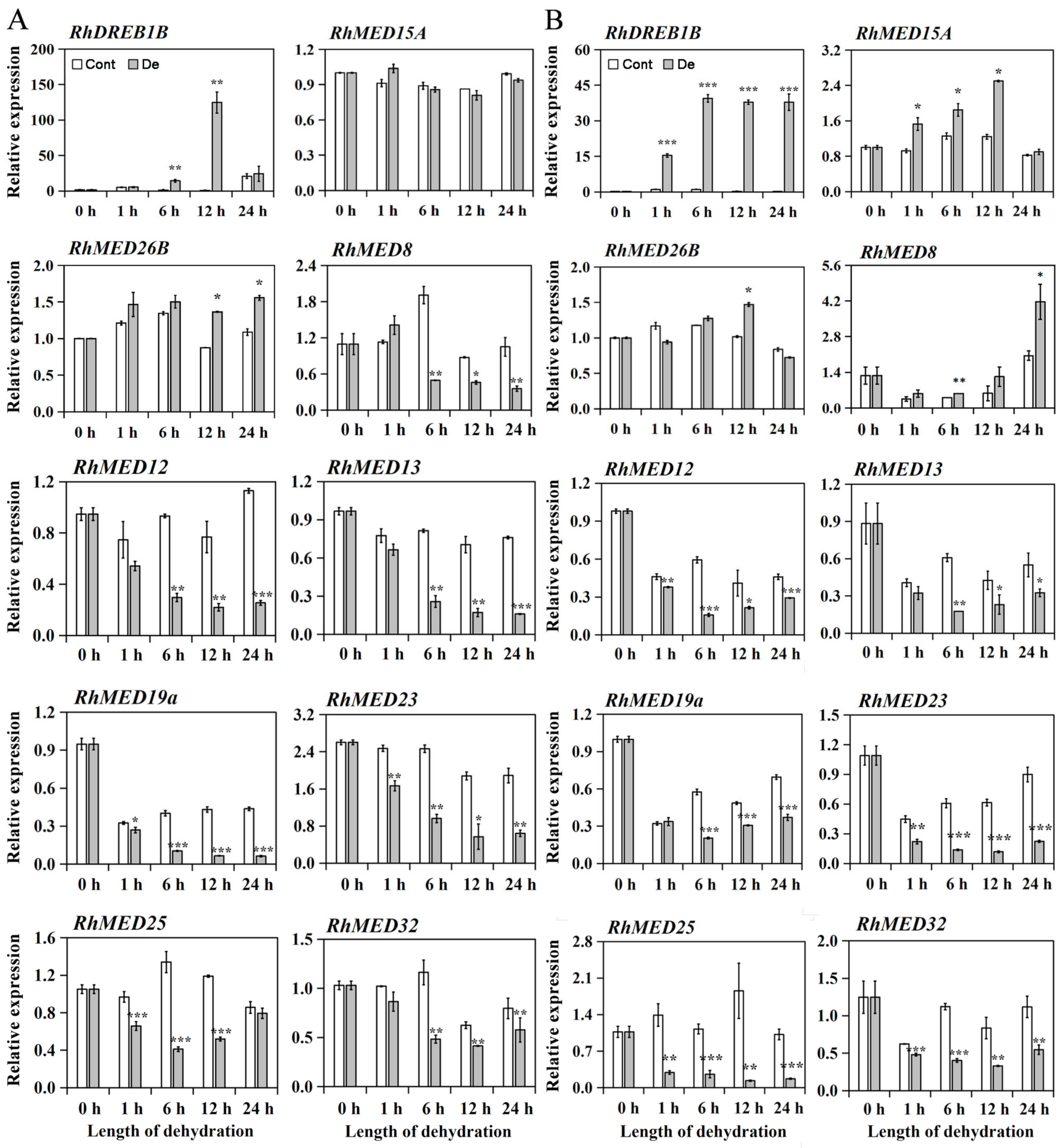
3.1. RhMED15a Was Induced by Dehydration Treatment in Roses

To investigate the role of Mediator subunits in the drought response of rose plants, nine differentially expressed RhMEDs from the transcriptome data of dehydration-treated flowers (unpublished data) were selected. Their expression profiles in rose seedlings during dehydration were confirmed using quantitative real-time PCR. Furthermore, the expression of the well-characterized water deficit-inducible gene, dehydration responsive element binding factor 1B (RhDREB1B), was detected as a positive control [36]. As depicted in Figure 1, both the leaves and roots expressed a significant increase in RhDREB1B during dehydration, suggesting the rose plants were subjected to moderate water stress. When compared to the control, the expression of six RhMED genes (RhMED12, RhMED13, RhMED19a, RhMED23, RhMED25, and RhMED32) significantly declined at least at three time points within the 24 h of dehydration in both the leaf and root. This indicates that dehydration treatment inhibits the expression of these RhMEDs in rose plants. Among the remaining RhMEDs, RhMED8’s expression profile differed between the leaf and root; its expression significantly increased in the root after six hours of dehydration treatment compared to the control, while concurrently, it substantially declined in the leaf. RhMED26b’s expression was not up-regulated until after 12 h of dehydration treatment in both the leaf and root. Notably, among all the tested RhMEDs, RhMED15a experienced the most significant dehydration-induced increase in root expression. Its abundance markedly increased at 1 h of dehydration treatment and reached an approximately two-fold value at 12 h compared to the control, despite no significant alteration in leaf expression during dehydration.

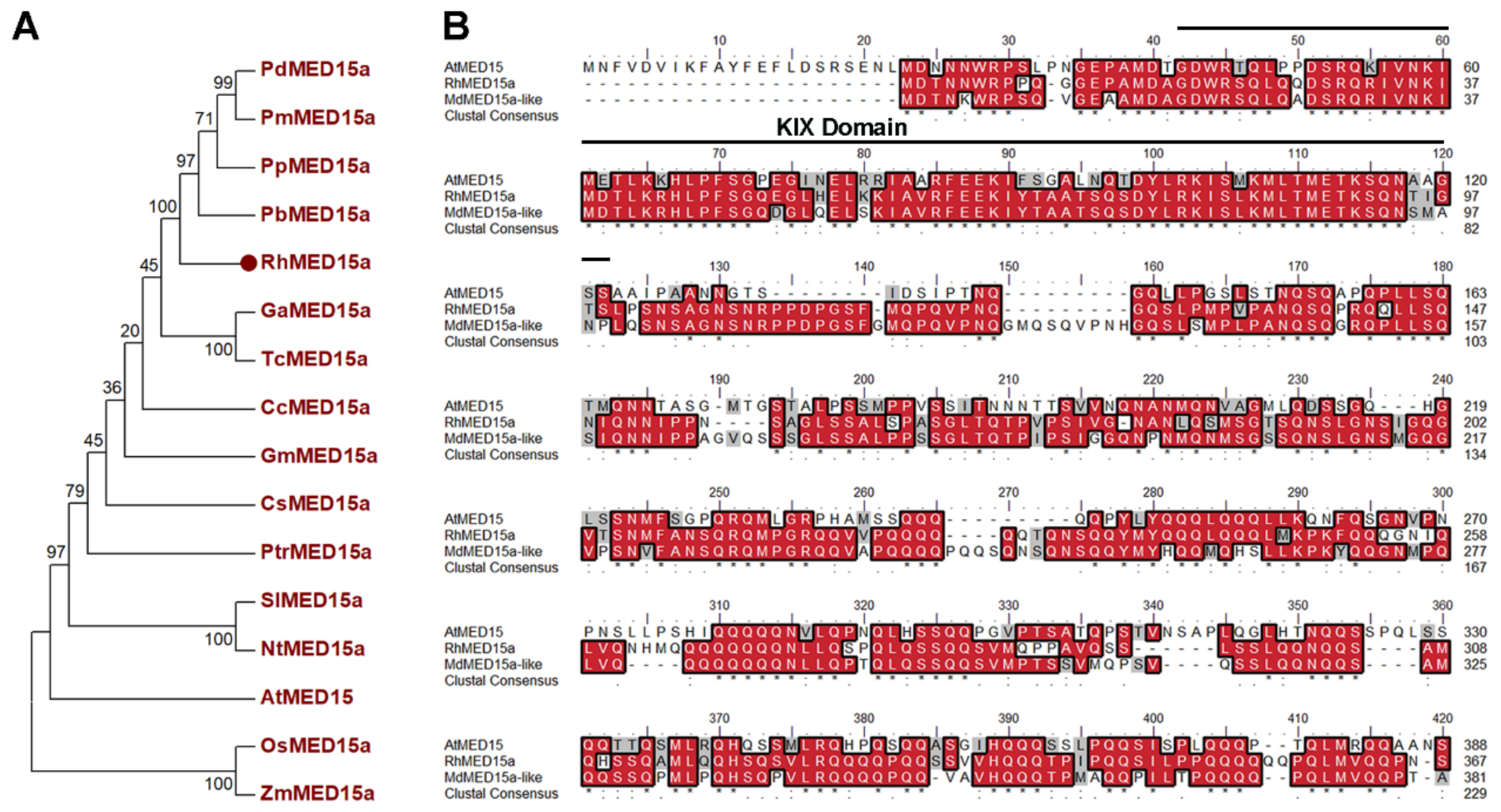
3.2. Isolation and Sequence Analysis of RhMED15a

The full-length cDNA of RhMED15a was isolated from rose plants and its structural features were analyzed. Its open reading frame is 4086 bp in length and codes for an inferred amino acid sequence of 1361 residues. The predicted relative molecular mass of these residues is 150.4 kDa, with a theoretical pI of 9.4. When the inferred amino acid sequences of RhMED15a were aligned with homologs from other plant species, high homology was observed (Figure 2B and Figure S1). Moreover, the RhMED15a protein was found to contain a KIX domain at the amino terminus. This domain has previously been seen as pivotal for the interaction between Med15 (a Mediator tail module subunit) and various transcription activators in yeast and other plants [37]. Phylogenetic analysis revealed that MED15a homologs from the Rosaceae form a clearly defined and substantially supported branch, distinct from homologs in other families (Figure 2A). In Arabidopsis, the closest ortholog to RhMED15a was found to be AtMED15 (NP_001321150.1), which shares 53.3% identity with the protein sequence of RhMED15a. Such results suggest that RhMED15a is a part of the tail module of the Mediator complex.

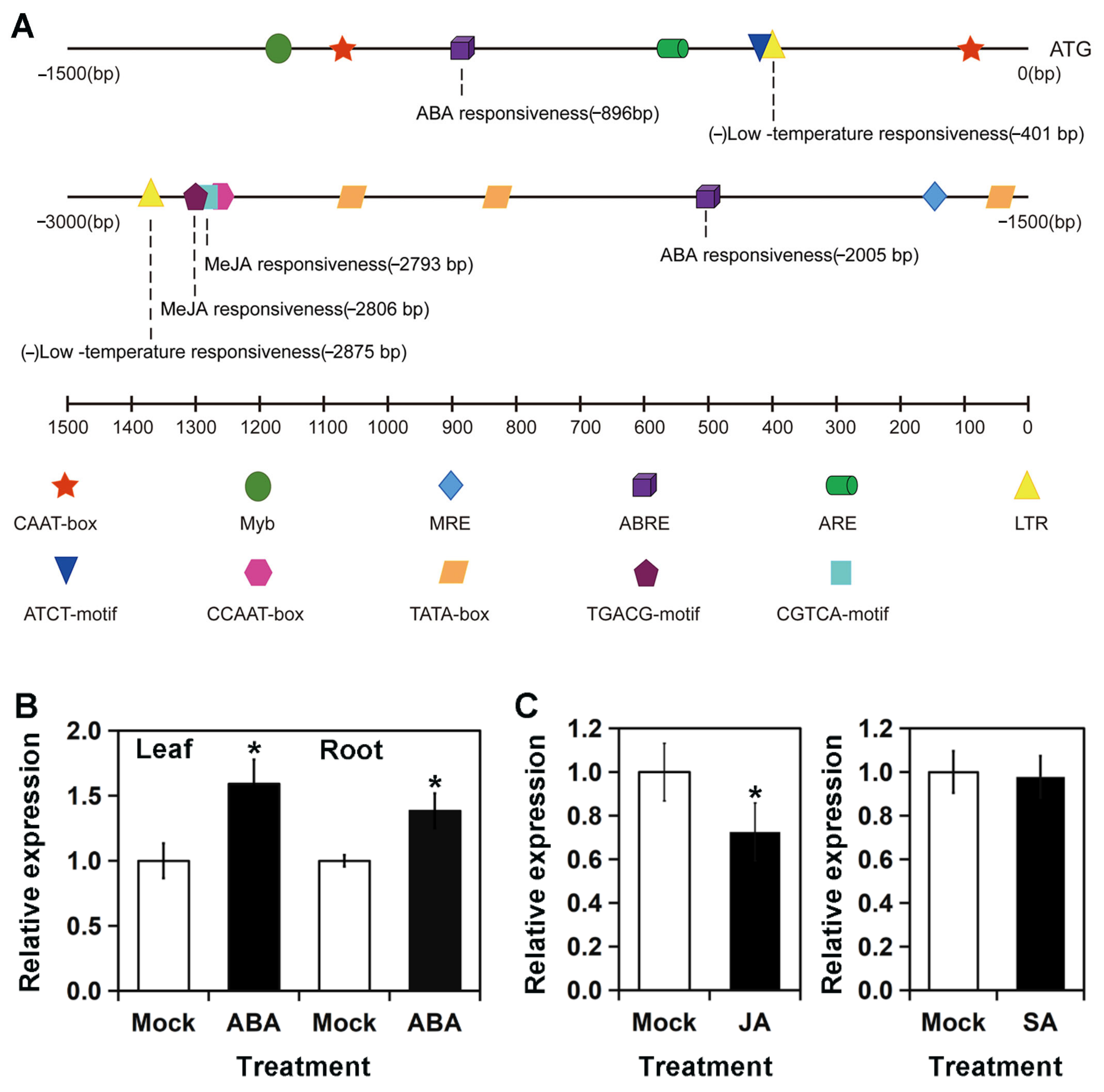
3.3. Expression of RhMED15a Was Significantly Up-Regulated by ABA but Down-Regulated by MeJA

To anticipate the expression profiles of RhMED15a, we analyzed nearly a 3kb promoter sequence of RhMED15a. PlantCare database was used to conduct bioinformatics analysis of this promoter sequence. As revealed in Figure 3A, the sequence exhibited typical regulatory elements found in plant gene promoters. These elements include those that are responsive to plant hormones and abiotic stress factors. There were four cis-acting motifs in the RhMED15a promoter that interact with two phytohormones: ABA and MeJA. This suggests the possibility of RhMED15a having a role in the stress response mediated by MeJA and ABA. In terms of abiotic stress, the sense strand of the promoter lacks any stress-response elements, although two LTRs (CCGAAA), which are regulatory elements for low-temperature stress, were found on the other strand. This indicates that the expression of RhMED15a in dehydration stress situations may be regulated by the stress hormones ABA or MeJA, both of which have been identified as potent mediators of plant response to water stress [9,38].
We further analyzed RhMED15a’s response to plant hormones using quantitative RT-PCR analysis. The focus was on ABA and MeJA, as these phytohormones were identified from the analysis of cis-elements in the RhMED15a promoter. We also studied the impact of SA on the expression of RhMED15a, since AtMED15 in Arabidopsis is known to play a part in the SA-mediated defense response [39]. As shown in Figure 3B, we found that ABA treatment led to a significant increase in the expression of RhMED15a by approximately 1.6-fold in leaves and 1.4-fold in roots after 12 h of exposure. Conversely, MeJA was found to suppress RhMED15a expression by about 27.4% in leaf tissue at 24 h stimulation when compared with the controls (Figure 3C). This result hints at RhMED15a’s potential involvement in ABA and JA-regulated development and abiotic stress response in roses. A 24 h treatment with SA did not significantly change RhMED15a expression in leaf tissue, aligning with our initial bioinformatics analysis of the RhMED15a promoter that found no SA-responsive cis-elements (Figure 3A).

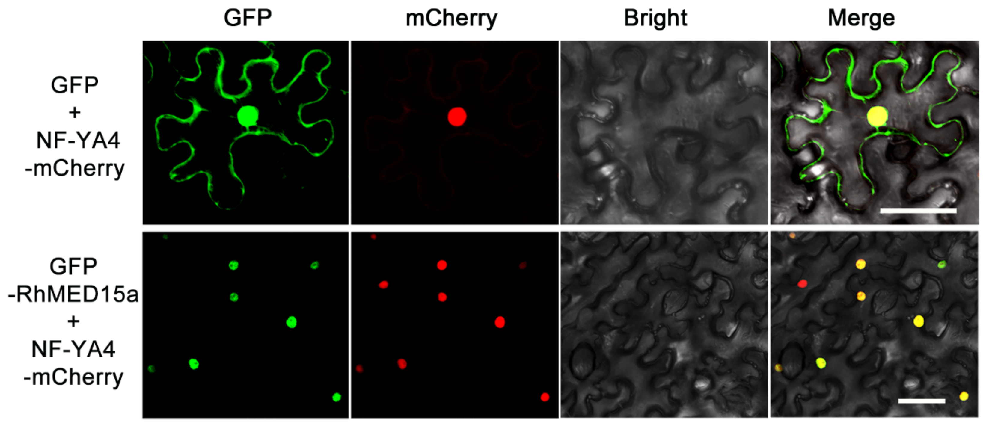
3.4. RhMED15a Was Localized in the Nucleus

We explored the subcellular location of RhMED15a in the epidermal cells of Nicotiana benthamiana leaves by executing a fusion of the ORF sequence of RhMED15a to the C-terminal side of the GFP reporter gene. The resultant construct, along with an mCherry-marked nuclear marker, was co-introduced into tobacco leaves. As delineated in Figure 4, green fluorescence signals from RhMED15a were located in the nucleus, overlapping significantly with the red fluorescence signal emanating from mCherry. This observation aligns with prior studies on Arabidopsis MED15 that pinpoint RhMED15a as a nuclear-localized protein [40].

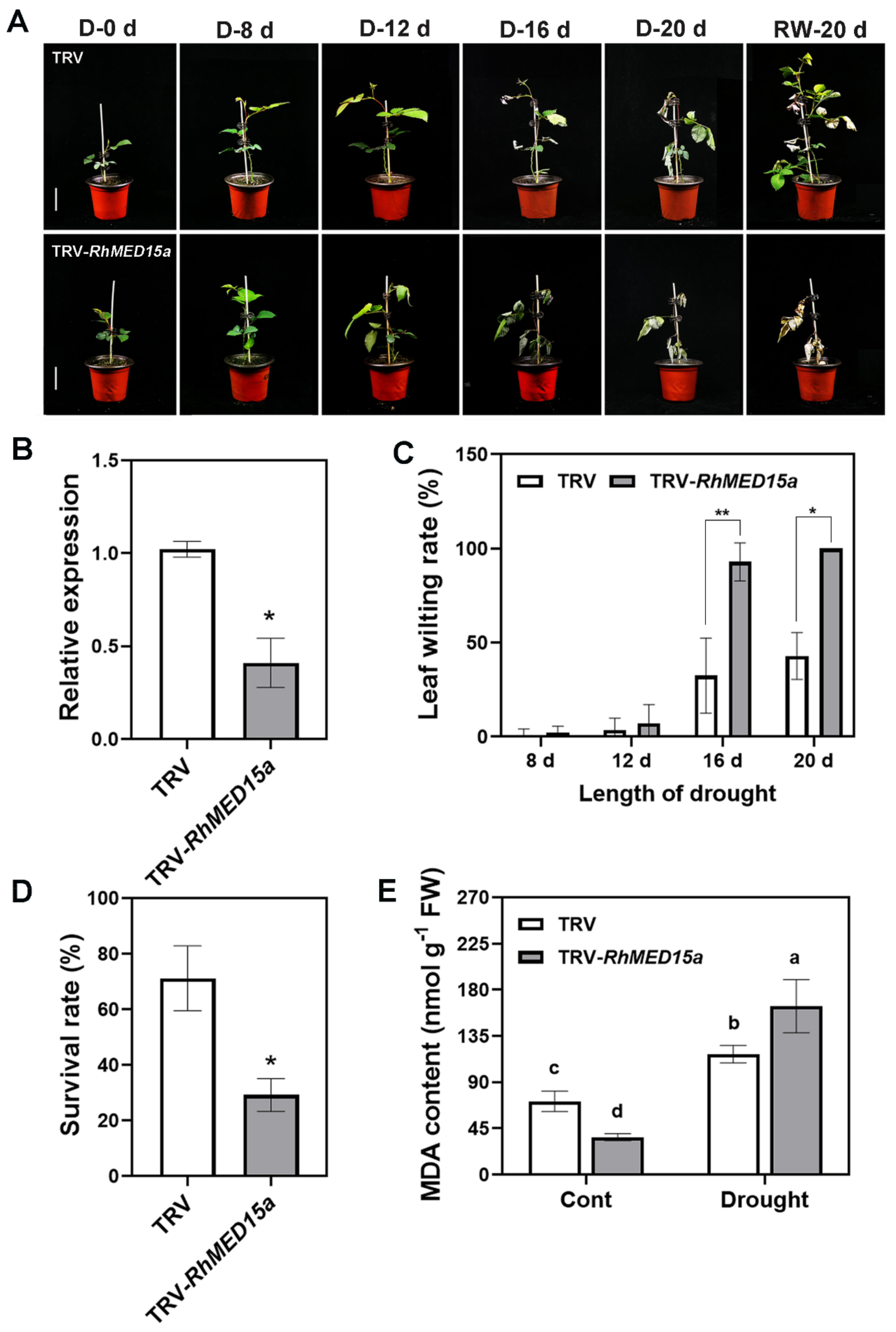
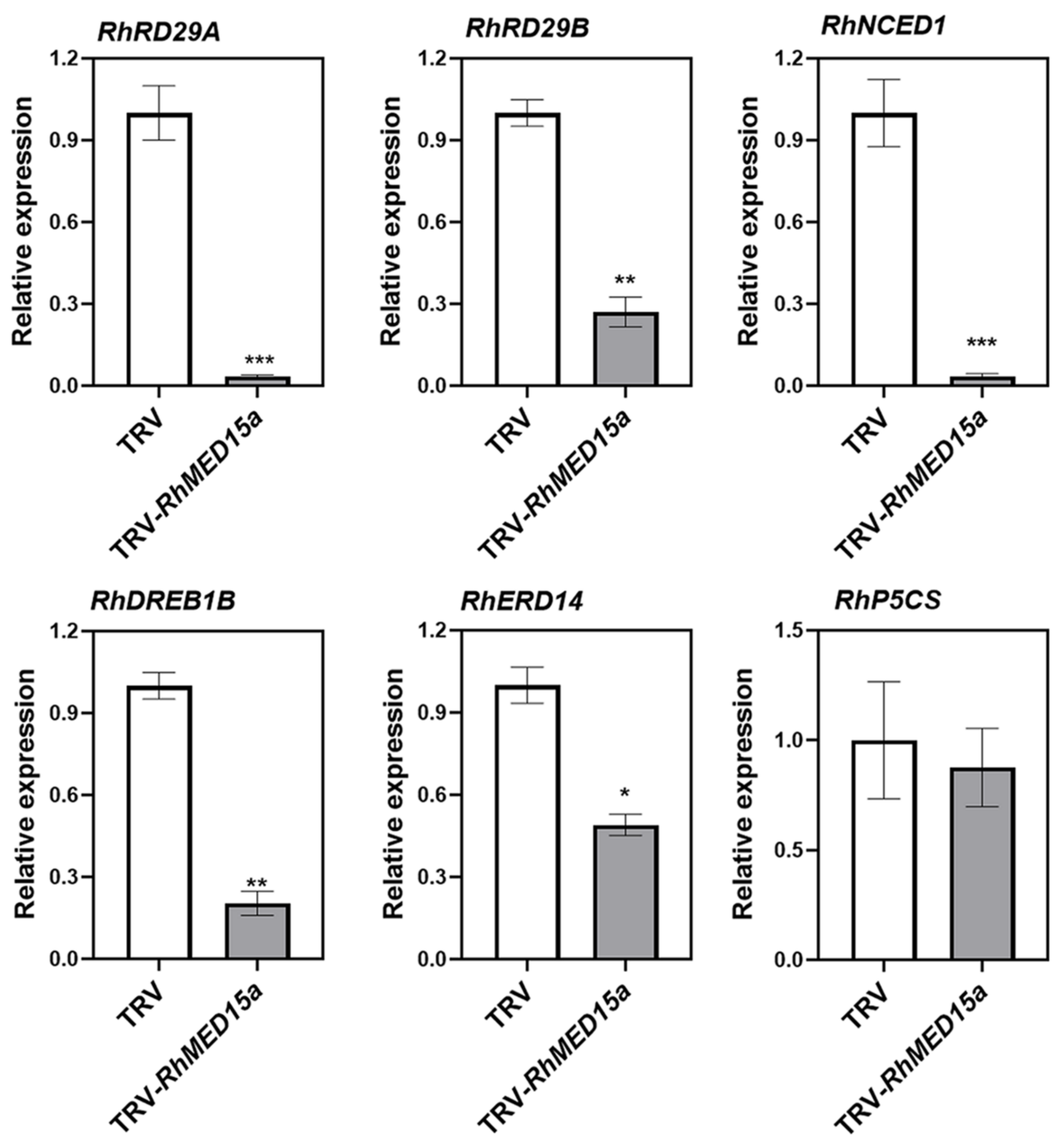
3.5. Silencing of RhMED15a Decreased Drought Tolerance in Rose Plants

To investigate the role of RhMED15a in response to water deficiency, we employed the VIGS approach using Tobacco Rattle Virus (TRV) to silence this gene in rose plants, following which, drought tolerance was gauged as outlined previously [35]. We leveraged a 350 bp gene-specific fragment of RhMED15a from the ORF region for pTRV2-RhMED15a assembly. Rose plantlets infiltrated with Agrobacterium incorporating either TRV control or TRV-RhMED15a were subsequently exposed to drought for 20 days and later reverted to standard watering regimes for an additional 20 days. Demonstrated in Figure 5B, we clinched a total of 7 successfully silenced plants exhibiting considerably lower expression of RhMED15a relative to the TRV control. Under standard growth conditions, there were no significant variances in plant height, leaf count, or leaf surface area between the TRV controls and RhMED15a-silenced plants, suggesting a likely non-involvement of RhMED15a in the regulation of plant growth in roses (Figure S2).
When subjected to drought stress, rose plants manifested drought-related phenotypes, including yellowing and curvature of leaf blades, wilted petioles, defoliation, and inhibited growth (Figure 5A). TRV plants began to wilt on the 16th day of drought treatment, becoming more severe by the 20th day. Contrastingly, RhMED15a-silenced plants exhibited noticeable wilting by the 12th day of drought treatment, with most leaves dried and defoliated by the 20th day (Figure 5A). RhMED15a-silenced plants demonstrated a significantly higher leaf wilting rate than the TRV controls at the 16th and 20th days of drought treatment (Figure 5C). Following re-watering, about 71.2% of the TRV control plants recovered, whereas only 29.2% of RhMED15a-silenced plants survived the drought stress (Figure 5D). MDA content was tested, as it is a product of peroxidation of unsaturated fatty acids in phospholipids and is commonly used as an indicator of cellular membrane lipid peroxidation [41]. The test results revealed that MDA levels in both groups displayed a rising trend post-drought stress (Figure 5E). On the 12th day of drought treatment, MDA levels of RhMED15a-silenced plants increased by approximately 4.5-fold, while TRV controls only showed about 1.6-fold induction, suggesting RhMED15a-silenced plants endured more severe damage during drought treatment.
Considering Mediator’s role in transcriptional regulation, we hypothesized that RhMED15a could impact plant tolerance to drought stress by controlling the expression of genes involved in drought response. Accordingly, we analyzed six drought-responsive genes, including responsive to desiccation stress 29A (RD29A), responsive to desiccation stress 29A (RD29B), 9-cis-epoxycarotenoid dioxygenase 1 (NCED1), DREB1B, early response to dehydration 14 (ERD14), and Δ1-pyrroline-5-carboxylate synthetase (P5CS) [42,43,44,45], via quantitative RT-PCR analysis on the 12th day of drought treatment in the leaves of TRV-RhMED15a plants and TRV controls. The findings revealed significant reductions in the expression levels of five genes (RD29A, RD29B, NCED1, DREB1B, and ERD14) in RhMED15a-silenced rose plants compared to TRV controls under drought stress. However, P5CS expression showed no significant difference (Figure 6). Particularly, in RhMED15a-silenced plants, expression levels of RD29A, NCED1, and DREB1B were markedly inhibited by roughly 97%, 96.5%, and 83.7%, respectively, compared to TRV controls. These results suggest that RhMED15a positively regulates the expression of these drought-responsive genes under drought conditions.

4. Discussion

The Mediator complex in plants functions as a transcription cofactor, and is involved in various growth, developmental, and stress-response processes [14]. The necessity for individual Mediator subunits typically varies within the complex, indicating distinct functions for different subunits in these processes [14,46,47]. Currently, research on plant Mediator subunits is primarily confined to Arabidopsis and rice [46,48]. Thus, it is crucial to identify potential Mediator subunits in crops that can influence yield, product quality, and stress tolerance [46]. In this study, we examined the expression profiles of nine Mediator subunits in roses during a water deficit scenario, with particular emphasis on the Mediator tail module subunit RhMED15a, which exhibited notable up-regulation following dehydration treatment. We explored RhMED15a’s role in drought tolerance in roses using VIGS, and evaluated the expression of drought-responsive genes within RhMED15a-silenced plants.

4.1. Dehydration Stress Responsive Mediator Subunits in Roses

The established role of the plant Mediator complex in immunity is well-recognized, with a few Mediator subunits implicated in responses to abiotic stresses [12,20,31,49]. It is evident that key Arabidopsis MEDs partake in the regulation of abiotic stress signaling pathways. For instance, AtMED16, AtMED14, and AtMED2 oversee the regulation of COLD ON-REGULATED (COR) genes, demonstrating sensitivity to cold stress [26]. AtMED25 and AtMED19a contribute to plant resistance against drought stress and control ABA-responsive transcription [19,22,49]. Evaluating RhMEDs’ expression profiles during water deficit situations sheds light on their functional roles within the drought response mechanism in rose plants. Nine RhMED expressions were confirmed during dehydration treatment in rose seedlings in this study. Six of the observed RhMEDs showed significant down-regulation in both leaf and root under dehydration, while three exhibited significant up-regulation in either leaf or root at varying degrees (Figure 1). The varied impacts of dehydration on RhMED expressions point to the diverse responsiveness of RhMEDs to water stress across Mediator subunits and plant organs. Notably, RhMED26b was induced at 12 h of dehydration treatment in both leaves and roots, in alignment with previous reports on tomato SlMED26b, suggesting a potential role in the regulation of plant dehydration tolerance [50]. Interestingly, RhMED25 demonstrated rapid and significant down-regulation in both leaf and root during dehydration treatment. Since AtMED25 can negatively regulate drought tolerance in Arabidopsis [19,22], such results hint that RhMED25 may perform a role similar to AtMED25 in plant drought tolerance. Another finding was the significant down-regulation of RhMED19a in both organs under dehydration, a surprising observation as AtMED19a reportedly bolsters plant resistance to drought stress [49]. This could potentially hint at the diverse roles MEDs play across different species and the decoupling of gene expression from its function due to multi-level molecular mechanisms interfacing with the regulatory process in stress responses [51,52]. Among the observations, the rapid rise of RhMED15a expression, significantly quicker and greater than other MEDs in the root, is notable, suggesting a potential role in the rose’s water stress response.

4.2. Insights into the Role of RhMED15a in Drought Stress Responses in Roses

The MED15 subunit of the mediator tail module in plants is reported to be an essential component involved in the transcriptional regulation of various processes, including floral transition [39], lipid biosynthesis in seeds [40], and plant immunity [39,53]. Existing studies reveal how AtMED15 in Arabidopsis functions downstream of NPR1 (Non-expresser-of Pathogenesis-Related-1) to regulate the SA-mediated defense response [39]. AtMED15 also activates glycolysis-related and fatty acid biosynthesis genes by interacting with the plant-specific transcription factor, Wrinkled 1 (WRI1) [40]. In rice plants, OsMED15a connects OsNAC024 and OsNAC02 to their target genes, modulating grain size and weight [54]. However, the role of MED15 in the drought stress response of plants remains undefined. Our study demonstrated that compared to controls, the silencing of RhMED15a through the VIGS approach induced quicker and more severe leaf wilting, increased MDA content, and decreased the survival rate of rose plants during drought stress (Figure 5). These findings genetically validate the conducive role of RhMED15a in the regulation of drought tolerance in roses, potentially revealing a new aspect of MEDs’ involvement in the plant response to drought stress. This implies that the MED15 mediator subunit might be instrumental in addressing both biotic and abiotic stresses in plants.
Additionally, we observed that the expression of five drought-responsive genes, including one ABA-independent gene (DREB1B) [55], three stress/ABA responsive genes (RD29A, RD29B, and ERD14) [56,57], and one ABA synthesis gene (NCED1) [58], were considerably down-regulated in RhMED15a-silenced plants under drought stress compared to TRV controls (Figure 6). This suggests that RhMED15a, as a transcriptional co-regulator, may enhance roses’ resistance to drought stress by regulating the expression of drought-stress-related genes. Thus, further studies, such as RNA-seq of TRV controls and RhMED15a-silenced plants, would help explicate the underlying molecular mechanisms of the role of RhMED15a in drought resistance in roses.
In addition, under normal conditions, we noted no statistically significant difference in plant height, the number of compound leaves, and leaf area between RhMED15a-silenced and TRV control plants on the 42nd day after infection (Figure S2). This suggests that RhMED15a may not regulate the growth rate of rose plants. However, compared to the wild type in Arabidopsis, the med15/nrb4-4 variant was smaller and had fewer flowers but more additional stems after bolting [39]. The phenotypic discrepancy between RhMED15a-silenced roses and Arabidopsis med15/nrb4-4 could be traced back to MED15’s diverse roles in different plant species, the various developmental periods of plants, and the effect of gene silencing. Consequently, the impact of RhMED15a on rose growth and development necessitates further investigation.
Alterations in hormonal content and corresponding signaling networks in plants are triggered by drought stress. These modifications aim to enhance tolerance or stimulate programed cell death within distinct cells, tissues, or organs, promoting survival under unfavorable conditions [10,59]. Certain phytohormones, such as ABA and JA, are crucial for regulating plant responses to water stress [38]. ABA, a primary stress-signaling phytohormone, tends to accumulate during drought periods and predominantly acts as a hormonal signaling molecule addressing drought stress [60,61]. In contrast, JA, a lipid-derived plant hormone, fine-tunes plant responses to various biotic and abiotic stresses through global transcriptional activation of JA-responsive genes [62]. Both the internally produced and externally introduced JAs contribute to drought stress tolerance in plants [63]. Specific Mediator subunits, including MED25, AtMED19a, and CDK8, critically participate in JA and/or ABA signaling. MED25 positively regulates JA signaling via interaction with MYC2, while negatively regulating ABA signaling and response through interacting with ABI5 [22]. AtMED19a acts as a positive regulator in ABA responses mediated by ABI5 [49]. CDK8 has been implicated as a critical regulator in the ABA signaling and drought response pathways. The cdk8 mutant demonstrated diminished sensitivity to ABA, compromised stomatal aperture, and hypersensitivity to drought stress compared to control [31]. In tomatoes, SlMED17, SlMED21, and SlMED23 were observed to have been up-regulated more than twice following 8 h of MeJA treatment, while the expression of SlMED18 significantly decreased in response to ABA treatment [50]. Our study found that RhMED15a displayed increased mRNA accumulation under ABA treatment and reduced accumulation under MeJA treatment (Figure 3B,C). Given its expression levels following ABA and MeJA treatments and the presence of ABA- and MeJA-related cis-acting elements in the promoter region (Figure 3A), we propose that RhMED15a could serve as a regulator of ABA and MeJA signaling in roses. Recently, it was reported that JA-Ile levels elevate in rose petals during dehydration, limiting the osmotic adjustment capability in petal cells and causing a decline in dehydration tolerance [9,35]. Nullifying RhMED15a function may deteriorate drought tolerance in rose plants due to its involvement in JA signaling, although this hypothesis demands more robust genetic evidence.
Considering the nature of the Mediator’s tail module, RhMED15a could potentially play a vital role in drought stress response via directed interactions with hormone signaling components and numerous TFs. For instance, the Mediator tail module subunit MED25 has been found to regulate drought tolerance negatively upon interacting with drought stress-induced proteins, including DREB2A, ZINC FINGER HOMEODOMAIN 1 (ZFHD1), and PHOSPHATE STARVATION RESPONSE LIKE1 (PHL1) [19,64]. Additionally, MED15a homologs were discovered interacting with multiple TFs [37]. In Arabidopsis, the AtMED15a interactome entailed 45 proteins interacting with the KIX domain of AtMED15a, including 11 TFs, three distinct nucleic acid-binding proteins, and a splicing factor [37]. A similar finding was recorded for RhMED15a, where the KIX domain was observed in its respective deduced protein (Figure 2B and Figure S1). Thus, there is a need to identify the interacting TFs of RhMED15a in roses that may contribute to water stress tolerance. In addition, Mediator subunits can interact to form flexible and diverse Mediator complexes that regulate the transcription of select genes. The specificity of these combinations is determined by both the nature of the environmental stimuli and the identity of the induced gene [14]. For instance, in Arabidopsis, the recruitment of RNA polymerase II to cold-regulated genes responsive to CBF requires the MED16, MED2, and MED14 subunits during cold acclimation-induced freezing tolerance [26]. These three subunits also manage the expression of certain cold-responsive genes, which are not recognized CBF targets, during low-temperature induction. Genes induced by darkness also necessitate MED16 but require a different Mediator subunit combination for their expression compared to the genes induced by cold. Consequently, it is worthwhile to further comprehend the unique Mediator complex composed of RhMED15a and other subunits affected by drought stress, as well as the specific downstream genes they regulate.

5. Conclusions

Drought stress profoundly impacts the growth and development of the rose plant, making it crucial to uncover its regulatory mechanisms. This research characterizes a rose-derived Mediator subunit gene, RhMED15a, providing insights into the role played by Mediator subunits in drought response in roses. RhMED15a operates as a positive regulator of drought tolerance as well as being a crucial transcriptional regulator mediating gene expression alterations associated with drought stress. This study contributes to understanding the role of Mediator subunits in plant responses to abiotic stresses and offers significant clues for investigating drought tolerance in roses.

Supplementary Materials

The following supporting information can be downloaded at: https://www.mdpi.com/article/10.3390/horticulturae10010084/s1, Figure S1: The alignment of the predicted amino acid sequences of RhMED15a and other homologs with the highest similarity from different plant species; Figure S2: The silencing of RhMED15a in roses does not affect growth; Table S1: Primers used in RT-PCR analysis and vector construction.

Author Contributions

Conceptualization, W.C., X.R. and Y.C.; performed the experiments, X.S., N.X., Y.L., Z.Z. and P.L.; writing and original draft preparation, W.C. and X.S.; approved the final manuscript for publication and agreed to be accountable for all aspects of the manuscript, W.C.; funding acquisition, W.C. All authors have read and agreed to the published version of the manuscript.

Funding

This work was funded by the National Natural Science Foundation of China, grant number 32072612.

Data Availability Statement

All data generated or analyzed during this study are included in this article and its Supplementary Information files.

Conflicts of Interest

The authors declare no conflicts of interest.

References

  1. Gleick, P.H. The world’s water, 2000–2001: The biennial report on freshwater resources. Electron. Green J. 2002, 1, 210–212. [Google Scholar] [CrossRef]
  2. Gupta, A.; Rico-Medina, A.; Caño-Delgado, A.I. The physiology of plant responses to drought. Science 2020, 368, 266–269. [Google Scholar] [CrossRef] [PubMed]
  3. Zhu, X.; Cao, B.; Zhao, S.; Wang, X.; Zhang, H.; Gao, D.; Duan, Y. Selection and evaluation of suitable tree species in dry and dusty mining areas of Northwest China. J. For. Res. 2022, 33, 1817–1828. [Google Scholar] [CrossRef]
  4. Li, W.; Fu, L.; Geng, Z.; Zhao, X.; Liu, Q.; Jiang, X. Physiological characteristic changes and full-length transcriptome of rose (Rosa chinensis) roots and leaves in response to drought stress. Plant Cell Physiol. 2021, 61, 2153–2166. [Google Scholar] [CrossRef] [PubMed]
  5. Dinneny, J.R. Developmental responses to water and salinity in root systems. Annu. Rev. Cell Dev. Biol. 2019, 35, 239–257. [Google Scholar] [CrossRef]
  6. Rellán-Álvarez, R.; Lobet, G.; Dinneny, J.R. Environmental control of root system biology. Annu. Rev. Plant Biol. 2016, 67, 619–642. [Google Scholar] [CrossRef] [PubMed]
  7. Bailey-Serres, J.; Parker, J.E.; Ainsworth, E.A.; Oldroyd, G.E.D.; Schroeder, J.I. Genetic strategies for improving crop yields. Nature 2019, 575, 109–118. [Google Scholar] [CrossRef]
  8. Sah, S.K.; Reddy, K.R.; Li, J. Abscisic acid and abiotic stress tolerance in crop plants. Front Plant Sci. 2016, 7, 571. [Google Scholar] [CrossRef]
  9. Fan, Y.; Liu, J.; Zou, J.; Zhang, X.; Jiang, L.; Liu, K.; Lü, P.; Gao, J.; Zhang, C. The RhHB1/RhLOX4 module affects the dehydration tolerance of rose flowers (Rosa hybrida) by fine-tuning jasmonic acid levels. Hortic. Res. 2020, 7, 74. [Google Scholar] [CrossRef]
  10. Llanes, A.S.; Andrade, A.M.; Alemano, S.G.; Luna, M.V. Alterations of endogenous hormonal levels in plants under drought and salinity. Am. J. Plant Sci. 2016, 7, 1357–1371. [Google Scholar] [CrossRef]
  11. Riechmann, J.L.; Heard, J.; Martin, G.; Reuber, L.; Jiang, C.; Keddie, J.; Adam, L.; Pineda, O.; Ratcliffe, O.J.; Samaha, R.R.; et al. Arabidopsis transcription factors: Genome-wide comparative analysis among eukaryotes. Science 2000, 290, 2105–2110. [Google Scholar] [CrossRef] [PubMed]
  12. Crawford, T.; Karamat, F.; Lehotai, N.; Rentoft, M.; Blomberg, J.; Strand, A.; Björklund, S. Specific functions for Mediator complex subunits from different modules in the transcriptional response of Arabidopsis thaliana to abiotic stress. Sci. Rep. 2020, 10, 5073. [Google Scholar] [CrossRef] [PubMed]
  13. Allen, B.L.; Taatjes, D.J. The Mediator complex: A central integrator of transcription. Nat. Rev. Mol. Cell Biol. 2015, 16, 155–166. [Google Scholar] [CrossRef] [PubMed]
  14. Buendía-Monreal, M.; Gillmor, C.S. Mediator: A key regulator of plant development. Dev. Biol. 2016, 419, 7–18. [Google Scholar] [CrossRef] [PubMed]
  15. Samanta, S.; Thakur, J.K. Importance of Mediator complex in the regulation and integration of diverse signaling pathways in plants. Front Plant Sci. 2015, 6, 757. [Google Scholar] [CrossRef] [PubMed]
  16. Robinson, P.J.; Trnka, M.J.; Bushnell, D.A.; Davis, R.E.; Mattei, P.J.; Burlingame, A.L.; Kornberg, R.D. Structure of a complete mediator-RNA polymerase II pre-Initiation complex. Cell 2016, 166, 1411–1422. [Google Scholar] [CrossRef] [PubMed]
  17. Yang, Y.; Li, L.; Qu, L.J. Plant Mediator complex and its critical functions in transcription regulation. J. Integr. Plant Biol. 2016, 58, 106–118. [Google Scholar] [CrossRef]
  18. Chen, J.; Yang, S.; Fan, B.; Zhu, C.; Chen, Z. The Mediator Complex: A central coordinator of plant adaptive responses to environmental stresses. Int. J. Mol. Sci. 2022, 23, 6170. [Google Scholar] [CrossRef]
  19. Elfving, N.; Davoine, C.; Benlloch, R.; Blomberg, J.; Brännström, K.; Müller, D.; Nilsson, A.; Ulfstedt, M.; Ronne, H.; Wingsle, G.; et al. The Arabidopsis thaliana Med25 mediator subunit integrates environmental cues to control plant development. Proc. Natl. Acad. Sci. USA 2011, 108, 8245–8250. [Google Scholar] [CrossRef]
  20. An, C.; Li, L.; Zhai, Q.; You, Y.; Deng, L.; Wu, F.; Chen, R.; Jiang, H.; Wang, H.; Chen, Q.; et al. Mediator subunit MED25 links the jasmonate receptor to transcriptionally active chromatin. Proc. Natl. Acad. Sci. USA 2017, 114, E8930–E8939. [Google Scholar] [CrossRef]
  21. Liu, Y.; Du, M.; Deng, L.; Shen, J.; Fang, M.; Chen, Q.; Lu, Y.; Wang, Q.; Li, C.; Zhai, Q. MYC2 regulates the termination of jasmonate signaling via an autoregulatory negative feedback loop. Plant Cell 2019, 31, 106–127. [Google Scholar] [CrossRef] [PubMed]
  22. Chen, R.; Jiang, H.; Li, L.; Zhai, Q.; Qi, L.; Zhou, W.; Liu, X.; Li, H.; Zheng, W.; Sun, J.; et al. The Arabidopsis mediator subunit MED25 differentially regulates jasmonate and abscisic acid signaling through interacting with the MYC2 and ABI5 transcription factors. Plant Cell 2012, 24, 2898–2916. [Google Scholar] [CrossRef] [PubMed]
  23. Knight, H.; Veale, E.L.; Warren, G.J.; Knight, M.R. The sfr6 mutation in Arabidopsis suppresses low-temperature induction of genes dependent on the CRT/DRE sequence motif. Plant Cell 1999, 11, 875–886. [Google Scholar] [CrossRef] [PubMed]
  24. Knight, H.; Mugford, S.G.; Ulker, B.; Gao, D.; Thorlby, G.; Knight, M.R. Identification of SFR6, a key component in cold acclimation acting post-translationally on CBF function. Plant J. 2009, 58, 97–108. [Google Scholar] [CrossRef] [PubMed]
  25. Boyce, J.M.; Knight, H.; Deyholos, M.; Openshaw, M.R.; Galbraith, D.W.; Warren, G.; Knight, M.R. The sfr6 mutant of Arabidopsis is defective in transcriptional activation via CBF/DREB1 and DREB2 and shows sensitivity to osmotic stress. Plant J. 2003, 34, 395–406. [Google Scholar] [CrossRef] [PubMed]
  26. Hemsley, P.A.; Hurst, C.H.; Kaliyadasa, E.; Lamb, R.; Knight, M.R.; De Cothi, E.A.; Steele, J.F.; Knight, H. The Arabidopsis mediator complex subunits MED16, MED14, and MED2 regulate mediator and RNA polymerase II recruitment to CBF-responsive cold-regulated genes. Plant Cell 2014, 26, 465–484. [Google Scholar] [CrossRef]
  27. Ohama, N.; Moo, T.L.; Chua, N.H. Differential requirement of MED14/17 recruitment for activation of heat inducible genes. New Phytol. 2020, 229, 3360–3376. [Google Scholar] [CrossRef]
  28. Zhang, Y.; Wu, H.; Wang, N.; Fan, H.; Chen, C.; Cui, Y.; Liu, H.; Ling, H.Q. Mediator subunit 16 functions in the regulation of iron uptake gene expression in Arabidopsis. New Phytol. 2014, 203, 770–783. [Google Scholar] [CrossRef]
  29. Yang, Y.; Ou, B.; Zhang, J.; Si, W.; Gu, H.; Qin, G.; Qu, L.J. The Arabidopsis Mediator subunit MED16 regulates iron homeostasis by associating with EIN3/EIL1 through subunit MED25. Plant J. 2014, 77, 838–851. [Google Scholar] [CrossRef]
  30. He, H.; Denecker, J.; Van Der Kelen, K.; Willems, P.; Pottie, R.; Phua, S.Y.; Hannah, M.A.; Vertommen, D.; Van Breusegem, F.; Mhamdi, A. The Arabidopsis mediator complex subunit 8 regulates oxidative stress responses. Plant Cell 2021, 33, 2032–2057. [Google Scholar] [CrossRef]
  31. Zhu, Y.; Huang, P.; Guo, P.; Chong, L.; Yu, G.; Sun, X.; Hu, T.; Li, Y.; Hsu, C.C.; Tang, K.; et al. CDK8 is associated with RAP2.6 and SnRK2.6 and positively modulates abscisic acid signaling and drought response in Arabidopsis. New Phytol. 2020, 228, 1573–1590. [Google Scholar] [CrossRef] [PubMed]
  32. Dai, F.; Zhang, C.; Jiang, X.; Kang, M.; Yin, X.; Lü, P.; Zhang, X.; Zheng, Y.; Gao, J. RhNAC2 and RhEXPA4 are involved in the regulation of dehydration tolerance during the expansion of rose petals. Plant Physiol. 2012, 160, 2064–2082. [Google Scholar] [CrossRef] [PubMed]
  33. Jiang, X.; Zhang, C.; Lü, P.; Jiang, G.; Liu, X.; Dai, F.; Gao, J. RhNAC3, a stress-associated NAC transcription factor, has a role in dehydration tolerance through regulating osmotic stress-related genes in rose petals. Plant Biotechnol. J. 2014, 12, 38–48. [Google Scholar] [CrossRef] [PubMed]
  34. Liu, J.; Fan, Y.; Zou, J.; Fang, Y.; Wang, L.; Wang, M.; Jiang, X.; Liu, Y.; Gao, J.; Zhang, C. A RhABF2/Ferritin module affects rose (Rosa hybrida) petal dehydration tolerance and senescence by modulating iron levels. Plant J. 2017, 92, 1157–1169. [Google Scholar] [CrossRef]
  35. Zhang, S.; Feng, M.; Chen, W.; Zhou, X.; Lu, J.; Wang, Y.; Li, Y.; Jiang, C.Z.; Gan, S.S.; Ma, N.; et al. In rose, transcription factor PTM balances growth and drought survival via PIP2;1 aquaporin. Nat. Plants 2019, 5, 290–299. [Google Scholar] [CrossRef]
  36. Wang, T.; Tong, Z.; Ma, N.; Gao, J. Isolation and expression analysis of Rh-DREB1s gene in cut roses (Rosa hybrida) under ethylene treatment and water deficit stress. Acta Hortic. Sin. 2009, 36, 65–72. [Google Scholar] [CrossRef]
  37. Kumar, V.; Waseem, M.; Dwivedi, N.; Maji, S.; Kumar, A.; Thakur, J.K. KIX domain of AtMed15a, a Mediator subunit of Arabidopsis, is required for its interaction with different proteins. Plant Signal. Behav. 2018, 13, e1428514. [Google Scholar] [CrossRef]
  38. Verma, V.; Ravindran, P.; Kumar, P.P. Plant hormone-mediated regulation of stress responses. BMC Plant Biol. 2016, 16, 86. [Google Scholar] [CrossRef]
  39. Canet, J.V.; Dobón, A.; Tornero, P. Non-recognition-of-BTH4, an Arabidopsis mediator subunit homolog, is necessary for development and response to salicylic acid. Plant Cell 2012, 24, 4220–4235. [Google Scholar] [CrossRef]
  40. Kim, M.J.; Jang, I.C.; Chua, N.H. The Mediator complex MED15 subunit mediates activation of downstream lipid-related genes by the WRINKLED1 transcription factor. Plant Physiol. 2016, 171, 1951–1964. [Google Scholar] [CrossRef]
  41. Sharma, P.; Jha, A.B.; Dubey, R.S.; Pessarakli, M. Reactive oxygen species, oxidative damage, and antioxidative defense mechanism in plants under stressful conditions. J. Bot. 2012, 2012, 1–26. [Google Scholar] [CrossRef]
  42. Yamaguchi-Shinozaki, K.; Shinozaki, K. A novel cis-acting element in an Arabidopsis gene is involved in responsiveness to drought, low-temperature, or high-salt stress. Plant Cell 1994, 6, 251–264. [Google Scholar] [CrossRef] [PubMed]
  43. Liu, Q.; Kasuga, M.; Sakuma, Y.; Abe, H.; Miura, S.; Yamaguchi-Shinozaki, K.; Shinozaki, K. Two transcription factors, DREB1 and DREB2, with an EREBP/AP2 DNA binding domain separate two cellular signal transduction pathways in drought- and low-temperature-responsive gene expression, respectively, in Arabidopsis. Plant Cell 1998, 10, 1391–1406. [Google Scholar] [CrossRef]
  44. Narusaka, Y.; Nakashima, K.; Shinwari, Z.K.; Sakuma, Y.; Furihata, T.; Abe, H.; Narusaka, M.; Shinozaki, K.; Yamaguchi-Shinozaki, K. Interaction between two cis-acting elements, ABRE and DRE, in ABA-dependent expression of Arabidopsis rd29A gene in response to dehydration and high-salinity stresses. Plant J. 2003, 34, 137–148. [Google Scholar] [CrossRef] [PubMed]
  45. Lehr, P.P.; Hernández-Montes, E.; Ludwig-Müller, J.; Keller, M.; Zrb, C. Abscisic acid and proline are not equivalent markers for heat, drought and combined stress in grapevines. Aust. J. Grape Wine Res. 2022, 28, 119–130. [Google Scholar] [CrossRef]
  46. Mathur, S.; Vyas, S.; Kapoor, S.; Tyagi, A.K. The Mediator complex in plants: Structure, phylogeny, and expression profiling of representative genes in a dicot (Arabidopsis) and a monocot (rice) during reproduction and abiotic stress. Plant Physiol. 2011, 157, 1609–1627. [Google Scholar] [CrossRef]
  47. Larivière, L.; Seizl, M.; Cramer, P. A structural perspective on Mediator function. Curr. Opin. Cell Biol. 2012, 24, 305–313. [Google Scholar] [CrossRef]
  48. Bäckström, S.; Elfving, N.; Nilsson, R.; Wingsle, G.; Björklund, S. Purification of a plant mediator from Arabidopsis thaliana identifies PFT1 as the Med25 subunit. Mol. Cell 2007, 26, 717–729. [Google Scholar] [CrossRef]
  49. Li, X.; Yang, R.; Gong, Y.; Chen, H. The Arabidopsis mediator complex subunit MED19a is involved in ABI5-mediated ABA responses. J. Plant Biol. 2018, 61, 97–110. [Google Scholar] [CrossRef]
  50. Wang, Y.; Liang, H.; Chen, G.; Liao, C.; Wang, Y.; Hu, Z.; Xie, Q. Molecular and phylogenetic analyses of the mediator subunit genes in Solanum lycopersicum. Front. Genet. 2019, 10, 1222. [Google Scholar] [CrossRef]
  51. Floris, M.; Mahgoub, H.; Lanet, E.; Robaglia, C.; Menand, B. Post-transcriptional regulation of gene expression in plants during abiotic stress. Int. J. Mol. Sci. 2009, 10, 3168–3185. [Google Scholar] [CrossRef] [PubMed]
  52. Guo, J.; Wang, S.; Valerius, O.; Hall, H.; Zeng, Q.; Li, J.F.; Weston, D.J.; Ellis, B.E.; Chen, J.G. Involvement of Arabidopsis RACK1 in protein translation and its regulation by abscisic acid. Plant Physiol. 2011, 155, 370–383. [Google Scholar] [CrossRef] [PubMed]
  53. Wang, C.; Du, X.; Mou, Z. The mediator complex subunits MED14, MED15, and MED16 are involved in defense signaling crosstalk in Arabidopsis. Front Plant Sci. 2016, 7, 1947. [Google Scholar] [CrossRef]
  54. Dwivedi, N.; Maji, S.; Waseem, M.; Thakur, P.; Kumar, V.; Parida, S.K.; Thakur, J.K. The mediator subunit OsMED15a is a transcriptional co-regulator of seed size/weight-modulating genes in rice. BBA Gene Regul. Mech. 2019, 1862, 194432. [Google Scholar] [CrossRef]
  55. Wei, T.; Deng, K.; Gao, Y.; Liu, Y.; Yang, M.; Zhang, L.; Zheng, X.; Wang, C.; Song, W.; Chen, C.; et al. Arabidopsis DREB1B in transgenic Salvia miltiorrhiza increased tolerance to drought stress without stunting growth. Plant Physiol. Biochem. 2016, 104, 17–28. [Google Scholar] [CrossRef] [PubMed]
  56. Kiyosue, T.; Yamaguchi-Shinozaki, K.; Shinozaki, K. Characterization of two cDNAs (ERD10 and ERD14) corresponding to genes that respond rapidly to dehydration stress in Arabidopsis thaliana. Plant Cell Physiol. 1994, 35, 225–231. [Google Scholar] [CrossRef]
  57. Msanne, J.; Lin, J.; Stone, J.M.; Awada, T. Characterization of abiotic stress-responsive Arabidopsis thaliana RD29A and RD29B genes and evaluation of transgenes. Planta 2011, 234, 97–107. [Google Scholar] [CrossRef]
  58. Lee, S.U.; Mun, B.G.; Bae, E.K.; Kim, J.Y.; Kim, H.H.; Shahid, M.; Choi, Y.I.; Hussain, A.; Yun, B.W. Drought stress-mediated transcriptome profile reveals NCED as a key player modulating drought tolerance in Populus davidiana. Front. Plant Sci. 2021, 12, 755539. [Google Scholar] [CrossRef]
  59. De Ollas, C.; Arbona, V.; Gómez-Cadenas, A.; Dodd, I.C. Attenuated accumulation of jasmonates modifies stomatal responses to water deficit. J. Exp. Bot. 2018, 69, 2103–2116. [Google Scholar] [CrossRef]
  60. Seki, M.; Umezawa, T.; Urano, K.; Shinozaki, K. Regulatory metabolic networks in drought stress responses. Curr. Opin. Plant Biol. 2007, 10, 296–302. [Google Scholar] [CrossRef]
  61. Chen, K.; Li, G.J.; Bressan, R.A.; Song, C.P.; Zhu, J.K.; Zhao, Y. Abscisic acid dynamics, signaling, and functions in plants. J. Integr. Plant Biol. 2020, 62, 25–54. [Google Scholar] [CrossRef] [PubMed]
  62. Howe, G.A.; Major, I.T.; Koo, A.J. Modularity in jasmonate signaling for multistress resilience. Annu. Rev. Plant Biol. 2018, 69, 387–415. [Google Scholar] [CrossRef] [PubMed]
  63. Wang, J.; Song, L.; Gong, X.; Xu, J.; Li, M. Functions of jasmonic acid in plant regulation and response to abiotic stress. Int. J. Mol. Sci. 2020, 21, 1446. [Google Scholar] [CrossRef] [PubMed]
  64. Zhai, Q.; Deng, L.; Li, C. Mediator subunit MED25: At the nexus of jasmonate signaling. Curr. Opin. Plant Biol. 2020, 57, 78–86. [Google Scholar] [CrossRef]
Figure 1. The expression profiles of RhMEDs in roses under dehydration stress. (A) The expression profiles of RhMEDs in rose leaf during dehydration stress. (B) The expression profiles of RhMEDs in rose root during dehydration stress. The samples for quantitative RT-PCR analysis were treated by dehydration for 0, 1, 6, 12, and 24 h, respectively. RhUBI2 was used as an internal control. The expression level of RhMEDs at 0 h was defined as 1. Values are means ± SD (n = 3). Cont, control; De, dehydration treatment. Asterisks indicate significant differences calculated with a t-test (* p < 0.05; ** p < 0.01; *** p < 0.001).
Horticulturae 10 00084 g001
Figure 2. Evolutionary analysis of RhMED15a. (A) Phylogenetic relationships of deduced RhMED15a amino acid sequences with homologs from other plants. The phylogenetic tree file was produced by MEGA7. RhMED15a is indicated by the red solid circle. Bootstrap values indicate the divergence of each branch, and the scale indicates branch length. PdMED15a (Prunus dulcis, XP_034219548.1), PbMED15a (Pyrus × bretschneideri, XP_048426092.1), PmMED15a (Prunus mume, XP_008218603.1), RhMED15a (Rosa hybrida, XM_024301679.2), PpMED15a (Prunus persica, XP_007205180.2), GmMED15a (Glycine max, XP_014623364.1), CcMED15a (Citrus clementina, XP_024036137.1), PtrMED15a (Populus trichocarpa, XP_024449454.1), GaMED15a (Gossypium arboreum, XP_017648934.1), SlMED15a (Solanum lycopersicum, XP_004236821.1), CsMED15a (Camellia sinensis, XP_028120173.1), TcMED15a (Theobroma cacao, XP_007023331.2), NtMED15a (Nicotiana tomentosiformis, XP_009616506.1), AtMED15 (Arabidopsis thaliana, NP_001321150.1), OsMED15a (Oryza sativa, XP_015649820.1), ZmMED15a (Zea mays, XP_035820115.1). (B) Multiple sequence alignment between N-terminal of RhMED15a, AtMED15, and MdMED15a-like (Malus domestica, XP_028955279.1). The red mark is the identical amino acid, and the grey mark is the similar amino acid. KIX domain was indicated by the line above the amino acids.
Horticulturae 10 00084 g002
Figure 3. Effect of plant hormones on the expression of RhMED15a. (A) Cis-acting elements analysis of RhMED15a promoter. The negative numbers indicate the upstream ends of the sequence before the ATG. The cis-acting elements on the antisense strand are indicated by a negative sign in parentheses. Myb, MYB transcription factor recognition sequence; MRE, MYB binding site involved in light responsiveness; ABRE, cis-acting element involved in abscisic acid responsiveness; ARE, cis-acting element essential for anaerobic induction; LTR, cis-acting element involved in low-temperature responsiveness. (B) RhMED15a expression in rose leaf (left) and root (right) in response to exogenous ABA treatment. Rose plantlets were sprayed with 100 µM ABA for 12 h. (C) RhMED15a expression in rose leaf in response to exogenous MeJA and SA treatments. Leaf discs from rose plant were soaked in 100 μM MeJA or 100 µM SA solution for 24 h. The expression level of RhMED15a in Mock was defined as 1. RhUBI2 was used as an internal control. Values are means ± SD (n = 3). Asterisks indicate significant differences calculated with a t-test (* p < 0.05).
Horticulturae 10 00084 g003
Figure 4. The subcellular localization of RhMED15a in the epidermis cells of Nicotiana benthamiana leaves. GFP-RhMED15a was colocalized with a mCherry-labelled nuclear marker (NF-YA4-mCherry). The experiment was performed independently three times, and representative results are shown. Bars = 50 μm.
Horticulturae 10 00084 g004
Figure 5. Silencing of RhMED15a reduced drought tolerance in rose plants. (A) Morphology of RhMED15a-silenced plants and TRV controls during drought stress. RhMED15a-silenced plants and TRV controls were grown for 40 days under normal conditions. Then, water was withheld for 20 days, followed by 20 days of re-watering. Scale bar, 4 cm. D-0 d, D-8 d, D-12 d, D-16 d, and D-20 d indicate drought treatment for 0, 8, 12, 16, and 20 days, respectively; RW-20 d indicates re-watering for 20 days. (B) Quantitative RT–PCR of RhMED15a in leaves of TRV and TRV- RhMED15a-infected plants at the 40th day after infiltration. (C) Leaf wilting rates of RhMED15a-silenced plants and TRV plants during drought stress. The leaf wilting rate was monitored every fourth day. (D) Survival rates of RhMED15a-silenced plants and TRV plants on 20th day of the re-watering treatment. The survival rate was calculated based on data from three independent experiments. (E) MDA contents of RhMED15a-silenced plants and TRV plants under normal conditions (control) and on the 12th day of the drought stress treatment. Asterisks indicate significant differences calculated with a t-test (* p < 0.05; ** p < 0.01). Lower-case letters indicate significant differences according to Duncan’s multiple range test (p < 0.05).
Horticulturae 10 00084 g005
Figure 6. Expression levels of drought-related genes in RhMED15a-silenced plants and TRV controls under drought stress by quantitative RT–PCR. Relative expression was normalized against the reference gene of RhUBI2. The expression level of the indicated genes in the TRV controls under drought treatment was set as 1. The mean values ± SD from three biological replicates (n = 3) are shown. Asterisks indicate significant differences calculated with a t-test (* p < 0.05; ** p < 0.01; *** p < 0.001).
Horticulturae 10 00084 g006
Disclaimer/Publisher’s Note: The statements, opinions and data contained in all publications are solely those of the individual author(s) and contributor(s) and not of MDPI and/or the editor(s). MDPI and/or the editor(s) disclaim responsibility for any injury to people or property resulting from any ideas, methods, instructions or products referred to in the content.

