Article

Short-Term Evaluation of Woodland Strawberry in Response to Melatonin Treatment under Low Light Environment

1
The Engineering Research Institute of Agriculture and Forestry, Ludong University, Yantai 264025, China
2
College of Agriculture, Ludong University, 186 Hongqizhong Road, Yantai 264025, China
3
Department of Plant Agriculture, University of Guelph, Guelph, ON N1G 2W1, Canada
*
Authors to whom correspondence should be addressed.
Horticulturae 2024, 10(2), 118; https://doi.org/10.3390/horticulturae10020118
Submission received: 9 November 2023 / Revised: 4 January 2024 / Accepted: 17 January 2024 / Published: 25 January 2024
(This article belongs to the Section Biotic and Abiotic Stress)

Abstract

:
The cultivation of strawberries in controlled environments presents challenges related to environmental stressors, especially insufficient light. Melatonin, as a widely investigated plant growth regulator, was considered as a potential candidate to mitigate damage, and enhance photosynthesis stability. However, whether melatonin can improve photosynthesis under light deficiency in woodland strawberry (Fragaria vesca) remains elusive. In this study, we evaluated gas exchange parameters, Chlorophyll fluorescence parameters, photochemical efficiency, and the related genes’ expression levels to decipher the multifaceted impact of melatonin on photosynthesis. We found concentration-dependent effects of melatonin on photosynthetic parameters, with potential benefits at lower concentration and inhibitory effects at higher concentration. Notably, melatonin increased non-photochemical quenching (NPQ), a mechanism for dissipating excess light energy, while leaving photochemical quenching (qP) relatively stable. Further analysis showed that melatonin up-regulated key xanthophyll cycle-related genes (DHAR, VDE, and PsbS), indicating its involvement in energy dissipation processes. In conclusion, our study uncovered the dual and complex role of melatonin in the short-term response of photosynthesis in woodland strawberries under low-light conditions.

1. Introduction

Photosynthesis is the most essential and crucial biological process in plants [1]. It is the basis of plant growth and a crucial factor for fruit yield and quality [2,3]. There are basically two ways to improve the photosynthetic efficiency, which are broadening the sources of income and reducing the expenditure. For the first part, a series of methods have been reported to positively regulate photosynthesis, such as enhancing the photosynthetic pigment content [4,5], maintaining the integrity of the photosystem [6] and controlling the stomatal movement [7]. On the other hand, alleviating the damage caused by intermediates also improves photosynthesis. Reactive oxygen species (ROS), which are generated from disrupted electron transfer chains in chloroplasts [8,9], are considered as the major negative factor in the photosynthetic system. ROS suppress carbon fixation by reducing the Calvin–Benson cycle enzymes’ activity, resulting in reduced photosynthesis [10,11]. Recently, plant growth regulators have been broadly investigated as exogenous chemicals to reduce the damage caused by ROS, and thus promote photosynthesis, such as 5-aminolevulinic acid (ALA) [12,13], spermidine [14], and melatonin [15,16,17].
Melatonin is a small molecule substance which functions as a hormone and efficient antioxidant that can regulate the rhythm of animals and plants [18,19,20]. It was first isolated from the bovine pineal gland in 1958 [21], and further identified in higher plants in ©1993 [22]. As a powerful antioxidant, melatonin was reported to have positive effects on photosynthesis. Current research shows that melatonin promotes the renewal of chlorophyll through the regulation of gene expression and it maintains the stability of chlorophyll in plant growth [23]. Application of melatonin improved the expression levels of chlorophyll synthesis genes, including protochlorophyllide oxidoreductase (POR), chlorophyll a oxygenase (CAO), and chlorophyll synthase (CHL G) [24]. Researchers also revealed that application of exogenous melatonin protects the PSII complex and significantly improves photosynthetic parameters [25,26,27]. Besides, melatonin mitigated photoinhibition by up-regulating the expression of violaxanthin de-epoxidase (VDE) and dehydroascorbate reductase (DHAR) genes and promoting energy-dependent quenching [28]. Nevertheless, there is still some evidence suggesting that melatonin treatment might lead to a reduction in photosynthetic efficiency [29,30].
According to the existing reports, the regulating roles of exogenous melatonin on photosynthesis can be intricate. Firstly, in high-light intensity environments, melatonin demonstrates a notable capacity to alleviate the damage inflicted by intense light [31,32]. Secondly, under normal light conditions, when plants are exposed to various abiotic stresses, melatonin acts as a protective agent, alleviating the photosystem damage caused by environmental stimuli. However, when plants are faced with a standard environment, the application of melatonin shows minimal benefit to photosynthesis [33,34]. Thirdly, the regulatory functions of melatonin under light-deficient conditions have rarely been explored and remain elusive.
Cultivated strawberries are grown globally in controlled environments such as greenhouses. Due to the cover material and shade in greenhouses, strawberry plants can easily suffer light deficiency, especially in rainy and winter seasons. To the best of our knowledge, whether melatonin can enhance the photosystem efficiency under low-light conditions has not been well-investigated. Consequently, we simulated the low-light condition in a plant growth chamber and evaluated the short-term response of woodland strawberries (Fragaria vesca) treated with or without exogenous melatonin. Further, by analyzing gas exchange and photosynthetic parameters, evaluating the photochemical efficiency, and testing the expression levels of related genes, we revealed the dual and complex role of melatonin in regulating photosynthesis. This research is an addition to investigating exogenous melatonin application in light-deficient environments and will facilitate the melatonin functional study as a plant growth regulator.

2. Materials and Methods

2.1. Plant Material and Treatment

The diploid woodland strawberry accession “Ruegen” was used in this study. Strawberry plants were cultivated in pots supplied with cultivation substrates (peat moss: perlite: vermiculite = 3:1:1), and watered every three days with distilled water. Plants were grown under LDs (16 h/8 h) shift at 20 ± 2 °C and 40 µmol·s−1·m2 light intensity (white light supplement by Philips 24T5 5000K 24W Fluorescent Tube). Light intensity was measured horizontally at the plant canopy by using LUYOR-3460PAR Handheld Quantum PAR Meter (LUYOR, Shanghai, China). Strawberry seedlings that are 12 weeks old and of similar size were chosen for exogenous melatonin treatments. Experimental groups were sprayed with 50, 100 and 500 µM melatonin (MT) separately. The control group was sprayed with pure water. All treatments were carried out once a day for three days. For low light treatment, similar sized 12-week-old strawberry seedlings were immediately transferred to a light-deficient chamber with 10 µmol·s−1·m2 light intensity [35,36] at 22 °C, 24 h light photoperiod, 70% humidity. Additional experimental and control groups were introduced in the same chamber supplied with 45 µmol·s−1·m2 light intensity. Strawberry leaves were collected at 0, 6, 12, and 24 h post treatment, respectively, for further experiments.

2.2. Gas Exchange and Chlorophyll Fluorescence Measurements

The net photosynthetic rate (Pn), intercellular CO2 concentration, transpiration rate, and stomatal conductivity were measured at 0, 6, 12, and 24 h after exposure to insufficient light treatment. The measurements were performed on paraxial third to fifth leaves under the growth chamber environment. Chlorophyll fluorescence data was measured at the corresponding four time points by using an Automatic Measurement System of Photosynthesis (Li-6800; LI-COR Biosciences, Lincoln, NE, USA). The setting of the Automatic Measurement System of Photosynthesis is carried out following the instrument manual. Minimum fluorescence in light (F0’), maximum fluorescence in light (Fm’), photochemical efficiency of PSII (Fv’/Fm’), electron transfer rate (ETR), non-photochemical quenching (NPQ), photochemical quenching (qP) and quantum yield of PSII (ΦPSII) were measured in the chamber, and the calculations of NPQ, qP, and ΦPSII were following the previous research [37], and the calculation methods of the regulated non-photochemical quantum yield (ΦNPQ) and non-regulatory quantum yield (ΦNO) were referenced as reported [38].

2.3. RNA Isolation and cDNA Synthesis

Total RNA was extracted from strawberry leaves using the E.Z.N.A. Plant RNA Kit (Omega, Norcross, GA, USA) according to the manufacturer’s instructions. cDNA was synthesized from 2 μg total RNA using HiScript III 1st Strand cDNA Synthesis Kit (with gDNA wiper) (Vazyme, Nanjing, China). The independent RNA extraction and cDNA synthesis of three biological replicates were carried out at different sampling points.

2.4. RNA Isolation and Quantitative RT-PCR

Quantitative PCR (qPCR) was carried out using Taq Pro Universal SYBR qPCR Master Mix (Vazyme) on the IQ5 real time PCR system (Bio-Rad, Hercules, CA, USA). Thermal cycling consisted of a hold at 95 °C for 30 s, followed by 40 cycles of 95 °C for 10 s and 58 °C for 15 s. After amplification, samples were kept at 50 °C for 30 s and the temperature was raised gradually by 0.5 °C every 10 s to perform the melting curve analysis. The GADPH2 gene was used as an internal control. Each relative expression level was analyzed with IQ5 software (Version 2.1) using the Normalized Expression Method. All reactions were performed in triplicate and three biological repeats were conducted. Primers used for RT-qPCR are listed in Table S1.

2.5. Statistical Analysis

The experiment was conducted with three replications. All statistical analyses were conducted using software SPSS 16. Data were statistically analyzed using analysis of variance (AVONA) and the means were compared for significant (p < 0.05) treatment differences using Tukey’s test.

3. Results

3.1. Effect of Melatonin Treatment on Photosynthetic Parameters

In this research, four independent concentrations (0, 50, 100 and 500 µM) of melatonin (MT) were sprayed on strawberry leaves. By determining the gas exchange relevant parameters, we found that high-level melatonin treatment influenced CO2 assimilation under low light treatment (Figure 1). The net photosynthetic rate is significantly decreased from 2.29 µmol CO2 m−2 s−1 (0 µM melatonin) to 1.34 µmol CO2 m−2 s−1 (500 µM) (Figure 1A). However, plants exposed to normal light did not show such a decrease. The net photosynthetic rate is 11.54 µmol CO2 m−2 s−1 at 500 µM melatonin, which shows no significant difference compared to the control 11.33 µmol CO2 m−2 s−1 (Figure 1A). For the transpiration rate, the parameters are 5.92 mmol H2O m−2 s−1 without melatonin treatment under normal light treatment and 0.46 mmol H2O m−2 s−1 under low light treatment (Figure 1B). Different concentrations of melatonin treatment show no significant difference among treatments, with only one exception (500 µM melatonin under low light treatment) which decreases to 0.33 mmol H2O m−2 s−1 (Figure 1B). Stomatal conductivity exhibits a similar tendency as the transpiration rate (Figure 1C). For intracellular CO2 concentration, plants exposed to normal light is 245.37 µmol CO2 mol−1, which shows no significant difference to plants exposed to low light as 252.77 µmol CO2 mol−1 (Figure 1D). With melatonin treatment, the intracellular CO2 concentration increases from 294.96 µmol CO2 mol−1 (50 µM melatonin) to 308.23 µmol CO2 mol−1 (500 µM melatonin) (Figure 1D) under normal light treatment. By contrast, under low light it achieves 278.46 µmol CO2 mol−1 (50 µM melatonin) and 284.53 µmol CO2 mol−1 (100 µM melatonin), as well as a remarkable decrease to 208.93 µmol CO2 mol−1 (500 µM melatonin). The net photosynthetic rate, transpiration rate, stomatal conductivity, and intercellular CO2 concentration were measured and found to be decrease by 41.36%, 29.54%, 41.29% and 11.03%, respectively, with 500 µM melatonin treatment compared to control under low light condition.

3.2. Effects of Melatonin on Chlorophyll Fluorescence Parameters

Chlorophyll fluorescence parameters are important indicators of photosynthesis, which can reflect the stability of the photosystem [35,36]. Common fluorescence parameters were selected to be analyzed after measurement. The correlation analysis revealed the relationship between various parameters (Figure 2).
Among these parameters, the concentration of melatonin application exhibited noteworthy correlations with net photosynthetic rate (Pn), NPQ, ΦNPQ, and ΦNO [38]. Parameters Pn and ΦNO demonstrated highly significant negative correlations with the concentration of melatonin, yielding correlation coefficients of −0.32 and −0.46, respectively. In contrast, NPQ and ΦNPQ exhibited positive correlations with the concentration of melatonin with the coefficient as 0.47. For the net photosynthetic rate parameter Pn, we observed a significant negative correlation with NPQ and ΦNPQ. Conversely, Pn displayed a positive correlation with ETR, ΦNO, and ΦPSII. These findings collectively suggest that melatonin exhibits complex effects on photosynthesis, and the negative effects of melatonin treatments on net photosynthetic rate (Figure 1) may be both indirect and multifaceted.
Our correlation analysis aimed to reveal the intricate relationships between these parameters. Taken together, the parameters NPQ, ΦNPQ, and ΦNO display contrasting correlations with melatonin concentration, indicating these parameters as the key indicators. Notably, the variable “time” did not display significant correlations with other parameters. Consequently, the following analyses were based on averages derived from the four time points. Detailed data of the distinct time points was shown in Figures S1–S3.

3.3. Effect of Melatonin on Photochemical Efficiency

Light energy absorbed by PSII is bifurcated into two essential components: photochemical quenching (qP) which fuels photosynthesis, and non-photochemical quenching (NPQ) [38,39] which leads to energy dissipation. Due to the insufficient light, NPQ and qP show significantly decreases under low light treatment compared to normal light (Figure 3). As shown in Figure 3A, when plants are exposed to normal light, the NPQ value is 2.85 without melatonin treatment, meanwhile it shows no significant difference compared to multiple melatonin treatments. However, under low light treatment, the NPQ value shows up-regulation correlated with the concentration of melatonin treatment. The NPQ value is 1.21 without melatonin treatment, which rises to 1.49, 1.68 and 1.95 under 50, 100 and 500 µM melatonin treatment, respectively (Figure 3A). By contrast, qP shows a relatively stable value under the multiple melatonin treatments. The average qP value ranges from 0.32 to 0.34 when plants are exposed to normal light and decreases to an average of 0.02 when exposed to low light (Figure 3B). The qP value shows no significant difference under multiple melatonin treatments. These results indicate that the dissipated energy is increased after melatonin treatments under low light conditions, while the other part of the energy that converts into photosynthetic products is insusceptible to melatonin treatments.
NPQ is a kind of mechanism which can consume excessive light energy to protect the photosystem [40,41], and it can be divided into two parts. For one thing, NPQ dissipates as heat energy via the xanthophyll cycle or alternative pathways, denoted as ΦNPQ in this study. For another, the energy triggers the synthesis of peroxides such as ROS [42,43,44] which is described as ΦNO [38].
As shown in Figure 4A, under normal light treatment the control group shows a close ratio value of ΦNPQ and ΦNO compared to NPQ, which is 0.51 and 0.49, respectively. The ratio value shows no significant difference under 50 µM melatonin treatment. However, when plants are treated with 100 or 500 µM melatonin, the ΦNPQ value reaches 0.52 and 0.53, and consequently ΦNO reaches 0.48 and 0.47, respectively (Figure 4A). By contrast, when plants exposed to low light, the ΦNPQ value shows continuous increase from 0.53 (0 µM melatonin) to 0.62 (500 µM melatonin) (Figure 4B). Meanwhile, ΦNO decreases from 0.47 to 0.38 (Figure 4B). Compared to the control, ΦNPQ/ΦNO is elevated by 15.8%, 31.7% and 59.7% when plants are treated with 50, 100 and 500 μM melatonin, respectively.

3.4. Gene Expression Analysis of Xanthophyll Cycle Genes

Given the observed significant relationship between melatonin and NPQ, and recognizing the vital role of the xanthophyll cycle in NPQ [28], we evaluated the transient gene expression levels of certain xanthophyll cycle-related genes, including DHAR [45,46], VDE [47] and PsbS [48]. Following melatonin pre-treatments, the up- or down-regulation of certain genes are observed (Figure 5). Relative expression levels of DHAR increases to 1.97, 2.01 and 2.62-fold under 50, 100 and 500 µM melatonin treatment, respectively. However, the expressions of DHAR return back to the identical levels as the control under 50 and 100 µM melatonin treatment at 6 h and 24 h. Under 500 µM melatonin treatment, the relative expression level exhibits 0.62 and 0.33-fold down-regulation compared to control at 6 h and 24 h. For the PsbS gene, the transcription level is up-regulated to 2.13-fold by 100 µM melatonin treatment at 0 h and increase to 2.65 or 3.12-fold by 50 or 100 µM melatonin treatment at 6 h. At 24 h post treatment, the expression of the PsbS gene shows decreases compared to the control by either melatonin treatment. For the VDE gene, 100 µM melatonin treatment increases the expression through 0 h to 24 h, from 2.73-fold to 3.15-fold. Besides, 50 µM melatonin treatment significantly increases the expression of VDE and reaches the peak of 3.31-fold. These results highlight a pronounced positive correlation between melatonin and the xanthophyll cycle, aligning with the previous research findings [28].

3.5. Effect of Melatonin on Gene Expression of the Antioxidant Enzyme System

Beyond its role in inducing the xanthophyll cycle, melatonin also plays a crucial part in enhancing the antioxidant enzyme system to alleviate various abiotic stresses [49]. Consequently, we tested three intriguing genes (superoxide dismutase (SOD), catalase isozyme 1 (CAT), and peroxidase 15 (POD) [50]), correspondingly responsible for the synthesis of SOD, CAT, and POD enzymes. As shown in Figure 6, melatonin treatment resulted in a varying degree of regulation on gene expression levels of the three genes. The SOD gene is significantly up-regulated by melatonin pre-treatment, with 2.25 (50 µM), 1.88 (100 µM) and 1.82 (500 µM)-fold increases compared to control. After 6 h post treatment, the relative expression level decreases and 500 µM melatonin treatment exhibits significant down-regulation. For the CAT gene, 50 µM melatonin treatment increases the relative expression level to a peak of 4.63-fold. Among the three genes, the POD gene is the most remarkable one. Pre-treatment of melatonin leads to significant up-regulations, which are 3.04, 3.21 and 4.45-fold changes. At 6 h post treatment, the relative expression increases to 11.15, 8.51 and 14.95-fold compared to the control. When it comes to 24 h, the relative expressions remain at high levels due to the 100 and 500 µM melatonin treatments, which are 4.56 and 3.66-fold, respectively. This signifies that melatonin pre-treatment elevates the gene expression levels of antioxidant enzymes’ genes, furnishing plants with resistance during the initial stages of abiotic stress.

4. Discussion

Plant growth is influenced by various factors, with the sensitivity of the photosynthetic system being a key determinant. Due to fluctuating environmental conditions, the photosynthetic system is flexible with photosynthetic efficiency [51]. The environmental stimuli often outpace corresponding adaptations in photosynthesis, resulting in reduced photosynthetic efficiency and, in some cases, the light suppression phenomena [48]. Recent research has revealed the antioxidative and chloroplast repair functions of melatonin [52,53,54], along with its significant effects in mitigating various stressors by reducing the accumulation of reactive oxygen species (ROS) [55,56,57,58]. Despite this, there is a paucity of reports on melatonin under low light conditions.
The impact of melatonin on gas exchange parameters and the net photosynthetic rate is evident. Low concentrations (50 and 100 μM) have no significant effects on these indicators, while high concentrations (500 μM) of melatonin lead to a notable reduction (Figure 1). Stomatal conductance is closely associated with transpiration and intercellular CO2 levels. Multiple physiological studies provide evidence supporting the idea that low concentrations of melatonin promote stomatal opening [53,57,59]. Our findings align with these reports, as we observe an upward trend in stomatal conductance under low-concentration melatonin treatment, contrasting with a significant decrease in high-concentration melatonin treatment.
Net photosynthetic rate is a crucial parameter for plant growth but is vulnerable to reduction under various stress conditions [60]. Melatonin plays a protective role in photosynthesis by mitigating oxidative damage or maintaining chlorophyll content. For example, it can enhance the expression of chlorophyll synthesis genes and reduces the expression of degradation genes in tomatoes [61]. It can also alleviate photosynthetic damage under cold stress by triggering induction of antioxidant enzyme activity in watermelons [58]. In strawberries, melatonin treatment increases antioxidant enzyme activity and mitigates the accumulation of excessive Na+ ions caused by salt stress [62]. These effects positively influence plant growth at low melatonin concentrations. However, at high concentrations, melatonin may inhibit growth and be harmful to photosynthesis as previously reported in apple [30]. In our research, the decrease of the net photosynthetic rate is also observed in high-concentration treatment (Figure 1A). This can be attributed to the increased consumption (Figure 3A) when treated with melatonin, while the absorbed light energy made to photosynthetic products remains insusceptible (Figure 3B) under low light condition.
Chlorophyll fluorescence serves as a crucial indicator of chloroplast status. In this study, we measured multiple photosynthetic parameters to evaluate the impact of melatonin treatment on the photosynthetic system. The parameter ETR, which stands for electron transport rate, is the key indicator measuring the product of the effective photochemical yield of PSII [63]. We find that the correlation between melatonin treatment and both ETR and ΦPSII is notably low (Figure 2). This finding along with the insusceptible qP value (Figure 3B) treated with different concentrations of melatonin, together suggests that melatonin treatment has no significant effect on electron transfer when exposed to light deficiency.
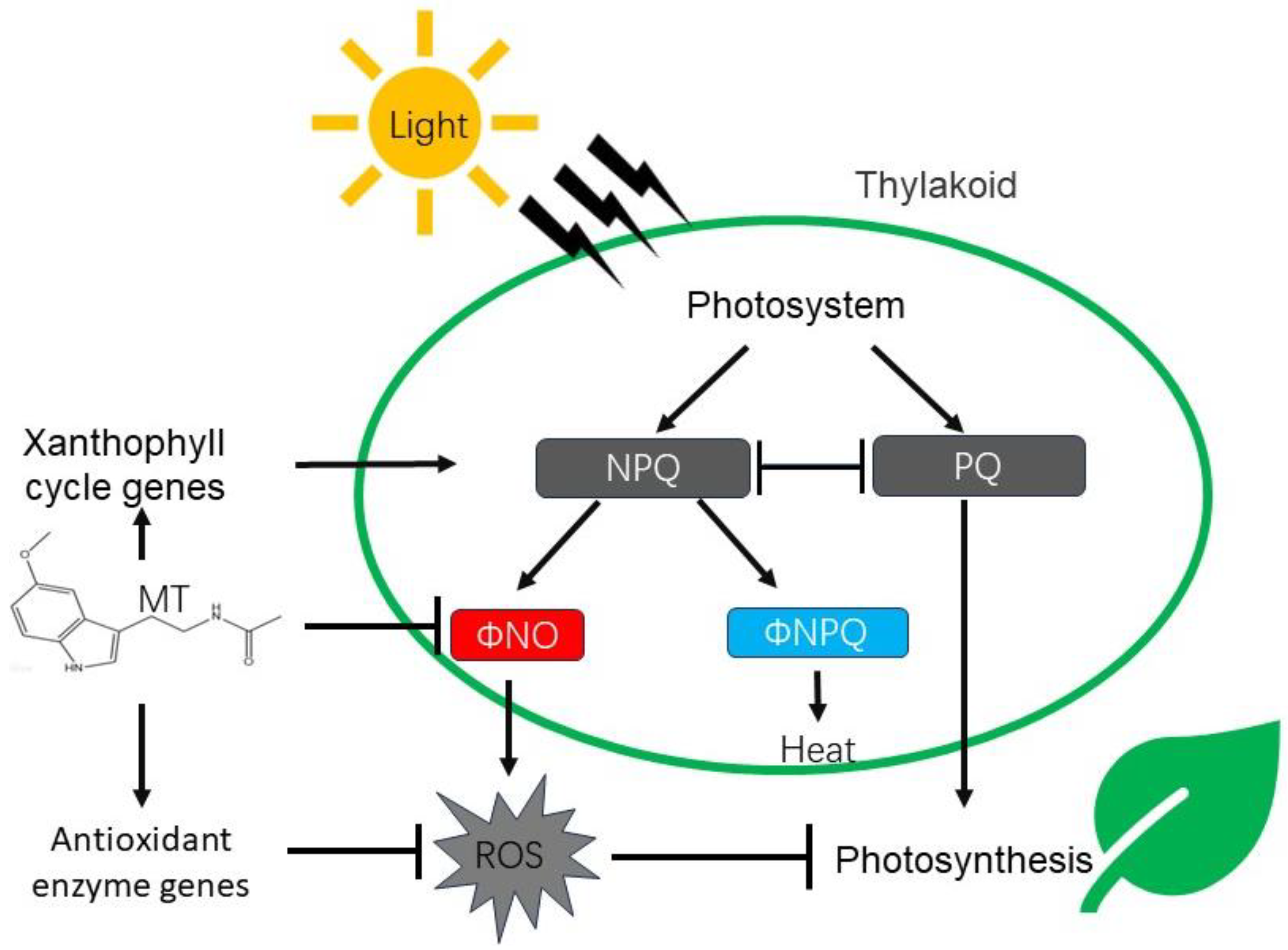
Non-photochemical quenching (NPQ) is an important component of the photosynthetic system; light energy absorbed by PSII is not only used for photochemical quenching but also contributes to non-photochemical quenching (NPQ) (Figure 7). Numerous studies have demonstrated that NPQ serves as a light protection mechanism developed by plants to counter stress conditions in light intensity [28,48,64]. Intriguingly, non-photochemical quenching (NPQ) is significantly increased by melatonin (Figure 3A). This increase could be one of the reasons for the observed decline in photosynthesis. NPQ is commonly associated with light protection mechanisms since it effectively dissipates excessive light energy [38]. Correlated research reported the function of melatonin to alleviate tomato chilling injury by accelerating NPQ [28]. Under light-deficient stress, the triggered and increased NPQ by melatonin consumes the light energy initially allocated for photosynthesis, thereby inhibiting a negative side effect.
Unlike the intriguing up-regulation of NPQ under melatonin treatment, the two components of NPQ, ΦNPQ and ΦNO, show distinct regulating ways (Figure 7). Melatonin treatment has a positive effect on ΦNPQ (Figure 4A) and a negative effect on ΦNO (Figure 4A), leading to an increasement of the ΦNPQ/ΦNO ratio approximately linearly with the melatonin concentration (Figure 4B). This suggests that melatonin plays a role in regulating energy distribution within the photosynthesis process. However, a comprehensive understanding of how melatonin impacts the ΦNPQ and ΦNO ratio requires further investigation.
To gain deeper insights into the regulating roles of melatonin, we tested the expression levels of multiple xanthophyll cycle-related genes and key synthetic genes of antioxidant enzymes. The xanthophyll cycle-related gene DHAR is induced by melatonin pre-treatment, but not by low light conditions (Figure 5). However, the other two genes, VDE and PsbS, exhibit inhomogenous induction at different concentrations of melatonin, but show significant up-regulation under low light conditions (Figure 5). These results are similar to previous studies [28], suggesting positive regulating roles on DHAR [45,46], VDE [47] and PsbS [48]. The gene expression test on the xanthophyll cycle indicates the induction activity of the xanthophyll cycle, and consequently leads to increased energy consumption (Figure 7). This might be the core reason for the NPQ increasement. Another pathway through which melatonin alleviates stress is by enhancing the antioxidant enzyme system (Figure 7). It is worth noting that the POD gene exhibits dramatic induction after melatonin treatment and is followed by significant up-regulation under low-light conditions. Quantitative gene expression data highlights the complexity of melatonin on photosynthesis, suggesting that the ultimate changes may vary from environmental conditions or treatment concentrations.
Overall, while melatonin is generally considered a positive regulator in photosynthesis and stress responses, our results showed that under a light-deficient environment, exogenous melatonin-repressed photosynthesis correlated with melatonin concentration. More research is needed to investigate the nuances of melatonin in plants to better understand its potential benefits and limitations.

Supplementary Materials

The following supporting information can be downloaded at: https://www.mdpi.com/article/10.3390/horticulturae10020118/s1, Figure S1: Detailed gas exchange relevant parameters under low light and normal light treatment; Figure S2: Detailed NPQ and qP values under low light and normal light treatment; Figure S3: Detailed ΦNPQ and ΦNO ratio values under low light and normal light treatment; Table S1: Primers used in this study.

Author Contributions

Conceptualization, K.Z. and H.Z.; methodology, Y.S. (Yunlong Shi) and K.Z.; software, Y.S. (Yahan Sun); validation, Y.S. (Yahan Sun), K.Z. and H.Z.; formal analysis, K.Z.; investigation, Y.S. (Yunlong Shi), X.F., Y.S. (Yahan Sun), Z.Y., Y.H. and D.L.; writing—original draft preparation, K.Z. and Y.S. (Yahan Sun); writing—review and editing, Z.S. and H.Z.; visualization, Y.S. (Yunlong Shi); supervision, K.Z. and H.Z.; project administration, K.Z.; funding acquisition, K.Z. and H.Z. All authors have read and agreed to the published version of the manuscript.

Funding

This research was funded by the National Natural Science Foundations of China (Grant No. 31801837), Natural Science Foundation of Shandong Province of China (ZR2019BC103) and the Cooperation Project of University and Local Enterprise in Yantai of Shandong Province (2021XDRHXMPT09). The authors acknowledge financial support from the China Scholarship Council (202008370047).

Data Availability Statement

Data are contained within the article and supplementary materials.

Conflicts of Interest

The authors declare no conflicts of interest.

References

  1. Ashraf, M.; Harris, P.J.C. Photosynthesis under Stressful Environments: An Overview. Photosynthetica 2013, 51, 163–190. [Google Scholar] [CrossRef]
  2. Long, S.P.; Zhu, X.G.; Naidu, S.L.; Ort, D.R. Can Improvement in Photosynthesis Increase Crop Yields? Plant Cell Environ. 2010, 29, 315–330. [Google Scholar] [CrossRef]
  3. Ort, D.R.; Merchant, S.S.; Alric, J.; Barkan, A.; Blankenship, R.E.; Bock, R.; Croce, R.; Hanson, M.R.; Hibberd, J.M.; Long, S.P.; et al. Bock Redesigning Photosynthesis to Sustainably Meet Global Food and Bioenergy Demand. Proc. Natl. Acad. Sci. USA 2015, 112, 8529–8536. [Google Scholar] [CrossRef] [PubMed]
  4. Erdal, S.; Turk, H. Cysteine-Induced Upregulation of Nitrogen Metabolism-Related Genes and Enzyme Activities Enhance Tolerance of Maize Seedlings to Cadmium Stress. Environ. Exp. Bot. 2016, 132, 92–99. [Google Scholar] [CrossRef]
  5. Duan, W.; Wang, Q.; Zhang, H.; Xie, B.; Li, A.; Hou, F.; Dong, S.; Wang, B.; Qin, Z.; Zhang, L. Comparative Study on Carbon–Nitrogen Metabolism and Endogenous Hormone Contents in Normal and Overgrown Sweetpotato. S. Afr. J. Bot. 2018, 115, 199–207. [Google Scholar] [CrossRef]
  6. Khan, M.N.; Zhang, J.; Luo, T.; Liu, J.; Rizwan, M.; Fahad, S.; Xu, Z.; Hu, L. Seed Priming with Melatonin Coping Drought Stress in Rapeseed by Regulating Reactive Oxygen Species Detoxification: Antioxidant Defense System, Osmotic Adjustment, Stomatal Traits and Chloroplast Ultrastructure Perseveration. Ind. Crops Prod. 2019, 140, 111597. [Google Scholar] [CrossRef]
  7. Xiong, D.; Flexas, J. From One Side to Two Sides: The Effects of Stomatal Distribution on Photosynthesis. New Phytol. 2020, 228, 1754–1766. [Google Scholar] [CrossRef]
  8. Allen, D.J.; Ort, D.R. Impacts of Chilling Temperatures on Photosynthesis in Warm-Climate Plants. Trends Plant Sci. 2001, 6, 36–42. [Google Scholar] [CrossRef] [PubMed]
  9. Apel, K. Reactive Oxygen Species: Metabolism, Oxidative Stress, and Signal Transduction. Annu. Rev. Plant Biol 2004, 55, 373–399. [Google Scholar] [CrossRef]
  10. Ding, F.; Wang, M.; Zhang, S. Overexpression of a Calvin Cycle Enzyme SBPase Improves Tolerance to Chilling-Induced Oxidative Stress in Tomato Plants. Sci. Hortic. 2017, 214, 27–33. [Google Scholar] [CrossRef]
  11. Ding, F.; Wang, M.; Zhang, S.; Ai, X. Changes in SBPase Activity Influence Photosynthetic Capacity, Growth, and Tolerance to Chilling Stress in Transgenic Tomato Plants. Sci. Rep. 2016, 6, 32741. [Google Scholar] [CrossRef]
  12. Long, S.; Liu, B.; Gong, J.; Wang, R.; Gao, S.; Zhu, T.; Guo, H.; Liu, T.; Xu, Y. 5-Aminolevulinic Acid Promotes Low-Light Tolerance by Regulating Chloroplast Ultrastructure, Photosynthesis, and Antioxidant Capacity in Tall Fescue. Plant Physiol. Biochem. 2022, 190, 248–261. [Google Scholar] [CrossRef]
  13. Brilli, F.; Pignattelli, S.; Baraldi, R.; Neri, L.; Pollastri, S.; Gonnelli, C.; Giovannelli, A.; Loreto, F.; Cocozza, C. Root Exposure to 5-Aminolevulinic Acid (ALA) Affects Leaf Element Accumulation, Isoprene Emission, Phytohormonal Balance, and Photosynthesis of Salt-Stressed Arundo Donax. Int. J. Mol. Sci. 2022, 23, 4311. [Google Scholar] [CrossRef]
  14. Li, L.; Gu, W.; Li, J.; Li, C.; Xie, T.; Qu, D.; Meng, Y.; Li, C.; Wei, S. Exogenously Applied Spermidine Alleviates Photosynthetic Inhibition under Drought Stress in Maize (Zea mays L.) Seedlings Associated with Changes in Endogenous Polyamines and Phytohormones. Plant Physiol. Biochem. 2018, 129, 35–55. [Google Scholar] [CrossRef]
  15. Jiang, D.; Lu, B.; Liu, L.; Duan, W.; Meng, Y.; Li, J.; Zhang, K.; Sun, H.; Zhang, Y.; Dong, H.; et al. Exogenous Melatonin Improves the Salt Tolerance of Cotton by Removing Active Oxygen and Protecting Photosynthetic Organs. BMC Plant Biol. 2021, 21, 1–19. [Google Scholar] [CrossRef]
  16. Yan, F.; Zhang, J.; Li, W.; Ding, Y.; Zhong, Q.; Xu, X.; Wei, H.; Li, G. Exogenous Melatonin Alleviates Salt Stress by Improving Leaf Photosynthesis in Rice Seedlings. Plant Physiol. Biochem. 2021, 163, 367–375. [Google Scholar] [CrossRef] [PubMed]
  17. Hasan, M.K.; Xing, Q.-F.; Zhou, C.-Y.; Wang, K.-X.; Xu, T.; Yang, P.; Qi, Z.-Y.; Shao, S.-J.; Ahammed, G.J.; Zhou, J. Melatonin Mediates Elevated Carbon Dioxide-Induced Photosynthesis and Thermotolerance in Tomato. J. Pineal Res. 2023, 74, e12858. [Google Scholar] [CrossRef] [PubMed]
  18. Murch, S.J.; Saxena, P.K. Mammalian Neurohormones: Potential Significance in Reproductive Physiology of St. John’s Wort (Hypericum perforatum L.)? Naturwissenschaften 2002, 89, 555–560. [Google Scholar] [CrossRef]
  19. Pelagio-Flores, R.; Muñoz-Parra, E.; Ortiz-Castro, R.; López-Bucio, J. Melatonin Regulates Arabidopsis Root System Architecture Likely Acting Independently of Auxin Signaling. J. Pineal Res. 2012, 53, 279–288. [Google Scholar] [CrossRef] [PubMed]
  20. Reiter, R.J.; Tan, D.X.; Galano, A. Melatonin: Exceeding Expectations. Physiology 2014, 29, 325–333. [Google Scholar] [CrossRef] [PubMed]
  21. Lerner, A.B.; Case, J.D.; Takahashi, Y.; Lee, T.H.; Mori, W. ISOLATION OF MELATONIN, THE PINEAL GLAND FACTOR THAT LIGHTENS MELANOCYTES1. J. Am. Chem. Soc. 1958, 80, 2587. [Google Scholar] [CrossRef]
  22. Vantassel, D.; Li, J.; Oneill, S. Melatonin-Identification of a Potential Dark Signal in Plants. Plant Physiol. 1993, 102, 117. [Google Scholar]
  23. Katarzyna, S.; Reiter, R.J.; Posmyk, M.M. Melatonin Improves the Photosynthetic Apparatus in Pea Leaves Stressed by Paraquat via Chlorophyll Breakdown Regulation and Its Accelerated de Novo Synthesis. Front. Plant Sci. 2017, 8, 878. [Google Scholar]
  24. Shah, M.; Guo, S.; Baloch, A.R.; Sun, J.; Kabir, K. Melatonin Alleviates Nickel Phytotoxicity by Improving Photosynthesis, Secondary Metabolism and Oxidative Stress Tolerance in Tomato Seedlings. Ecotoxicol. Environ. Saf. 2020, 197, 110593. [Google Scholar]
  25. Ayyaz, A.; Amir, M.; Umer, S.; Iqbal, M.; Bano, H.; Gul, H.S.; Noor, Y.; Kanwal, A.; Khalid, A.; Javed, M.; et al. Melatonin Induced Changes in Photosynthetic Efficiency as Probed by OJIP Associated with Improved Chromium Stress Tolerance in Canola (Brassica napus L.). Heliyon 2020, 6, e04364. [Google Scholar] [CrossRef] [PubMed]
  26. Can, H. Melatonin Application at Different Doses Changes the Physiological Responses in Favor of Cabbage Seedlings (Brassica oleracea var. Capitata) Against Flooding Stress. Gesunde Pflanz. 2023, 75, 2733–2745. [Google Scholar] [CrossRef]
  27. Kurt-Celebi, A.; Colak, N.; Torun, H.; Dosedělová, V.; Tarkowski, P.; Ayaz, F.A. Exogenous Melatonin Ameliorates Ionizing Radiation-Induced Damage by Modulating Growth, Osmotic Adjustment and Photosynthetic Capacity in Wheat Seedlings. Plant Physiol. Biochem. 2022, 187, 67–76. [Google Scholar] [CrossRef] [PubMed]
  28. Ding, F.; Wang, M.; Liu, B.; Zhang, S. Exogenous Melatonin Mitigates Photoinhibition by Accelerating Non-Photochemical Quenching in Tomato Seedlings Exposed to Moderate Light during Chilling. Front. Plant Sci. 2017, 8, 244. [Google Scholar] [CrossRef]
  29. Zhao, H.; Su, T.; Huo, L.; Wei, H.; Jiang, Y.; Xu, L.; Ma, F. Unveiling the Mechanism of Melatonin Impacts on Maize Seedling Growth: Sugar Metabolism as a Case. J. Pineal Res. 2015, 59, 255–266. [Google Scholar] [CrossRef]
  30. Yang, J.; Zhang, C.; Wang, Z.; Sun, S.; Zhan, R.; Zhao, Y.; Ma, B.; Ma, F.; Li, M. Melatonin-Mediated Sugar Accumulation and Growth Inhibition in Apple Plants Involves Down-Regulation of Fructokinase 2 Expression and Activity. Front. Plant Sci. 2019, 10, 150. [Google Scholar] [CrossRef]
  31. Lee, H.Y.; Back, K. Melatonin Induction and Its Role in High Light Stress Tolerance in Arabidopsis Thaliana. J. Pineal Res. 2018, 65, e12504. [Google Scholar] [CrossRef]
  32. Yang, S.-J.; Huang, B.; Zhao, Y.-Q.; Hu, D.; Chen, T.; Ding, C.-B.; Chen, Y.-E.; Yuan, S.; Yuan, M. Melatonin Enhanced the Tolerance of Arabidopsis Thaliana to High Light Through Improving Anti-Oxidative System and Photosynthesis. Front. Plant Sci. 2021, 12, 752584. [Google Scholar] [CrossRef]
  33. Manafi, H.; Baninasab, B.; Gholami, M.; Talebi, M.; Khanizadeh, S. Exogenous Melatonin Alleviates Heat-Induced Oxidative Damage in Strawberry (Fragaria × ananassa Duch. cv. Ventana) Plant. J. Plant Growth Regul. 2022, 41, 52–64. [Google Scholar] [CrossRef]
  34. Zhou, X.; Zhao, H.; Cao, K.; Hu, L.; Du, T.; Baluška, F.; Zou, Z. Beneficial Roles of Melatonin on Redox Regulation of Photosynthetic Electron Transport and Synthesis of D1 Protein in Tomato Seedlings under Salt Stress. Front. Plant Sci. 2016, 7, 1823. [Google Scholar] [CrossRef]
  35. Choi, H.G. Correlation among Phenotypic Parameters Related to the Growth and Photosynthesis of Strawberry (Fragaria × ananassa Duch.) Grown under Various Light Intensity Conditions. Front. Plant Sci. 2021, 12, 647585. [Google Scholar] [CrossRef]
  36. Kouřil, R.; Wientjes, E.; Bultema, J.B.; Croce, R.; Boekema, E.J. High-Light vs. Low-Light: Effect of Light Acclimation on Photosystem II Composition and Organization in Arabidopsis Thaliana. Biochim. Biophys. Acta (BBA)-Bioenerg. 2013, 1827, 411–419. [Google Scholar] [CrossRef]
  37. Cheng, J.-S.; Duan, W.; Tang, X.-L.; Zhang, Y.-G.; Li, B.; Wang, Y.-J.; Yang, C.-X.; Song, Z.-Z.; Wang, L.-J.; Yang, J.; et al. Low Sink Demand Caused Net Photosynthetic Rate Decrease Is Closely Related to the Irrecoverable Damage of Oxygen-Releasing Complex and Electron Receptor in Peach Trees. J. Plant Physiol. 2021, 266, 153510. [Google Scholar] [CrossRef]
  38. Kramer, D.M.; Johnson, G.; Kiirats, O.; Edwards, G.E. New Fluorescence Parameters for the Determination of Q A Redox State and Excitation Energy Fluxes. Photosynth. Res. 2004, 79, 209–218. [Google Scholar] [CrossRef]
  39. Tietz, S.; Hall, C.C.; Cruz, J.A.; Kramer, D.M. NPQ (T): A Chlorophyll Fluorescence Parameter for Rapid Estimation and Imaging of Non-Photochemical Quenching of Excitons in Photosystem-II-Associated Antenna Complexes: New, Rapid Probe of Non-Photochemical Quenching. Plant Cell Environ. 2017, 40, 1243–1255. [Google Scholar] [CrossRef]
  40. Demmig-Adams, B.; Adams, W.W. Photoprotection and Other Responses of Plants to High Light Stress. Ann. Rev. Plant Physiol. Plant Mol. Biol 1992, 43, 599–626. [Google Scholar] [CrossRef]
  41. Müller, P.; Li, X.P.; Niyogi, K.K. Non-Photochemical Quenching. A Response to Excess Light Energy. Plant Physiol. 2001, 125, 1558–1566. [Google Scholar] [CrossRef]
  42. Crofts, A.R.; Yerkes, C.T. A Molecular Mechanism for qE-Quenching. Febs Lett. 1994, 352, 265–270. [Google Scholar] [CrossRef]
  43. Demmig-Adams, B.; Adams III, W.W. Photoprotection in an Ecological Context: The Remarkable Complexity of Thermal Energy Dissipation. New Phytol. 2006, 172, 11–21. [Google Scholar] [CrossRef]
  44. Takahashi, S.; Badger, M.R. Photoprotection in Plants: A New Light on Photosystem II Damage. Trends Plant Sci. 2011, 16, 53–60. [Google Scholar] [CrossRef]
  45. Smirnoff, N. Ascorbate Biosynthesis and Function in Photoprotection. Philos. Trans. R. Soc. London. Ser. B Biol. Sci. 2000, 355, 1455–1464. [Google Scholar] [CrossRef]
  46. Smirnoff, N. Ascorbic Acid: Metabolism and Functions of a Multi-Facetted Molecule. Curr. Opin. Plant Biol. 2000, 3, 229–235. [Google Scholar] [CrossRef]
  47. Hager, A.; Holocher, K. Localization of the Xanthophyll-Cycle Enzyme Violaxanthin de-Epoxidase within the Thylakoid Lumen and Abolition of Its Mobility by a (Light-Dependent) pH Decrease. Planta 1994, 192, 581–589. [Google Scholar] [CrossRef]
  48. Li, X.-P.; Müller-Moulé, P.; Gilmore, A.M.; Niyogi, K.K. PsbS-Dependent Enhancement of Feedback de-Excitation Protects Photosystem II from Photoinhibition. Proc. Natl. Acad. Sci. USA 2002, 99, 15222–15227. [Google Scholar] [CrossRef]
  49. Wu, X.; Ren, J.; Huang, X.; Zheng, X.; Tian, Y.; Shi, L.; Dong, P.; Li, Z. Melatonin: Biosynthesis, Content, and Function in Horticultural Plants and Potential Application. Sci. Hortic. 2021, 288, 110392. [Google Scholar] [CrossRef]
  50. Li, M.; Li, X.; Li, J.; Ji, Y.; Han, C.; Jin, P.; Zheng, Y. Responses of Fresh-Cut Strawberries to Ethanol Vapor Pretreatment: Improved Quality Maintenance and Associated Antioxidant Metabolism in Gene Expression and Enzyme Activity Levels. J. Agric. Food Chem. 2018, 66, 8382–8390. [Google Scholar] [CrossRef]
  51. Messant, M.; Krieger-Liszkay, A.; Shimakawa, G. Dynamic Changes in Protein-Membrane Association for Regulating Photosynthetic Electron Transport. Cells 2021, 10, 1216. [Google Scholar] [CrossRef]
  52. Allan, A.C.; Zuo, B.; Zhao, Y.; Reiter, R.J.; Wang, L.; Wang, Z.; Guo, Y.; Zhou, J.; Shan, D.; Li, Q.; et al. Chloroplastic Biosynthesis of Melatonin and Its Involvement in Protection of Plants from Salt Stress. Sci. Rep. 2017, 7, 41236. [Google Scholar] [CrossRef]
  53. Han, Q.-H.; Huang, B.; Ding, C.-B.; Zhang, Z.-W.; Chen, Y.-E.; Hu, C.; Zhou, L.-J.; Huang, Y.; Liao, J.-Q.; Yuan, S.; et al. Effects of Melatonin on Anti-Oxidative Systems and Photosystem II in Cold-Stressed Rice Seedlings. Front. Plant Sci. 2017, 8, 785. [Google Scholar] [CrossRef]
  54. Ding, F.; Wang, G.; Zhang, S. Exogenous Melatonin Mitigates Methyl Viologen-Triggered Oxidative Stress in Poplar Leaf. Molecules 2018, 23, 2852. [Google Scholar] [CrossRef]
  55. Cao, Y.-Y.; Qi, C.-D.; Li, S.; Wang, Z.; Wang, X.; Wang, J.; Ren, S.; Li, X.; Zhang, N.; Guo, Y.-D. Melatonin Alleviates Copper Toxicity via Improving Copper Sequestration and ROS Scavenging in Cucumber. Plant Cell Physiol. 2019, 60, 562–574. [Google Scholar] [CrossRef]
  56. Park, H.-S.; Kazerooni, E.A.; Kang, S.-M.; Al-Sadi, A.M.; Lee, I.-J. Melatonin Enhances the Tolerance and Recovery Mechanisms in Brassica Juncea (L.) Czern. Under Saline Conditions. Front. Plant Sci. 2021, 12, 593717. [Google Scholar] [CrossRef]
  57. Cui, G.; Sun, F.; Gao, X.; Xie, K.; Zhang, C.; Liu, S.; Xi, Y. Proteomic Analysis of Melatonin-Mediated Osmotic Tolerance by Improving Energy Metabolism and Autophagy in Wheat (Triticum aestivum L.). Planta 2018, 248, 69–87. [Google Scholar] [CrossRef]
  58. Li, H.; Chang, J.; Zheng, J.; Dong, Y.; Liu, Q.; Yang, X.; Wei, C.; Zhang, Y.; Ma, J.; Zhang, X. Local Melatonin Application Induces Cold Tolerance in Distant Organs of Citrullus lanatus L. via Long Distance Transport. Sci. Rep. 2017, 7, 40858. [Google Scholar] [CrossRef]
  59. Li, C.; Tan, D.-X.; Liang, D.; Chang, C.; Jia, D.; Ma, F. Melatonin Mediates the Regulation of ABA Metabolism, Free-Radical Scavenging, and Stomatal Behaviour in Two Malus Species under Drought Stress. J. Exp. Bot. 2015, 66, 669–680. [Google Scholar] [CrossRef]
  60. Sharma, A.; Kumar, V.; Shahzad, B.; Ramakrishnan, M.; Singh Sidhu, G.P.; Bali, A.S.; Handa, N.; Kapoor, D.; Yadav, P.; Khanna, K.; et al. Photosynthetic Response of Plants under Different Abiotic Stresses: A Review. J. Plant Growth Regul. 2020, 39, 509–531. [Google Scholar] [CrossRef]
  61. Jahan, M.S.; Guo, S.; Sun, J.; Shu, S.; Wang, Y.; El-Yazied, A.A.; Alabdallah, N.M.; Hikal, M.; Mohamed, M.H.M.; Ibrahim, M.F.M.; et al. Melatonin-Mediated Photosynthetic Performance of Tomato Seedlings under High-Temperature Stress. Plant Physiol. Biochem. 2021, 167, 309–320. [Google Scholar] [CrossRef] [PubMed]
  62. Zahedi, S.M.; Hosseini, M.S.; Abadía, J.; Marjani, M. Melatonin Foliar Sprays Elicit Salinity Stress Tolerance and Enhance Fruit Yield and Quality in Strawberry (Fragaria × ananassa Duch.). Plant Physiol. Biochem. 2020, 149, 313–323. [Google Scholar] [CrossRef]
  63. Maxwell, K.; Johnson, G.N. Chlorophyll Fluorescence—A Practical Guide. J. Exp. Bot. 2000, 51, 659–668. [Google Scholar] [CrossRef] [PubMed]
  64. Mallick, N.; Mohn, F.H. Use of Chlorophyll Fluorescence in Metal-Stress Research: A Case Study with the Green Microalga Scenedesmus. Ecotoxicol. Environ. Saf. 2003, 55, 64–69. [Google Scholar] [CrossRef] [PubMed]
Figure 1. Gas exchange relevant parameters under low light and normal light treatment. (A) Net photosynthetic rate; (B) transpiration rate; (C) stomatal conductance; and (D) intercellular CO2 concentration. NL (blank column) represents normal light treatment and LL (dark column) represents low light treatment. The values 0, 50, 100, and 500 µM represent the four independent concentrations of melatonin treatment. Data was collected at 0, 6, 12, and 24 h post treatment. And the value represents the average of the four time points. Error bars represent the standard error of the mean and letters indicate significant differences calculated by Tukey’s test (p < 0.05).
Horticulturae 10 00118 g001
Figure 2. Correlation analysis of multiple chlorophyll fluorescence parameters. Independent t-test was used to determine statistical differences. ***, **, and * represent significant correlations of p < 0.001, p < 0.01 and p < 0.05, respectively.
Horticulturae 10 00118 g002
Figure 3. Evaluation of photochemical efficiency on melatonin treatments under normal light and low light environments. NL (blank column) represents normal light treatment and LL (dark column) represents low light treatment. NPQ (A) and qP (B) were calculated from chlorophyll fluorescence parameters and indicated as an absolute value. The values 0, 50, 100, and 500 µM represent the four independent concentrations of melatonin treatment. Data was collected at 0, 6, 12, and 24 h post treatment. And the value represents the average of the four time points. Error bars represent the standard error of the mean and letters indicate significant differences calculated by Tukey’s test (p < 0.05).
Horticulturae 10 00118 g003
Figure 4. Evaluation of non-photochemical quenching on melatonin treatments under normal light and low light environments. NL represents normal light treatment (A) and LL represents low light treatment (B). Data shows the ratio value of ΦNPQ (solid line) and ΦNO (dashed line) to NPQ. The values 0, 50, 100, and 500 µM represent the four independent concentrations of melatonin treatment. Data was collected at 0, 6, 12, and 24 h post treatment. And the value represents the average of the four time points. Error bars represent the standard error of the mean and letters indicate significant differences calculated by Tukey’s test (p < 0.05).
Horticulturae 10 00118 g004
Figure 5. Relative expression levels of xanthophyll cycle genes under different concentrations of melatonin treatments. The value indicates the fold change of experimental treatments relative to control samples. GADPH2 gene was used as internal control. The values 0, 50, 100, and 500 µM represent the four independent concentrations of melatonin treatment. Error bars represent the standard error of the mean and letters indicate significant differences calculated by Tukey’s test (p < 0.05).
Horticulturae 10 00118 g005
Figure 6. Relative expression levels of selected antioxidant enzyme genes under different concentrations of melatonin treatments. The value indicates fold-change of experimental treatments relative to control samples. GADPH2 gene was used as internal control. The values 0, 50, 100, and 500 µM represent the four independent concentrations of melatonin treatment. Error bars represent the standard error of the mean and letters indicate significant differences calculated by Tukey’s test (p < 0.05).
Horticulturae 10 00118 g006
Figure 7. Diagram of melatonin in mediating photosynthesis, energy metabolism and activating antioxidant enzyme genes. The arrows indicate promotion and the inhibitory arrow indicates suppression. NPQ, non-photochemical quenching; PQ, photochemical quenching; and ROS, reactive oxygen species.
Horticulturae 10 00118 g007
Disclaimer/Publisher’s Note: The statements, opinions and data contained in all publications are solely those of the individual author(s) and contributor(s) and not of MDPI and/or the editor(s). MDPI and/or the editor(s) disclaim responsibility for any injury to people or property resulting from any ideas, methods, instructions or products referred to in the content.