Article

Identification of Laccase Genes in Grapevine and Their Roles in Response to Botrytis cinerea

1
College of Horticulture, Henan Agricultural University, Zhengzhou 450002, China
2
Henan Key Laboratory of Fruit and Cucurbit Biology, College of Horticulture, Henan Agricultural University, Zhengzhou 450002, China
*
Authors to whom correspondence should be addressed.
These authors contributed equally to this work.
Horticulturae 2024, 10(4), 376; https://doi.org/10.3390/horticulturae10040376
Submission received: 24 February 2024 / Revised: 24 March 2024 / Accepted: 3 April 2024 / Published: 9 April 2024
(This article belongs to the Special Issue Advances in Developmental Biology in Tree Fruit and Nut Crops)

Abstract

:
Laccases are the key enzymes responsible for plant lignin biosynthesis and responses to environment stress. However, the roles of LAC genes in plant disease resistance are still largely unknown, especially in grapevine, one of the most important horticultural crops in the world. Its quality and yield are very vulnerable to gray mold disease caused by Botrytis cinerea. In total, 30 VvLAC genes were identified and found to be unevenly distributed on seven chromosomes; they were classified into seven groups based on phylogenetic analysis according to the criteria applied in Arabidopsis thaliana. Collinearity and synteny analyses identified some orthologous gene pairs in Vitis vinifera and a few paralogous gene pairs among grape and peach. The VvLAC gene family has diverse gene structures and a highly conserved motif composition. The prominent presence of the MYB cis-elements in each VvLAC promoter highlighted MYB transcriptional factors as the main regulators of VvLAC genes. Furthermore, twenty-five VvLAC genes with functional redundancy are probably implicated in grape lignin biosynthesis. The expression patterns of the LAC genes in grape leaves of Chinese wild V. amurensis ‘Shuangyou’ (SY), a germplasm highly resistant to B. cinerea, were investigated through transcriptomic data and qRT-PCR verification. Combined with the phylogenetic analysis, with AtLACs participating in lignin metabolism, and the cis-element analysis, VaLAC14, VaLAC19, VaLAC24 and VaLAC30 were identified as key candidate genes for lignin biosynthesis in the grape response to B. cinerea. This study supplies a comprehensive understanding of the classification, evolution, structure and responses of the grape LAC genes against B. cinerea. It also provides valuable genetic resources for functional characterization towards enhancing grapevine disease resistance.

1. Introduction

Grape is an important fruit crop worldwide with huge economic value. With the increase in grape planting years, the base number of pathogenic seedlings and insect population is gradually increasing [1]. The occurrence of Botrytis cinerea on grape, the ubiquitous necrotrophic fungal pathogen, is becoming more and more serious, resulting in a prominent problem of yield and quality decline [1,2]. B. cinerea causes serious damage to young leaves, young tendril, flower buds and ripened fruit of grape; it is generally called gray mold and is the second most important plant disease in the world [1,2,3,4].
B. cinerea has a very powerful ‘arsenal’ for causing disease in more than 1000 plant species which is especially characterized by massive secretion of cell wall degrading enzymes, such as BcPGs (endo-polygalacturonase), BcPMEs (pectin methylesterase), BcCBHs (cellobiohydrolase) and so on, as one of the primary infection strategies [5,6,7]. The cell wall matrix is one of the first and largest plant structures that pathogens encounter when interacting with potential hosts [8]. Lignin is one of the main components of the plant cell wall, and as a complex phenolic polymer produced by the phenylalanine/tyrosine metabolic pathway, it plays important roles in plant defense by enhancing plant cell wall rigidity, protecting cell wall integrity, acting as phytoalexins and acting as a physical barrier [9,10,11]. Previous studies have revealed that the cell wall strengthening through lignin accumulation mediated by the ethylene- and abscisic acid-associated pathways is significantly involved in the plant response to B. cinerea [12,13]. Lignin biosynthesis was also observed in grapevine defense against trunk diseases, downy mildew, Pierce’s disease and postharvest infections [14,15,16]. Particularly, grapevine resistance to B. cinerea is also suggested to be associated with lignin metabolism. Higher lignin content was detected in Greek grape berries of the highly resistant ‘Limnio’ variety than in those of the susceptible ‘Roditis’ variety [17]. The genes associated with grapevine lignin resistant against B. cinerea remain unexplored.
In recent years, laccases (LACs), as one of the classes of key enzymes, have been found to have the important function of polymerizing lignin monomers into lignin in the cell wall [9,18]. A total of 17 laccase genes have been identified in Arabidopsis thaliana, and among them, AtLAC4, AtLAC17, AtLAC2 and AtLAC15 have been shown to participate in lignin polymerization in leaves, seed coat and roots [19,20,21,22,23]. The expression of AtLAC4 and AtLAC17 contributes to constitutive lignification in stems and to lignin monomer deposition in fibers; the lac4 lac17 mutant increases their monolignol glucosides and display dwarfism, while lignin deposition in the roots is almost completely abolished in the lac11 lac4 lac17 triple mutant [19,20,21,22,23]. For other species, Miscanthus MsLAC1 and Pyrus bretschneideri PbrLAC1, PbrLAC2 and PbrLAC18, as well as Populus tomentosa PtoLAC14 and Cleome hassleriana ChLAC8, were all reported to be significantly involved in lignin polymerization in various tissue types [24,25,26,27].
Laccases associated with lignin biosynthesis have been a prominent research focus in recent years for their significant disease resistance function. Among the 84 laccase genes in cotton (Gossypium hirsutum), GhLAC1, GhLAC4 and GhLAC15 are all implicated in disease resistance to verticillium wilt caused by Verticillium dahliae by enhancing G-lignin biosynthesis and lignification in the cell wall [28,29,30,31]. Laccase-induced lignification is suggested to be one of the major processes associated with the defense response in apple plant diseases, in which both MhLAC7 and MdLAC7 have an important function [32,33,34]. Tobacco overexpressing EuLAC1 from Eucommia ulmoides Oliver. displayed significantly higher laccase activity and resistance to gray mold compared with wild-type tobacco [35]. However, the implication of laccase genes in grapevine lignin associated with disease resistance is little understood. Here, we characterized the laccase gene family in grapevine through bio-information analysis and analyzed the potential roles of these genes in the defense response based on the expression profile of grape leaves under infection by B. cinerea. The results highlight four important candidate genes, VaLAC14, VaLAC19, VaLAC24 and VaLAC30, associated with lignin biosynthesis in the response against B. cinerea. Our work would strengthen the understanding of grape laccase genes and provide an important basis for further studying their implication in disease resistance mechanisms.

2. Materials and Methods

2.1. Plant and Fungus Materials

The leaves of Chinese wild grape (V. amurensis Rupr.) ‘Shuangyou’ (SY), a germplasm highly resistant to B. cinerea [36], were used as experimental plant materials. They were planted in a plant incubator at 23 °C and 75% HR (relative humidity) under a cycle of 16 h light/8 h darkness. The B. cinerea fungus was isolated and purified from infected grape berries. Conidia suspension, inoculation and detached leaf assay were all performed according to the description by Wan et al. [36]. The experiment was repeated three times, and 8–10 leaves were collected repeatedly in each experiment. All treated and control (inoculated with sterile water) leaves were collected at 4, 8, 18, 36 and 72 h post-inoculation (hpi). All collected samples were immediately frozen in liquid nitrogen and stored at −80 °C for further use.

2.2. Identification of the VvLAC Gene Family

The sequence distributions of V. vinifera L. and Arabidopsis thaliana were downloaded from Ensembl Plants (http://plants.ensembl.org/index.html, accessed on 18 November 2023) and the TAIR database (http://www.arabidopsis.org/, accessed on 18 November 2023). Then, according to the conserved domain protein sequences retrieved from the Pfam database (http://pfam.sanger.ac.uk/, accessed on 18 November 2023), HMMER and BLASTP tools were used to screen the grape candidate LAC genes. The NCBI CDD database (Conserved Domain Database; http://www.ncbi.nlm.nih.gov//Structure/bwrpsb/bwrpsb.cgi, accessed on 18 November 2023) was used to analyze and identify the conserved domains of candidate VvLAC genes. The chromosome location information of the identified VvLAC genes was mapped by genome annotation files, and all the identified VvLAC genes were named according to the chromosome (Chr) locations. For further exploring the protein characteristics, the ExPASy protein online tool (https://web.expasy.org/compute_pi/, accessed on 22 November 2023) was used to predict the isoelectric point (pI) and molecular weight (MW) of the VvLAC proteins. WoLF PSORT (https://www.genscript.com/wolf-psort.html, accessed on 22 November 2023) was used to predict the subcellular localizations of VvLACs.

2.3. Chromosomal Location, Phylogenetic Analysis and Collinearity Analysis

MEGA software (version 10.0; Mega Limited, Auckland, New Zealand) was used to construct a phylogenetic tree with the maximum likelihood (ML) method, and the bootstrap test was repeated 1000 times [37]. Finally, the tree was annotated and decorated by ITOL online tools (http://itol.embl.de/, accessed on 10 December 2023) [38]. The phylogenetic tree was constructed based on the full-length protein sequences of LACs in Arabidopsis thaliana [39], Populus trichocarpa [40], Citrus reticulata Blanco [41] and V. vinifera. TBtools software (SCAU; version 1.0687) was used to visualize the position of grape chromosomes. The Multiple Collinearity Scan tool kit with default parameters in TBtools (version 1.0687; SCAU, Guangzhou, Guangdong, China) was used to analyze the collinearity between grape and the following herbaceous plants: Glycine max, Oryza sativa Japonica, Gossypium raimondii and Arabidopsis thaliana, and ligneous plants, Populus trichocarpa, Malus domestica Golden and Prunus persica.

2.4. Gene Structures, Conserved Motifs and Cis-Elements

The exon–intron structures of the VvLAC genes were visualized by using TBtools software (version 1.0687; SCAU, Guangzhou, Guangdong, China) by referring to the downloaded genomic information. The Multiple Em for Motif Elicitation (MEME; https://meme-suite.org/meme/tools/meme, accessed on 13 December 2023) online tool was used to analyze the conserved motifs in the VvLAC genes. The NCBI CDD database (Conserved Domain Database; https://www.ncbi.nlm.nih.gov/Structure/bwrpsb/bwrpsb.cgi, accessed on 13 December 2023) was used to analyze and identify the conserved domains of VvLACs. To analyze the cis-acting elements of VvLACs, we intercepted a sequence of 2000 bp upstream of the start codon of each VvLAC and then used the PlantCARE (https://bioinformatics.psb.ugent.be/webtools/plantcare/html/, accessed on 20 December 2023) online tool [42] to predict and analyze it; finally, we visualized it with TBtools (version 1.0687; SCAU, Guangzhou, Guangdong, China).

2.5. Protein Interaction Network Analysis

The homologous proteins of the AtLAC genes in Arabidopsis thaliana were obtained by the STRING online tool (http://string-db.org, accessed on 22 January 2024), and protein interaction network (PPI) prediction analysis was carried out. The results were visualized by Cytoscape software (version 3.10.1; USA).

2.6. Expression Profile of Grape LAC Genes in SY Leaves against B. cinerea

RNA-seq data about the response of VaLACs in SY leaves against B. cinerea were derived from our previously published study [36].

2.7. RNA Extraction and qRT-PCR Analysis

The quantitative primers of the candidate genes were designed by Primer Premier 5.0 software (PREMIER Biosoft, San Francisco, CA, USA). The specificity of the primers was determined through NCBI (Table S1), and they were synthesized by Bioengineering Co., Ltd. (Shanghai, China). The RNA extraction kit E.Z.N.A.® Plant RNA Kit # R6827-0 (Omega Bio-tek, Dallas, TX, USA) was used to extract total RNA from SY leaves in the indicated infection stages. The NanoDrop 2000 instrument (Thermo Fisher Scientific, Shanghai, China) was used to check the purity and concentration of RNA, and 1% agarose gel electrophoresis was used to determine its quality. Then, according to the reverse transcription kit HiScript III qRT Super Mix for q PCR (+gDNA wiper) (Vazyme Biotech Co., Ltd., Nanjing, Jiangsu, China), the cDNAs of the corresponding samples were obtained by reverse transcription. Finally, the fluorescence quantitative PCR reaction system was configured according to the fluorescence quantitative kit ChamQ Universal SYBR qPCR Master Mix (Vazyme Biotech Co., Ltd., Nanjing, China), and qRT-PCR was performed on the CFX96 real-time system (Bio-Rad, Hercules, CA, USA). The reaction procedure was performed at 95 °C for 5 min, followed by 40 cycles at 95 °C for 15 s, 60 °C for 1 min and 72 °C for 5 min. The relative expression level of each gene was calculated by using the 2−ΔΔt method.

2.8. Determination of Laccase Activity and Lignin Content

Each leaf sample collected at the indicated time points was dried at 80 °C to constant weight, ground to powder, and sieved with a 0.425 mm aperture sieve. Then, 2 mg samples were evaluated in terms of lignin content (expressed as mg g−1) by using the MZS-1-G Kit (Suzhou Comin Biotechnology Co. Ltd., Suzhou, Jiangsu, China) [43]. Then, homogenates made with 0.1 g samples with 1 mL of extracting solution in an ice bath were centrifuged at 1200 g at 4 °C for 30 min to obtain supernatant liquid. Afterwards, 45 μL of supernatant liquid was used to determine the laccase activity of each sample (expressed as nmol min−1 g−1) by using the QM-1-G Kit (Suzhou Comin Biotechnology Co., Ltd., Suzhou, Jiangsu, China). The determination was repeated three times, and the results were expressed as means ± SEs.

2.9. Data Analysis

The data were analyzed by using SPSS version 20 (IBM SPSS Inc., Chicago, IL, USA). Statistical significance was analyzed by Duncan’s test. Different lowercase letters indicated that the difference among treatments reached a significant level (p ≤ 0.05).

3. Results

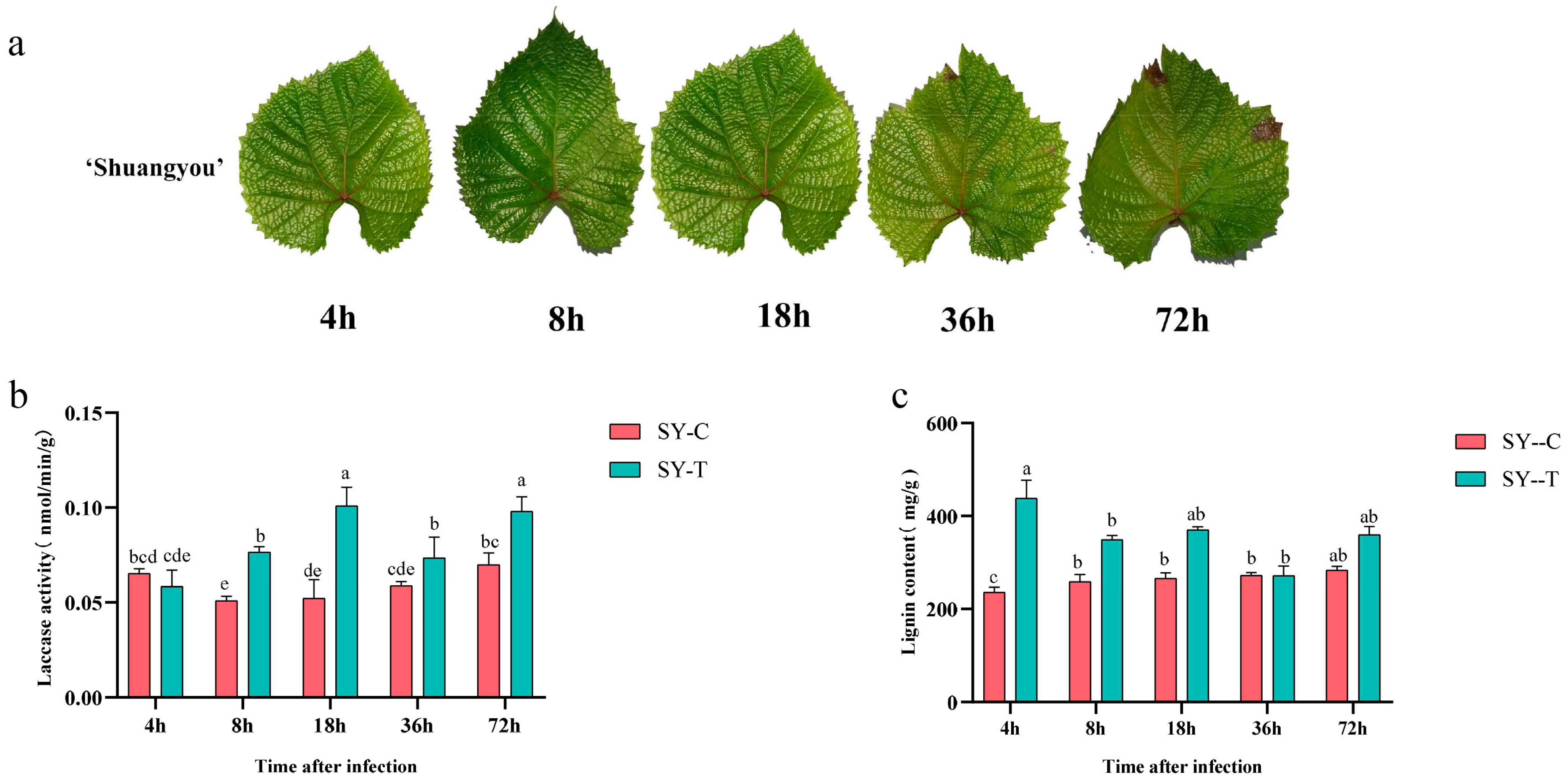
3.1. Laccase Activity and Lignin Content in SY Leaves in Response to B. cinerea

The phenotypic characteristics of grape leaves inoculated with B. cinerea conidia 4–72 hpi verified that SY was resistant against the pathogen (Figure 1a). In control samples, laccase activity and lignin biosynthesis almost showed no significant change. After infection, laccase activity showed a significant increase 18 hpi and was significant higher in the infected SY leaves than in the control samples from 8 hpi onwards. Lignin content in the B. cinerea-infected SY leaves was increased 4 hpi and was also significantly higher than in the control samples from then on, except at 36 hpi (Figure 1b,c).

3.2. Identification of VvLAC Genes in Grapevine

A total of 30 members of the VvLAC gene family were retrieved from the ‘Pinot Noir’ Vitis genome (PN40024.v4) and were renamed from VvLAC1 to VvLAC30 according to their positions on the Chrs (Table 1). The length of their encoding proteins ranged from 386 to 627 amino acids, and the molecular weights of the proteins ranged from 42.46 to 69.76 KDa (Table 1). The theoretical isoelectric points of these proteins ranged from 4.79 to 9.79, with VvLAC28 showing the lowest value and VvLAC11 the highest one; they comprised 24 basic proteins and 6 acidic proteins distributed on Chr 18, except for VvLAC14, which was found on Chr 8 (Table 1). Moreover, the subcellular localization displayed that 17 VvLAC proteins were located on chloroplasts, followed by the vacuolar membrane and extracellular matrix, each with 4 VvLAC proteins; the cytoskeleton, with 3 proteins; and the plasma membrane and peroxisome, with the lowest number of protein, with 1 VvLAC protein each.

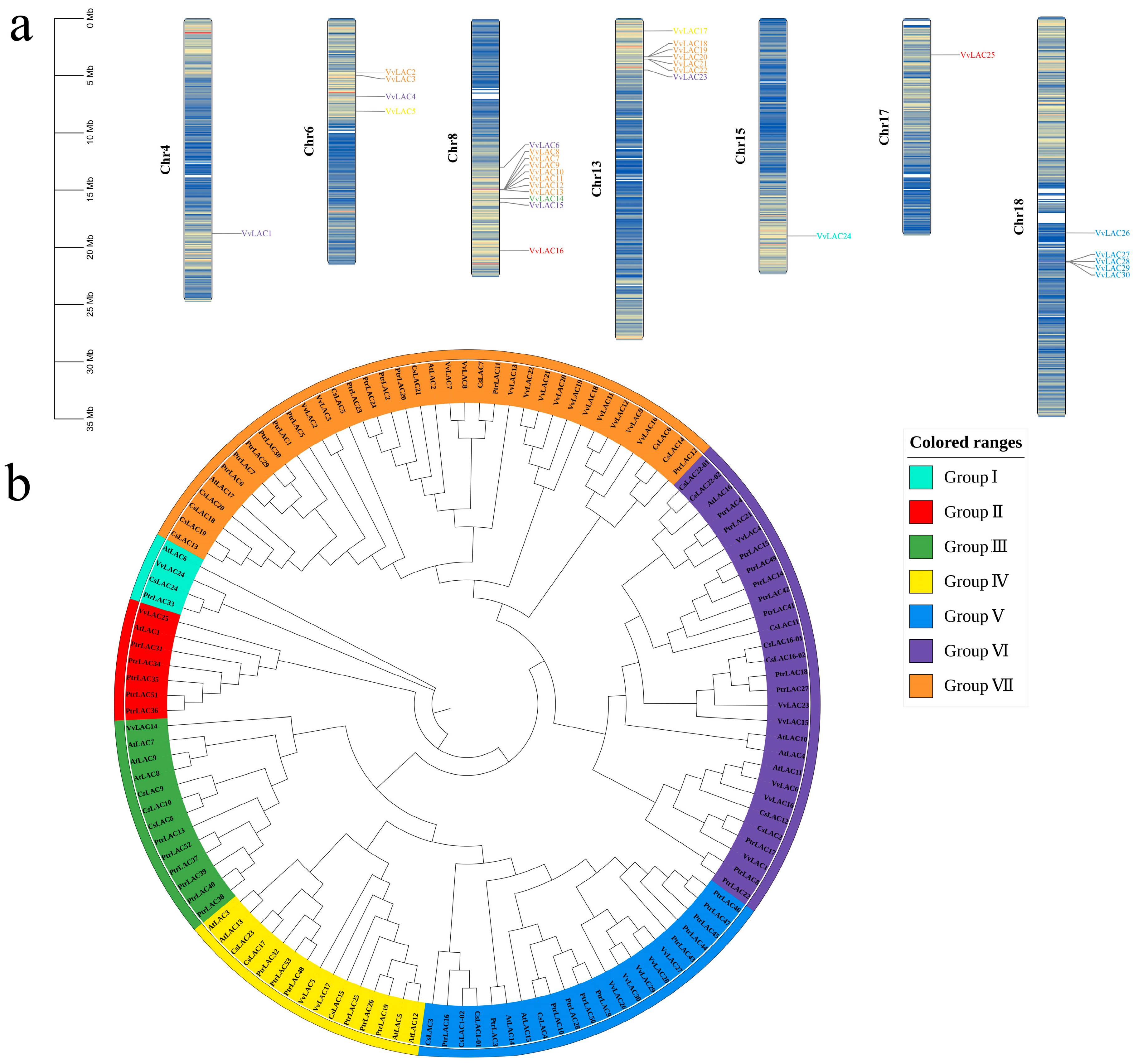
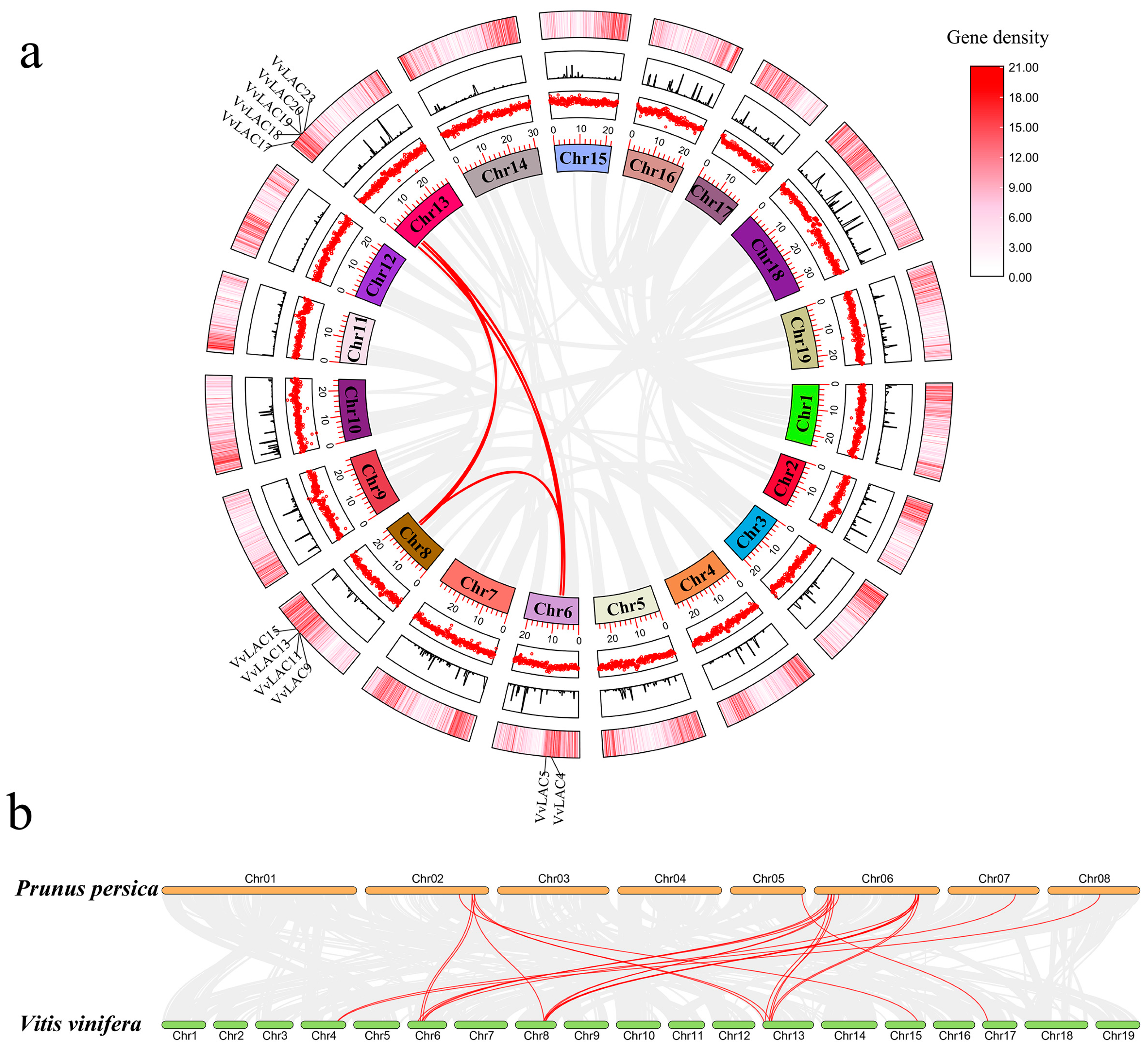
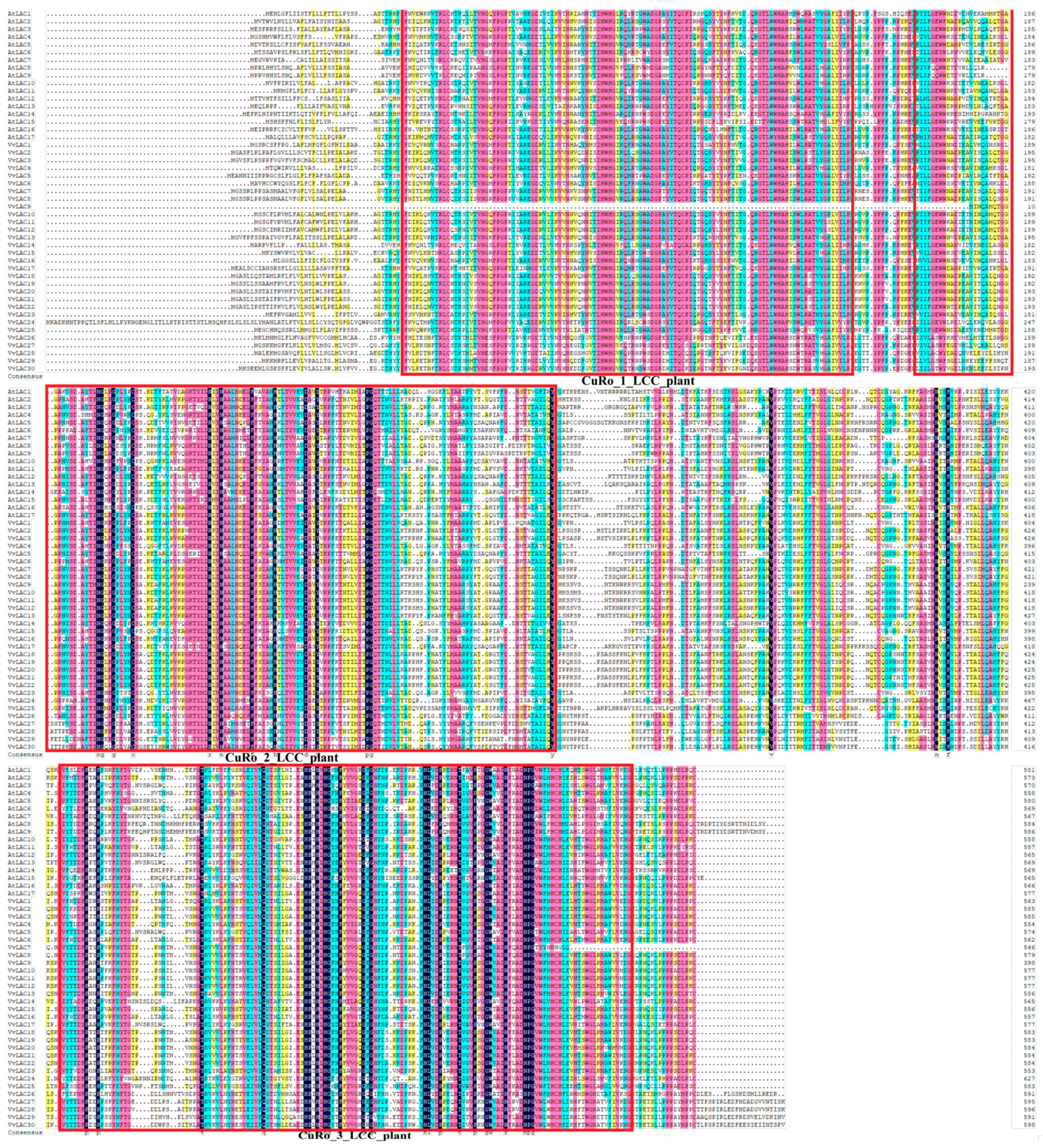
3.3. Chromosomal Locations and Phylogenetic Analysis

In the present study, 30 VvLAC genes were distributed on seven different chromosomes (Figure 2a). It was found that 11 of them were mainly located on Chr 8 (VvLAC6–16), 7 on Chr 13 (VvLAC17–23), 5 on Chr 18 (VvLAC26–30) and 4 on Chr 6 (VvLAC2–5), together containing 27 VvLAC genes, while Chr 4, Chr 15 and Chr 17 only contained 1 VvLAC gene each. The results show no positive correlation between chromosome length and number of VvLAC genes. Moreover, some members of the VvLAC family on Chr 8, Chr 13 and Chr 18 exist in the form of gene clusters.
To explore the evolutionary relationships of VvLACs, a phylogenetic analysis was performed based on the full-length amino acid sequences of 30 VvLACs and the LAC proteins from other plant species, which included 17 LACs from Arabidopsis thaliana, 53 from Populus trichocarpa and 27 from Citrus reticulata. According to the classification standard of Arabidopsis thaliana laccases, 30 VvLACs and 97 LACs were divided into seven groups, and their distribution in each group was rather uneven (Figure 2b). In detail, only 1 VvLAC was clustered with 2, 6 and 12 LACs in Group I, Group II and Group III, respectively. Also, Group IV contained 2 VvLACs, VvLAC5 and VvLAC17, together with 15 LACs (Figure 2b). Moreover, 22 and 29 LACs were classified in Group V and Group VI, each with 5 VvLAC, whereas Group VII contained the maximum number of LAC proteins, up to 37 LACs, in which there were 15 VvLACs (Figure 2b). In addition, it was found that all the 5 VvLACs clustered in Group V were located on Chr 18, while 8 out of the 11 VvLACs on Chr 13 all belonged to Group VII (Figure 2). These results indicate that specific evolutionary events occurred among VvLAC genes after the divergence of grapevine and the other three plants.

3.4. Gene Duplication and Syntenic Analysis

Generally, gene duplication, including tandem repeats, and segmental and tandem duplication, is one of the most important driving forces leading gene expansion and evolution during genome evolution [44,45]. It plays important roles in improving plant adaptability to various environmental stresses [45]. To understand the gene duplication pattern of VvLACs, a collinearity analysis was performed in this study. As a result, 11 VvLAC duplicated geneswere distributed on seven chromosomes (including Chr 6, Chr 13 and Chr 8), among which VvlAC18–20 were found to be tandem repeat genes, and so were VvlAC9, VvLAC11 and VvLAC13 (Figure 3a).
Furthermore, a syntenic analysis was performed for predicting the function of VvLACs through the homology analysis of LAC genes from four herbaceous plants and three ligneous plants. In total, 22 syntenic gene pairs between grape and Prunus persica were observed; however, no syntenic gene pairs were present (Figure 3b), which may relate to the weaker phylogenetic relationships between grape and the other six plant species.

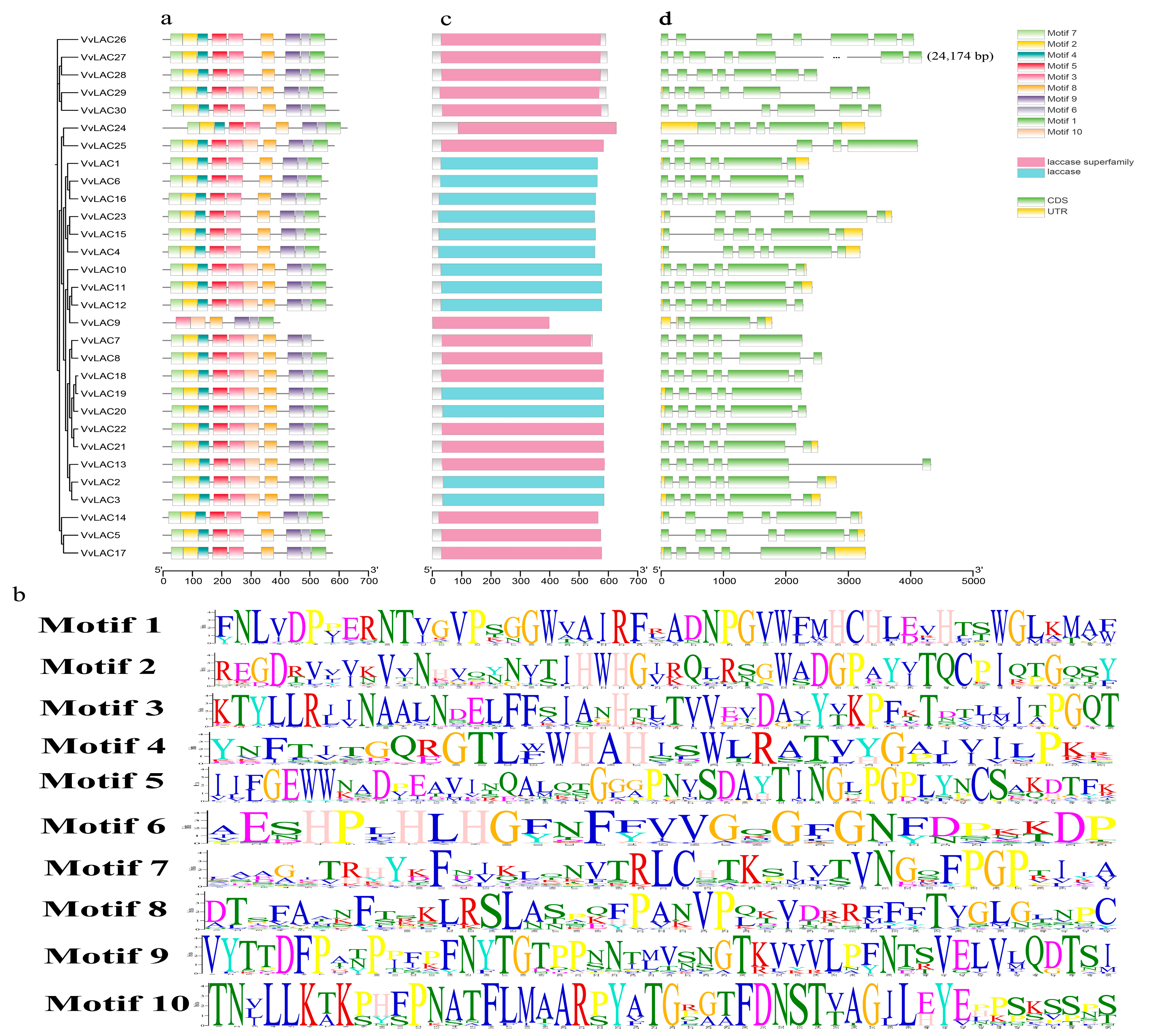
3.5. Motif Composition and Gene Structure

A total of 10 types of conserved motifs of the VvLAC family genes were explored by using the MEME program (Figure 4). Many classes of VvLAC proteins were found to have quite consistent motif compositions, except for VvLAC9, which was composed of only six motifs (Figure 4a). This suggests that functional redundancy possibly exist among these genes. Also, motif variations in numbers, amino acids and lengths across VvLACs were observed (Figure 4b), and as a result, their functional divergence was hypothesized. As expected, all the identified VvLAC proteins contained the conserved functional laccase domain (Figure 4c).
To further investigate gene evolution, the exon–intron distribution of VvLACs was analyzed by aligning coding sequences against their corresponding genomic sequences. The results show that the gene structures of VvLACs exhibit diverse intron–exon patterns (Figure 4d). For example, except for VvLAC9, which has only four exons, the exon number varies from five to seven among the VvLAC genes. Notably, there are five genes, all in Group V, containing the highest number of exons, seven, with VvLAC27 being the longest gene, with 24,174 bp in total. Generally, VvLACs displaying high homology have highly similar gene structures (Figure 4).
Subsequently, the amino acid sequences of all the AtLACs and VvLACs were aligned together. The results show that the VvLAC proteins have higher similarity with the AtLAC proteins and have three canonical plant laccase Cu oxidase domains, namely, Cu RO_1_LCC_plant, Cu RO_2_LCC_plant and Cu RO_3_LCC_plant (Figure 5).

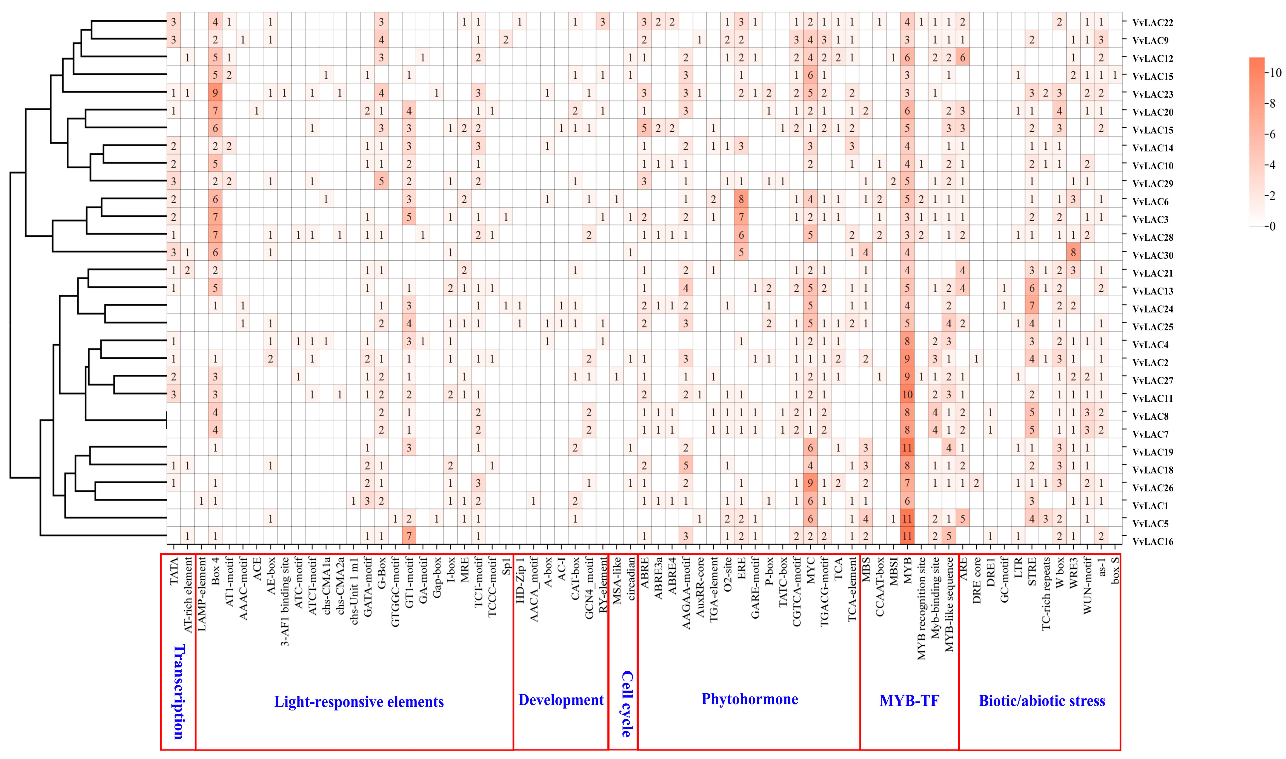
3.6. Cis-Elements in Promoters of VvLAC Genes

To explain their transcriptional differences, we analyzed the cis-elements in the promoter regions of the laccase gene family. Sixty–eight cis-elements were observed in VvLAC promoters and classified into the seven categories shown in Figure 6. The light-responsive elements, phytohormone elements, biotic/abiotic stress elements and MYB-TF (transcriptional factor) elements were the most frequently identified in the promoter regions of the grape laccase genes. Among all the elements, the MYB elements were the most prominent, and the only ones present in each VvLAC promoter, reaching the maximum of 11 in VvLAC6, VvLAC16 and VvLAC19 (Figure 6). Meanwhile, the MYC element was specially noticed, being present the most among phytohormone elements, except in the promoters of VvLAC29 and VvLAC30. Also, STRE (stress response promoter element), W-box (WRKY-binding site [36]) and WRE3 were relatively abundant among biotic/abiotic stress elements (Figure 6). Moreover, there were 22 types of light-responsive elements, and Box 4, GT1-motif, G-box and GATA-motif were abundantly detected (Figure 6).

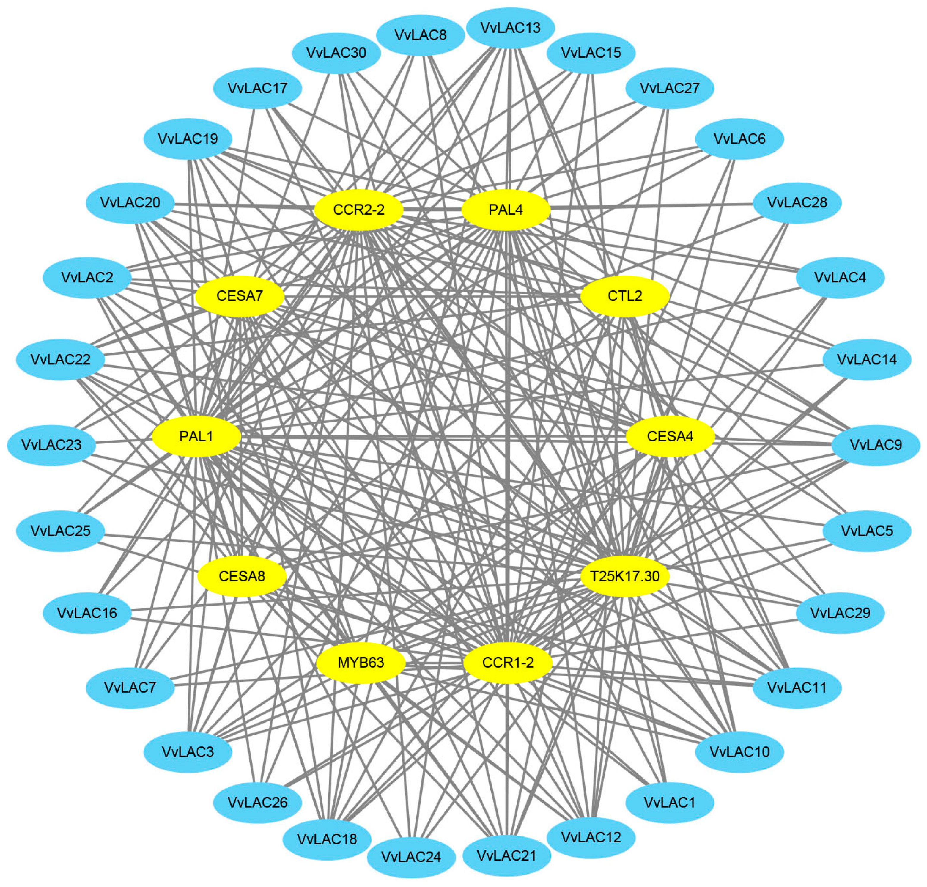
3.7. Protein–Protein Interaction Network

To investigate whether the VvLAC proteins might function by forming homo- or hetero-protein complexes, we constructed a protein interaction network for VvLACs based on their orthology with AtLAC proteins (Figure 7, Table S2). A total of 62 interacting protein pairs composed of 30 VvLACs and 11 VtLACs were predicted and divided into 11 subfamilies. There were no direct interactions between the VvLACs themselves, and the interacting proteins mainly included six types, PAL1-4 and CCR1/2, except for the 11th subfamily, which had distinct types, including PAL1/4, CESA4/7/8 and MYB63, T25K17.30, CESA4/7/8, PAL1, PAL1/2/4 and CCR1/2 were all predicted to interact with the VvLAC2, VvLAC7/8 and VvLAC1/6/16 proteins in the first, second and eighth subfamilies, respectively (Figure 7, Table S2). The fifth, sixth and seventh subfamilies, including VvLAC24, VvLAC14 and VvLAC15, respectively, revealed the same interaction proteins, PAL1-4 and CCR1. Additionally, both the third (VvLAC17) and tenth (VvLAC4 and VvLAC23) subfamilies contained T25K17.30 and PAL1-4. Additionally, PAL1-3 and CCR1/2 were predicted as the interaction proteins of VvLAC29-30 in the ninth subfamily, while VvLAC17 in the fourth subfamily was linked with the T25K17.30, PAL1/2 and CCR1/2 interaction proteins (Figure 7, Table S2).

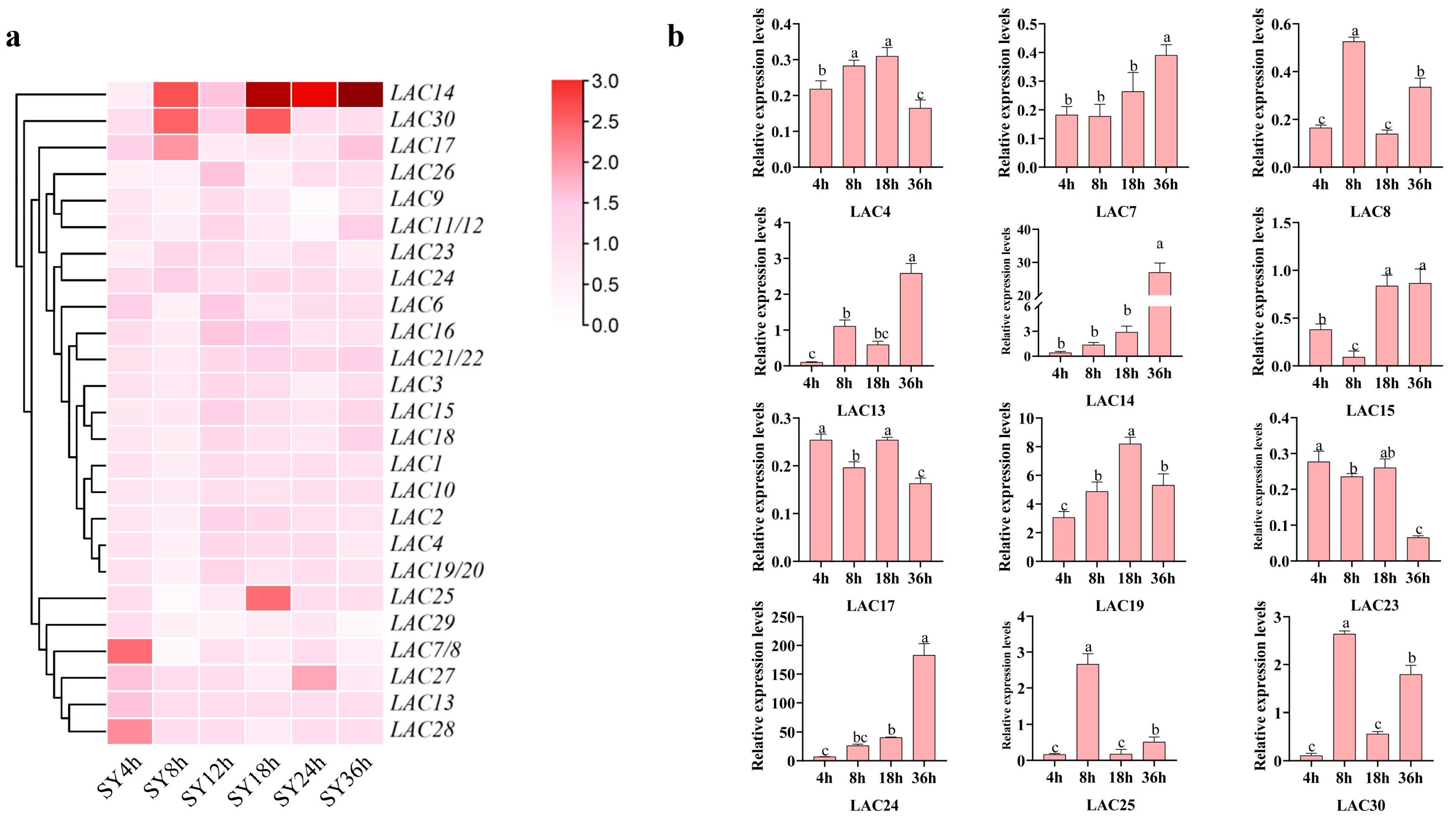
3.8. Expression Profiles of LAC Genes in SY Leaves in Response against Botrytis cinerea

The expression patterns of all the 30 LAC genes were analyzed based on the RNA-seq data of the SY leaves in the early interaction stages, 4–36 after inoculation with B. cinerea and sterile water [36]. There were four genes previously annotated as LACs according to the RNA-seq data; however, in the present study, each was actually identified to be containing two LACs according to the latest released grape genomic annotation version (PN40024. v4). These grape genes are LAC7/8, LAC11/12, LAC19/20 and LAC21/22. Even so, we obviously found several genes with different expression in the infected SY leaves compared with the control leaves, for example, VaLAC14, VaLAC24, VaLAC30, VaLAC17 and so on (Figure 8a). Relative quantification was used to confirm the expression levels of the selected genes as shown in Figure 8b. The results show that in contrast with other VaLAC genes, the similar trends of expression of VaLAC14 and VaLAC24 were significantly high in the SY leaves during infection by B. cinerea (Figure 8b). After B. cinerea infection, the expression levels of VaLAC13, VaLAC30 and VaLAC25 significantly increased, especially from 4 hpi to 8 hpi and following 36 hpi. Also, VaLAC19 expression was significantly increased from 4 hpi to 18 hpi (Figure 8b). Despite displaying significant changes, the relative expression of the remaining VaLAC genes following infection was very low (Figure 8b).

4. Discussion

Laccase is a multi-copper oxidase that catalyzes the oxidation of a wide range of phenolic compounds and has been widely and traditionally researched for potential applications in the food, pharmaceutical and environmental industries for its eco-friendly catalysis [46]. In recent decades, increasing numerous studies have demonstrated that plant LACs, a family of key enzymes polymerizing lignin monomers into lignins, play multiple roles in development and response to biotic and abiotic stresses [19,28,33,34,44]. Systematic analyses have been conducted to identify laccase gene families in many model plants, as well as crop, woody and horticultural plants. The laccase gene families in Arabidopsis thaliana (17) [47], Oryza sativa (30) [48], Populus trichocarpa (49) [40], Brassica rapa var. pekinensis (27) [49], Brassica rapa var. rapa (27) [49], Bretschneideri (45) [25], Citrus sinensis (41) [41] and Punica granatum (57) [43] have been revealed. Comparatively, Triticum aestivum (95) [50], soybean (93) and Gossypium hirsutum (84) [28] have more LAC members.
Grape is one of the most important horticultural crops in the world. We found that the B. cinerea-resistant grape germplasm SY significantly increased the lignin levels in accordance with the increase in laccase activity after infection. Considering the crucial roles of lignin in plant disease resistance [9,11], it might be an important characteristic for SY resistance to B. cinerea. Thus far, the laccase gene family members have been little studied in grapevine. Here, a total of 30 VvLACs were identified based on the grapevine genome in public databases, and a comprehensive analysis of the VvLAC gene family was performed. In grapevine, 30 VvLACs are unevenly distributed on seven chromosomes, including 6 tandem repeat genes and 5 segmentally duplicated genes on three chromosomes. Gene duplication events, including tandem repeat and segmental duplication, contributed to grape LAC gene family expansion and evolution. Moreover, syntenic pairs were only observed between grape and peach LAC genes, and they might have little evolution relationship with the other six species, including Arabidopsis thaliana and Malus domestica, in the present study. Motif and gene structure analyses showed that the closely related grape LAC members in the phylogenetic tree had similar exon–intron structures and common motif compositions, indicating a possible functional similarity. Diversities in gene structures and motif composition across different groups were also found, indicating functional differentiation in grape laccases.
Here, 30 VvLACs were classified into seven groups according to the classification criteria applied in Arabidopsis thaliana [39,47]. AtLAC15 in Group V, AtLAC4 and AtLAC11 in Group VI, and AtLAC2 and AtLAC17 in Group VII have been demonstrated to have a defined function in lignin biosynthesis [19,20,21,22,23]. PbrLAC1 and PbrLAC2 homologous with AtLAC4 and PbrLAC18 homologous with AtLAC17 have been proved to regulate lignin biosynthesis in pear stone cells [25]. This implies that as many as 25 VvLACs in the three groups are probably implicated in grape lignin biosynthesis. Combined for their quite consistent motif compositions and structures, these grape LAC genes possibly present functional redundancy. Recent research results also support the point that functional redundance exist in multiple LAC genes from one species. In Arabidopsis thaliana or pear, each single-laccase-gene-mutant or -silenced strain only slightly reduced the lignin content, whereas the simultaneous mutation or silencing of at least two laccase genes led to a significant decrease in lignin content [23,26].
V. amurensis, the most cold-tolerant Vitis species, is suggested to be highly resistant to B. cinerea and to share a common ancestor with V. vinifera dating approximately 2 million years ago [3,45,51]. VaERF16, VaMYB306 and VaERF20 from V. amurensis have been revealed to have a resistant function in B. cinerea infection [52,53]. Here, the expression profile of V. amurensis SY LAC genes highlighted the candidates VaLAC14, VaLAC24, VaLAC30, VaLAC13, VaLAC19 and VaLAC25 as being significantly involved in the grape response to B. cinerea. In Group III, VvLAC14 was clustered with AtLAC7, AtLAC8 and AtLAC9, whose expression is induced under environmental stress [39]. VaLAC14 and VaLAC24, having similar expression trends, might both play potential roles in the response of grapevine to B. cinerea. In Group V, VvLAC30 was clustered with AtLAC15. In cotton, GhLAC15, which is phylogenetically related to AtLAC15, was reported to positively regulate disease-induced lignification to enhance resistance to broad-spectrum biotic stress response [30]. Here, VaLAC30 was suggested to be involved in the SY defense against the fungus through lignin enhancement. Grape LAC13 and LAC19 were both clustered with AtLAC17 and PtrLAC23 in Group VII, implying their possible roles in grape lignin biosynthesis to resist to the fungus. PtrLAC23 was confirmed to have the function of lignin deposition in Populus trichocarpa stem transects through transgenic experiments [40]. VaLAC25 was in Group II with AtLAC1, PtrLAC31 and PtrLAC34, which are mainly expressed in plant roots and leaves [39,40], while their implication in lignin biosynthesis and plant disease, including VaLAC25, in the grape response to B. cinerea remains to be revealed in future.
To understand the potential regulation of grape LAC expression, we analyzed the cis-elements of VvLACs, and seven classes of cis-elements were obtained. The putative cis-elements suggest that VvLACs participate in many physiological processes, such as development, and light and stress responses. MYB and WRKY are significant regulators in the plant defense response to B. cinerea, and they can interact with genes associated with cell wall metabolism [36,54]. Notably, the MYB cis-element was present in all VvLAC promoters. AtMYB58 and AtMYB63, directly interacting with AtLAC4, are specific transcriptional activators of lignin biosynthesis [55]. Therefore, we predicted that MYB may have crucial roles in B. cinerea resistance by targeting LACs in grape leaves, but further research is needed. The VvLAC19 promoter had three WRKY-targeting W-box cis-elements, and MdLAC7 can be bound by MdWRKY75 to improve apple resistance to A. alternata by increasing the biosynthesis of laccase and lignin [33]. There might be other regulators besides MYB and WRKY in this response of grapevine. The stress cis-element STRE was also observed in candidate VvLAC13, VvLAC24 and VvLAC25 promoters. The STRE cis-element is a stress response promoter element widely involved in responding to various stressors, e.g., salt, water and heat, and is regulated by the transcriptional factors of zinc finger proteins and heat shock factors [56,57].

5. Conclusions

Laccases have been revealed to play important roles in plant disease resistance associated with lignin biosynthesis in recent years. However, information on grape laccase activity in the defense against B. cinerea is largely unknown. In the present study, a total of 30 grape laccase genes from the V. vinifera genome were identified and divided into seven groups. The proteins containing three typical plant Cu-oxidase domains displayed high conservation among each other. The phylogenetic analysis indicated that up to 25 VvLACs might be related to grape lignin biosynthesis, also implying a possible redundant function. The cis-element analysis highlighted MYB’s and WRKY’s regulating roles in grape LAC expression. Further investigation of LAC gene expression in B. cinerea-resistant Chinese wild V. amurensis ‘Shuangyou’ revealed that VaLAC14, VaLAC19, VaLAC24 and VaLAC30 could potentially act as the key candidate genes in lignin biosynthesis in the grape response to B. cinerea. This study lays the foundation for understanding the grape LAC genes and their roles in the response to B. cinerea. How these candidate genes participate in B. cinerea resistance by regulating lignification needs to be further verified.

Supplementary Materials

The following supporting information can be downloaded at: https://www.mdpi.com/article/10.3390/horticulturae10040376/s1, Table S1: Primers of selected VaLAC genes used for qRT-PCR, Table S2: Information on protein–protein network prediction.

Author Contributions

Conceptualization, R.W., Z.Y., J.S. and X.Z.; formal analysis, Z.Y., J.L., M.Z. and J.J.; visualization, R.W., Z.Y., M.W. and C.S.; investigation, Z.Y., J.L., J.S., Y.L. and K.Z.; resources, R.W., X.Z., P.H. and T.B.; writing—original draft, R.W. and Z.Y.; writing—review and editing, R.W., S.S., J.S. and X.Z. All authors have read and agreed to the published version of the manuscript.

Funding

We appreciate the support from Natural Science Foundation of Henan province (222300420182) and National Natural Science Foundation of China (32302495).

Data Availability Statement

The original contributions presented in the study are included in the article and supplementary materials.

Conflicts of Interest

The authors declare no conflicts of interest.

References

  1. Shen, F.; Wu, W.; Han, X.; Wang, J.; Li, Y.; Liu, D. Study on the occurrence law and green control of grape gray mold from the perspective of ecological balance. Bioengineered 2021, 12, 779–790. [Google Scholar] [CrossRef] [PubMed]
  2. De Simone, N.; Pace, B.; Grieco, F.; Chimienti, M.; Tyibilika, V.; Santoro, V.; Capozzi, V.; Colelli, G.; Spano, G.; Russo, P. Botrytis cinerea and table grapes: A review of the main physical, chemical, and bio-based control treatments in post-harvest. Foods 2020, 9, 1138. [Google Scholar] [CrossRef] [PubMed]
  3. Wan, R.; Hou, X.; Wang, X.; Qu, J.; Singer, S.D.; Wang, Y.; Wang, X. Resistance evaluation of Chinese wild Vitis genotypes against Botrytis cinerea and different responses of resistant and susceptible hosts to the infection. Front. Plant Sci. 2015, 6, 160149. [Google Scholar] [CrossRef] [PubMed]
  4. Harper, L.A.; Paton, S.; Hall, B.; McKay, S.; Oliver, R.P.; Lopez-Ruiz, F.J. Fungicide resistance characterized across seven modes of action in Botrytis cinerea isolated from Australian vineyards. Pest Manag. Sci. 2022, 78, 1326–1340. [Google Scholar] [CrossRef] [PubMed]
  5. Bi, K.; Liang, Y.; Mengiste, T.; Sharon, A. Killing softly: A roadmap of Botrytis cinerea pathogenicity. Trends Plant Sci. 2023, 28, 211–222. [Google Scholar] [CrossRef]
  6. Blanco-Ulate, B.; Morales-Cruz, A.; Amrine, K.C.H.; Labavitch, J.M.; Powell, A.L.T.; Cantu, D. Genome-wide transcriptional profiling of Botrytis cinerea genes targeting plant cell walls during infections of different hosts. Front. Plant Sci. 2014, 5, 98301. [Google Scholar] [CrossRef] [PubMed]
  7. Veloso, J.; van Kan, J.A.L. Many shades of grey in Botrytis-host plant interactions. Trends Plant Sci. 2018, 23, 613–622. [Google Scholar] [CrossRef] [PubMed]
  8. Goetz, G.; Fkyerat, A.; Métais, N.; Kunz, M.; Tabacchi, R.; Pezet, R.; Pont, V. Resistance factors to grey mould in grape berries: Identification of some phenolics inhibitors of Botrytis cinerea stilbene oxidase. Phytochemistry 1999, 52, 759–767. [Google Scholar] [CrossRef]
  9. Liu, Q.; Luo, L.; Zheng, L. Lignins: Biosynthesis and biological functions in plants. Int. J. Mol. Sci. 2018, 19, 335. [Google Scholar] [CrossRef] [PubMed]
  10. Lazniewska, J.; Macioszek, V.K.; Kononowicz, A.K. Plant-fungus interface: The role of surface structures in plant resistance and susceptibility to pathogenic fungi. Physiol. Mol. Plant Pathol. 2012, 78, 24–30. [Google Scholar] [CrossRef]
  11. Lee, M.-H.; Jeon, H.S.; Kim, S.H.; Chung, J.H.; Roppolo, D.; Lee, H.-J.; Cho, H.J.; Tobimatsu, Y.; Ralph, J.; Park, O.K. Lignin-based barrier restricts pathogens to the infection site and confers resistance in plants. Embo J. 2019, 38, e101948. [Google Scholar] [CrossRef] [PubMed]
  12. Asselbergh, B.; Curvers, K.; Franca, S.C.; Audenaert, K.; Vuylsteke, M.; Van Breusegem, F.; Hoefte, M. Resistance to Botrytis cinerea in sitiens, an abscisic acid-deficient tomato mutant, involves timely production of hydrogen peroxide and cell wall modifications in the epidermis. Plant Physiol. 2007, 144, 1863–1877. [Google Scholar] [CrossRef] [PubMed]
  13. Lloyd, A.J.; Allwood, J.W.; Winder, C.L.; Dunn, W.B.; Heald, J.K.; Cristescu, S.M.; Sivakumaran, A.; Harren, F.J.M.; Mulema, J.; Denby, K.; et al. Metabolomic approaches reveal that cell wall modifications play a major role in ethylene-mediated resistance against Botrytis cinerea. Plant J. 2011, 67, 852–868. [Google Scholar] [CrossRef] [PubMed]
  14. Xue, M.; Yi, H. Induction of disease resistance providing new insight into sulfur dioxide preservation in Vitis vinifera L. Sci. Hortic. 2017, 225, 567–573. [Google Scholar] [CrossRef]
  15. Hamiduzzaman, M.M.; Jakab, G.; Barnavon, L.; Neuhaus, J.M.; Mauch-Mani, B. β-Aminobutyric acid-induced resistance against downy mildew in grapevine acts through the potentiation of callose formation and jasmonic acid signaling. Mol. Plant-Microbe Interact. 2005, 18, 819–829. [Google Scholar] [CrossRef] [PubMed]
  16. Wallis, C.M.; Chen, J. Grapevine phenolic compounds in xylem sap and tissues are significantly altered during infection by Xylella Fastidiosa. Phytopathology 2012, 102, 816–826. [Google Scholar] [CrossRef] [PubMed]
  17. Tziros, G.T.; Ainalidou, A.; Samaras, A.; Kollaros, M.; Karamanoli, K.; Menkissoglu-Spiroudi, U.; Karaoglanidis, G.S. Differences in defence-related gene expression and metabolite accumulation reveal insights into the resistance of Greek grape wine cultivars to Botrytis bunch rot. Oeno One 2022, 56, 111–123. [Google Scholar] [CrossRef]
  18. Perkins, M.L.; Schuetz, M.; Unda, F.; Chen, K.T.; Bally, M.B.; Kulkarni, J.A.; Yan, Y.; Pico, J.; Castellarin, S.D.; Mansfield, S.D.; et al. Monolignol export by diffusion down a polymerization-induced concentration gradient. Plant Cell 2022, 34, 2080–2095. [Google Scholar] [CrossRef] [PubMed]
  19. Yu, Y. LACCASE2 negatively regulates lignin deposition of Arabidopsis Roots. Plant Physiol. 2020, 182, 1190–1191. [Google Scholar] [CrossRef] [PubMed]
  20. Zhao, Q.; Nakashima, J.; Chen, F.; Yin, Y.; Fu, C.; Yun, J.; Shao, H.; Wang, X.; Wang, Z.-Y.; Dixon, R.A. LACCASE is necessary and nonredundant with PEROXIDASE for lignin polymerization during vascular development in Arabidopsis. Plant Cell 2013, 25, 3976–3987. [Google Scholar] [CrossRef] [PubMed]
  21. Liang, M.; Davis, E.; Gardner, D.; Cai, X.; Wu, Y. Involvement of AtLAC15 in lignin synthesis in seeds and in root elongation of Arabidopsis. Planta 2006, 224, 1185–1196. [Google Scholar] [CrossRef] [PubMed]
  22. Berthet, S.; Demont-Caulet, N.; Pollet, B.; Bidzinski, P.; Cezard, L.; Le Bris, P.; Borrega, N.; Herve, J.; Blondet, E.; Balzergue, S.; et al. Disruption of LACCASE4 and 17 results in tissue-specific alterations to lignification of Arabidopsis thaliana stems. Plant Cell 2011, 23, 1124–1137. [Google Scholar] [CrossRef] [PubMed]
  23. Perkins, M.L.; Schuetz, M.; Unda, F.; Smith, R.A.; Sibout, R.; Hoffmann, N.J.; Wong, D.C.J.; Castellarin, S.D.; Mansfield, S.D.; Samuels, L. Dwarfism of high-monolignol Arabidopsis plants is rescued by ectopic LACCASE overexpression. Plant Direct 2020, 4, e00265. [Google Scholar] [CrossRef] [PubMed]
  24. He, F.; Machemer-Noonan, K.; Golfier, P.; Unda, F.; Dechert, J.; Zhang, W.; Hoffmann, N.; Samuels, L.; Mansfield, S.D.; Rausch, T.; et al. The in vivo impact of MsLAC1, a Miscanthus laccase isoform, on lignification and lignin composition contrasts with its in vitro substrate preference. BMC Plant Biol. 2019, 19, 552. [Google Scholar] [CrossRef]
  25. Xue, C.; Yao, J.-L.; Qin, M.-F.; Zhang, M.-Y.; Allan, A.C.; Wang, D.-F.; Wu, J. PbrmiR397a regulates lignification during stone cell development in pear fruit. Plant Biotechnol. J. 2019, 17, 103–117. [Google Scholar] [CrossRef] [PubMed]
  26. Qin, S.; Fan, C.; Li, X.; Li, Y.; Hu, J.; Li, C.; Luo, K. LACCASE14 is required for the deposition of guaiacyl lignin and affects cell wall digestibility in poplar. Biotechnol. Biofuels 2020, 13, 197. [Google Scholar] [CrossRef] [PubMed]
  27. Wang, X.; Zhuo, C.; Xiao, X.; Wang, X.; Docampo-Palacios, M.; Chen, F.; Dixon, R.A. Substrate specificity of LACCASE8 facilitates polymerization of caffeyl alcohol for C-lignin biosynthesis in the seed coat of Cleome hassleriana. Plant Cell 2020, 32, 3825–3845. [Google Scholar] [CrossRef] [PubMed]
  28. Balasubramanian, V.K.; Rai, K.M.; Thu, S.W.; Hii, M.M.; Mendu, V. Genome-wide identification of multifunctional laccase gene family in cotton (Gossypium spp.); expression and biochemical analysis during fiber development. Sci. Rep. 2016, 6, 34309. [Google Scholar] [CrossRef] [PubMed]
  29. Wei, T.; Tang, Y.; Jia, P.; Zeng, Y.; Wang, B.; Wu, P.; Quan, Y.; Chen, A.; Li, Y.; Wu, J. A Cotton lignin biosynthesis gene, GhLAC4, fine-tuned by Ghr-miR397 modulates plant resistance against Verticillium dahliae. Front. Plant Sci. 2021, 12, 743795. [Google Scholar] [CrossRef] [PubMed]
  30. Zhang, Y.; Wu, L.; Wang, X.; Chen, B.; Zhao, J.; Cui, J.; Li, Z.; Yang, J.; Wu, L.; Wu, J.; et al. The cotton laccase gene GhLAC15 enhances Verticillium wilt resistance via an increase in defence-induced lignification and lignin components in the cell walls of plants. Mol. Plant Pathol. 2019, 20, 309–322. [Google Scholar] [CrossRef] [PubMed]
  31. Hu, Q.; Min, L.; Yang, X.; Jin, S.; Zhang, L.; Li, Y.; Ma, Y.; Qi, X.; Li, D.; Liu, H.; et al. Laccase GhLac1 modulates broad-spectrum biotic stress tolerance via manipulating phenylpropanoid pathway and jasmonic acid synthesis. Plant Physiol. 2018, 176, 1808–1823. [Google Scholar] [CrossRef] [PubMed]
  32. Yu, X.; Gong, H.; Cao, L.; Hou, Y.; Qu, S. MicroRNA397b negatively regulates resistance of Malus hupehensis to Botryosphaeria dothidea by modulating MhLAC7 involved in lignin biosynthesis. Plant Sci. 2020, 292, 110390. [Google Scholar] [CrossRef] [PubMed]
  33. Hou, Y.; Yu, X.; Chen, W.; Zhuang, W.; Wang, S.; Sun, C.; Cao, L.; Zhou, T.; Qu, S. MdWRKY75e enhances resistance to Alternaria alternata in Malus domestica. Hortic. Res. 2021, 8, 225. [Google Scholar] [CrossRef] [PubMed]
  34. Zhu, Y.; Li, G.; Singh, J.; Khan, A.; Fazio, G.; Saltzgiver, M.; Xia, R. Laccase directed lignification is one of the major processes associated with the defense response against Pythium ultimum infection in apple roots. Front. Plant Sci. 2021, 12, 629776. [Google Scholar] [CrossRef] [PubMed]
  35. Zhao, Y.; Liu, Y.; Dong, X.; Liu, J.-J.; Zhao, D.-G. Identification of a novel laccase gene EuLAC1 and its potential resistance against Botrytis cinerea. Transgenic Res. 2022, 31, 215–225. [Google Scholar] [CrossRef] [PubMed]
  36. Wan, R.; Guo, C.; Hou, X.; Zhu, Y.; Gao, M.; Hu, X.; Zhang, S.; Jiao, C.; Guo, R.; Li, Z.; et al. Comparative transcriptomic analysis highlights contrasting levels of resistance of Vitis vinifera and Vitis amurensis to Botrytis cinerea. Hortic. Res. 2021, 8, 103. [Google Scholar] [CrossRef] [PubMed]
  37. Kumar, S.; Stecher, G.; Li, M.; Knyaz, C.; Tamura, K. MEGA X: Molecular evolutionary genetics analysis across computing platforms. Mol. Biol. Evol. 2018, 35, 1547–1549. [Google Scholar] [CrossRef] [PubMed]
  38. Letunic, I.; Bork, P. Interactive tree of life (iTOL) v3: An online tool for the display and annotation of phylogenetic and other trees. Nucleic Acids Res. 2016, 44, W242–W245. [Google Scholar] [CrossRef] [PubMed]
  39. McCaig, B.C.; Meagher, R.B.; Dean, J.F.D. Gene structure and molecular analysis of the laccase-like multicopper oxidase (LMCO) gene family in Arabidopsis thaliana. Planta 2005, 221, 619–636. [Google Scholar] [CrossRef] [PubMed]
  40. Liao, B.; Wang, C.; Li, X.; Man, Y.; Ruan, H.; Zhao, Y. Genome-wide analysis of the Populus trichocarpa laccase gene family and functional identification of PtrLAC23. Front. Plant Sci. 2023, 13, 1063813. [Google Scholar] [CrossRef]
  41. Xu, X.; Zhou, Y.; Wang, B.; Ding, L.; Wang, Y.; Luo, L.; Zhang, Y.; Kong, W. Genome-wide identification and characterization of laccase gene family in Citrus sinensis. Gene 2019, 689, 114–123. [Google Scholar] [CrossRef] [PubMed]
  42. Lescot, M.; Déhais, P.; Thijs, G.; Marchal, K.; Moreau, Y.; Van de Peer, Y.; Rouzé, P.; Rombauts, S. PlantCARE, a database of plant cis-acting regulatory elements and a portal to tools for in silico analysis of promoter sequences. Nucleic Acids Res. 2002, 30, 325–327. [Google Scholar] [CrossRef] [PubMed]
  43. Shi, J.; Yao, J.; Tong, R.; Wang, S.; Li, M.; Song, C.; Wan, R.; Jiao, J.; Zheng, X. Genome-wide identification of laccase gene family from Punica granatum and functional analysis towards potential involvement in lignin biosynthesis. Horticulturae 2023, 9, 918. [Google Scholar] [CrossRef]
  44. Zhu, J.; Zhang, H.; Huang, K.; Guo, R.; Zhao, J.; Xie, H.; Zhu, J.; Gu, H.; Chen, H.; Li, G.; et al. Comprehensive analysis of the laccase gene family in tea plant highlights its roles in development and stress responses. BMC Plant Biol. 2023, 23, 129. [Google Scholar] [CrossRef] [PubMed]
  45. Wan, R.; Song, J.; Lv, Z.; Qi, X.; Han, X.; Guo, Q.; Wang, S.; Shi, J.; Jian, Z.; Hu, Q.; et al. Genome-wide identification and comprehensive analysis of the AP2/ERF gene family in pomegranate fruit development and postharvest preservation. Genes 2022, 13, 895. [Google Scholar] [CrossRef]
  46. Mayolo-Deloisa, K.; Gonzalez-Gonzalez, M.; Rito-Palomares, M. Laccases in food industry: Bioprocessing, potential industrial and biotechnological applications. Front. Bioeng. Biotechnol. 2020, 8, 222. [Google Scholar] [CrossRef] [PubMed]
  47. Cai, X.; Davis, E.J.; Ballif, J.; Liang, M.; Bushman, E.; Haroldsen, V.; Torabinejad, J.; Wu, Y. Mutant identification and characterization of the laccase gene family in Arabidopsis. J. Exp. Bot. 2006, 57, 2563–2569. [Google Scholar] [CrossRef] [PubMed]
  48. Liu, Q.; Luo, L.; Wang, X.; Shen, Z.; Zheng, L. Comprehensive Analysis of Rice Laccase Gene (OsLAC) Family and ectopic expression of OsLAC10 enhances tolerance to copper stress in Arabidopsis. Int. J. Mol. Sci. 2017, 18, 209. [Google Scholar] [CrossRef] [PubMed]
  49. Wen, J.; Liu, Y.; Yang, S.; Yang, Y.; Wang, C. Genome-wide characterization of laccase gene family from turnip and chinese cabbage and the role in xylem lignification in hypocotyls. Horticulturae 2022, 8, 522. [Google Scholar] [CrossRef]
  50. Sun, Z.; Zhou, Y.; Hu, Y.; Jiang, N.; Hu, S.; Li, L.; Li, T. Identification of wheat LACCASEs in response to Fusarium graminearum as potential deoxynivalenol trappers. Front. Plant Sci. 2022, 13, 832800. [Google Scholar] [CrossRef] [PubMed]
  51. Ramirez, V.; Garcia-Andrade, J.; Vera, P. Enhanced disease resistance to Botrytis cinerea in myb46 Arabidopsis plants is associated to an early down-regulation of CesA genes. Plant Signal. Behav. 2011, 6, 911–913. [Google Scholar] [CrossRef] [PubMed]
  52. Wang, Y.; Xin, H.; Fan, P.; Zhang, J.; Liu, Y.; Dong, Y.; Wang, Z.; Yang, Y.; Zhang, Q.; Ming, Y.; et al. The genome of Shanputao (Vitis amurensis) provides a new insight into cold tolerance of grapevine. Plant Fournal 2021, 105, 1495–1506. [Google Scholar] [CrossRef] [PubMed]
  53. Zhu, Y.; Zhang, X.; Zhang, Q.; Chai, S.; Yin, W.; Gao, M.; Li, Z.; Wang, X. The transcription factors VaERF16 and VaMYB306 interact to enhance resistance of grapevine to Botrytis cinerea infection. Mol. Plant Pathol. 2022, 23, 1415–1432. [Google Scholar] [CrossRef] [PubMed]
  54. Wang, M.; Zhu, Y.; Han, R.; Yin, W.; Guo, C.; Li, Z.; Wang, X. Expression of Vitis amurensis VaERF20 in Arabidopsis thaliana improves resistance to Botrytis cinerea and Pseudomonas syringae pv. tomato DC3000. Int. J. Mol. Sci. 2018, 19, 696. [Google Scholar] [CrossRef] [PubMed]
  55. Zhou, J.; Lee, C.; Zhong, R.; Ye, Z. MYB58 and MYB63 are transcriptional activators of the lignin biosynthetic pathway during secondary cell wall formation in Arabidopsis. Plant Cell 2009, 21, 248–266. [Google Scholar] [CrossRef] [PubMed]
  56. Fu, J.; Huang, S.; Qian, J.; Qing, H.; Wan, Z.; Cheng, H.; Zhang, C. Genome-Wide identification of Petunia HSF genes and potential function of PhHSF19 in benzenoid/phenylpropanoid biosynthesis. Int. J. Mol. Sci. 2022, 23, 2974. [Google Scholar] [CrossRef] [PubMed]
  57. Schmitt, A.P.; McEntee, K. Msn2p, a zinc finger DNA-binding protein, is the transcriptional activator of the multistress response in Saccharomyces cerevisiae. Proc. Natl. Acad. Sci. USA 1996, 93, 5777–5782. [Google Scholar] [CrossRef] [PubMed]
Figure 1. Changes in resistant ‘Shuangyou’ grape leaves. (a) Phenotypic observation. (b) Laccase activity and (c) lignin content. C: control (inoculation with sterile water); T: treatment (inoculation with Botrytis cinerea). SY: Chinese wild V. amurensis ‘Shuangyou’, a germplasm highly resistant to B. cinerea. The results are the means (SDs) of three biological replicates. There were significant differences between the results marked with different lowercase letters (p ≤ 0.05).
Horticulturae 10 00376 g001
Figure 2. Chromosomal localization and phylogenetic analysis. (a) The chromosomal localization of the VvLAC gene family. Genes in the same subgroup are represented by the same color. The left ruler indicates the length of the grape chromosomes. (b) The phylogenetic tree was constructed based on the protein sequences from Arabidopsis thaliana, Populus trichocarpa and Citrus reticulata Blanco. The adjacency tree was constructed by using MEGA10.0. The seven groups are distinguished by different colors.
Horticulturae 10 00376 g002
Figure 3. Interspecific and intraspecific collinearity analyses of the VvLAC genes. (a) Intraspecific collinearity analysis. The innermost circle shows the 19 chromosomes of Vitis vinifera. Different chromosomes are represented by different colors, followed by N ratio and G/C ratio. The outermost circle shows the gene densities on the corresponding chromosomers, and a redder color represents a higher gene densities. The identified VvLAC gene pairs are linked by red lines. (b) Syntenic analysis of Prunus persica and Vitis vinifera LAC genes. The different chromosomes of the two are represented by different colors. The gray lines represent the collinear lines in the genome of the two plants, while the red lines highlight the same line of the LAC gene pair.
Horticulturae 10 00376 g003
Figure 4. Motif composition and gene structure analyses of VvLACs. (a,b) Motif information. (c) Conserved domain. (d) Intron–exon patterns. UTR and CDS are represented by yellow boxes and green boxes, respectively.
Horticulturae 10 00376 g004
Figure 5. VvLACs and AtLAC protein sequence alignment analysis.
Horticulturae 10 00376 g005
Figure 6. Identification of cis-elements in the upstream promoters of the indicated VvLAC genes. The heat map was generated based on the numbers and predicted functions of the cis-elements. Different colors and numbers in the grid represent the number of the corresponding cis-elements in each VvLAC gene promoter.
Horticulturae 10 00376 g006
Figure 7. Prediction of protein–protein interaction network of VvLACs based on AtLACs in Arabidopsis thaliana. VvLAC proteins are shown in blue circles and the predicted interaction proteins are shown in yellow circles.
Horticulturae 10 00376 g007
Figure 8. The expression analysis of the VaLACs in the SY response to B. cinerea. (a) The ratios of VaLAC expression after infection compared with the control, which were derived from previously published RNA-Seq data on the interaction between SY leaves and B. cinerea at the indicated time points after infection [36]. (b) The relative expression levels of the selected VaLAC genes determined through qRT-PCR. SY: Chinese wild V. amurensis ‘Shuangyou’, a germplasm highly resistant to B. cinerea. The expression value was reported as the mean ± standard deviation of three independent biological replicates, where each biological replicate contained three technical replicates. Different letters in the figure indicate significant differences, p < 0.05. C: control (inoculation with sterile water); T: treatment (inoculation with B. cinerea).
Horticulturae 10 00376 g008
Table 1. Information of the VvLAC gene family.
NameGene IDChromosomeGene Length (bp)pIMW (Kda)Amino Acid (aa)Subcellular Localization
VvLAC1Vitvi04g01317_t001Chr416928.4962.23563vacu
VvLAC2Vitvi06g00378_t001Chr617589.0264.39585chlo
VvLAC3Vitvi06g00405_t001Chr617588.7164.30585extr
VvLAC4Vitvi06g00591_t001Chr616659.2660.70554chlo
VvLAC5Vitvi06g00728_t001Chr617258.5963.59574vacu
VvLAC6Vitvi08g01031_t001Chr816898.3762.49562extr
VvLAC7Vitvi08g01223_t002Chr816419.3259.90546chlo
VvLAC8Vitvi08g01223_t001Chr817409.2463.69579chlo
VvLAC9Vitvi08g04233_t001Chr811979.7443.53398chlo
VvLAC10Vitvi08g02201_t001Chr817349.7764.04577chlo
VvLAC11Vitvi08g01228_t001Chr817349.7964.04577chlo
VvLAC12Vitvi08g04234_t001Chr817349.7864.08577chlo
VvLAC13Vitvi08g01229_t001Chr817619.2264.96586chlo
VvLAC14Vitvi08g01299_t001Chr816986.7962.00565vacu
VvLAC15Vitvi08g01335_t001Chr816719.1260.48556chlo
VvLAC16Vitvi08g01735_t001Chr816748.0461.69557extr
VvLAC17Vitvi13g00117_t001Chr1317347.6363.18577cyto
VvLAC18Vitvi13g00321_t001Chr1317529.4464.30583chlo
VvLAC19Vitvi13g00322_t001Chr1317529.5364.39583chlo
VvLAC20Vitvi13g00323_t001Chr1317559.3564.61584vacu
VvLAC21Vitvi13g00341_t001Chr1317559.464.50584chlo
VvLAC22Vitvi13g00342_t001Chr1317559.2264.45584chlo
VvLAC23Vitvi13g00509_t001Chr1316628.6760.40553chlo
VvLAC24Vitvi15g00941_t001Chr1518848.1969.76627chlo
VvLAC25Vitvi17g00227_t001Chr1717528.565.26583chlo
VvLAC26Vitvi18g01488_t001Chr1817765.366.34591extr
VvLAC27Vitvi18g02924_t001Chr1817914.8966.31596cyto
VvLAC28Vitvi18g02922_t001Chr1817944.7966.25597pero
VvLAC29Vitvi18g02927_t001Chr1817795.5266.58592plas
VvLAC30Vitvi18g02928_t001Chr1818004.9267.64599cyto
Note: vacu: vacuolar membrane; chlo: chloroplast; extr: eextracellular matrix; cyto: cytoskeleton; pero: peroxisome; plas: plasma membrance.
Disclaimer/Publisher’s Note: The statements, opinions and data contained in all publications are solely those of the individual author(s) and contributor(s) and not of MDPI and/or the editor(s). MDPI and/or the editor(s) disclaim responsibility for any injury to people or property resulting from any ideas, methods, instructions or products referred to in the content.

