Article

Preliminary Study on Programmed Cell Death during Calyx Abscission of Korla Fragrant Pear

1
College of Horticulture, Xinjiang Agricultural University, Urumqi 830052, China
2
Korla Fragrant Pear Research Centre, Korla 841000, China
*
Author to whom correspondence should be addressed.
These authors contributed equally to this work.
Horticulturae 2024, 10(6), 637; https://doi.org/10.3390/horticulturae10060637
Submission received: 7 May 2024 / Revised: 4 June 2024 / Accepted: 7 June 2024 / Published: 13 June 2024

Abstract

:
Programmed cell death (PCD) is common in plant growth and development, such as xylem development, organ senescence, and abscission. Calyx abscission in Korla fragrant pear contributes to fruit quality, while it was not clear whether PCD occurred during calyx abscission and which signals regulated the process. Therefore, it is imperative to clarify the process of PCD in the process of calyx abscission in Korla fragrant pear under natural conditions to enrich the mechanism of calyx abscission. The results showed that the total time of calyx abscission of Korla fragrant pear began from 6 days after pollination (DAP) to 13 DAP, and the peak of calyx abscission occurred 10 DAP. During the whole process of calyx abscission, PCD started 6 DAP. At 9 DAP, the degree of PCD deepened. At 12 DAP, the cells in the abscission zone showed asymmetry on both sides, the organelles in the distal cells of the abscission zone degraded into apoptotic fragments, and the protective layer of the normal development of cells located at the proximal end of the abscission zone region ended the PCD process. ETH concentrations in the abscission zone of the decalyx fruit were significantly higher than those of the persistent calyx fruits in each period during calyx abscission, and high levels of ethylene and hydrogen peroxide and low contents of the GA3, ZT, and hydroxyl radicals promoted calyx abscission before the formation of the abscission zone. At 3 DAP, the ethylene concentration (43.97 ppm) and H2O2 content (8.49 μmol/g) of decalyx fruit in the abscission zone were significantly higher than those of persistent calyx fruit by 67.69% and 27.86%, respectively; however, the GA3, ZT, and hydroxyl radicals showed the opposite. Overall, PCD in the abscission zone of decalyx fruits did occur during the calyx abscission of Korla fragrant pear, and ethylene and H2O2 might play major roles in initiating the PCD process during Korla fragrant pear calyx abscission.

1. Introduction

In plants, programmed cell death (PCD) is a cell death process that is spontaneous and genetically regulated in a specific time and space through the action of endogenous developmental signals or exogenous environmental signals and is prevalent in all processes of plant growth and development [1]; for example, PCD controls root organ size and promotes optimal root growth [2]. Past studies have found that PCD was present throughout the flower opening and senescence of petals [3], and during seed germination, endosperm cells first underwent degradation and death, followed by the death of aleurone layer cells, which PCD mediated in a tightly controlled spatiotemporal pattern [4]. Furthermore, the plant PCD process is regulated by a range of signals, such as plant hormones, reactive oxygen species (ROS), calcium ions (Ca2+), and nitric oxide (NO), which cross-react extensively with each other to induce PCD through different pathways [5,6]. Among them, plant hormones as important factors regulating plant organ abscission play a crucial role in regulating PCD [7]. Ethylene (ETH) is an important factor in the regulation of plant PCD. Meir et al. [8] found that during tomato flower stalk abscission, the expression of a number of genes induced by ethylene was upregulated, including the ethylene signaling and abscission regulators (ETR4, CTR1, ERF1c, TAGL12, and LRR-RLK), programmed cell death (LX), cell wall remodeling (TAPG and Cel), and protective-layer-formation-related genes (WRKY TFs, ERT10), ultimately leading to abscission and the formation of a protective layer at the proximal end of the break. In addition, abscisic acid (ABA) promted PCD in wheat, whereas gibberellins (GA3) had the opposite effect [9]. Furthermore, zeatin (ZT) acts as a cytokinin (CTK), which responds to both abiotic and biotic plant stresses and indirectly affects organ decay by reducing the rate of synthesis of the ethylene synthesis precursor 1-aminocyclopropane carboxylic acid (ACC) [10].
ROS are products of normal metabolism in organisms and play a key role in regulating the PCD process [11]. Among the types of ROS, O2 is the earliest key ROS produced in plants to induce cell death. When cell death occurs, O2 first accumulates at a site and subsequently spreads to adjacent living cells, causing surrounding cell damage and triggering massive cell death [12]. Furthermore, H2O2 is essential as a signaling molecule during many developmental and environmental responses in the plant body, where cells can sense sublethal doses of H2O2, activate peroxide detoxification mechanisms, and trigger the PCD process [13]. It has also been reported that hydroxyl radicals (·OH) produced in the plant cell wall have distinct localization properties, producing ·OH at a dedicated site in the cell wall when the cell elongates, which directly mediates the shear break of polysaccharide polymers in the cell wall, loosening the cell wall and promoting cell volume or length growth. In addition, regulation by specific genes is important evidence of the hypersensitive response (HR) as a type of PCD. The PCD process in the HR is accompanied by the accumulation of ROS (including O2, H2O2, and ·OH), and PCD is triggered only when different abiotic stress stimuli directly/indirectly induce ROS production and accumulate to a certain concentration [14].
Korla fragrant pear (Pyrus sinkiangensis Yü) is one of the most characteristic fruits of Xinjiang in China, and its fruits can be divided into decalyx and persistent calyx fruits, the latter of which are generally of poor quality and low economic efficiency compared to decalyx fruit [15]. Therefore, the study of the calyx abscission mechanism of Korla fragrant pear is key to improving the fruit quality. Lers et al. [16] showed that inhibition of the expression of the ribonuclease gene LX, which is one of the genes regulating PCD, resulted in delayed leaf abscission in tomato, indicating that PCD was involved in the process of leaf abscission. In addition, studies have demonstrated that the presence of PCD is often associated with the loss of cell viability, altered nuclear morphology, and DNA breakage when plant organs are shed to form an abscission zone [17]. However, it is not clear whether PCD occurs during calyx abscission in Korla fragrant pear and its associated regulatory signals. Therefore, in this study, we determined the external morphological characteristics and cellular ultrastructure, DNA gradient characteristics (through terminal deoxynucleotidyl transferase (TdT) dUTP nick-end labeling (TUNEL)), plant hormone content, and ROS levels during calyx abscission in the abscission zone of Korla fragrant pear. The objectives of this study were (1) to determine whether the typical characteristics of PCD occur during calyx abscission and (2) to determine how some plant hormones and ROS regulate the occurrence of PCD during calyx abscission in Korla fragrant pear. This study lays the foundation for analyzing the molecular mechanism of PCD occurrence during the abscission of plant organs, further elucidating the mechanism of calyx abscission in Korla fragrant pear and providing a theoretical basis for improving the rate of its calyx abscission by using physiological and molecular techniques.

2. Materials and Methods

2.1. Description of the Experimental Site and Plant Material

The experimental site is located at the experimental base of the Korla Fragrant Pear Research Centre, Bayingolin Mongolian Autonomous Prefecture, Xinjiang (86°00′39.09″ E, 41°36′53.86″ N), with an average annual temperature of 11.4 °C, an average of 2986 h of sunshine annually, and an average annual precipitation of 58.6 mm. The plant material was the Korla fragrant pear tree aged 10 years with a sparse layered tree shape, and all the selected trees had uniform growth potential. Figure 1 presents a photograph of the Korla fragrant pear tree.

2.2. Sampling Methods

Samples for this experiment were collected in 2022, and young fruits were collected 3, 6, 9, and 12 days after pollination. For sample preparation, a scalpel was used to sever the calyx tube at its base where it attached to the fruit, followed by the manual removal of the petals and calyx. In the case of decalyx fruits, the tissue in the abscission zone, encompassing the calyx tube, and several layers of distal cells and adjacent cells along the separation line were carefully extracted. For the persistent fruits, the equivalent area in the distal and proximal region of the calyx was similarly extracted. Some samples were wrapped in aluminum foil, frozen in liquid nitrogen, brought back to the laboratory, and stored in an ultralow-temperature refrigerator for the determination of DNA fragmentation, plant hormones, and ROS. Some samples were fixed in formalin–acetic acid–alcohol (FAA) fixative for 4′,6-diamidino-2-phenylindole (DAPI) fluorescence and TUNEL assays. The calyx parts of decalyx fruits and persistent fruits were cut into small pieces of 1 mm3 and fixed in glutaraldehyde solution to observe the cellular ultrastructure.

2.3. Investigation of Korla Fragrant Pear Calyx Abscission Patterns

In 2021 and 2022, 6 trees with the same tree vigor were selected in the flower-bud period of Korla fragrant pear; the development degree of similar branches of the crown proceeded in the east, west, south, north directions for hanging marks; and each tree marked 4 main branches (a total of 24 big branches, 139 inflorescences). The number of calyxes shed by the Korla fragrant pear was counted daily from the first flowering stage until the calyx abscission stopped. Calyx abscission rate (%) = the number of new abscissions per day on the marked main branches/the total number of young fruits on the marked main branches × 100%.

2.4. Characteristics of PCD during Korla Fragrant Pear Calyx Abscission

2.4.1. Cellular Ultrastructure Observation

The fixed material was postfixed, dehydrated, embedded, polymerized, and block repaired according to the methods of Bar-Dror et al. [17]; then, it was sectioned using a Leica UC7 ultrathin sectioning machine with a section thickness of 70 nm and stained with a 2% uranyl acetate saturated alcohol solution (avoiding light) and a 2.6% lead citrate solution (avoiding carbon dioxide) double staining method, before being stained and observed under an HT7800 transmission electron microscope and photographed.

2.4.2. DAPI Fluorescence with TUNEL Assay

The slides were placed in phosphate-buffered saline (PBS) (pH 7.4) and washed 3 times for 5 min each time by shaking on a decolorization shaker. After breaking the membrane and allowing equilibration at room temperature, a reaction solution (an appropriate amount of TDT enzyme) was added. The DAPI restaining of cell nuclei was conducted as follows: Slides were washed 3 times with PBS (pH 7.4) for 5 min each time. After removing the PBS, DAPI staining solution was added dropwise to the circle and incubated for 10 min at room temperature and protected from light. Blocking was conducted as follows: The slides were washed 3 times with shaking in PBS (pH 7.4) on a decolorization shaker for 5 min each time. The sections were slightly shaken and then blocked with an anti-fluorescence quenching blocker. Microscopic examination and photography were conducted as follows: the sections were observed under a fluorescence microscope, and images were taken. The DAPI UV excitation wavelength was 330–380 nm and the emission wavelength was 420 nm (blue light), while the CY3 excitation wavelength was 510–561 nm and the emission wavelength was 590 nm (red light).

2.4.3. DNA Fragmentation Assay

DNA isolation was performed using the Universal Genomic DNA Extraction Kit Ver. 3.0 (TaKaRa, Dalian, China). To observe DNA fragmentation, samples were run on a 1.0% ethidium bromide agarose gel with a 2000 bp molecular weight standard at a constant 60 V for approximately 1.5 h, followed by photographing using a gel imaging system.

2.5. Regulatory Signals of PCD during Korla Fragrant Pear Calyx Abscission

2.5.1. Determination of Endogenous Hormones

The specific method for the determination of ethylene was modified from that of Rodriguez et al. [18]. An amount of 5 g of sample was weighed into a glass vial and corked with a rubber stopper, and the gap was sealed with a sealing film and stored for 24 h in a room at 25 °C. The glass container was gently shaken to completely remove the air from the syringe, and the gas-tight needle bar was repeatedly pushed and pulled four times before a volume of 30 mL in the glass container was collected by draining and injection into the gas chromatographic assay. The ethylene standard curve was calculated using the concentration of the standard (ppm) as the horizontal coordinate and the peak area as the vertical coordinate. The peak area of the sample was substituted into the standard curve to calculate the concentration of ethylene in the sample.
The determination of remaining plant hormones, including ABA, GA3, and ZT, was determined by High-Performance Liquid Chromatography (HPLC) as follows: (1) 0.1 g of the plant sample was accurately weighed in a 2 mL centrifuge tube on ice; (2) 400 μL of methanol solution was added and vortex shaken for 1 min; (3) 2 steel beads were added and the samples were ground at 50 Hz 2–3 times at low temperature before removing the beads; (4) ice bath ultrasonic extraction was performed to allow it to be fully extracted; (5) the samples were centrifuged at 14,000× g for 15 min at 4 °C; and (6) 200 μL of supernatant was aspirated into a new centrifuge tube for analysis. The measurements were repeated three times for each sample.

2.5.2. ROS Determination

Active oxygen species were determined using three kits, SAQ-1-G, H2O2-1-Y, and QZQ-1-G (Comin Ltd., Suzhou, China), with an extract volume (mL) of 1:5 (a sample of approximately 0.1 g of tissue was weighed and added to 1 mL of extract) for homogenization in an ice bath, followed by centrifugation at 10,000× g and at 4 °C for 20 min, with the supernatant placed on ice for testing. The assay was performed according to the instructions of the kit. The measurements were repeated three times for each sample.

2.6. Statistical Analysis

SPSS statistical software package version 25.0 (SPSS Inc., Chicago, IL, USA) was used for statistical analysis of all the data, and the t-test was used for determining the significance of differences. Microsoft Excel 2019 and Origin 2018 were used for graphing, and Adobe Photoshop CC 2019 was used to integrate the images.

3. Results

3.1. Apparent Appearance of Korla Fragrant Pear during Calyx Abscission

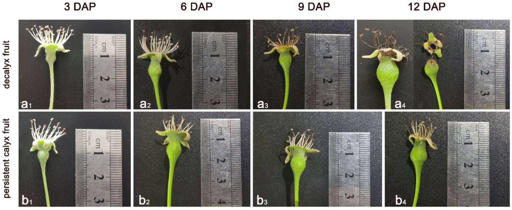
3.1.1. Changes in External Morphological Characteristics during Calyx Abscission

The external morphological characteristics of the decalyx and persistent calyx fruits during calyx abscission differed markedly in Korla fragrant pear (Figure 2). The external changes in decalyx fruit were as follows: within 3 days after pollination, the stamens and calyx developed normally, epidermal hairs were observed on the surface of the calyx and young fruit, and the fruit gradually increased in size (Figure 2(a1)). By 6 days after pollination, the anthers of the stamens began to turn brown, the calyx and young fruit were still growing normally, a relatively faint ring-like abscission zone could be observed, and the epidermal hairs on the surface of the young fruit began to decrease (Figure 2(a2)). By 9 days after pollination, the filaments of the stamens began to turn brown, the calyx was still growing normally, and a more obvious yellow ring of abscission could be observed, which made it possible to determine whether the calyx was that of the decalyx fruit (Figure 2(a3)). By 12 days after pollination, the anthers and filaments of the stamens turned completely dark brown, the lower part of the calyx tube turned dark brown, and marginal wilting and drying features were evident. Moreover, the calyx began to separate from the young fruit in the abscission zone, but there was still some of the calyx attached, after which the abscission was naturally completed under the action of wind and rain, leaving no calyx present in the abscission zone (Figure 2(a4)).
The external changes in persistent calyx fruit were as follows: By 3 days after pollination, little differences were observed in appearance between decalyx fruits and persistent calyx fruits (Figure 2(b1)). During calyx growth and development, the anthers of the stamens began to turn brown 6 days after pollination (Figure 2(b2)), but the overall drying and water loss of the stamens were slow, the epidermal hairs of the young fruit gradually decreased, and the fruit grew in size (Figure 2(b3,b4)). Moreover, the calyx always grew and developed normally, showing no signs of water loss, wilting, and falling off, and it became part of the fruit apex as the fruit grew and developed.

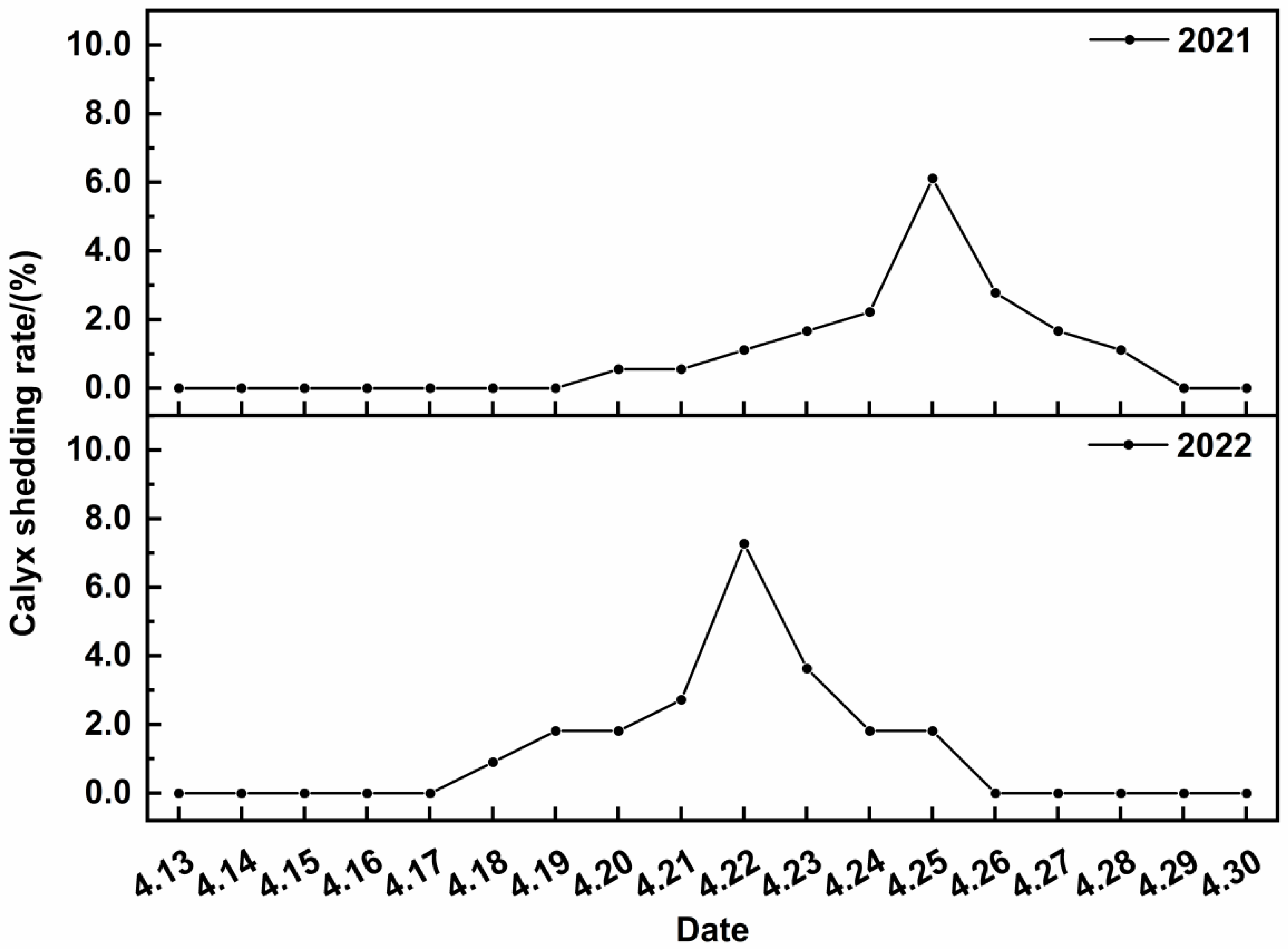
3.1.2. Calyx Abscission Patterns in 2021 and 2022

The calyx abscission started on 20 April (6 days after pollination) and peaked at 6.11% on 25 April in 2021, while the start of calyx abscission was on 18 April (6 days after pollination), and the peak abscission rate was 7.27% on 22 April in 2022 (Figure 3). The exact time of calyx abscission varied according to the temperature of each year, and for 2021–2022, in the natural state, the total time of calyx abscission of Korla fragrant pear began from about 6 days after pollination to about 13 days after pollination, and the peak of calyx abscission occurred at about 10 days after pollination.

3.2. Characteristics of Programmed Cell Death during Korla Fragrant Pear Calyx Abscission

3.2.1. Cellular Ultrastructure

At 3 days after pollination, there were no significant differences in cell morphology and outer wall structure between the decalyx and persistent calyx fruit, which both showed normal nuclei and most of the mitochondria. The difference was that the Golgi apparatus was found in the cells of the abscission zone of the decalyx fruit, while the cells of the corresponding part of the abscission zone of the persistent calyx fruit were filled with most of the chloroplasts (Figure 4a,f). At 6 days after pollination, the cells in the abscission zone of the decalyx fruit were slightly edematous, with intact cell membranes and cell walls that were continuous and of uniform thickness, with no obvious separation of the plasmolysis. The nucleus was an oval shape, with the nuclear membrane becoming blurred. Moreover, the chloroplasts were moderately numerous and had an intact membrane structure and a homogeneous matrix, and plastoglobuli were abundant. The mitochondria were moderately numerous and slightly swollen, and the vesicles had a local membrane break (Figure 4b). The cytoskeleton of the part of the persistent calyx fruit that corresponded to the abscission zone of the decalyx fruit was normal, and the nucleus was enlarged; the number of chloroplasts was moderate, the membrane structure was intact, and the number of plastoglobulus was abundant; and the number of mitochondria was passable and the morphology was normal (Figure 4g). At 9 days after pollination, the outer cell wall structure of the abscission zone of the decalyx fruit was deformed, and the number of Golgi vesicles increased and their size became larger, with the vesicle membrane border becoming blurred. The cells were vacuolated, with dark and round granules of different sizes present. Moreover, the number of mitochondria decreased and showed clear marginalization (Figure 4c). The cytoskeleton of the part of the persistent calyx fruit that corresponded to the abscission zone of the decalyx fruit was normal, with a moderate number of mitochondria and an intact membrane structure. Furthermore, the number of chloroplasts increased, with an abundance of plastoglobuli and a homogeneous stroma, and with obvious starch granules filling the chloroplasts (Figure 4h). At 12 days after pollination, the outer wall structure of the cell at the distal end of the abscission zone of the decalyx fruit was fractured and severely deformed. The vesicles had been completely degraded, the nucleus was wrinkled and severely degraded, and the organelles, including mitochondria, were degraded to residues concentrated in the cell in clusters of varying sizes and to apoptotic fragments, which is typical of the ultrastructural features of programmed cell death (Figure 4d). The outer wall of the cell at the proximal end of the abscission zone appeared significantly thickened. Additionally, the number of vesicles increased significantly, but the vesicles decreased in size, with a small amount of flocculent distribution visible. The chloroplasts were less numerous and filled with abundant plastoglobuli, and there was no obvious mitochondrial structure visible (Figure 4e). The part of the persistent calyx fruit that corresponded to the abscission zone of the decalyx fruit appeared to be visibly thickened within the cell wall, and no wrinkling or deformation of the cell wall was found. The number of vesicles increased, and the membrane boundary was relatively blurred. The number of other organelles, such as chloroplasts and mitochondria, was moderate, and the organelle structure was morphologically normal and distributed at the cell margins. In the part of the persistent calyx fruit that corresponded to the abscission zone of the decalyx fruit entered a relatively quiescent and inactive state after maturation, thus not moving towards apoptosis (Figure 4i).

3.2.2. DAPI Fluorescence and TUNEL Assay

By 9 days after pollination, blue-stained nuclei could be clearly observed along the anatomical structure of the abscission zone in both the abscission part of the decalyx fruit and the corresponding part in the persistent calyx fruit, with the cells appearing regularly rounded and closely arranged (Figure 5(a1–f1)). On day 12 after pollination, the number of blue-stained nuclei observed at the distal end of the abscission zone of the decalyx fruit was significantly reduced, and the brightness of the blue-stained nuclei was lower (Figure 5(g1)), whereas the number of blue-stained nuclei observed at the proximal end of the abscission zone was moderate, indicating that the cells that had become the protective layer developed normally after calyx abscission and programmed cell death were complete (Figure 5(h1)). In the field of view, fewer blue nuclei were observed in the corresponding parts in the persistent calyx fruit, which may be explained by the formation of a thickened layer on the inner wall of the cells in this part, the reduction in the volume of the nuclei and their distribution at the cell margins, and the separation of the mass wall in some cells, resulting in fewer nuclei and lower staining intensity (Figure 5(i1)).
At 6 days after pollination, a few nuclei with red fluorescence were observed in the decalyx fruit abscission zone cells, showing irregular morphology, and the abscission zone tissue was in good cellular condition, showing the presence of a few apoptotic cells around the normal cells, at which point an early apoptotic cell phenomenon was observed (Figure 5(c2,c3)). At 9 days after pollination, a significant increase in nuclei with red fluorescence was observed in the decalyx fruit abscission zone cells, with disorganized and loose cell-to-cell arrangements, and the gaps widened, showing significant apoptotic features, at which point a mid-to-late-stage apoptotic cell phenomenon was observed (Figure 5(e2,e3)). Twelve days after pollination, no obvious apoptosis-positive cells were observed in the field of view (Figure 5(g2,g3)). During the different periods of Korla fragrant pear calyx abscission, the cells in corresponding zone in the persistent calyx fruit under the field of view were relatively uniformly stained, and no apoptotic-positive cells were observed, indicating that their nuclear morphology was normal and that no nuclear deformation, solidification, or other phenomena occurred (Figure 5b,d,f,i).

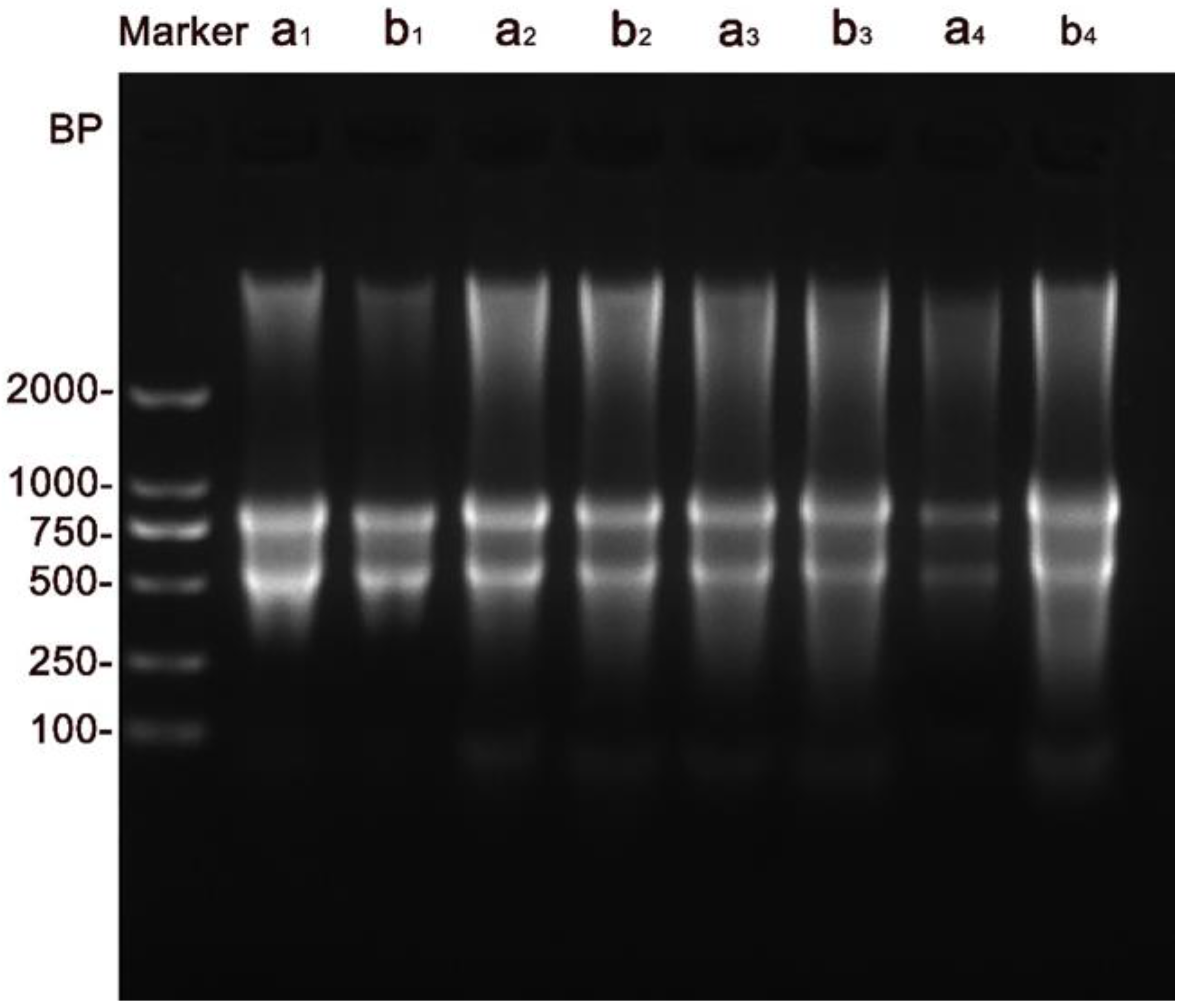
3.2.3. DNA Fragmentation Assay

At 3 days after pollination, the DNA gel electrophoresis results showed a slight trailing effect (Figure 6(a1,b1)). At 6 days after pollination, the DNA gel electrophoresis results showed significant trailing (Figure 6(a2,b2)). At 9 days after pollination, the DNA gel electrophoresis results showed a significant trailing phenomenon, which tended to intensify compared to results from the previous day (Figure 6(a3,b3)). At 12 days after pollination, the trailing phenomenon of the DNA gel electrophoresis results disappeared, and the degradation of the DNA deepened, resulting in lower DNA integrity. Moreover, the previously bright bands became very light and difficult to identify, which was probably caused by the death of most cells in the calyx abscission zone in the later stage, indicating that the cells in this zone were degraded, a typical biochemical feature of programmed cell death (Figure 6(a4)). The DNA gel electrophoresis for the persistent calyx fruit, however, was still deepening, but the brightness of the bands had increased, indicating that although there was the occurrence of DNA degradation, the DNA integrity was good and no apoptosis occurred (Figure 6(b4)).

3.3. PCD-Regulated Signals during Korla Fragrant Pear Calyx Abscission

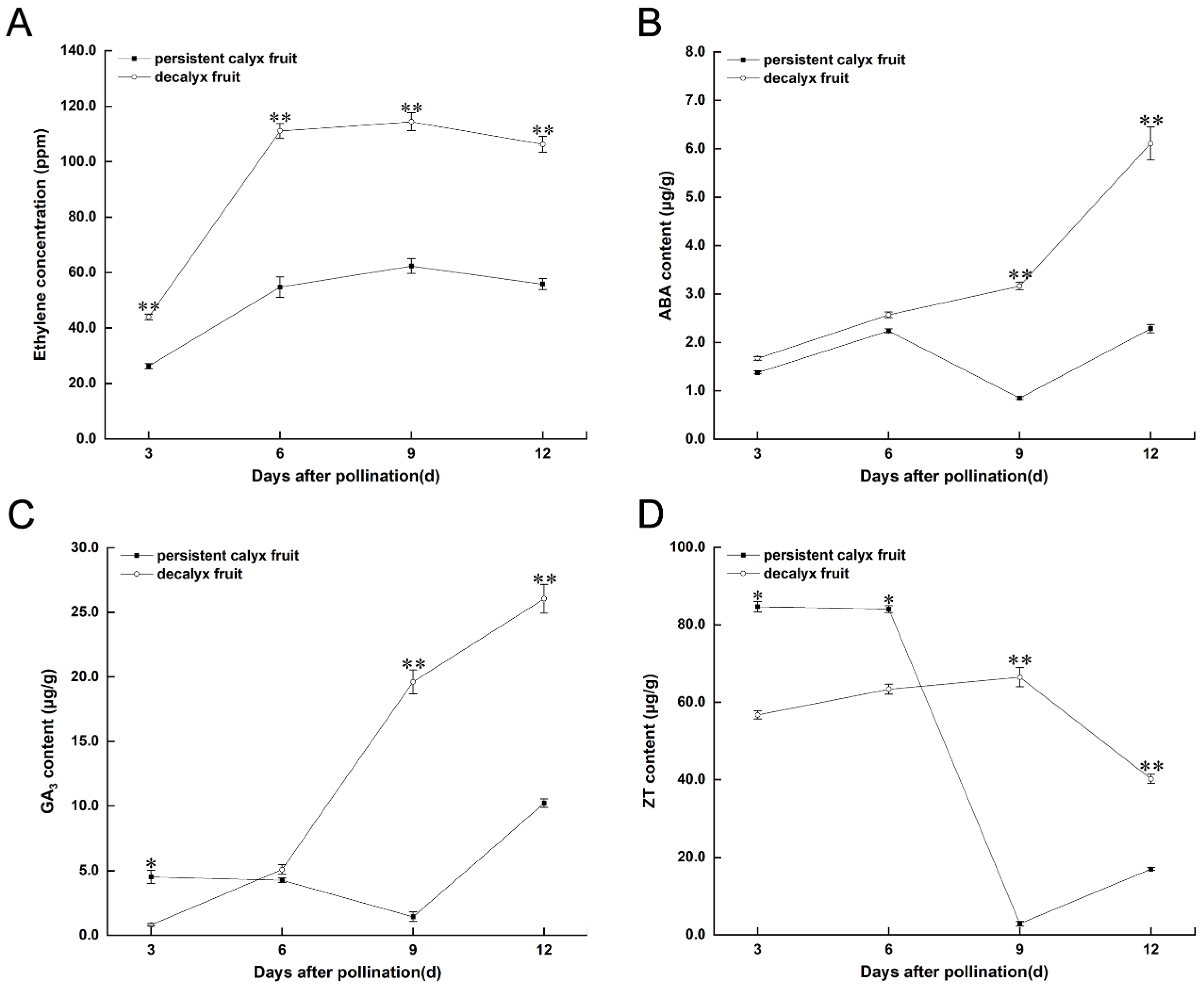
3.3.1. Endogenous Hormone

During the calyx abscission in Korla fragrant pear calyxes, the ETH concentration of the decalyx fruit was significantly higher than that of the persistent calyx fruit in different stages, decreasing after a peak 9 days after pollination (Figure 7A). Moreover, the ETH concentrations of the decalyx fruit 3, 6, 9, and 12 days after pollination were 43.97 ppm, 111.03 ppm, 114.36 ppm, and 106.24 ppm, respectively, which were 67.69%, 102.83%, 83.50%, and 90.39% higher than those of the persistent calyx fruits. The ABA content showed an increasing trend in the decalyx calyx fruit during calyx abscission (Figure 7B), while that of the persistent calyx fruit varied little. The ABA content of the decalyx fruits was 3.17 μg/g and 6.11 μg/g 9 and 12 days after pollination, respectively, and was significantly higher by 272.94% and 167.98% compared to those of the persistent calyx fruits, respectively. The GA3 content of the persistent calyx fruit was significantly higher than that of the decalyx fruit 3 days after pollination, and then gradually decreased and increased significantly 12 days after pollination (Figure 7C). Furthermore, the GA3 content of the decalyx fruit gradually increased and was significantly higher than that of the persistent calyx fruit 9 and 12 days after pollination; the GA3 content of the decalyx fruit was 19.61 ng/g and 26.05 ng/g 9 and 12 days after pollination, respectively, which were 126.18% and 154.89% higher than that of the persistent calyx fruit. The ZT content of the persistent calyx fruit was significantly higher than those of the decalyx fruits 3 and 6 days post pollination, decreasing significantly at 9 days and then increasing (Figure 7D). Furthermore, the ZT content of the decalyx fruit gradually increased and peaked 9 days post pollination and was significantly higher than that of the persistent calyx fruit. The ZT content of the decalyx fruits was 66.45 ng/g and 40.18 ng/g 9 and 12 days post pollination, which were 221.53% and 136.91% higher than those of the persistent calyx fruits, respectively.

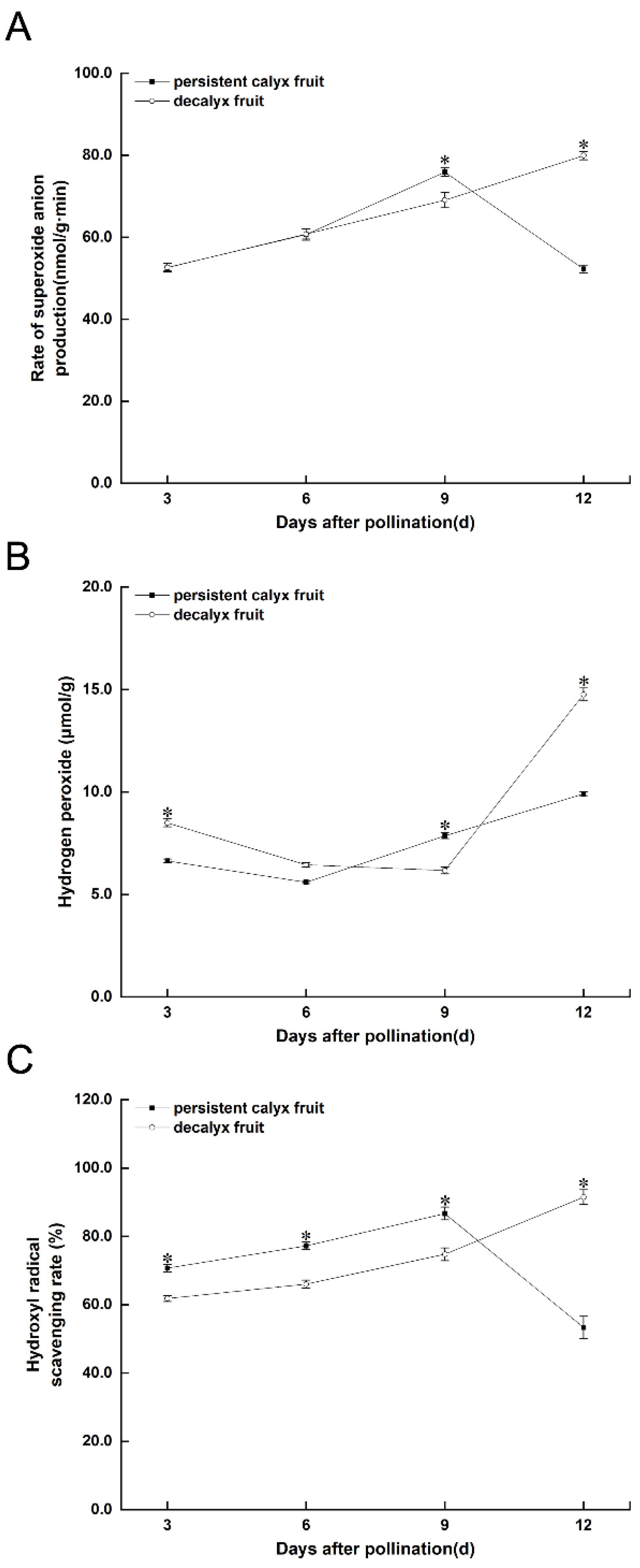
3.3.2. Reactive Oxygen Species

The rate of superoxide anion production in the decalyx fruit and persistent calyx fruit of Korla fragrant pear gradually increased during 3 to 9 days after pollination, being significantly higher in the persistent calyx than in the decalyx fruit 9 days after pollination. The rate of superoxide anion production in the decalyx fruit gradually increased, peaking 12 days after pollination, and was significantly higher than that in the persistent calyx fruit (Figure 8A). The hydrogen peroxide content in the persistent calyx fruit showed a decrease and then an increase, which was significantly higher than that of the decalyx fruit 9 days post pollination and which peaked 12 days post pollination. Meanwhile, the hydrogen peroxide content of the decalyx fruit was significantly higher than that of the persistent calyx fruit 3 days post pollination specifically at 8.49 μmol/g, which was 27.86% higher than that of the persistent calyx fruit, and then declined until 12 days post pollination when it rapidly increased and peaked, becoming significantly higher than that of the persistent calyx fruit (Figure 8B). The rate of hydroxyl radical scavenging in the persistent calyx fruit increased gradually from 3 to 9 days after pollination, peaking 9 days after pollination and decreasing thereafter. The rate of hydroxyl radical scavenging in the decalyx fruit showed a gradual increase, peaking 12 days after pollination, and was significantly higher than that in the persistent calyx fruit (Figure 8C).

4. Discussion

4.1. Apparent Characteristics of Calyx Abscission in Korla Fragrant Pear

Abscission is a widespread phenomenon in plants, and the process of abscission includes abscission zone formation and separation [19], during which changes in tissue structure, physiology, biochemistry, metabolism, and gene expression occur [20,21]. The process of programmed cell death in plant organs is often accompanied by features such as wilting, browning, and chlorosis [22]. Related studies have reported that yellow ring-shaped abscission zones can be observed in the calyx of young fruits of Korla fragrant pear during calyx development 8 days after the flowers are in full bloom to determine whether the calyx is undergoing abscission [23,24]. Previous studies showed that only vessels were present in the microstructure of the vascular bundle of cells in the abscission zone, and that no sieve tubes or heterocells were formed, so there was a lack of nutrient and water supply in the calyx, leading to the emergence of the abscission zone in the late development of the calyx [24]. This also indicated that the abscission process had completely finished, and ultimately, the decalyx fruit was formed. Previously, we found that calyx abscission in Korla fragrant pear could be divided into three periods: before, during, and after the formation of the abscission zone [25]. On this basis, to understand in detail the reasons for the occurrence of PCD during the calyx abscission process, we divided the process into more detailed divisions and observed whether there were external features of PCD during the process to grasp the details of its developmental changes. The results of this study show that the total time of calyx abscission of Korla fragrant pear began from about 6 days after pollination to about 13 days after pollination, and the peak of calyx abscission occurred at about 10 days after pollination. The external morphological characteristics of the decay and abscission of the calyx in the decalyx fruit over time were as follows: First, normal development of the calyx. Then, the calyx appeared as a faint ring-like abscission zone, followed by a more obvious abscission zone with a yellow ring (only vessels were present in the vascular bundle). Next, the lower part of the calyx tube turned dark brown and the edge wilted (at the distal end of the abscission zone, most of the tissues appeared extruded, wrinkled, and distorted). Ultimately, calyx abscission spontaneously occurred (a protective layer was produced near the axial end where the abscission zone was broken).
In addition, in pollination work, we intentionally bagged a part of Korla fragrant pears in the big bud stage in order to prevent their pollination and fertilization, and after 20 days, the bags were removed, and no PCD was found in Korla fragrant pears. We therefore hypothesize that pollination may be an important factor affecting the PCD phenomenon during Korla fragrant pear calyx abscission, and more research should be focused on the effects of pollination on PCD in future research.

4.2. PCD Was Involved in Calyx Abscission in Korla Fragrant Pear

Cells that undergo PCD in plant organs have distinctive morphological features, which are usually characterized by cytoskeletal disruption, reduced cell size, nuclear condensation, chromatin margination, the degradation of nuclear DNA from nucleosomes by induced nucleic acid endonucleases, and the production of oligomeric nucleosome fragments of different sizes [26]. These fragments can be seen on gel electrophoresis as “ladder” DNA strips multiplied by 180–200 bp. In addition, DNA fragmentation is often considered a “hallmark” of PCD in plants and animals [27]. TUNEL has long been widely adopted in the study of apoptosis as a research method combining molecular biology and morphology and accurately reflecting the typical biochemical and morphological characteristics of programmed cell death [28]. Our results revealed that 3 days post pollination, cells in the abscission zone of Korla fragrant pear calyx did not show typical PCD features; by 6 days post pollination, a few red fluorescent nuclei were observed in the abscission zone of the calyx, which showed an irregular morphology, indicating the presence of a few apoptotic cells around normal cells, at which point the PCD process was initiated, showing early PCD. At 9 days post pollination in the abscission zone of calyx, some typical PCD features were detected, including disorganized and loose cell-to-cell arrangements in the abscission zone, widened gaps, positive TUNEL tests, and significant trailing in the DNA gel electrophoresis indicators, which have been reported in the occurrence of PCD in barley aleurone layer cells, at which time the degree of PCD further deepened and the phenomenon of intermediate PCD was manifested [29]. By 12 days post pollination, the decalyx fruit showed a fracture of the outer wall structure at the distal end of the abscission zone and severe deformation. Additionally, the vesicles were completely degraded, the nucleus was wrinkled and severely degraded, and the organelles, including mitochondria, were degraded to residues concentrated in the cell in clusters of different sizes and to apoptotic debris, which is a typical ultrastructural feature of programmed cell death [27]. However, no obvious apoptosis-positive cells were observed in the field of view at this point in the TUNEL assay, probably because of the limitations of TUNEL fluorescence staining, where apoptotic cells and cell necrosis undergo DNA breaks and are labeled by dUTP but do not stain well for apoptotic cells after the late stages. At the same time, the DNA gel electrophoresis of the decalyx fruit lost its tailing phenomenon, the degradation of DNA deepened (resulting in lower DNA integrity), and the previously bright bands became light and difficult to identify, likely because most of the cells in the decalyx fruit abscission zone had died. This indicated that the cells in the calyx abscission zone were degraded, a typical biochemical feature of programmed cell death, which could indicate that this was a late PCD phenomenon; in contrast, the outer wall of the cells in the proximal end of the abscission zone appeared significantly thickened, with a moderate number of nuclei stained in blue, indicating that programmed cell death had ended at the completion of the calyx abscission, when the cells that become the protective layer develop normally [30]. Notably, in this study, microscopic and ultrastructural observations of the cells in the abscission zone of the calyx revealed that both sides of the cells in the decalyx fruit exhibited asymmetry during calyx abscission. Feng et al. [31] reported that programmed cell death in the distal part of chestnut male flowers facilitated the distribution and balance of nutrients between female and male flowers, and in the study by Bar-Dror et al. [17] on the mechanism of leaf abscission in tomato, the expression of PCD-related genes such as LX and nuclease genes was mainly expressed in the distal tissues of the abscission zone, showing an asymmetry between the two sides of the cells in the abscission zone. Thus, there was an asymmetry in the occurrence of PCD, a key mechanism, in the normal abscission process.
In summary, we compared the period of Korla fragrant pear calyx abscission with the PCD process and found that the period before the formation of the abscission zone corresponded to 3–6 days after pollination. At 6 days after pollination, the abscission zone of decalyx fruits showing the early PCD phenomenon and the PCD process started, the period of abscission zone formation corresponded to 9 days after pollination (showing the intermediate PCD phenomenon) when the PCD process further deepened, and the point after the formation of the abscission zone corresponded to 12 days after pollination (showing the late PCD phenomenon at the distal end) when the PCD process entered its final stage. In the early stages (3–6 days post pollination), cells in the calyx of Korla fragrant pear did not show typical PCD morphology characteristics, which was the key stage of PCD regulation in the process of calyx abscission of Korla fragrant pear.

4.3. Relationship between PCD and Plant Hormone during Calyx Abscission in Korla Fragrant Pear

The onset of PCD during plant organ or tissue abscission is regulated spatially and temporally by ethylene [32]. Ethylene can affect the fragmentation of plant cell DNA, and early PCD can be induced in wheat and maize endosperm by ethylene treatment, while treatment with substances that inhibit ethylene synthesis can significantly delay endosperm PCD [4]. The induction of PCD in the rice root epidermis is dependent on ethylene signaling and is further promoted by GA, whereas treatment with GA alone fails to induce PCD and requires ethylene and GA to act in a synergistic manner for induction [33], while the combined effect of ethylene and GA can be reversed by CTK and ABA [34]. The results of the present study revealed that the ETH concentrations of the decalyx fruit were significantly higher than those of the persistent calyx fruits in each period during calyx abscission, and when the cells of the Korla fragrant pear calyx abscission zone did not exhibit the typical morphological and biochemical characteristics of PCD, during the early stage of abscission zone formation (3–6 days after pollination), it was also significantly higher in decalyx fruits than in the persistent calyx fruit during this period, while ABA did not show significant differences between the decalyx fruits and persistent calyx fruit. In contrast, GA3 was significantly lower at 3 days and ZT was significantly lower at 3 and 6 days after pollination in the decalyx fruits than in the persistent calyx fruits. Moreover, compared to other plant hormones, ethylene concentration was highest throughout the calyx abscission period. Therefore, it was speculated that ethylene acts as the primary signal, most likely in concert with other signaling molecules, to control PCD development [35], and high concentrations of ethylene promoted calyx abscission, while GA3 and ZT had opposite effects. This suggested that ethylene signaling was the main signaling pathway leading to PCD in the calyx abscission of Korla fragrant pear, and played a positive regulatory role, in agreement with the findings of Lombardi et al. [36]. Previous research has found that ethylene signals MPK6 and EIN3 are activated during the preparation phase of PCD and regulate downstream target genes [37]. Among them, EIN3 plays a central role in the process of PCD, promoting the expression of positive regulatory factors ORE1 and AtNAP, and reducing miR164 to relieve the post-transcriptional inhibitory effect on ORE1, thereby promoting programmed cell death [38].

4.4. Relationship between PCD and Reactive Oxygen Species during Calyx Abscission in Korla Fragrant Pear

ROS, including O2, H2O2, and ·OH, are important signals for the induction of PCD. In the present study, we found that when the cells in the Korla fragrant pear calyx abscission zone did not show the typical morphological characteristics of PCD, during the pre-abscission zone formation period (3 days after pollination), only the H2O2 content was significantly higher in the decalyx fruit than in the persistent calyx fruit. This is presumably because high concentrations of ethylene in the decalyx fruit’s abscission zone regulated the expression of genes related to H2O2 metabolism and activate enzymes involved in H2O2 synthesis to promote the accumulation of H2O2 [28,39,40], and H2O2 could directly or indirectly oxidize intracellular nucleic acids, proteins, and other biomolecules and cause damage to cell membranes, thus accelerating the aging and disintegration of cells [41]. Furthermore, H2O2 has better stability compared to other reactive oxygen species, and at high concentrations, it induces oxidative damage to biomolecules, ultimately leading to cell death, making it an important regulatory signal in the process of programmed cell death [42,43]. Therefore, we speculated that H2O2 was the main regulatory signal that induced PCD in the calyx abscission of Korla fragrant pear and played a positive regulatory role, which was consistent with the results of Pan et al. [44].
Notably, calyx abscission cells of decalyx fruits in Korla fragrant pear exhibited the morphological and biochemical characteristics of late PCD in the late stage of abscission zone formation (12 days after pollination), and the levels of the three reactive oxygen species continued to rise and reached their peak, which indicated that it was likely in the late stage of programmed cell death that the damage caused by high levels of reactive oxygen species to the plant exceeded the ability of the plant cells to regulate themselves. Under normal conditions, the generation and removal of reactive oxygen species in plants were in dynamic balance, and plants can maintain normal life activities. However, once this balance was disturbed, their cellular state would be affected, resulting in plant damage or even death.

5. Conclusions

In this study, we systematically observed the typical morphological characteristics of programmed cell death and the dynamics of regulatory signals (plant hormones and ROS) during the Korla fragrant pear calyx abscission process, and we demonstrated from multiple perspectives that PCD was involved in the process of Korla fragrant pear calyx abscission and that PCD was influenced by plant hormones and ROS, among which ethylene and H2O2 were the main signals initiating the calyx PCD process of Korla fragrant pear and thus affect calyx abscission.

Author Contributions

Y.W., B.S. and J.T. conceived the structure and idea of this paper. Z.H., T.S. and Y.H. were involved in collecting data. C.W. and F.Z. were involved in revising the manuscript. Y.W. and B.S. took charge of data processing, data analysis, and the writing of this paper. All authors have read and agreed to the published version of the manuscript.

Funding

This study was supported by the Natural Science Foundation of Xinjiang Uygur Autonomous Region (grant number: 2022D01A178), the National Natural Science Foundation of China (grant number: 32160686), and the Xinjiang Agricultural University High-level Talent Cultivation Program.

Institutional Review Board Statement

Not applicable.

Informed Consent Statement

Not applicable.

Data Availability Statement

All data that support the findings of this study are available from the corresponding author upon reasonable request.

Conflicts of Interest

The authors declare no conflicts of interest.

References

  1. Locato, V.; De Gara, L. Programmed Cell Death in Plants: An Overview. In Plant Programmed Cell Death: Methods and Protocols; Humana Press: New York, NY, USA, 2018; pp. 1–8. [Google Scholar]
  2. Huysmans, M.; Buono, R.A.; Skorzinski, N.; Radio, M.C.; De Winter, F.; Parizot, B.; Mertens, J.; Karimi, M.; Fendrych, M.; Nowack, M.K. NAC Transcription Factors ANAC087 and ANAC046 Control Distinct Aspects of Programmed Cell Death in the Arabidopsis Columella and Lateral Root Cap. Plant Cell 2018, 30, 2197–2213. [Google Scholar] [CrossRef] [PubMed]
  3. Parveen, S.; Altaf, F.; Farooq, S.; Lone, M.L.; Haq, A.U.; Tahir, I. The Swansong of Petal Cell Death: Insights into the mechanism and regulation of ethylene-mediated flower senescence. J. Exp. Bot. 2023, 74, 3961–3974. [Google Scholar] [CrossRef] [PubMed]
  4. Li, C.; Zhang, X.; Guo, W.; Fu, K.; Li, C.; Li, C. Impact of Ethylene and Abscisic Acid on Programmed Cell Death in Endosperm and Grain Filling of Wheat (Triticum aestivum L.). J. Plant Growth Regul. 2023, 42, 4275–4289. [Google Scholar] [CrossRef]
  5. Huang, L.; Liu, Y.; Wang, X.; Jiang, C.; Zhao, Y.; Lu, M.; Zhang, J. Peroxisome-Mediated Reactive Oxygen Species Signals Modulate Programmed Cell Death in Plants. Int. J. Mol. Sci. 2022, 23, 10087. [Google Scholar] [CrossRef] [PubMed]
  6. Ren, H.; Zhao, X.; Li, W.; Hussain, J.; Qi, G.; Liu, S. Calcium Signaling in Plant Programmed Cell Death. Cells 2021, 10, 1089. [Google Scholar] [CrossRef] [PubMed]
  7. Gao, Y.; Liu, Y.; Liang, Y.; Lu, J.; Jiang, C.; Fei, Z.; Jiang, C.Z.; Ma, C.; Gao, J. Rosa hybrida Rh ERF 1 and Rh ERF 4 Mediate Ethylene-and Auxin-Regulated Petal Abscission by Influencing Pectin Degradation. Plant J. 2019, 99, 1159–1171. [Google Scholar] [CrossRef] [PubMed]
  8. Meir, S.; Philosoph-Hadas, S.; Sundaresan, S.; Selvaraj, K.V.; Burd, S.; Ophir, R.; Kochanek, B.; Reid, M.S.; Jiang, C.-Z.; Lers, A. Microarray Analysis of the Abscission-Related Transcriptome in the Tomato Flower Abscission Zone in Response to Auxin Depletion. Plant Physiol. 2010, 154, 1929–1956. [Google Scholar] [CrossRef] [PubMed]
  9. Xie, Y.; Zhang, C.; Lai, D.; Sun, Y.; Samma, M.K.; Zhang, J.; Shen, W. Hydrogen Sulfide Delays GA-Triggered Programmed Cell Death in Wheat Aleurone Layers by the Modulation of Glutathione Homeostasis and Heme Oxygenase-1 Expression. J. Plant Physiol. 2014, 171, 53–62. [Google Scholar] [CrossRef] [PubMed]
  10. Cortleven, A.; Leuendorf, J.E.; Frank, M.; Pezzetta, D.; Bolt, S.; Schmülling, T. Cytokinin Action in Response to Abiotic and Biotic Stresses in Plants. Plant Cell Environ. 2019, 42, 998–1018. [Google Scholar] [CrossRef]
  11. Ye, C.; Zheng, S.; Jiang, D.; Lu, J.; Huang, Z.; Liu, Z.; Zhou, H.; Zhuang, C.; Li, J. Initiation and Execution of Programmed Cell Death and Regulation of Reactive Oxygen Species in Plants. Int. J. Mol. Sci. 2021, 22, 12942. [Google Scholar] [CrossRef]
  12. Wang, Y.; Shirakawa, M.; Ito, T. Dynamic Changes in Reactive Oxygen Species in the Shoot Apex Contribute to Stem Cell Death in Arabidopsis thaliana. Int. J. Mol. Sci. 2022, 23, 3864. [Google Scholar] [CrossRef] [PubMed]
  13. Sun, Y.; Tian, X.; Wu, H.; Hao, X.; Gao, B.; Zhang, H.; Feng, J.; Han, L. H2O2 Signaling Modulates Glycoprotein-1 Induced Programmed Cell Death in Tobacco Suspension Cells. Pestic. Biochem. Physiol. 2021, 171, 104697. [Google Scholar] [CrossRef] [PubMed]
  14. Chang, Y.; Yang, H.; Ren, D.; Li, Y. Activation of ZmMKK10, a Maize Mitogen-Activated Protein Kinase Kinase, Induces Ethylene-Dependent Cell Death. Plant Sci. 2017, 264, 129–137. [Google Scholar] [CrossRef] [PubMed]
  15. Jiang, W.; Yan, P.; Zheng, Q.; Wang, Z.; Chen, Q.; Wang, Y. Changes in the Metabolome and Nutritional Quality of Pulp from Three Types of Korla Fragrant Pears with Different Appearances as Revealed by Widely Targeted Metabolomics. Plants 2023, 12, 3981. [Google Scholar] [CrossRef] [PubMed]
  16. Lers, A.; Sonego, L.; Green, P.J.; Burd, S. Suppression of LX Ribonuclease in Tomato Results in a Delay of Leaf Senescence and Abscission. Plant Physiol. 2006, 142, 710–721. [Google Scholar] [CrossRef] [PubMed]
  17. Bar-Dror, T.; Dermastia, M.; Kladnik, A.; Žnidarič, M.T.; Novak, M.P.; Meir, S.; Burd, S.; Philosoph-Hadas, S.; Ori, N.; Sonego, L.; et al. Programmed Cell Death Occurs Asymmetrically during Abscission in Tomato. Plant Cell 2011, 23, 4146–4163. [Google Scholar] [CrossRef] [PubMed]
  18. Rodriguez, J.M.; Morcillo, R.L.; Vierheilig, H.; Ocampo, J.A.; Ludwig-Müller, J.; Garrido, J.M.G. Mycorrhization of the Notabilis and Sitiens Tomato Mutants in Relation to Abscisic Acid and Ethylene Contents. J. Plant Physiol. 2010, 167, 606–613. [Google Scholar] [CrossRef] [PubMed]
  19. Liu, X.; Chen, R.; Ma, J. Detection of Antioxidant Enzymes and DNA Dynamic Changes During Senescence of Eucalyptuses Phellogen Zone. Acta Bot. Boreali-Occident. Sin. 2012, 32, 940–947. [Google Scholar]
  20. Heo, S.; Chung, Y.S. Validation of MADS-Box Genes from Apple Fruit Pedicels during Early Fruit Abscission by Transcriptome Analysis and Real-Time PCR. Genes Genom. 2019, 41, 1241–1251. [Google Scholar] [CrossRef]
  21. Pei, M.; Niu, J.; Li, C.; Cao, F.; Quan, S. Identification and Expression Analysis of Genes Related to Calyx Persistence in Korla Fragrant Pear. BMC Genom. 2016, 17, 132. [Google Scholar] [CrossRef]
  22. Kirschner, G.K. A matter of Life and Death: Networks regulating programmed cell death in Arabidopsis. Plant J. 2023, 115, 1463–1464. [Google Scholar] [CrossRef] [PubMed]
  23. Ma, L.; Zhou, L.; Quan, S.; Xu, H.; Yang, J.; Niu, J. Integrated Analysis of mRNA-Seq and miRNA-seq in Calyx Abscission Zone of Korla Fragrant Pear Involved in Calyx Persistence. BMC Plant Biol. 2019, 19, 192. [Google Scholar] [CrossRef]
  24. Ma, H.; Wang, Y.; Wen, X.; Qi, M.; Li, J. Effects of Different Reagent Treatments on the Microstructure of Calyx Tubes with or without Calyx of Korla Fragrant Pear. J. Fruit Sci. 2011, 28, 518–520. [Google Scholar]
  25. Ma, L.; Zhou, L.; Quan, S.; Xu, H.; Yang, J.; Niu, J. Relationship between Abscission of Calyx of Korla Fragrant Pear and Plant Hormones in Different Parts of the Separation Zone. Plant Physiol. J. 2022, 58, 1369–1380. [Google Scholar]
  26. Lee, J.; Chen, H.; Lee, G.; Emonet, A.; Kim, S.; Shim, D.; Lee, Y. MSD2-Mediated ROS Metabolism Fine-Tunes the Timing of Floral Organ Abscission in Arabidopsis. New Phytol. 2022, 235, 2466–2480. [Google Scholar] [CrossRef]
  27. Van Doorn, W.G. Classes of Programmed Cell Death in Plants, Compared to Those in Animals. J. Exp. Bot. 2011, 62, 4749–4761. [Google Scholar] [CrossRef] [PubMed]
  28. Ni, X.-L.; Gui, M.-Y.; Tan, L.-L.; Zhu, Q.; Liu, W.-Z.; Li, C.-X. Programmed Cell Death and Aerenchyma Formation in Water-Logged Sunflower Stems and Its Promotion by Ethylene and ROS. Front. Plant Sci. 2019, 9, 1928. [Google Scholar] [CrossRef]
  29. Wang, M.; Oppedijk, B.J.; Lu, X.; Van Duijn, B.; Schilperoort, R.A. Apoptosis in Barley Aleurone during Germination and Its Inhibition by Abscisic Acid. Plant Mol. Biol. 1996, 32, 1125–1134. [Google Scholar] [CrossRef] [PubMed]
  30. Ni, X.L.; Su, H.; Zhou, Y.F.; Wang, F.H.; Liu, W.Z. Leaf-Shape Remodeling: Programmed Cell Death in Fistular Leaves of a Llium fistulosum. Physiol. Plant. 2015, 153, 419–431. [Google Scholar] [CrossRef]
  31. Feng, Y.-Q.; Shen, Y.-Y.; Qin, L.; Cao, Q.-Q.; Han, Z.-H. Short catkin1, a Novel Mutant of Castanea mollissima, is Associated with Programmed Cell Death during Chestnut Staminate Flower Differentiation. Sci. Hortic. 2011, 130, 431–435. [Google Scholar] [CrossRef]
  32. Trobacher, C.P. Ethylene and Programmed Cell Death in Plants. Botany 2009, 87, 757–769. [Google Scholar] [CrossRef]
  33. Steffens, B.; Sauter, M. Epidermal Cell Death in Rice is Regulated by Ethylene, Gibberellin, and Abscisic Acid. Plant Physiol. 2005, 139, 713–721. [Google Scholar] [CrossRef] [PubMed]
  34. Orzáez, D.; Granell, A. DNA Fragmentation is Regulated by Ethylene during Carpel Senescence in Pisum sativum. Plant J. 1997, 11, 137–144. [Google Scholar] [CrossRef]
  35. Li, N.; Han, X.; Feng, D.; Yuan, D.; Huang, L.-J. Signaling Crosstalk between Salicylic Acid and Ethylene/Jasmonate in Plant Defense: Do We Understand What They Are Whispering? Int. J. Mol. Sci. 2019, 20, 671. [Google Scholar] [CrossRef] [PubMed]
  36. Lombardi, L.; Mariotti, L.; Picciarelli, P.; Ceccarelli, N.; Lorenzi, R. Ethylene Produced by the Endosperm is Involved in the Regulation of Nucellus Programmed Cell Death in Sechium edule Sw. Plant Sci. 2012, 187, 31–38. [Google Scholar] [CrossRef] [PubMed]
  37. Li, D.; Wu, D.; Li, S.; Guo, N.; Gao, J.; Sun, X.; Cai, Y. Transcriptomic Profiling Identifies Differentially Expressed Genes Associated with Programmed Cell Death of Nucellar Cells in Ginkgo biloba L. BMC Plant Biol. 2019, 19, 91. [Google Scholar] [CrossRef] [PubMed]
  38. Li, Z.; Peng, J.; Wen, X.; Guo, H. Ethylene-Insensitive3 is a Senescence-associated Gene that Accelerates Age-dependent Leaf Senescence by Directly Repressing miR164 Transcription in Arabidopsis. Plant Cell 2013, 25, 3311–3328. [Google Scholar] [CrossRef] [PubMed]
  39. Wang, G.; Xiao, Y.; Deng, X.; Zhang, H.; Li, T.; Chen, H. Exogenous Hydrogen Peroxide Contributes to Heme Oxygenase-1 Delaying Programmed Cell Death in Isolated Aleurone Layers of Rice Subjected to Drought Stress in a cGMP-dependent Manner. Front. Plant Sci. 2018, 9, 84. [Google Scholar] [CrossRef] [PubMed]
  40. Gechev, T.S.; Hille, J. Hydrogen Peroxide as a Signal Controlling Plant Programmed Cell Death. J. Cell Biol. 2005, 168, 17–20. [Google Scholar] [CrossRef]
  41. Baxter, A.; Mittler, R.; Suzuki, N. ROS as Key Players in Plant Stress Signalling. J. Exp. Bot. 2013, 65, 1229–1240. [Google Scholar] [CrossRef]
  42. Černý, M.; Habánová, H.; Berka, M.; Luklová, M.; Brzobohatý, B. Hydrogen Peroxide: Its Role in Plant Biology and Crosstalk with Signalling Networks. Int. J. Mol. Sci. 2018, 19, 2812. [Google Scholar] [CrossRef] [PubMed]
  43. Tang, X.; Liu, M.; Chen, G.; Yuan, L.; Hou, J.; Zhu, S.; Zhang, B.; Li, G.; Pang, X.; Wang, C. TMT-Based Comparative Proteomic Analysis of the Male-Sterile Mutant ms01 sheds Light on Sporopollenin Production and Pollen Development in Wucai (Brassica campestris L.). J. Proteom. 2022, 254, 104475. [Google Scholar] [CrossRef] [PubMed]
  44. Pan, R.; Han, H.; Medison, M.B.; Abou-Elwafa, S.F.; Liu, Y.; Yang, X.; Zhang, W. Aerenchyma Formation in the Root of Leaf-Vegetable Sweet Potato: Programmed Cell Death Initiated by Ethylene-Mediated H2O2 Accumulation. Physiol. Plant. 2021, 173, 2361–2375. [Google Scholar] [CrossRef] [PubMed]
Figure 1. Photograph of the Korla fragrant pear tree.
Horticulturae 10 00637 g001
Figure 2. Changes in external morphological characteristics during Korla fragrant pear calyx abscission. Note: samples collected 3, 6, 9, and 12 days after pollination of decalyx fruit are referred to as (a1a4), respectively; samples collected 3, 6, 9, and 12 days after persistent calyx fruit pollination are referred to as (b1b4), respectively.
Horticulturae 10 00637 g002
Figure 3. Daily dynamics of Korla fragrant pear calyx abscission in 2021 and 2022.
Horticulturae 10 00637 g003
Figure 4. Cellular ultrastructure of programmed cell death during Korla fragrant pear calyx abscission. Note: (ae) are the ultrastructures of cells in 4 stages (3, 6, 9, and 12 days after pollination) of decaylx fruit, (d,e) represent cells at the distal and proximal ends of the abscission zone in decalyx fruit on day 12 after pollination, respectively; (fi) are the ultrastructures of cells in 4 stages (3, 6, 9, and 12 days after pollination) of persistent calyx fruit. N, nucleus; Nu, nucleolus; S, starch granule; M, mitochondrion; GA, Golgi apparatus; Chl, chloroplast; V, vesicle (Golgi vesicle); Pm, plasma membrane; Pb, plastoglobulus; CW, cell wall. Scale bar = 5.0 μm.
Horticulturae 10 00637 g004
Figure 5. DAPI fluorescence and TUNEL assay of PCD during Korla fragrant pear calyx abscission. Note: (ae) were detected by DAPI fluorescence and TUNEL in 4 stages (3, 6, 9, and 12 days after pollination) of decaylx fruit; (fi) were detected by DAPI fluorescence and TUNEL in 4 stages (3, 6, 9, and 12 days after pollination) of persistent calyx fruit; blue fluorescence is DAPI staining, and red fluorescence is positive apoptotic cells after TUNEL staining. First 2 columns of plots: scale bar = 80 μm. Column 3 is an equal-scale enlargement of the white-boxed portion of column 2—a representative picture of apoptotic cells: scale bar = 20 μm.
Horticulturae 10 00637 g005
Figure 6. DNA agarose electrophoresis profiles during Korla fragrant pear calyx abscission. Note: samples collected 3, 6, 9, and 12 days after decalyx fruit pollination are referred to as (a1a4), respectively; samples collected 3, 6, 9, and 12 days after persistent calyx fruit pollination are referred to as (b1b4), respectively.
Horticulturae 10 00637 g006
Figure 7. Changes in endogenous hormone content during Korla fragrant pear calyx abscission. Note: (A) ethylene; (B) abscisic acid; (C) gibberellin; (D) zeatin. A t-test was used to test the significance of differences. ** indicates that the relevant indicators for the same period are significantly different at the 0.01 level; * indicates that the relevant indicators for the same period are significantly different at the 0.05 level.
Horticulturae 10 00637 g007
Figure 8. Changes in reactive oxygen species during Korla fragrant pear calyx abscission. Note: (A) rate of superoxide anion production; (B) hydrogen peroxide; (C) hydroxyl radical scavenging rate. A t-test was used to test the significance of differences. * indicates that the relevant indicators for the same period were significantly different at the 0.05 level.
Horticulturae 10 00637 g008
Disclaimer/Publisher’s Note: The statements, opinions and data contained in all publications are solely those of the individual author(s) and contributor(s) and not of MDPI and/or the editor(s). MDPI and/or the editor(s) disclaim responsibility for any injury to people or property resulting from any ideas, methods, instructions or products referred to in the content.

