Article

Functional Analysis of RMA3 in Response to Xanthomonas citri subsp. citri Infection in Citron C-05 (Citrus medica)

by
Mingming Zhao
1,2,
Rongchun Ye
1,2,
Yi Li
1,2,
Lian Liu
1,2,
Hanying Su
1,2,
Xianfeng Ma
1,2,* and
Ziniu Deng
1,3,*
1
College of Horticulture, Hunan Agricultural University, Changsha 410128, China
2
Pomology Variety Innovation Center, Yuelushan Laboratory, Changsha 410128, China
3
Nanling Institute of Citrus Industry, Chenjiang Laboratory, Chenzhou 423000, China
*
Authors to whom correspondence should be addressed.
Horticulturae 2024, 10(7), 693; https://doi.org/10.3390/horticulturae10070693
Submission received: 21 May 2024 / Revised: 27 June 2024 / Accepted: 29 June 2024 / Published: 1 July 2024
(This article belongs to the Section Plant Pathology and Disease Management (PPDM))

Abstract

:
Citrus bacterial canker disease, caused by Xanthomonas citri subsp. citri (Xcc), poses a significant global threat to the citrus industry. Lateral organ boundaries 1 (Lob1) is confirmed as a citrus susceptibility gene that induces pathogenesis by interaction with the PthA4 effector of Xcc. Citron C-05 (Citrus medica) is a Citrus genotype resistant to Xcc. However, there is little information available on the regulation of Lob1 in resistant genotypes, which is important for the breeding of citrus cultivars resistant to canker disease. This study aimed to identify upstream regulatory factors of Lob1 in Citron C-05 and to investigate its function in disease resistance. ‘Bingtang’ sweet orange (C. sinensis), a susceptible genotype, was utilized as the control. cDNA yeast libraries of Xcc-induced Citron C-05 and ‘Bingtang’ sweet orange were constructed. The capacities of ‘Bingtang’ and Citron C-05 were 1.896 × 10⁷ and 2.154 × 10⁷ CFU, respectively. The inserted fragments ranged from 500 to 2000 bp with a 100% recombination rate. The promoter of Lob1 was segmented into two pieces and the P1 fragment from both genotypes was used to construct a bait yeast (PAbAi-CsLob1-P1; PAbAi-CmLob1-P1). Through library screening with the bait yeast, upstream regulators interacting with the Lob1-P1 promoter were identified and then validated using Y1H and dual-luciferase tests. The expression analysis of the three transcript factors indicated that RMA3 was upregulated by inoculation with Xcc in the resistant Citron C-05, but not in the susceptible sweet orange. The overexpression of CsRMA3 in ‘Bingtang’ sweet orange led to reduced canker symptoms, with a significantly lower pathogen density in the leaves following Xcc inoculation. When CmRMA3 was silenced by virus-induced gene silencing (VIGS) in Citron C-05, typical canker symptoms appeared on the CmRMA3-silenced leaves at 15 days post-inoculation with Xcc. Further expression analyses revealed that the CmRMA3 transcription factor suppressed the expression of Lob1. These results suggest that RMA3 participates in the resistant reaction of Citron C-05 to Xcc infection, and such a response might be in relation to its suppression of the expression of the pathogenic gene Lob1.

1. Introduction

Citrus is one of the most important fruit crops in the world [1]. However, citrus canker, caused by the bacterium Xanthomonas citri subsp. citri (Xcc), is one of the most destructive diseases in the citrus industry [2,3,4]. This disease is widely distributed in most citrus-producing areas except Australia and the Mediterranean region, and most commercial citrus varieties are susceptible to Xcc [5,6,7,8]. At present, the management of citrus canker is mainly based on copper bactericide sprays, which have limited effects or cause the resistance of the pathogen to these chemicals [9,10,11,12,13]. Therefore, the genetic improvement of citrus cultivars resistant to canker disease is essential to eliminate this threat.
Previous studies have confirmed that CsLob1 is a key susceptibility gene of citrus canker [14]. A subcellular localization analysis showed that CsLob1 was mainly located in the nucleus [15]. A single allele of CsLob1 activated by PthA4, the Xcc pathogenicity effector, is sufficient to induce citrus canker [16]. Nonetheless, there have been scarce reports on the regulation of Lob1 expression, which may provide a possible mechanism for the resistance to this disease.
After years of screening among citrus accessions, a Citrus medica genotype resistant to Xcc was identified in our laboratory, named ‘Citron C-05’ [17]. It showed high resistance to Xcc under field and indoor conditions with a necrosis reaction after being inoculated with Xcc. The serial spatiotemporal observation of the dynamics of Xcc infection indicated that Citron C-05 was able to initiate the defense response to the infection by effectively limiting the reproduction of Xcc in mesophyll cells [18].
In order to breed resistant citrus varieties, it is necessary to clarify the molecular mechanism of the response to Xcc infection in Citrus. Previously, the transcript factor RAP2-13 was identified to interact with BAK1 and its expression was upregulated by Xcc infection [19]. A better understanding of the interaction between the resistant genotype Citron C-05 and the susceptibility gene CsLob1 would allow us to more closely clearly understand the molecular mechanism of resistance to Xcc infection.
Yeast one- and two-hybrid systems are often used to screen interactions between target proteins and bait molecules [20]. Based on high-throughput screening with strict screening pressure, many studies have obtained candidate prey proteins through yeast one- and two-hybrid systems, such as in wheat [21,22], Arabidopsis thaliana [23], rice [24], poplar [25], kiwifruit [26] and citrus [19].
In the present study, two cDNA libraries were constructed with Xcc-induced cDNA from the resistant Citron C-05 and the susceptible ‘Bingtang’ sweet orange (C. sinensis). The promoter of the pathogenetic gene Lob1 from two genotypes was used as bait to screen the cDNA yeast library. The goal was to identify the upstream transcription factors that regulate the expression of the Lob1 promoter in resistant and susceptible genotypes.

2. Materials and Methods

2.1. Inoculation of Citrus Leaves with Xcc

The resistant Citron C-05 and the susceptible ‘Bingtang’ sweet orange were chosen as test genotypes. Potted plants aged 2 years and grafted on trifoliate orange (Poncirus trifoliata) rootstock were used for inoculation with Xcc. All plants were grown in a greenhouse at 28 °C with a natural photoperiod.
The Xcc strain DL509, previously isolated in the laboratory, was prepared as described by Wu Qi [19]. The final concentration of the bacterial solution was determined by plate counting. According to the previous inoculation experiments, 104 CFU/mL was utilized as the pathogen inoculum concentration.
Three expanding young leaves from both citrus genotypes were inoculated with the Xcc inoculum by injection. According to the leaf size, 6–8 injections per leaf were performed. Canker symptom development was continuously observed at 1–2-day intervals until 17 days post-inoculation (dpi), when the disease symptoms had completely appeared.
No symptoms appeared at 2–6 dpi in both genotypes. Later, water-soaked spots began to appear on the leaves of ‘Bingtang’, and sunken tissue necrosis occurred in the leaves of Citron C-05. The disease symptoms continued to develop, and, at 17 dpi, typical canker occurred on ‘Bingtang’ leaves, while necrosis appeared on Citron C-05 leaves. Upon careful examination of symptom development, at 8 dpi, the symptoms started to show clear differences between the susceptible and resistant genotypes (Figure 1). The inoculated leaves of ‘Bingtang’ sweet orange and Citron C-05 at 8 dpi were sampled for library construction.

2.2. Construction of cDNA Library

Total RNA was isolated from the sampled leaves of both genotypes using TRIzol reagent (Invitrogen, Grand Island, NY, USA), following the manufacturer’s instructions. Subsequently, 1 μg of extracted total RNA was used for reverse transcription using the FastQuant RT kit (Tiangen biotech, Beijing, Co., Ltd., Beijing, China) to synthesize the first-strand cDNA, and the double-stranded cDNA (ds cDNA) was synthesized using the CloneMiner II cDNA Library Construction Kit (Invitrogen), according to the manufacturers’ protocols.
The obtained ds cDNA was linked with the attB1 adapter in a mixture containing adapter buffer and DTT, which was incubated at 16 °C for 16–24 h. The cDNA with the attB1 adapter was collected from cDNA size fraction columns and cloned into the pDONR222 plasmid through a BP recombination reaction. The ligated product was then transferred into competent Escherichia coli cells and incubated at 37 °C on a shaker at 220 rpm for 1 h to obtain the cDNA library. The obtained E. coli solution was diluted 1000 times, and 50 μL was spread onto LB plates supplemented with the Amp (ampicillin) antibiotic to determine the recombination rate of the inserted fragments in the library. The number of colonies was recorded on the second day. Then, 24 clones were randomly picked from each library for PCR identification to assess the recombination rate.

2.3. Construction of Lob1 Promoter Bait and Detection of Autonomous Activation

The Lob1 promoters of ‘Bingtang’ and Citron C-05 were amplified with pAbAi-CsLob1-P-F and pAbAi-CmLob1-P-F, respectively, with the pAbAi-Lob1-P-R primers (Appendix A Table A1), using Phanta Max Super Fidelity DNA Polymerase PCR (Vazyme, Nanjing, China). In order to prevent the full length of the Lob1 promoter from exhibiting auto-activation, the promoters of both genotypes were segmented into two pieces, i.e., P1 and P2. These two fragments were also amplified with the corresponding primers (Appendix A Table A1).
The PCR products of the full length and the P1 and P2 fragments of the Lob1 promoter were recovered and cloned, respectively, into pAbAi, which had been digested with restriction endonucleases HindIII and KpnI at 37 °C for 25 min. The ligation was performed using T4 DNA ligase and they were incubated overnight at 16 °C. Sequencing was performed using primers pAbAi seq-F/pAbAi seq-R (Appendix A Table A1) for bait verification. Plasmids containing the Lob1 promoter fragment were extracted.
PBait Lob1 AbAi, PBait Lob1-P1 AbAi, and PBait Lob1-P2 AbAi, as well as p53 AbAi (positive control), were linearized and separated on 1.5% agarose gel at 37 °C. All plasmids were then purified from the gel and separately transferred to Y1H Gold. The transformed yeast cells were cultured on Synthetic Dextrose Minimal Medium without Uracil (SD/-Ura)-deficient plates for 3–5 days. The successfully transformed yeast clones were designated as bait yeast and subjected to subsequent autonomous activation analysis. Following validation, the positive clones were agitated and preserved in a −80 °C freezer.
The yeast bait strains were cultured on SD/-Ura culture dishes to assess their auto-activation according to the background expression levels of Aureobasidin A (AbA). The OD600 was adjusted to around 0.002 using an appropriate volume of 0.9% NaCl suspension, and 100 μL of the yeast suspension was applied to the SD/-Ura solid culture medium with varying AbA concentrations (0–1000 ng/mL) and incubated at 30 °C for 2–3 days. Yeast growth observation indicated that the full length and the P2 segment of the Lob1 promoter possessed autonomous activation. Growth inhibition was observed for P1 from sweet orange (PAbAi-CsLob1-P1) on SD/-Ura medium containing 125 mg/mL of AbA and that from Citron C-05 (PAbAi-CmLob1-P1) on SD/-Ura medium containing 100 mg/L of AbA (Figure S1). Therefore, the P1 segment was utilized for successive library screening.

2.4. Screening Xcc-Induced Citrus cDNA Library with PAbAi-Lob1-P1

The bait yeast PAbAi-Lob1-P1 from sweet orange and Citron C-05 was further used for the screening of their corresponding cDNA libraries. Y1H Gold containing the suitable bait was adjusted to the yeast-receptive state; subsequently, the cDNA library plasmids of Citron C-05 and ‘Bingtang’ sweet orange were introduced into the Y1H Gold receptive cells for library screening. The transformation solution coated with SD/-Ura was incubated for 3–5 days, and positive clones were detected using PCR. Subsequently, 10 μL of the PCR amplification products was separated on the gel for size verification and the remaining PCR products were subjected to sequencing.

2.5. Verification of Candidate Interacting Proteins by Yeast One-Hybrid System

The full-length sequences of the identified positive clones from ‘Bingtang’ and Citron C-05 were amplified and then ligated into enzyme-cleaved prey vector pGADT7-AD. The constructed AD prey vector was introduced into the Y1H Gold yeast containing the Lob1 promoter fragment in the receptive states for the identification of the positive yeast clone.

2.6. Validation of Interaction with Dual-Luciferase Enzyme Assay

By employing homologous recombination, the CmLob1-p1 promoter was cloned into the pGreen II 0800-LUC vector to generate the reporter construct pGreen II 0800-LUC-CmLob1-p1, which was then transformed into Agrobacterium GV3101 (pSoup). Simultaneously, the CmRMA3 coding sequence (CDS) was cloned into the Pcambia1300-35S vector as the effector construct and transformed into Agrobacterium GV3101. Utilizing empty vectors pGreen II 0800-LUC and p1300-35S as the controls, a tobacco transient injection system was employed to co-inject the reporter and effector constructs into tobacco (Nicotiana benthamiana) leaves at a 1:5 volume ratio. After 2 days, the dual-luciferase enzyme activity was assessed, the LUC/REN ratio was calculated, and the relative LUC activity was determined.

2.7. Expression Analysis of Candidate Transcription Factors

Xcc was injected into both Citron C-05 and ‘Bingtang’ leaves as described in Section 2.1, and the inoculation concentration of Xcc was 105 CFU/mL, with sterile water as the control. The leaves at 0, 2, 4, 6, and 8 dpi were sampled and promptly transferred into pre-labeled 2 mL centrifuge tubes containing steel beads, and then they were immediately frozen in liquid nitrogen. Subsequently, total RNA extraction and cDNA synthesis were carried out as described in Section 2.2. The cDNA products were subjected to quantitative real-time PCR analysis using a Bio-Rad CFX96 qPCR system. All qPCR assays were conducted in duplicate following the SYBR Green protocol using the qPCR primers for RMA3, TFGTE, and NAC (Appendix A Table A1).

2.8. RMA3 Cloning and Sequence Comparison in Different Citrus Genotypes

The RMA3 sequences of American citron, Aiguo citron, Nanchuan citron, Danna citron, ‘Shatian’ pomelo, and ‘Eureka’ lemon were obtained through citrus genome query websites (http://citrus.hzau.edu.cn/index.php; https://www.citrusgenomedb.org/; and http://10.100.128.171:4567/, accessed on 20 August 2023).

2.9. Subcellular Localization of Citrus RMA3

The E. coli strain carrying the pCambia1300 vector was streaked on LB solid medium containing 50 mg/L of kanamycin for activation. The pCambia1300 plasmid was extracted and digested with a KpnI restriction enzyme. Then, CmRMA3 was cloned into the plasmid through a homologous recombination enzyme at 37 °C for 45 min. Single positive colonies were picked, and the desired plasmid 35S::CmRMA3::eYFP was extracted. After verification by sequencing, the target plasmids were introduced into the Agrobacterium GV3101 strain. Simultaneously, the original pCambia1300 vector was introduced into the GV3101 strain as an empty vector control for subsequent experiments. Positive clones were identified and bacterial cultures were preserved. The activated cultures were grown at 28 °C to maintain an OD600 of 0.5–0.8. Bacterial suspensions were harvested in 50 mL centrifuge tubes and resuspended in an equal volume of injection medium: 1.98 g/L of MgCl2, 0.98 g/L of 2-morpholinoethanesulphonic acid (MES), and 0.029 g/L of acetosyringone (AS). The adjusted bacterial suspension was injected into Nicotiana benthamiana, and, after 48 h, a fluorescence microscope was used for observation.

2.10. Transient Overexpression of Candidate Genes

The target plasmid was introduced into the Agrobacterium EHA105 strain via electroporation according to the procedures described in Section 2.9. Simultaneously, the original vector Pcambia1300 was also introduced into the EHA105 strain as an empty control. Citrus leaves were infiltrated with the transformed EHA105 and, 24 h later, inoculated with 105 CFU/mL of Xcc, as mentioned in Section 2.7. Leaf symptoms were regularly monitored and the quantity of bacteria in the leaf was recorded.

2.11. RMA3 Gene Silencing in Citron C-05 through VIGS

Citron C-05 seedlings at 8 days after seed germination were utilized as virus-induced gene silencing (VIGS) test plants. A 300 bp fragment from the conserved region of the RMA3 CDS was ligated to EcoRI and the BamHI enzyme-cleaved TRV2 vector. The RMA3-TRV2 plasmid was transformed into Agrobacterium EHA105 using the electroporation method. Positive monoclonal colonies were preserved in 50% sterile glycerol at −80 °C. The bacterial suspensions of TRV1, RMA3-TRV2, and Empty-TRV2 were cultured on LB solid medium at 28 °C for 2 days. Colonies resistant to both kanamycin (50 μg/mL) and rifampicin (20 μg/mL) were selected and cultured in LB liquid medium until an OD600 of 1.2 was reached. The resuspended bacterial cells were mixed at a 1:1 ratio of TRV1 and RMA3-TRV2 suspensions in an osmotic solution (10 mM MgCl2, 10 mM MES, 150 μM AS) and incubated at 24 °C for 3 h. The bacterial solution was then applied to 8-day-old seedlings of ‘Bingtang’ and Citron C-05 under a vacuum for 3 min to facilitate penetration. Subsequently, the seedlings were washed, transferred to MT medium, and kept in the dark for 3 days before being moved to a controlled environment and transplanted into soil. After 2–3 weeks of growth, RNA was isolated and reverse transcription was conducted on the leaves of the silenced ‘Bingtang’ and Citron C-05 clones. The expression of relevant genes was analyzed. Xcc inoculation was performed as described in Section 2.7 to observe leaf symptoms at regular intervals for the evaluation of the resistance to Xcc infection.

2.12. Data Analysis

The Microsoft Excel 2013 software was used for data processing, and all of the presented data are shown as average values and standard errors. Differences between the means were evaluated using Tukey’s Honest Significant Difference test.

3. Results

3.1. Screening Xcc-Induced Citrus cDNA Library

3.1.1. Quality Determination of Xcc-Induced cDNA Library of Yeast Hybridization

pGADT7-DEST containing Xcc-induced Citron C-05 and ‘Bingtang’ sweet orange cDNA fragments was transformed into E. coli. After being diluted 1000 times, the transformed E. coli solution was plated on Amp + LB plates and the number of grown clones was recorded. The total library capacity for ‘Bingtang’ was estimated at about 1.896×10⁷ CFU, and that for Citron C-05 was 2.154 × 10⁷ CFU. Twenty-four clones of E. coli containing both Citron C-05 and ‘Bingtang’ cDNA were randomly selected and identified by PCR amplification. The inserted fragments in the library ranged from 500 to 2000 bp, and the 24 checked colonies for each library showed a recombination rate of 100%.

3.1.2. Screening Xcc-Induced cDNA Library with PAbAi-Lob1-P1

The plasmids extracted from the cDNA library were transformed into Y1H yeast cells containing the P1 bait fragment of the Lob1 promoter. The transformed yeast culture was diluted 10 times and spread on SD/-Leu plates for 3 days, and the library screening efficiency was calculated (Figure S2A,B). The remaining yeast culture solution was spread on SD/-Leu 100 ng/mL AbA plates and incubated at 30 °C for 3–5 days to screen candidate interacting proteins in yeast single colonies (Figure S2C). Each single yeast colony grown was separately resuspended in 0.9% NaCl solution and transferred to SD/-Leu plates with an increased AbA concentration of 50 ng/mL for repeated screening (Figure S2D). Colonies that continued to grow were identified by PCR (Figure S2E), and the amplified bands ranged from 500 to 1500 bp.
From the screening of the Xcc-induced ‘Bingtang’ library with PAbAi-CsLob1-P1, eight interacting proteins were detected, while, from the Citron C-05 library with PAbAi-CmLob1-P1, 11 interacting proteins were identified, among which CmNAC (CmLP1-11), CmTFGTE (CmLP1-7), and CmRMA3 (CmLP1-8) were transcription factors (Table 1).

3.2. Verification of Candidate Interacting Proteins by Yeast One-Hybrid System

The three transcript factors (CmTFGTE, CmRMA3, and CmNAC) detected from the Citron C-05 cDNA library were chosen for further verification. The full-length DNA sequences coding for the three candidate interacting proteins were amplified and ligated into the prey vector pGADT7-AD. The constructed AD-prey vector plasmids were then transformed into Y1H-PAbAi-CmLob1-P1 bait yeast cells. The results revealed that all three candidate proteins exhibited interactions with PAbAi-CmLob1-P1 (Figure 2).
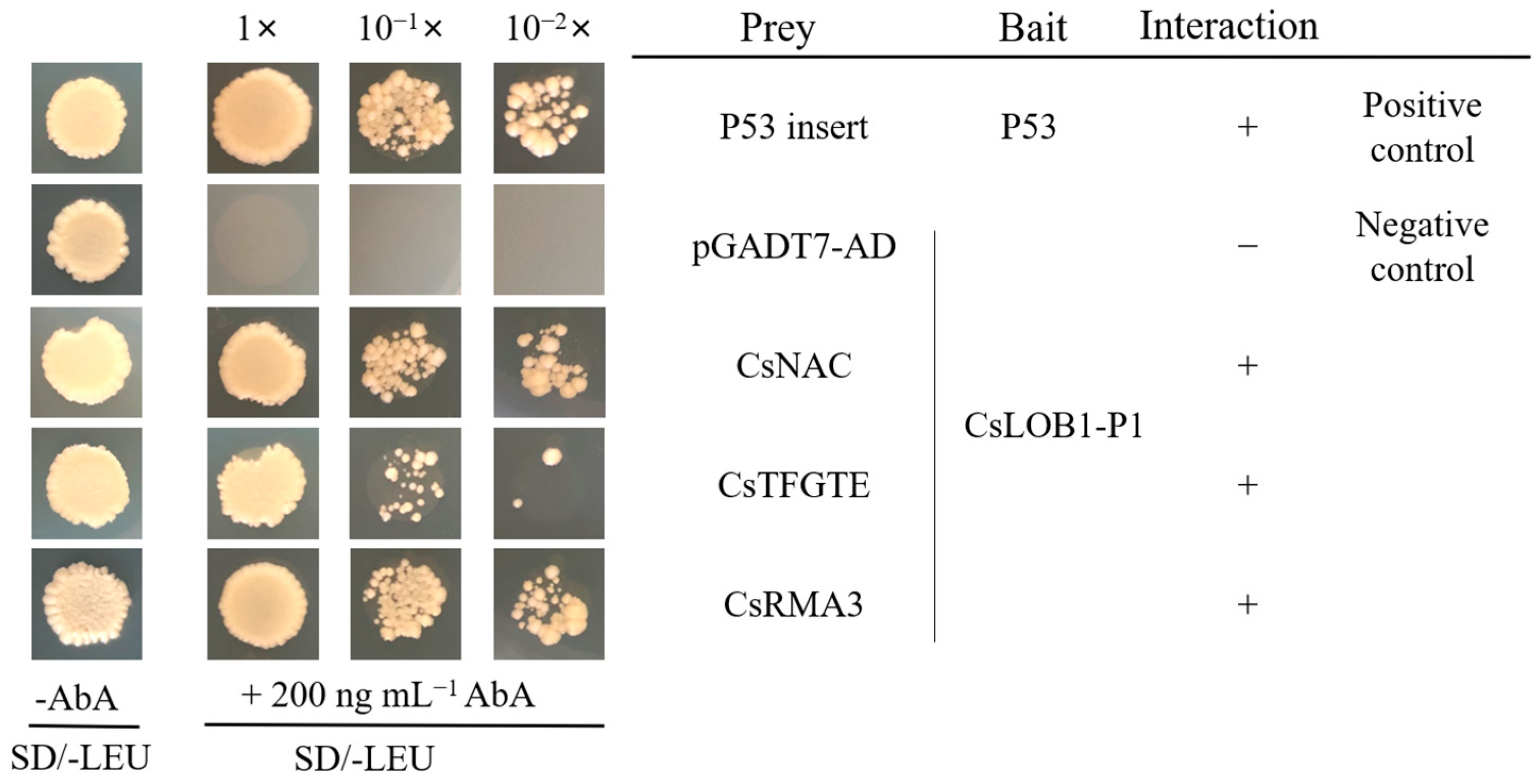
The homologous genes from sweet orange with the three identified transcript factors in citron were introduced into sweet orange yeast cells (Y1H-PAbAi CsLob1-P1) to detect the possible interactions with PAbAi CsLob1-P1. CsNAC, CsTFGTE, and CsRMA3 demonstrated interactions with PAbAi CsLob1-P1 (Figure 3), even though their interactions had not been detected in the previous screening (Table 1).

3.3. Interaction Validation with Dual-Luciferase Enzyme Assay

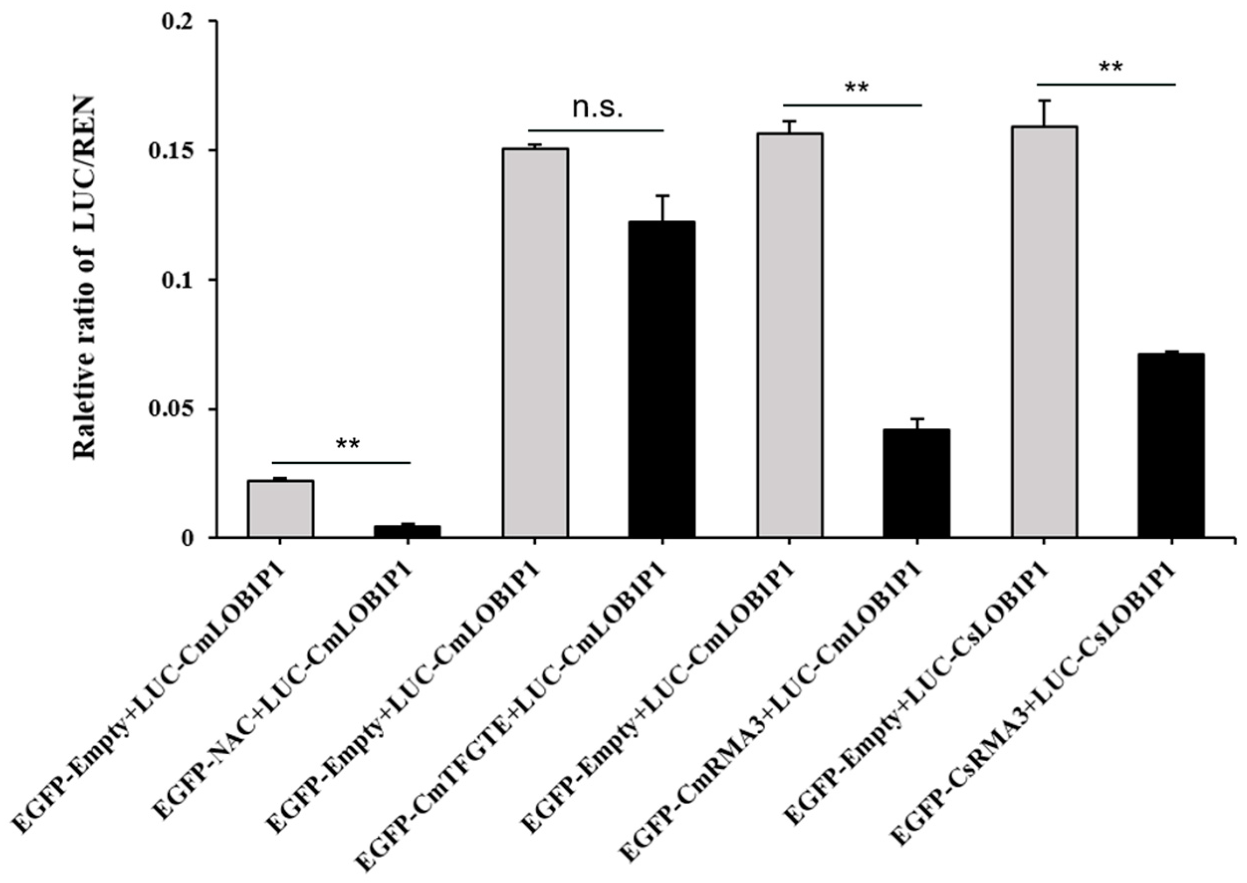
The results of the dual-luciferase assay indicated that the CmNAC transcription factors had very low expression, even though they significantly reduced the expression of Lob1. CmTFGTE did not show the suppression of the expression of Lob1. Moreover, the CmRMA3 and CsRMA3 transcription factors bound to the promoter of Lob1, thereby significantly downregulating the expression of the Lob1 gene (Figure 4).

3.4. Expression Analysis of Candidate Genes following Infection with Xcc

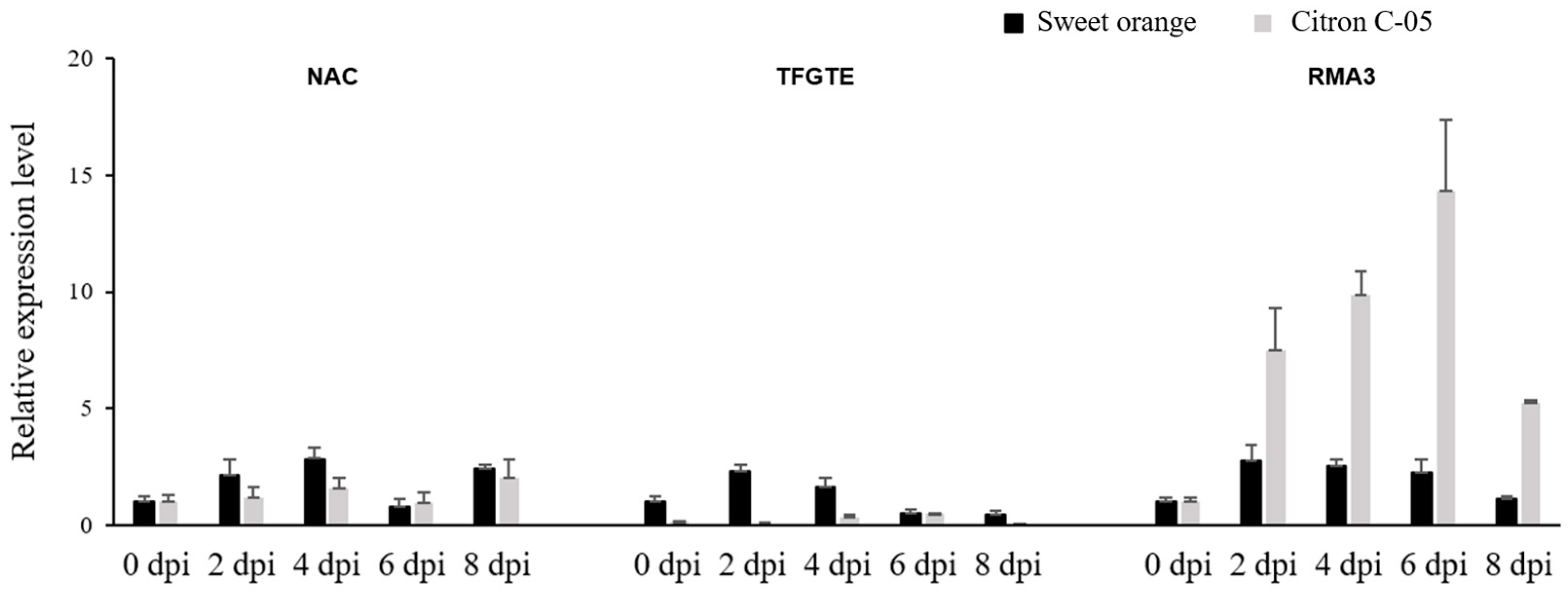
To investigate the expression profiles of the NAC, TFGTE20, and RMA3 genes induced by Xcc in ‘Bingtang’ sweet orange and Citron C-05, qPCR validation was conducted using cDNA from the two genotypes inoculated with Xcc at different intervals following inoculation with 105 CFU/mL of Xcc. The expression of RMA3 started to be upregulated at 2 dpi and peaked at 6 dpi, with significantly higher expression in Citron C-05 compared with ‘Bingtang’. The expression levels of the NAC and TFGTE20 genes were low, displaying higher expression levels in the susceptible ‘Bingtang’ than in the resistant Citron C-05 (Figure 5). The results clearly indicated that the expression of the RMA3 gene was stimulated by Xcc infection; therefore, RMA3 was considered the candidate transcript factor for the subsequent functional analysis.

3.5. Comparison of RMA3 Sequences among Different Citrus Genotypes

The RMA3 CDS was cloned from pomelo (C. grandis), sweet orange (C. sinensis), lemon (C. limon), and five citrons (C. medica) with different behaviors regarding Xcc infection. Sequence alignment revealed 91.17% homology among the eight genotypes with only 14 nucleotide mutations. The RMA3 amino acid sequences in the different citrus genotypes showed a high degree of homology (90.05%). Focusing on ‘Bingtang’ sweet orange and Citron C-05, both citrus genotypes contained 260 amino acid residues, with differences at six amino acid positions. The RMA3 proteins presented a C3HC4-RING finger domain that allowed them to function as E3 ubiquitin ligases (Figure S3).

3.6. Subcellular Localization of RMA3

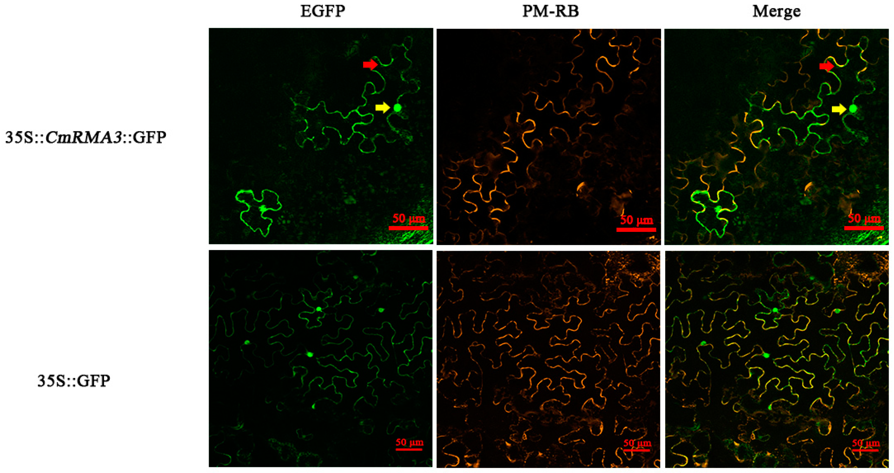
The constructed 35S::CmRMA3::GFP vectors were transformed into GV3101 and subsequently infiltrated into N. benthamiana leaves. Fluorescence microscopy showed green fluorescence signals emanating from the cell nuclei and membranes, potentially suggesting the presence of RMA3 in both the nucleus and cell membrane (Figure 6).

3.7. Transient Overexpression of RMA3 in ‘Bingtang’ Sweet Orange

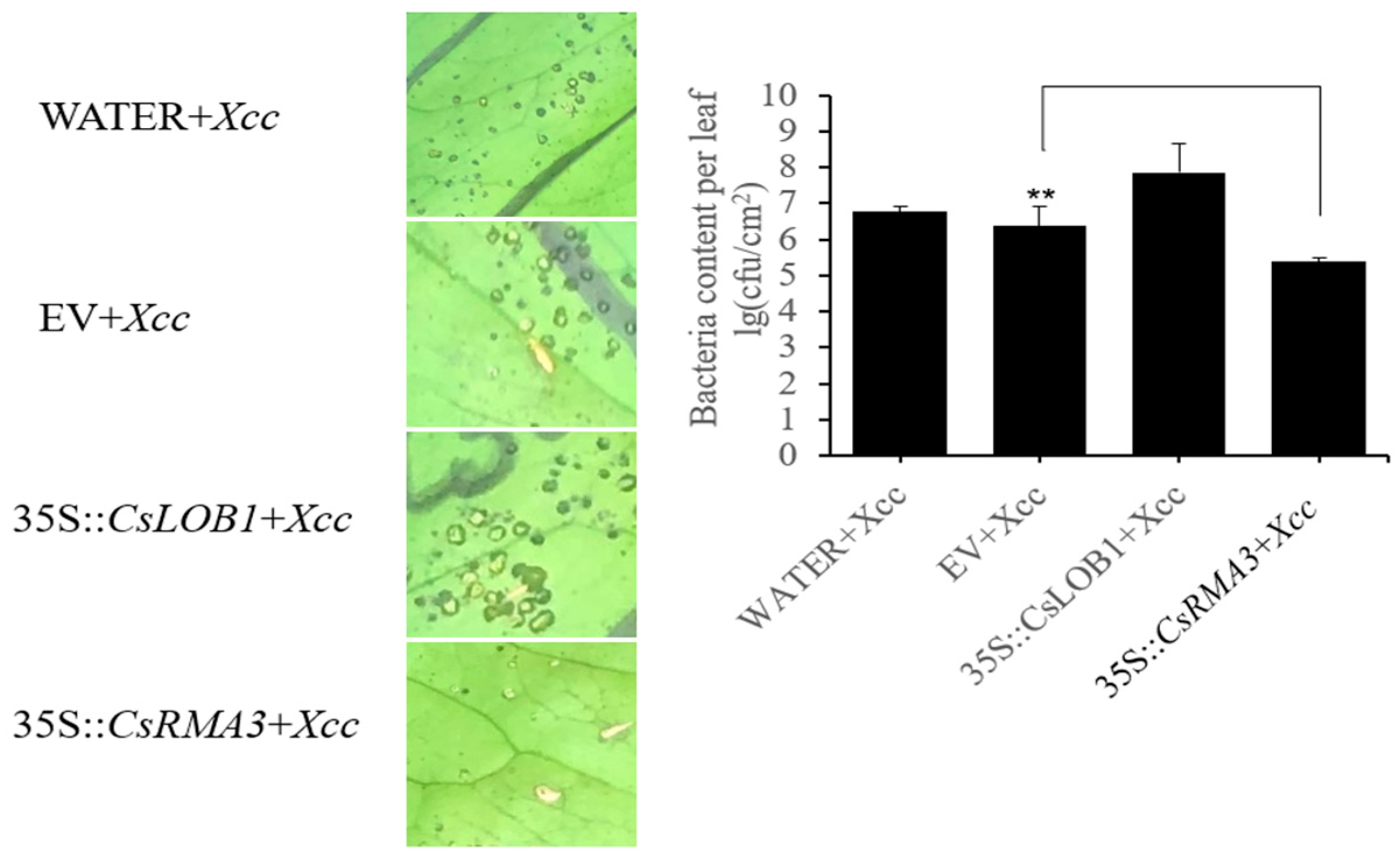
The 35S::CsRMA3::GFP vector was introduced into Agrobacterium EHA105, which was then injected into the susceptible ‘Bingtang’ leaves inoculated with Xcc. The transient overexpression of RMA3 inhibited Xcc colonization in the ‘Bingtang’ leaves. The number of lesions per square centimeter area in the treatment group was 0.136 times that of the control group. Bacterial quantification recorded 2.31 × 10⁵ CFU/cm2 for RMA3-overexpressed leaves and 2.37 × 10⁶ CFU/cm2 for the control, thus confirming the significant inhibitory effects on Xcc by RMA (Figure 7).
The expression analysis of Lob1 demonstrated that the transient overexpression of RMA3 suppressed the expression of the Lob1 gene. At 3 days following inoculation with Xcc in both sweet orange and citron leaves overexpressing RMA3, the expression of Lob1 in both genotypes was significantly reduced compared with the controls (Figure 8).

3.8. Silencing RMA3 Gene in Citron C-05 Using VIGS

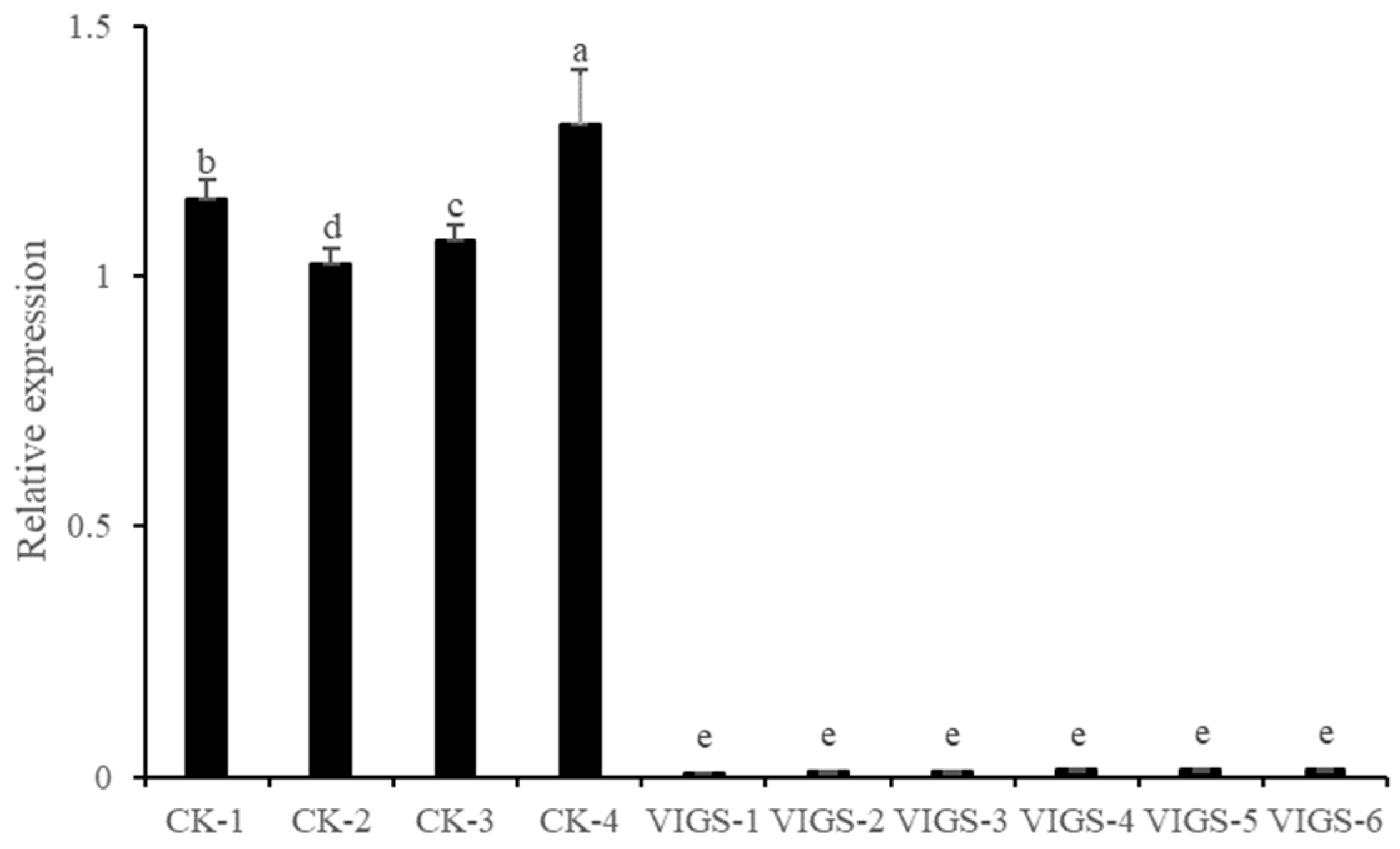
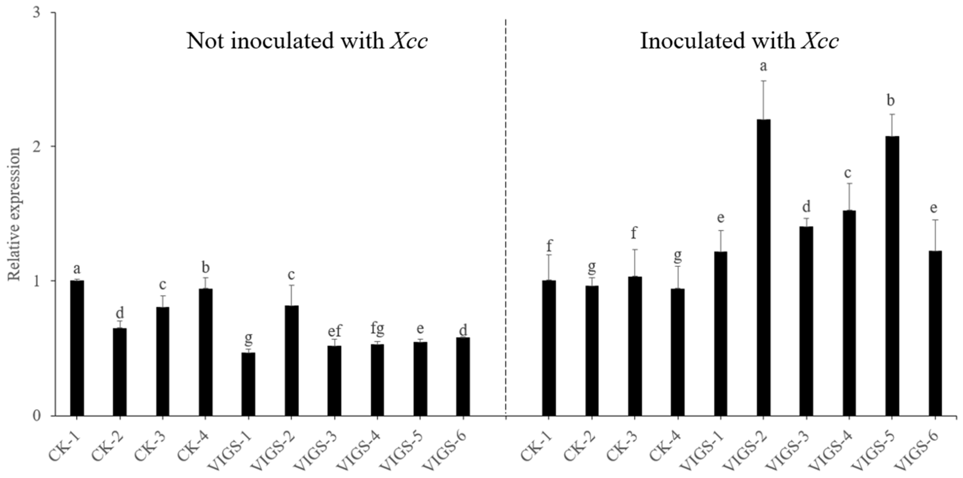
RMA3 was silenced by VIGS in Citron C-05 and the expression of RMA3 was downregulated in the six tested RMA3-silenced Citron C-05 clones (Figure 9). Additionally, the relative expression levels of Lob1 in four out of six silenced clones were significantly lower than those in the control plants (Figure 10). Nonetheless, after being inoculated with Xcc, the silenced plants at 3 dpi increased the relative expression levels of Lob1, significantly higher than those of the control plants (Figure 10).

3.9. Canker Symptom Observation of RMA3-Silenced Plants following Inoculation with Xcc

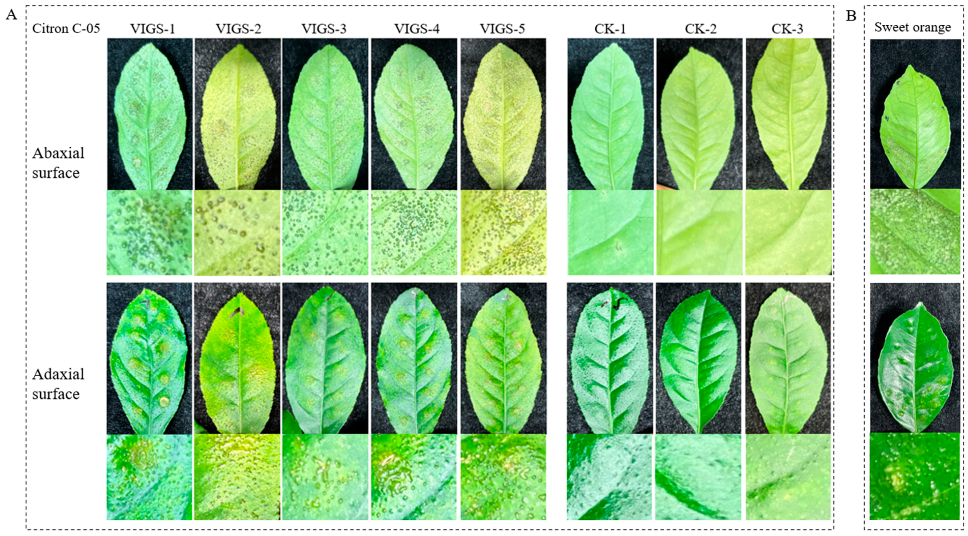
To evaluate the resistance of RMA3-silenced plants to Xcc infection, the RMA3-silenced Citron C-05 clones were inoculated with 105 cfu/mL of Xcc by injection. At 7 dpi, symptoms began to manifest on the leaves, characterized by pinpoint raised lesions. Over time, the lesions increased in both their number and spot size. At 15 dpi, numerous typical canker symptoms occurred on both sides of the inoculated leaves. The sweet orange, used as a susceptible genotype for the positive control, exhibited the first callus symptoms (Figure 11). It seemed that the silencing of RMA3 rendered the resistant Citron C-05 susceptible.

4. Discussion

The yeast hybrid system is effective for the identification of interacting proteins [27]. A high-quality cDNA library and an accurate bait protein are essential for the successful discovery of the target gene. The present study aimed to identify the transcription factors regulating the resistance to canker disease in Citron C-05. The cDNA library of resistant and susceptible genotypes was separately constructed using leaf samples collected when the Xcc symptoms started to exhibit a difference between the two tested citrus accessions, thus enabling us to identify resistance genes.
Lob1 has been confirmed as a pathogenic gene for citrus canker. We chose the promoter of Lob1 as bait to screen the resistant and susceptible citrus cDNA libraries and to detect possible upstream regulatory factors in relation to resistance to canker disease. After the initial screening, eight interacting proteins were identified in the susceptible ‘Bingtang’ sweet orange library induced by Xcc with PAbAi-CsLob1-P1, but none of them were useful transcription factors. However, 11 interacting proteins were detected in the resistant Citron C-05 library with PAbAi-CmLob1-P1, among which three were transcription factors. In the yeast one-hybrid validation, not only did the three transcription factors confirm the interaction with CmLob1-P1 for Citron C-05, but the three homolog transcription factors in ‘Bingtang’ also revealed an interaction with CsLob1-P1. Such double confirmation indicated the interaction between the transcription factors and the Lob1 promoter.
Among the three candidate transcription factors, RMA3 solely exhibited significant upregulation in the resistant citrus genotype Citron C-05 upon Xcc induction, which was not observed in the susceptible sweet orange in the qPCR analysis. RMA3 is a helicase-like transcription factor (HLTF) with E3 ubiquitin ligase activity, containing the zinc finger C3HC4 ring 2 domain. Ubiquitination plays a crucial role in the degradation of abnormal proteins under metal stress [28]. It is also involved in the recognition and regulation of pathogen-associated molecular patterns (PAMPs) by cell membrane receptors during pattern-triggered immunity (PTI). Furthermore, ubiquitination modulates the levels of intracellular immune receptors to prevent the constitutive activation of effector-triggered immunity (ETI), which is typically associated with hypersensitive cell death (HR) at the infection site [29]. E3 ubiquitin ligase has been reported to play an important role in various stages of the plant immune pathway [30,31]. In the present study, the interaction between RMA3 and the LOB1 promoter was confirmed, and it was observed that the overexpression of RMA3 suppressed the expression of LOB1, thus inducing resistance in the susceptible sweet orange. Conversely, RMA3 silencing in the resistant Citron C-05 caused canker symptoms. These results suggest that RMA3 plays an important role in the resistance to citrus canker by inhibiting the expression of the pathogenic gene Lob1.
The expression analysis revealed that RMA3 was induced by Xcc infection, which indicated its involvement in the response to pathogen invasion. The reaction mechanism, however, is not clear and needs further investigation. The E3 ligases of the RING family are considered essential for fundamental defense mechanisms in plants [32,33]. It might be hypothesized that RMA3 participates in the basic defense response of Citron C-05 against Xcc.
Citron C-05 is the first identified genotype resistant to canker disease in the Citrus genus [17]. Citron (C. medica), as one of the true citrus species [34], has a unique genetic background, differing from other citrus genotypes. All of the analyzed citrus genotypes have RMA3 with high homology; however, they demonstrate quite different behaviors in response to Xcc infection. It seems that there may be other factors regulating RMA3’s function. Therefore, more extensive studies are needed to clarify the functional mechanism of RMA3 in resistance to canker disease in Citron C-05, and whether or not such a mechanism is also valid in other citrus genotypes.

5. Conclusions

Through yeast hybrid screening in the Xcc-induced Citron C-05 cDNA yeast library, transcription factor RMA3 was identified using the Lob1 promoter P1 fragment as the bait protein. The yeast one-hybrid assay and dual-luciferase tests further validated the binding of RMA3 to the Lob1 promoter P1 fragment. The transient overexpression of CsRMA3 in the susceptible ‘Bingtang’ sweet orange significantly inhibited Xcc growth and reduced canker symptoms, while the silencing of CmRMA3 in the resistant Citron C-05 promoted symptom development. Furthermore, the expression analysis indicated that RMA3 suppressed Lob1 expression in both genotypes. Overall, these results suggest that RMA3 may participate in the resistance to citrus canker in Citron C-05 by inhibiting Lob1 expression. Further study is necessary to clarify the mechanism of citrus’ response to Xcc invasion. The overexpression of the RMA3 gene in susceptible citrus genotypes should be realized by genetic transformation in order to thoroughly comprehend its function in resistance to canker disease. The knock-out of the RMA3 gene in the resistant Citron C-05 using CRISPR/Cas9 would give a complete picture of RMA3 in response to invasion by the pathogen.

Supplementary Materials

The following supporting information can be downloaded at: https://www.mdpi.com/article/10.3390/horticulturae10070693/s1, Figure S1: Autonomous activation screening of P1 segment of Lob1 promoter; Figure S2: Screening of Xanthomonas citri subsp. citri-induced cDNA library with PAbAi-Lob1-P1; Figure S3: Schematic illustration of the protein domains of RMA3 in ‘Bingtang’ sweet orange and Citron C-05.

Author Contributions

Conceptualization, X.M. and Z.D.; methodology and experimentation, M.Z., R.Y., Y.L., L.L. and H.S.; formal analysis, M.Z., R.Y. and Z.D.; writing—original draft preparation, M.Z. and Z.D.; writing—review and editing, Z.D.; supervision, X.M. and Z.D. All authors have read and agreed to the published version of the manuscript.

Funding

This work was supported by the Provincial Special Project of the Chenzhou National Sustainable Agenda Innovation Demonstration Area ‘Chenjiang Laboratory Construction’ (2023sfq08) and the Key Project of International Cooperation and Exchange of the National Natural Science Foundation of China (No. 31720103915).

Data Availability Statement

The raw data supporting the conclusions of this article will be made available by the authors on request.

Conflicts of Interest

The authors declare no conflicts of interest.

Appendix A

Table A1. Primers used in this study.
Primer NameSequence (5′-3′)
pAbAi-CsLOB1-P-FGCTTGAATTCGAGCTGACCTCTGCCTACCCTATTGC
pAbAi-CmLOB1-P-FGCTTGAATTCGAGCTGACCTCTGCCTACCCTATTAC
pAbAi-LOB1-P-RTACAGAGCACATGCCCAAAGACAGTAAGGGATGAGG
pAbAi-CsLOB1-P1-FGAAAAGCTTGAATTCGGACCTCTGCCTACCCTATTGCC
pAbAi-CsLOB1-P1-RATACAGAGCACATGCCGAACTTCGAGGTTCATTGCATTG
pAbAi-CsLOB1-P2-FGAAAAGCTTGAATTCGCCAGGTTATAAGTAAACAGAACGAC
pAbAi-CsLOB1-P2-RATACAGAGCACATGCCCATCAAAGACAGTAAGGGATGAGG
pAbAi-CmLOB1-P1-FGAAAAGCTTGAATTCGGACCTCTGCCTACCCTATTACC
pAbAi-CmLOB1-P1-RATACAGAGCACATGCCGAACTTCGAGGTTCATTGGATTG
pAbAi-CmLOB1-P2-FGAAAAGCTTGAATTCGCCAGGTTATAAGTAAACAGCACGAC
pAbAi-CmLOB1-P2-RATACAGAGCACATGCCCAAAGACAGTAAGGGATGAGG
pGADT7-RMA3-FGTACCAGATTACGCTCAATGGAACAGAACCTCTTTGAGCC
pGADT7-RMA3-RATGCCCACCCGGGTGGTCAGAACAAAAGAAGACACAGGACTAGG
35S RMA3-FCACGGGGGACGAGCTCATGGAACAGAACCTCTTTGAGCC
35S RMA3-RCCTTGCTCACCATGGTACCGAACAAAAGAAGACACAGGACTAGG
qLOB1-FCTGCCAGAATCTCAACGAGC
qLOB1-R TTGGCTAACTGAGCCTGAAGC
q actin-FCATCCCTCAGCACCTTCC
q actin-R CCAACCTTAGCACTTCTCC
qRMA3-FTCAATCACAGGCACCAGCAT
qRMA3-RTAGAAGACGCCAAAGCTGCA
qNAC-FGATGTGGCGCAAGAGAGGTA
qNAC-RTGGCCCTGTTCGATCTGTTC
qTFGTE-FTTGGTGCAGCACTTACCAGA
qTFGTE-RAGCCTGTGTTACTCTGAGCA
VIGS RMA3-FTGAGTAAGGTTACCGAATTCGAACAGAACCTCTTTGAGCCTGAGA
VIGS RMA3-RGTGAGCTCGGTACCGGATCCTTGGGCTTCTTGGAGTCGGA

References

  1. Wu, G.A.; Terol, J.; Ibanez, V.; López-García, A.; Pérez-Román, E.; Borredá, C.; Domingo, C.; Tadeo, F.R.; Carbonell-Caballero, J.; Alonso, R.; et al. Genomics of the origin and evolution of Citrus. Nature 2018, 554, 311–316. [Google Scholar] [CrossRef] [PubMed]
  2. Rai, R.; Pasion, J.; Majumdar, T.; Green, C.E.; Hind, S.R. Genome sequencing and functional characterization of Xanthomonas cucurbitae, the causal agent of bacterial spot disease of cucurbits. Phytopathology 2021, 111, 1289–1300. [Google Scholar] [CrossRef] [PubMed]
  3. Ference, C.M.; Gochez, A.M.; Behlau, F.; Wang, N.; Graham, J.H.; Jones, J.B. Recent advances in the understanding of Xanthomonas citri ssp. citri pathogenesis and citrus canker disease management. Mol. Plant Pathol. 2018, 19, 1302–1318. [Google Scholar] [PubMed]
  4. An, S.Q.; Potnis, N.; Dow, M.; Vorhölter, F.J.; He, Y.Q.; Becker, A.; Teper, D.; Li, Y.; Wang, N.; Bleris, L.; et al. Mechanistic insights into host adaptation, virulence and epidemiology of the phytopathogen Xanthomonas. FEMS Microbiol. Rev. 2020, 44, 1–32. [Google Scholar] [CrossRef] [PubMed]
  5. Graham, J.H.; Gottwald, T.R.; Cubero, J.; Achor, D.S. Xanthomonas axonopodis pv. citri: Factors affecting successful eradication of citrus canker. Mol. Plant Pathol. 2004, 5, 1–15. [Google Scholar] [PubMed]
  6. Favaro, M.A.; Molina, M.C.; Roeschlin, R.A.; Gadea, J.; Gariglio, N.; Marano, M.R. Different responses in mandarin cultivars uncover a role of cuticular waxes in the resistance to citrus canker. Phytopathology 2020, 110, 1791–1801. [Google Scholar] [CrossRef] [PubMed]
  7. Ference, C.M.; Manthey, J.A.; Narciso, J.A.; Jones, J.B.; Baldwin, E.A. Detection of phenylpropanoids in citrus leaves produced in response to Xanthomonas citri subsp. citri. Phytopathology 2020, 110, 287–296. [Google Scholar] [PubMed]
  8. Lanza, F.E.; Marti, W.; Silva-Junior, G.J.; Behlau, F. Characteristics of citrus canker lesions associated with premature drop of sweet orange fruit. Phytopathology 2019, 109, 44–51. [Google Scholar] [CrossRef]
  9. Gochez, A.M.; Huguet-Tapia, J.C.; Minsavage, G.V.; Shantaraj, D.; Jalan, N.; Strauß, A.; Lahaye, T.; Wang, N.; Canteros, B.I.; Jones, J.B.; et al. Pacbio sequencing of copper-tolerant Xanthomonas citri reveals presence of a chimeric plasmid structure and provides insights into reassortment and shuffling of transcription activator-like effectors among X. citri strains. BMC Genom. 2018, 19, 16–29. [Google Scholar] [CrossRef]
  10. Li, J.; Pang, Z.; Duan, S.; Lee, D.; Kolbasov, V.; Wang, N. The in planta effective concentration of oxytetracycline against Candidatus Liberibacter asiaticus for suppression of citrus Huanglongbing. Phytopathology 2019, 109, 2046–2054. [Google Scholar] [CrossRef]
  11. Li, J.; Kolbasov, V.; Lee, D.; Pang, Z.; Huang, Y.; Collins, N.; Wang, N. Residue dynamics of streptomycin in citrus delivered by foliar spray and trunk injection and effect on ‘Candidatus Liberibacter asiaticus’ titer. Phytopathology 2020, 111, 1095–1103. [Google Scholar] [CrossRef]
  12. Burlakoti, R.; Hsu, C.F.; Chen, J.R.; Wang, J.F. Population dynamics of Xanthomonads associated with bacterial spot of tomato and pepper during twenty-seven years across Taiwan. Plant Dis. 2018, 102, 1348–1356. [Google Scholar] [CrossRef]
  13. Strayer-Scherer, A.; Liao, Y.Y.; Young, M.; Ritchie, L.; Vallad, G.E.; Santra, S.; Freeman, J.H.; Clark, D.; Jones, J.B.; Paret, M.L. Advanced copper composites against copper-tolerant Xanthomonas perfo-rans and tomato bacterial spot. Phytopathology 2018, 108, 196–205. [Google Scholar] [CrossRef] [PubMed]
  14. Hu, Y.; Zhang, J.; Jia, H.; Sosso, D.; Li, T.; Frommer, W.B.; Yang, B. Lateral organ boundaries 1 is a disease susceptibility gene for citrus bacterial canker disease. Proc. Natl. Acad. Sci. USA 2014, 111, E521–E529. [Google Scholar] [CrossRef]
  15. Duan, S.; Jia, H.; Pang, Z. Functional characterization of the citrus canker susceptibility gene CsLOB1. Mol. Plant Pathol. 2018, 19, 1908–1916. [Google Scholar] [CrossRef] [PubMed]
  16. Jia, H.; Orbovic, V.; Jones, J.B.; Wang, N. Modification of the PthA4 effector binding elements in Type I CsLOB1 promoter usingCas9/sgRNA to produce transgenic Duncan grapefruit alleviating XccDpthA4:dCsLOB1.3 infection. Plant Biotechnol. J. 2015, 14, 1291–1301. [Google Scholar] [CrossRef]
  17. Deng, Z.N.; Xu, L.; Li, D.Z.; Long, G.Y.; Liu, L.P.; Fang, F.; Shu, G.P. Screening citrus genotypes for resistance to canker disease (Xanthomonas axonopodis pv. citri). Plant Breed. 2010, 129, 341–345. [Google Scholar]
  18. Fu, H.Y.; Zhao, M.M.; Xu, J.; Tan, L.M.; Han, J.; Li, D.Z.; Wang, M.J.; Xiao, S.Y.; Ma, X.F.; Deng, Z.N. Citron C-05 inhibits both the penetration and colonization of Xanthomonas citri subsp. citri to achieve resistance to citrus canker disease. Hortic. Res. 2020, 7, 58–69. [Google Scholar] [PubMed]
  19. Wu, Q.; Zhao, M.M.; Li, Y.; Li, D.Z.; Ma, X.F.; Deng, Z.N. Identification of the transcription factors RAP2-13 activating the expression of CsBAK1 in citrus defence response to Xanthomonas citri subsp. Citri. Horticult. 2022, 8, 1012–1026. [Google Scholar]
  20. Sun, Y.; Li, Y.; Huang, G.; Wu, Q.; Wang, L. Application of the yeast one-hybridtechnique to plant functional genomics studies. Biotechnol. Biotechnol. Equip. 2017, 31, 1087–1092. [Google Scholar] [CrossRef]
  21. Jiang, W.; Liu, T.; Nan, W.; Jeewani, D.C.; Niu, Y.; Li, C.; Wang, Y.; Shi, X.; Wang, C.; Wang, J.; et al. Two transcription factors TaPpm1 and TaPpb1co-regulate anthocyanin biosynthesis in purple pericarps of wheat. J. Exp. Bot. 2018, 69, 2555–2567. [Google Scholar] [CrossRef] [PubMed]
  22. Lopato, S.; Bazanova, N.; Morran, S.; Milligan, A.S.; Shirley, N.; Langridge, P. Isolation of plant transcription factors using a modified yeast one-hybridsystem. Plant Methods 2006, 2, 45–66. [Google Scholar] [CrossRef] [PubMed]
  23. Mitsuda, N.; Ikeda, M.; Takada, S.; Takiguchi, Y.; Kondou, Y.; Yoshizumi, T.; Fujita, M.; Shinozaki, K.; Matsui, M.; Ohme-Takagi, M. Efficient yeast one−/two-hybrid screening using a library composed only of transcription factors in Arabidopsis thaliana. Plant Cell Physiol. 2010, 51, 2145–2151. [Google Scholar] [CrossRef] [PubMed]
  24. Zhan, Y.; Sun, X.; Rong, G.; Hou, C.; Huang, Y.; Jiang, D.; Weng, X. Identification of two transcription factors activating the expression of OsXIP in rice defense response. BMC Biotechnol. 2017, 17, 26–36. [Google Scholar] [CrossRef] [PubMed]
  25. Petzold, H.E.; Rigoulot, S.B.; Zhao, C.; Chanda, B.; Sheng, X.; Zhao, M.; Jia, X.; Dickerman, A.W.; Beers, E.P.; Brunner, A.M. Identification of new protein- proteinand protein- DNA interactions linked with wood formation in Populus trichocarpa. Tree Physiol. 2017, 38, 362–377. [Google Scholar] [CrossRef] [PubMed]
  26. Dharmaraj, K.; Cui, W.; EHA, R.; Templeton, M.D. Construction of a kiwifruit yeast two-hybrid cDNA library to identify host targets of the Pseudomonas syringae pv. actinidiae effector AvrPto5. BMC Res. Notes 2019, 12, 63–68. [Google Scholar] [PubMed]
  27. Hou, W.Q.; Yan, P.; Shi, T.Y.; Lu, P.Z.; Zhao, W.W.; Yang, H.M.; Zeng, L.Q.; Yang, J.; Li, Z.Y.; Fan, W.J.; et al. Modulation of anthocyanin accumulation in storage roots of sweet potato by transcription factor ibmyb1-2 through direct binding to anthocyanin biosynthetic gene promoters. Plant Physiol. Biochem. PPB 2023, 196, 868–879. [Google Scholar] [CrossRef] [PubMed]
  28. Lim, S.D.; Hwang, J.G.; Han, A.R.; Park, Y.C.; Jang, C.S. Positive regulation of rice RING E3 ligase OsHIR1 in arsenic and cadmium uptakes. Plant Mol. Biol. 2014, 85, 365–379. [Google Scholar] [CrossRef]
  29. Stael, S.; Kmiecik, P.; Willems, P.; VD-Kelen, K.; Coll, N.S.; Teige, M.; Van Breusegem, F. Plant innate immunity-sunny side up. Trends Plant Sci. 2015, 20, 3–11. [Google Scholar] [CrossRef]
  30. Shu, K.; Liu, X.D.; Xie, Q.; He, Z.H. Two faces of one seed: Hormonal regulation of dormancy and germination. Mol. Plant 2016, 9, 34–45. [Google Scholar] [CrossRef]
  31. Zhang, X.; Garreton, V.; Chua, N.H. The AIP2 E3 ligase acts as a novel negative regulator of ABA signaling by promoting ABI3 degradation. Genes Dev. 2005, 19, 1532–1543. [Google Scholar] [CrossRef] [PubMed]
  32. Lee, D.H.; Choi, H.W.; Hwang, B.K. The pepper E3 ubiquitin ligase ring1 gene, CaRING1, is required for cell death and the salicylic acid-dependent defense response. Plant Physiol. 2011, 156, 2011–2025. [Google Scholar] [CrossRef] [PubMed]
  33. Lin, S.S.; Martin, R.; Mongrand, S.; Vandenabeele, S.; Chen, K.C.; Jang, I.C.; Chua, N.H. RING1 E3 ligase localizes to plasma membrane lipid rafts to trigger FB1-induced programmed cell death in Arabidopsis. Plant J. 2008, 56, 550–561. [Google Scholar] [CrossRef] [PubMed]
  34. Xu, Q.; Huang, Y.; Deng, X.X. The definition, classification and evolution of citrus based on whole genome sequence information. Sci. Sin. Vitae 2024, 54, 525–536. (In Chinese) [Google Scholar] [CrossRef]
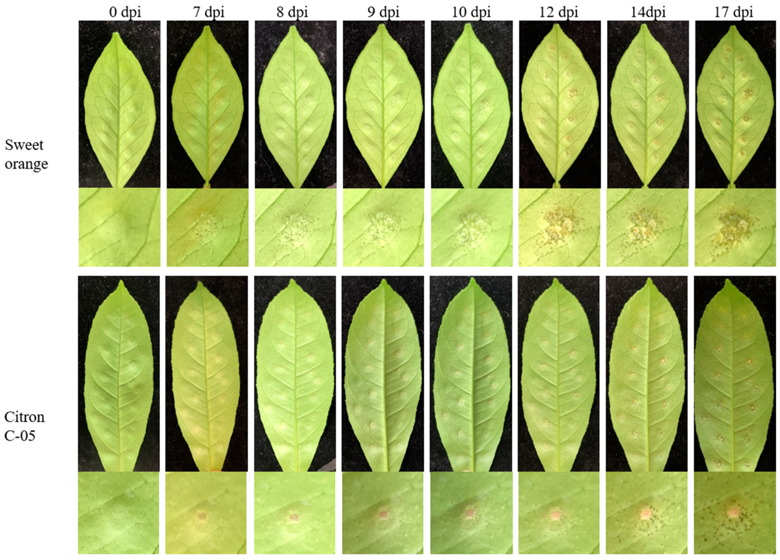
Figure 1. Leaf symptoms on the two citrus genotypes (‘Bingtang’ sweet orange = susceptible, Citron C-05 = resistant) after inoculation with 104 CFU/mL Xanthomonas citri subsp. citri. dpi = days post-inoculation.
Horticulturae 10 00693 g001
Figure 2. Validation of the interacting protein in Citron C-05 by CmLob1-P1 in Y1H system. pGADT7-AD: empty vector introduced into the same yeast cells as the negative control; pGADT7-p53: plasmid containing the p53 antibody introduced into yeast cells as the positive control; ‘+’: yeast containing Citron C-05 cDNA able to grow normally in SD/-LEU medium, representing the candidate protein interacting with CmLob1-P1; ‘−’: yeast did not grow in SD/-LEU medium, meaning that the protein had no interaction with CmLob1-P1. The concentration of AbA was 200 ng/mL.
Horticulturae 10 00693 g002
Figure 3. Identification of the interacting proteins in sweet orange and CsLob1-P1 in the Y1H system. pGADT7-AD: empty vector introduced into the same yeast cells as the negative control; pGADT7-p53: plasmid containing the p53 antibody introduced into yeast cells as the positive control. ‘+’: yeast containing sweet orange cDNA able to grow normally in SD/-LEU medium, indicating that the candidate proteins interact with CmLob1-P1; ‘−’: the same yeast did not grow in SD/-LEU medium, meaning that the protein had no interaction with CmLob1-P1. The concentration of AbA was 200 ng/mL.
Horticulturae 10 00693 g003
Figure 4. Identification of proteins interacting with the Lob1-P1 promoter by dual-luciferase assay. The CmNAC transcription factors had very low expression, even though they significantly reduced the expression of Lob1. CmTFGTE did not show the suppression of the expression of Lob1. The CmRMA3 and CsRMA3 transcription factors bound to the promoter of Lob1, thereby significantly downregulating the expression of the Lob1 gene. CsNAC and CsTFGTE were not analyzed for interactions with CsLob1-P1. ‘**’ indicates a significant difference (p < 0.01). ‘n.s.’ indicates no significant difference.
Horticulturae 10 00693 g004
Figure 5. Relative expression of three candidate genes in ‘Bingtang’ sweet orange (susceptible) and Citron C-05 (resistant) following inoculation with 105 CFU/mL of Xanthomonas citri subsp. citri.
Horticulturae 10 00693 g005
Figure 6. The subcellular localization of CmRMA3 of Citron C-05. Yellow arrows indicate the nucleus; the red arrow points to the cell membrane; PM-RB is a membrane-targeting mark; 35S::GFP: an empty control, localized to the cell membrane and nucleus.
Horticulturae 10 00693 g006
Figure 7. Effects on the inhibition of Xanthomonas citri subsp. citri (Xcc) infection following RMA3 overexpression in susceptible ‘Bingtang’ sweet orange leaves. On the left, upon inoculation with Xcc, canker symptoms were observed in sweet orange plants with the transient overexpression of the RMA3 gene; on the right, quantitative counting plate count data of Xcc bacteria. The concentration of Xcc was 105 CFU/mL. ‘**’ indicates a significant difference between ‘EV + Xcc’ and ‘35S::CsRMA3 + Xcc’ (p < 0.01).
Horticulturae 10 00693 g007
Figure 8. Relative expression of the Lob1 gene in citrus leaves following the transient overexpression of RMA3 at 3 dpi. (A) Leaf of ‘Bingtang’ sweet orange; (B) leaf of Citron C-05. The concentration of Xanthomonas citri subsp. citri was 105 CFU/mL. ‘**’ indicates a significant difference(p < 0.01) between the transient clone and the control.
Horticulturae 10 00693 g008
Figure 9. Relative expression of the RMA3 gene in Citron C-05 with RMA3 silenced by VIGS. CK-1 to CK-4 are clones transferred with the Empty-TRV2 plasmid as controls; VIGS-1 to VIGS-6 are RMA3-silenced plants transferred with RMA3-TRV2. The different letters in the columns indicate significant differences (p < 0.05).
Horticulturae 10 00693 g009
Figure 10. Relative expression of Lob1 in Citron C-05 with RMA3 silenced by VIGS. On the left, the plants were not inoculated with Xanthomonas citri subsp. citri sampled at the same time as the inoculated ones; on the right, the plants were inoculated with Xcc and evaluated at 3 days post-inoculation. The concentration of Xcc was 105 CFU/mL. CK-1 to CK-4 are clones transferred with the Empty-TRV2 plasmid as controls; VIGS-1 to VIGS-6 are RMA3-silenced plants. The different letters in the columns indicate significant differences (p < 0.05).
Horticulturae 10 00693 g010
Figure 11. Leaf canker symptom observation of RMA3-silenced Citron C-05 at 15 days post-inoculation with Xanthomonas citri subsp. citri. (A) Leaf canker symptoms of RMA3-silenced Citron C-05. (B) Leaf symptoms of ‘Bingtang’ sweet orange as a positive control for disease susceptibility genotypes. The inoculation concentration of Xcc was 105 CFU/mL.
Horticulturae 10 00693 g011
Table 1. Interacting proteins obtained from the screening of cDNA libraries of ‘Bingtang’ sweet orange (susceptible) and Citron C-05 (resistant) induced by Xanthomonas citri subsp. citri with Lob1-P1 promoter.
Bait
Name
Library
Genotype
Gene CodeFunction
PAbAi-CsLob1-P1‘Bingtang’ sweet
orange
CsLP1-1Bispecific inhibitors; lipid transfer proteins; seed storage 2S albumins.
CsLP1-2Oxygen-evolving enhancer protein 2; chloroplast.
CsLP1-3Composition of ribosomal protein S9; cytoplasmic 80S ribosome.
CsLP1-4E3 ubiquitin protein ligase RING1; RING finger protein 126; E3 ubiquitin ligase.
CsLP1-5Zinc finger protein and putative TFIIIA-like zinc finger protein.
CsLP1-6Oxygen-evolving enhancer protein 3; chloroplast.
CsLP1-7Wound-induced protein 1; putative unidentified protein.
CsLP1-8Uncharacterized protein At4g01150 in chloroplast.
PAbAi-CmLob1-P1Citron
C-05
CmLP1-1Protein binding chlorophyll a and b.
CmLP1-2Alkaline chitinase A and lectin.
CmLP1-3Unidentified protein.
CmLP1-4Bifunctional dihydroflavonol 4-reductase plays crucial role in reduction of flavonoids.
CmLP1-560S ribosomal protein L10, a fragment of the same.
CmLP1-6Nuclear transcription factor Y subunit C-1.
CmLP1-7Transcription factor GTE1; chloroplast transcription factor GTE3.
CmLP1-8The first variation could be ‘E3 ubiquitin protein ligase RMA3, peroxisome generating factor 10, and helicase-like transcription factor are the proteins under study’.
CmLP1-9Receptor kinase 1 associated with brassinosteroid insensitivity 1.
CmLP1-102-Acetamido-2-deoxy-d-galactose-bound seed agglutinin 2, also known as lectin.
CmLP1-11NAC domain referred to as Protein 29.
Disclaimer/Publisher’s Note: The statements, opinions and data contained in all publications are solely those of the individual author(s) and contributor(s) and not of MDPI and/or the editor(s). MDPI and/or the editor(s) disclaim responsibility for any injury to people or property resulting from any ideas, methods, instructions or products referred to in the content.