Article

PhSPEAR1 Participates in Regulating the Branch Development of Petunia

1
Agricultural Engineering Research Institute, Anhui Academy of Agricultural Sciences, Hefei 230001, China
2
Department of Ornamental Horticulture, Anhui Agricultural University, Hefei 230061, China
*
Author to whom correspondence should be addressed.
These authors contributed equally to this work.
Horticulturae 2024, 10(9), 912; https://doi.org/10.3390/horticulturae10090912
Submission received: 2 July 2024 / Revised: 5 August 2024 / Accepted: 22 August 2024 / Published: 27 August 2024
(This article belongs to the Section Floriculture, Nursery and Landscape, and Turf)

Abstract

:
Petunia hybrida is an economically important ornamental plant species. Branching in ornamental plants is closely associated with their ornamental traits, and branching is a significant agronomic trait in petunia, which shapes plant architecture and production cost. Although there are few studies regarding the involvement of the SPEAR genes in lateral branch development, they are known to regulate the development of plant organs. The PhSPEAR1 gene in petunia, an ortholog of Arabidopsis SPEAR1, was isolated for study. According to the results of real-time quantitative PCR (qRT-PCR), PhSPEAR1 was primarily expressed in the roots. The nucleus-specific fluorescence signal indicated that PhSPEAR1 was localized in the nucleus. An increase in PhSPEAR1 expression was induced by cytokinin or decapitation. Overexpression of PhSPEAR1 increased lateral branches in Arabidopsis. Based on our findings, PhSPEAR1 participates significantly in the regulation of branch number in petunia.

1. Introduction

Branching is a crucial aspect of plant growth and development that influences plant morphogenesis, crop yield, and ornamental characteristics. The regulation of branch development involves pathways associated with strigolactone (SL), gibberellin (GA), auxin, and cytokinin (CK) [1,2,3,4]. Moreover, factors like sugar, temperature, nutrient availability, and light conditions can influence branch development [5,6,7,8,9]. The primary determinant of branching ability, however, is the genetic makeup of the plant itself. Thus, a key area of research for branch development is the study of gene function.
Over the last ten years, our knowledge of controlling shoot branching has become increasingly detailed. Auxin is regarded as the main trigger for apical dominance. The growing, immature leaves at the apex of the shoot produce auxin. Auxin influx and efflux carriers, which are polarly localized in plasma membranes, help to transport it basipetally [10]. However, lateral buds are not directly affected by auxin, which is derived from the shoot apex. It is therefore believed that the mechanism of auxin action is exerted through various methods, including canalization and secondary messengers. CK promotes bud outgrowth and is considered the second messenger of auxin. It is transported from the roots to the shoots [11], and its local synthesis in the stem is likely the primary means of regulating shoot branching [12]. Another class of factors implicated in regulating shoot branching in a variety of plant species is SLs [13]. SLs in the roots can be transported acropetally to shoots and inhibit shoot branching locally by inducing BRC1/TB1 [14,15]. Research conducted on a diverse range of species revealed a negative correlation between bud activity and the abundance of ABA in buds [16]. In many species, including tomatoes and Ipomoea nil, exogenous ABA hinders the growth of buds [17]. In a study by Arend et al. [18], it was observed that lowering ABA sensitivity led to greater lateral bud growth in Populus × canescens. These investigations point to ABA’s role in regulating the development of branches.
In addition to the well-characterized transcriptional regulator BRC1, a number of genes also control branch processes, and their respective mechanisms of action have been elucidated. We still lack a thorough understanding of the control mechanisms underlying branching patterns, despite the identification and characterization of many of the genes that regulate them. SPL-like, EAR-containing (SPEAR) proteins are unique to embryophytes and not present in other organisms; therefore, it has been hypothesized that they are embryophyte-specific genes. They have the same general structure: an N-terminal NLS, which is followed by the SPL motif, and a C-terminal EAR motif (LxLxL) [19]. TCP interactors containing EAR motif protein 1 (TIE1) and TIE2, as members of SPEAR proteins, can interact with ARABIDOPSIS RESPONSE REGULATORs (ARRs) to modulate CK responses in roots [20,21]. TIE1 has also been found to be involved in the regulation of Arabidopsis branching. SPOROCYTELESS (SPL), another member of the SPEAR family, can interact with the TOPLESS (TPL) co-repressors in Arabidopsis and transcription factors of the TCP family to perform its transcription repressive function [19]. These existing studies indicate that SPEARs are involved in multiple developmental processes in plants. Although SPEARs have been studied in several species, they have not been reported in petunia yet.
Petunia hybrida Vilm. is an extremely important ornamental plant, and its branching characteristics directly affect its ornamental and commercial value. Therefore, the study of branch regulation in petunia has important practical application value. Secondly, petunia, as a model plant, can help to quickly and accurately explore the regulatory mechanisms of branch development. To investigate the function of SPEAR1 in petunia, we cloned it and studied its expression characteristics, subcellular localization, and transgenic plant phenotype. This research will lay the groundwork for further understanding the regulatory mechanism of SPEAR1.

2. Materials and Methods

2.1. Plant Materials

The Petunia × hybrida cv. Mitchell Diploid, Arabidopsis thaliana Col-0, Nicotiana benthamiana, and the transgenic plants utilized in this research were all placed in the plant growth room of the School of Horticulture, Anhui Agricultural University. The growing conditions were adjusted to 25 ± 2 °C, with 3500 LX and a 16/8 h light/dark cycle.

2.2. Isolation of PhSPEAR1

We isolated total RNA from pooled leaves of 10 seedlings of Petunia × hybrida cv. Mitchell Diploid (40-day-old) utilizing the RNAprep Pure Plant Plus Kit (TIANGEN Biotech Co., Ltd., Beijing, China). The Prime-Script™ RT reagent kit with gDNA Eraser (TaKaRa Biotechnology Co., Ltd., Dalian, China) was then used to synthesize first-strand complementary deoxyribonucleic acid (cDNA) and remove DNA. Based on the genome sequence of petunia available on the Solanaceae Genome Network [22], primers FSPEAR1 (5′-3′): ATGGGCAGCAATTATTTTGGGGA and RSPEAR1 (5′-3′): TCAAAGGGAAAGTCTG AGCTCCA were used to amplify the PhSPEAR1 with MegaFiTM Fidelity 2 × PCR Master Mix (Toyobo Co., Ltd., Shanghai, China), and the amplified products were then purified and sequenced at General Biotechnology Co., Ltd. (Chuzhou, China). Sequencing was repeated three times to ultimately determine the correct sequence.

2.3. Bioinformatic Analysis

The protein physicochemical properties of PhSPEAR1 were predicted with the ExPASy-ProtParam tool (https://web.expasy.org/protparam/, accessed on 3 August 2023). MEGA software (version 7.0.14) was used to run the ClustalW program and perform multiple sequence alignment. Phylogenetic trees based on Arabidopsis SPEARs and PhSPEAR1 were generated using MEGA v7.0.14 software and the maximum-likelihood method with a bootstrapping value of 1000. Arabidopsis SPEARs sequences were downloaded from NCBI (https://www.ncbi.nlm.nih.gov/, accessed on 4 August 2023). The conserved domains were identified using the online software Pfam (http://www.sanger.ac.uk/Software/Pfam/, accessed on 5 August 2023).

2.4. Subcellular Localization Analysis

To generate the PhSPEAR1-enhanced green fluorescent protein (eGFP) fusion protein, the PhSPEAR1 lacking the stop codon was amplified using primers SPEAR1-1300-F (5′-3′): GAGAACACGGGGACTCTAGAATGGGCAGCAATTATTTTG GGGA and SPEAR1-1300-R (5′-3′): GCCCTTGCTCACCATGGATCCTCAAAGGGAAA GTCTGAGCTCCA. The amplified PCR product was digested with Hind III (TaKaRa Biotechnology Co., Ltd., Dalian, China) and Sal I (TaKaRa Biotechnology Co., Ltd., Dalian, China). The recombinant reaction was performed according to the instructions of the NovoRec® plus One step PCR Cloning Kit (Novoprotein, Shanghai, China). The recombinant product was transformed into competent cells of Escherichia coli DH5α (Shanghai Tolo Biotechnology Co., Ltd., Shanghai, China). After screening for positive clones, the plasmid of pSuper1300-PhSPEAR1-eGFP was extracted and transformed into an A. tumefaciens GV3101 strain (Shanghai Tolo Biotechnology Co., Ltd., Shanghai, China) using the freeze–thaw method, and positive clones were selected for temporary infiltration into N. benthamiana leaves using a previously described procedure [23]. After 48 h of culture at 25 °C, the eGFP fluorescence was observed using a confocal microscope (Olympus FV1200, Tokyo, Japan).

2.5. qRT-PCR Analysis

Four plant parts—roots, stems, leaves, and buds—from 75-day-old plants of Petunia × hybrida cv. Mitchell Diploid were obtained to perform a tissue expression experiment. Notably, samples of the leaves, buds, and stems were taken from the shoot tip downward within the range of nodes 2 to 6. Furthermore, newly grown, tender roots were chosen.
In the treatment experiment, 40-day-old plants of Petunia × hybrida cv. Mitchell Diploid were split into three groups, including control, decapitated, and the third group that was treated with 10 µL of 50 µM 6-benzylaminopurine (6-BA) (Sinopharm Chemical Reagent Co., Ltd., Shanghai, China) at the axillary buds at the top 4 th nodes. Buds at the first node (decapitation) or fourth node (6-BA treatment) were collected after six hours. For the control plants, the fourth bud from top to bottom was collected. Then, all the tissues were promptly submerged in liquid nitrogen and kept at −80 °C.
There were three biological and three technical replicates in every experiment, unless stipulated otherwise. For every technical replicate, 20–40 plants were pooled to provide tissue samples.
Two micrograms of DNase I (TaKaRa Biotechnology Co., Ltd., Dalian, China)-digested RNA from various samples were reverse-transcribed into cDNA. qRT-PCR was conducted with the Thermo PIKO REAL96 Real-Time PCR System employing a GoTaq qPCR Master Mix (Promega, Madison, WA, USA). Amplification was conducted by an initial denaturation at 95 °C for 30 s, then 40 cycles of amplification (denaturation at 95 °C for 15 s, annealing at 58 °C for 30 s, and extension at 72 °C for 1 min) were performed. P. hybrida Glyceraldehyde-3-phosphate dehydrogenase (PhGAPDH) was employed as the reference gene with primers FGAPDH (5′-3′): CAAGGCTGGAATTGCTTTGAG and RGAPDH (5′-3′): CACCACTTTACTCCACTGATGCA. The 2−ΔΔCT method was utilized to calculate the relative expression [24]. In addition, all primers used in this study were designed and validated using Primer 5 software.

2.6. Construction and Transformation of 35S::PhSPEAR1

The primers SPEAR1-1300-F and SPEAR1-1300-R were synthesized by adding Hind III and Sal I restriction sites, and the target gene product was obtained by amplification with high-fidelity KOD DNA polymerase (Toyobo, Osaka, Japan). Furthermore, the pCAMBIA1300 vector was double-digested. The enzyme digestion vector and the target gene product were connected by homologous recombination. The recombinant product was transformed into E. coli-competent cells using the freeze–thaw method and cultured in LB medium containing kanamycin. After successful detection, the plasmid was extracted, and the 35S::PhSPEAR1 vector was obtained.

2.7. Arabidopsis Transformation

Using the A. tumefaciens-mediated flower-dipping method, the constructed pCAMBIA1300-PhSPEAR1-eGFP was introduced into Arabidopsis WT [25]. After the seeds of the infiltrated plants were harvested, they were transferred to a Murashige and Skoog (MS) medium (Solarbio Biotech Co., Ltd., Beijing, China) with 50 mg/L of kanamycin (Sinopharm Chemical Reagent Co., Ltd., Shanghai, China). Kanamycin-resistant seedlings were cultivated in a growth chamber and transplanted into the soil after 15 days of germination.

2.8. Phenotype Analysis and Statistical Analysis

The number of rosette branches (bud length ≥ 10 mm) and the height of the main stems were measured for phenotypic analysis. All the data in this study was expressed as the mean value ± standard deviation.

2.9. Sample Preparation and Plant Hormone Extraction

The rosette sections of 25-day-old WT and transgenic plants were harvested, instantly frozen in liquid nitrogen, ground into powder, and kept at −80 °C until needed. About 50 mg of the plant sample was transferred into a 2 mL centrifuge tube kept in frozen liquid nitrogen and then dissolved in 1 mL of methanol/water/formic acid (15:4:1, v/v/v). To quantify the extraction solution as the internal standard, 10 μL of an internal standard solution (100 ng/mL) was added. After 10 min of vortexing, the mixture was centrifuged for 5 min at 6000× g and 4 °C. After centrifugation, the supernatant was transferred to clean plastic microtubes and dried before being dissolved in 100 μL of 80% methanol (v/v) (Sinopharm Chemical Reagent Co., Ltd., Shanghai, China). Following filtration through a 0.22 μm membrane filter (Beijing Heng Odd Instrument Co., Ltd., Beijing, China), LC-MS/MS culture analysis was conducted. MetWare (http://www.metware.cn/, accessed on 10 May 2024) was used to detect phytohormone contents utilizing the QTRAP 6500 LC-MS/MS platform (AB Sciex, Concord, ON, Canada).

2.10. Statistical Analysis

All the data were subjected to analysis of Student’s t-tests using IBM SPSS Statistics software (version 27.0).

3. Results

3.1. Identification of SPEAR1 in Petunia

Five Arabidopsis SPEARs were used with the petunia SPEAR to generate a phylogenetic tree. Evolutionary analysis combined with sequence alignment suggested that the petunia SPEAR and Arabidopsis SPEAR1 were closely related (Figure 1), suggesting that the petunia SPEAR gene is a potential homolog of the Arabidopsis SPEAR1 homolog, hence the name PhSPEAR1. Sequence alignment of the PhSPEAR1 and Arabidopsis SPEAR homologs was performed, and the results showed that the N-terminus of PhSPEAR1 protein contained an SPL motif (residues 33 and 49) and the NLS region (residues 16 and 25). A typical EAR motif sequence (DLELRL) is present in the C-terminal region (residues 185 and 192) (Figure 1B).
The full length of the PhSPEAR1 gene was 579 bp, encoding a protein of 192 amino acids. Computationally, the molecular formula of the PhSPEAR1 protein was C896H1417N273O309S8, the relative molecular weight was 21.2 KD, the isoelectric point was 8.54, and the instability parameter was 69.80. The highest content of PhSPEAR1 was Ser (19.8%, 38), and the lowest content was Trp (0.05%, 1) and Cys (1.0%, 2). Asp + Glu (total negatively charged residues) had a total of 18, and Arg + Lys (total positively charged residues) had a total of 20. The theoretical half-life was 30 h.

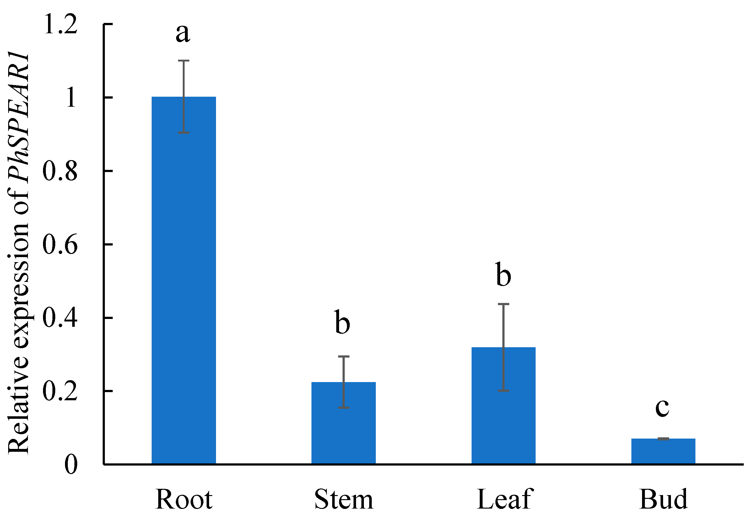
3.2. Tissue-Specific Expression Analysis of PhSPEAR1

The expression levels of the PhSPEAR1 gene in the roots, stems, leaves, and axillary buds of petunia were determined by qRT-PCR to establish the expression level of the gene in various tissues. PhSPEAR1 was expressed in all tested tissues. The PhSPEAR1 gene was expressed highest in roots, followed by leaves, and was least expressed in buds. The expression level of PhSPEAR1 in the buds was 1/13 of the expression in the roots (Figure 2).

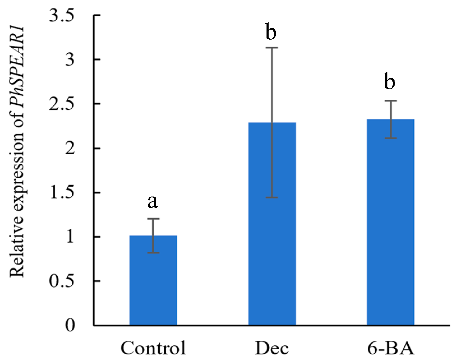
3.3. Expression Analysis of PhSPEAR1 under Branch-Inducing Treatments

To investigate the response of the PhSPEAR1 gene to decapitation and CK, 6-BA was applied to the axil of petunia seedlings. qRT-PCR was used to assess the expression levels of PhSPEAR1. Both 6-BA and decapitation both doubled the expression level of PhSPEAR1 (Figure 3).

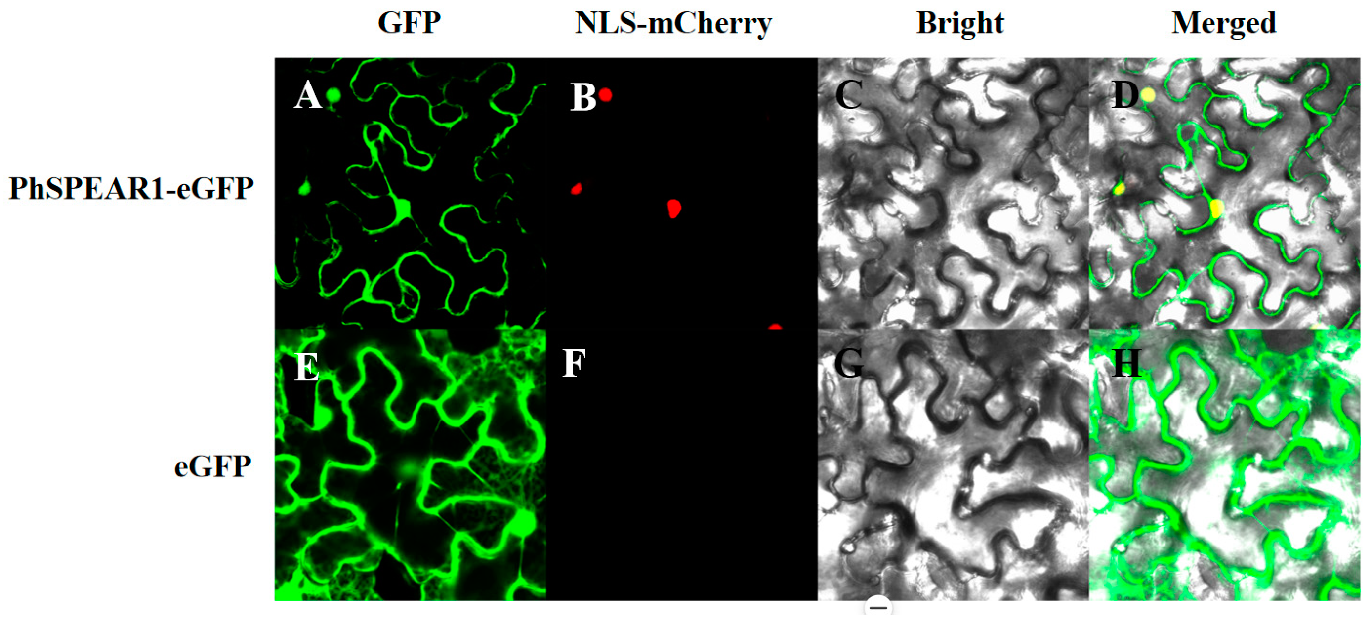
3.4. Subcellular Localization Analysis of PhSPEAR1

To examine PhSPEAR1’s subcellular location, the 35S::PhSPEAR1:eGFP vector was constructed, transformed into Agrobacterium, and injected into tobacco leaves. The fluorescence was observed by confocal microscopy after 48 h. PhSPEAR1 fused with eGFP only produced green fluorescence in the nucleus, being that PhSPEAR1 was localized to the nucleus (Figure 4).

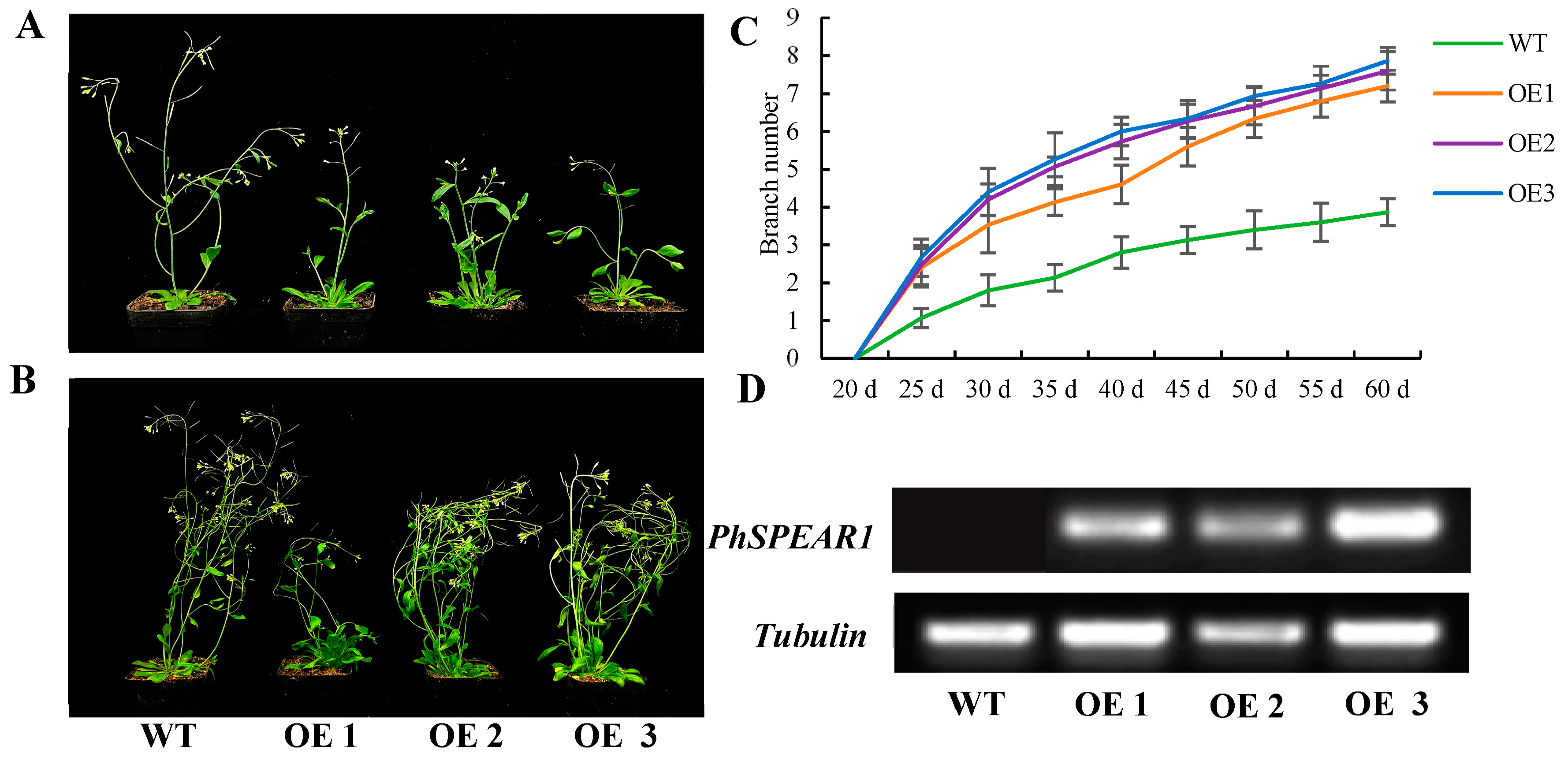
3.5. Overexpression of PhSPEAR1 Increases Branch Number in Arabidopsis

To analyze the function of the PhSPEAR1 gene, construct 35S::PhSPEAR1 was transformed into Arabidopsis, and the transgenic plants were screened. Our findings demonstrated that 35S::PhSPEAR1 plants produced more first-order rosette branches than the WT (Figure 5A–C). Compared with the WT, the average number of branches of transgenic plants increased from 1.8 to 3.5, 4.2, and 4.4, respectively (Figure 5A,C). WT plants displayed about four lateral branches after 60 d of growth, whereas 35S::PhSPEAR1 lines 1, 3, and 3 displayed about seven, eight, and eight lateral branches, respectively. RT-PCR revealed no PhSPEAR1 expression in the leaves of 28-day-old WT plants, and in 35S::PhSPEAR1 transgenic plants, PhSPEAR1 transcript abundance was detected (Figure 5D).

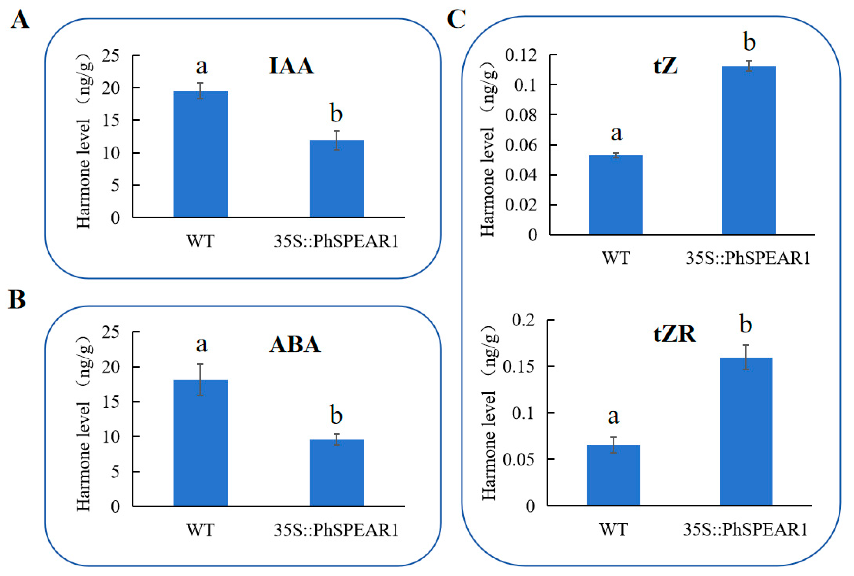
3.6. PhSPEAR1 Regulates the Synthesis of CKs, IAA, and ABA

To analyze the downstream regulatory pathway of PhSPEAR1, hormone levels were detected in the OE2 line plants of transgenic Arabidopsis overexpressing PhSPEAR1, which exhibited a more pronounced phenotype compared to other transgenic strains. As shown in Figure 6, overexpression of PhSPEAR1 led to significant downregulation of IAA and upregulation of the important cytokinin types trans-zeatin (tZ) and trans-zeatin riboside (tZR). ABA levels were also significantly decreased. Based on the above conclusions, we speculate that PhSPEAR1 affected branch development by promoting the synthesis of CK and inhibiting the synthesis of hormones such as IAA and ABA.

4. Discussion

SPEAR refers to proteins with NLS, SPL motif, and EAR motif. SPL is the most extensively studied of the reported SPEAR proteins and is involved in multiple developmental processes in plants, such as sporocyte, ovule, fruit initiation, lateral organs, and stamen identity [26,27,28,29,30]. However, less research has been carried out on other SPEAR members, including AthSPEAR1 (AT2G20080), AthSPEAR2 (AT2G34010), AthSPEAR3 (AT4G28840), and AthSPEAR4 (AT1G29010). To enrich our understanding of the function of SPEAR proteins, we cloned a homologous gene of SPEAR1 in petunia and analyzed its function.
We isolated one SPEAR gene in Petunia × hybrida cv. Mitchell Diploid, which contained three structural domains of SPEAR proteins. Phylogenetic tree and sequence alignment analyses show AtSPEAR1 was more closely related to PhSPEAR1, suggesting that this gene may be a homologous gene of AtSPEAR1.
Analysis of the expression characteristics of the PhSPEAR1 gene in various tissues revealed that the gene was significantly expressed in the roots, followed by the leaves. These findings raise the possibility that PhSPEAR1 plays a significant role in the development of roots and leaves in petunia.
Since there are reports showing that the SPEAR gene is involved in regulating plant branch development, in order to analyze whether PhSPEAR1 is involved in branching regulation, we conducted branch-inducing treatments on it. Studies have shown that decapitation triggers axillary bud germination by activating multiple downstream pathways [31,32,33] and cytokinin functions by directly promoting axillary bud germination by regulating multiple genes, such as BRC1 [2]. Our findings demonstrated that 6-BA and decapitation both upregulated the PhSPEAR1 gene approximately twofold, suggesting that cytokinin and decapitation influence petunia bud outgrowth through upregulating PhSPEAR1. As a gene regulator, PhSPEAR1 is likely localized in the nucleus. To verify our hypothesis, we observed the fluorescence of PhSPEAR1-GFP. We found that it was localized to the nucleus. Therefore, it likely interacts with genetic material.
While the candidate gene in petunia was divergent from Arabidopsis, key protein structures of the SPEAR family were maintained. Therefore, we tested the gene’s function by overexpressing PhSPEAR1 in Arabidopsis. The results showed that the overexpression of the PhSPEAR1 gene resulted in a marked increase in the number of branches. The main stem of Arabidopsis WT was visible at 30 days old, with fewer rosette branches and shorter lengths. Transgenic plants, however, were relatively short in height overall and exhibited clear lateral branches growing at the base, along with weaker apical advantages. Transgenic plants typically had seven or eight branches when they reached 60 days of growth, compared to roughly 3.8 for wild types. It was evident that PhSPEAR1 stimulated more dormant axillary buds, which caused them to germinate and produce more branches.
To determine whether PhSPEAR1 controls the synthesis of hormones, we used transgenic Arabidopsis plants that overexpressed PhSPEAR1 to perform hormone detection. The synthesis of CK was found to be significantly increased upon overexpression of PhSPEAR1, while the levels of IAA and ABA were found to be significantly decreased. These findings suggest that PhSPEAR1 regulates branch development by influencing the synthesis of these three hormones. Considering that CKs can regulate the expression of PhSPEAR1, it is speculated that PhSPEAR1 may have feedback regulation with CKs. The regulation of downstream hormone pathways or genes by PhSPEAR1 at the molecular level will be analyzed through transcriptome sequencing in the future. This study established the biological role of PhSPEAR1 in petunia, paving the way for additional analyses of its regulatory mechanism and the provision of genetic resources for the breeding of new petunia varieties.

Author Contributions

Conceptualization, L.D.; methodology, W.W. and C.L.; formal analysis, W.W., C.L. and Q.Z.; data curation, Y.W., X.L. and Q.Y.; writing—original draft preparation, W.W. and C.L.; writing—review and editing, L.D. All authors have read and agreed to the published version of the manuscript.

Funding

This research was funded by the Tourism Agriculture Innovation Research and Service Team of the Anhui Academy of Agricultural Sciences 2024 Institute Research Project (grant number: 2024YL011) and the Natural Science Foundation of Anhui Province of China (grant number: 1808085MC85).

Data Availability Statement

Data are contained within the article.

Conflicts of Interest

The authors declare no conflicts of interest. The funders had no role in the design of the study; in the collection, analyses, or interpretation of data; in the writing of the manuscript; or in the decision to publish the results.

References

  1. Hou, M.; Wu, D.; Li, Y.; Tao, W.; Chao, L.; Zhang, Y. The role of auxin in nitrogen-modulated shoot branching. Plant Signal. Behav. 2021, 16, 1885888. [Google Scholar] [CrossRef] [PubMed]
  2. Del Rosario Cárdenas-Aquino, M.; Sarria-Guzmán, Y.; Martínez-Antonio, A. Review: Isoprenoid and aromatic cytokinins in shoot branching. Plant Sci. 2022, 319, 111240. [Google Scholar] [CrossRef] [PubMed]
  3. Dun, E.A.; Brewer, P.B.; Gillam, E.M.J.; Beveridge, C.A. Strigolactones and Shoot Branching: What Is the Real Hormone and How Does It Work? Plant Cell Physiol. 2023, 64, 967–983. [Google Scholar] [CrossRef] [PubMed]
  4. Katyayini, N.U.; Rinne, P.L.H.; Tarkowská, D.; Strnad, M.; van der Schoot, C. Dual Role of Gibberellin in Perennial Shoot Branching: Inhibition and Activation. Front. Plant Sci. 2020, 11, 736. [Google Scholar] [CrossRef]
  5. Salam, B.B.; Barbier, F.; Danieli, R.; Teper-Bamnolker, P.; Ziv, C.; Spíchal, L.; Aruchamy, K.; Shnaider, Y.; Leibman, D.; Shaya, F.; et al. Sucrose promotes stem branching through cytokinin. Plant Physiol. 2021, 185, 1708–1721. [Google Scholar] [CrossRef]
  6. Fichtner, F.; Barbier, F.F.; Annunziata, M.G.; Feil, R.; Olas, J.J.; Mueller-Roeber, B.; Stitt, M.; Beveridge, C.A.; Lunn, J.E. Regulation of shoot branching in arabidopsis by trehalose 6-phosphate. New Phytol. 2021, 229, 2135–2151. [Google Scholar] [CrossRef]
  7. de Jong, M.; Tavares, H.; Pasam, R.K.; Butler, R.; Ward, S.; George, G.; Melnyk, C.W.; Challis, R.; Kover, P.X.; Leyser, O. Natural variation in Arabidopsis shoot branching plasticity in response to nitrate supply affects fitness. PLoS Genet. 2019, 15, e1008366. [Google Scholar] [CrossRef]
  8. Holalu, S.V.; Finlayson, S.A. The ratio of red light to far red light alters Arabidopsis axillary bud growth and abscisic acid signalling before stem auxin changes. J. Exp. Bot. 2017, 68, 943–952. [Google Scholar] [CrossRef]
  9. Yang, Y.; Ahmad, S.; Yang, Q.; Yuan, C.; Zhang, Q. Decapitation Experiments Combined with the Transcriptome Analysis Reveal the Mechanism of High Temperature on Chrysanthemum Axillary Bud Formation. Int. J. Mol. Sci. 2021, 22, 9704. [Google Scholar] [CrossRef]
  10. Semeradova, H.; Montesinos, J.C.; Benkova, E. All Roads Lead to Auxin: Post-translational Regulation of Auxin Transport by Multiple Hormonal Pathways. Plant Commun. 2020, 1, 100048. [Google Scholar] [CrossRef]
  11. Dun, E.A.; Brewer, P.B.; Beveridge, C.A. Strigolactones: Discovery of the elusive shoot branching hormone. Trends Plant Sci. 2009, 14, 364–372. [Google Scholar] [CrossRef] [PubMed]
  12. Müller, D.; Leyser, O. Auxin, cytokinin and the control of shoot branching. Ann. Bot. 2011, 107, 1203–1212. [Google Scholar] [CrossRef]
  13. Gomez-Roldan, V.; Fermas, S.; Brewer, P.B.; Puech-Pagès, V.; Dun, E.A.; Pillot, J.P.; Letisse, F.; Matusova, R.; Danoun, S.; Portais, J.C.; et al. Strigolactone inhibition of shoot branching. Nature 2008, 455, 189–194. [Google Scholar] [CrossRef] [PubMed]
  14. Sasse, J.; Simon, S.; Gübeli, C.; Liu, G.W.; Cheng, X.; Friml, J.; Bouwmeester, H.; Martinoia, E.; Borghi, L. Asymmetric localizations of the ABC transporter PaPDR1 trace paths of directional strigolactone transport. Curr. Biol. 2015, 25, 647–655. [Google Scholar] [CrossRef]
  15. Seale, M.; Bennett, T.; Leyser, O. BRC1 expression regulates bud activation potential but is not necessary or sufficient for bud growth inhibition in Arabidopsis. Development 2017, 144, 1661–1673. [Google Scholar] [CrossRef]
  16. Yao, C.; Finlayson, S.A. Abscisic Acid Is a General Negative Regulator of Arabidopsis Axillary Bud Growth. Plant Physiol. 2015, 169, 611–626. [Google Scholar] [CrossRef] [PubMed]
  17. Cline, M.G.; Oh, C. A reappraisal of the role of abscisic acid and its interaction with auxin in apical dominance. Ann. Bot. 2006, 98, 891–897. [Google Scholar] [CrossRef] [PubMed]
  18. Arend, M.; Schnitzler, J.P.; Ehlting, B.; Hänsch, R.; Lange, T.; Rennenberg, H.; Himmelbach, A.; Grill, E.; Fromm, J. Expression of the Arabidopsis mutant abi1 gene alters abscisic acid sensitivity, stomatal development, and growth morphology in gray poplars. Plant Physiol. 2009, 151, 2110–2119. [Google Scholar] [CrossRef]
  19. Chen, G.H.; Sun, J.Y.; Liu, M.; Liu, J.; Yang, W.C. SPOROCYTELESS is a novel embryophyte-specific transcription repressor that interacts with TPL and TCP proteins in Arabidopsis. J. Genet. Genom. 2014, 41, 617–625. [Google Scholar] [CrossRef]
  20. Tao, Q.; Guo, D.; Wei, B.; Zhang, F.; Pang, C.; Jiang, H.; Zhang, J.; Wei, T.; Gu, H.; Qu, L.J.; et al. The TIE1 transcriptional repressor links TCP transcription factors with TOPLESS/TOPLESS-RELATED corepressors and modulates leaf development in Arabidopsis. Plant Cell 2013, 25, 421–437. [Google Scholar] [CrossRef]
  21. He, Q.; Yuan, R.; Zhang, T.; An, F.; Wang, N.; Lan, J.; Wang, X.; Zhang, Z.; Pan, Y.; Wang, X.; et al. Arabidopsis TIE1 and TIE2 transcriptional repressors dampen cytokinin response during root development. Sci. Adv. 2022, 8, eabn5057. [Google Scholar] [CrossRef] [PubMed]
  22. Bombarely, A.; Moser, M.; Amrad, A.; Bao, M.; Bapaume, L.; Barry, C.S.; Bliek, M.; Boersma, M.R.; Borghi, L.; Bruggmann, R.; et al. Insight into the evolution of the Solanaceae from the parental genomes of Petunia hybrida. Nat. Plants 2016, 2, 16074. [Google Scholar] [CrossRef] [PubMed]
  23. Hoshikawa, K.; Fujita, S.; Renhu, N.; Ezura, K.; Yamamoto, T.; Nonaka, S.; Ezura, H.; Miura, K. Efficient transient protein expression in tomato cultivars and wild species using agroinfiltration-mediated high expression system. Plant Cell Rep. 2019, 38, 75–84. [Google Scholar] [CrossRef]
  24. Livak, K.J.; Schmittgen, T.D. Analysis of relative gene expression data using real-time quantitative PCR and the 2−ΔΔCT Method. Methods 2001, 25, 402–408. [Google Scholar] [CrossRef] [PubMed]
  25. Clough, S.J.; Bent, A.F. Floral dip: A simplified method for Agrobacterium-mediated transformation of Arabidopsis thaliana. Plant J. 1998, 16, 735–743. [Google Scholar] [CrossRef]
  26. Yang, W.C.; Ye, D.; Xu, J.; Sundaresan, V. The SPOROCYTELESS gene of Arabidopsis is required for initiation of sporogenesis and encodes a novel nuclear protein. Genes Dev. 1999, 13, 2108–2117. [Google Scholar] [CrossRef]
  27. Wei, B.; Zhang, J.; Pang, C.; Yu, H.; Guo, D.; Jiang, H.; Ding, M.; Chen, Z.; Tao, Q.; Gu, H.; et al. The molecular mechanism of sporocyteless/nozzle in controlling Arabidopsis ovule development. Cell Res. 2015, 25, 121–134. [Google Scholar] [CrossRef]
  28. Rojas-Gracia, P.; Roque, E.; Medina, M.; Rochina, M.; Hamza, R.; Angarita-Díaz, M.P.; Moreno, V.; Pérez-Martín, F.; Lozano, R.; Cañas, L.; et al. The parthenocarpic hydra mutant reveals a new function for a SPOROCYTELESS-like gene in the control of fruit set in tomato. New Phytol. 2017, 214, 1198–1212. [Google Scholar] [CrossRef]
  29. Li, L.C.; Qin, G.J.; Tsuge, T.; Hou, X.H.; Ding, M.Y.; Aoyama, T.; Oka, A.; Chen, Z.; Gu, H.; Zhao, Y.; et al. SPOROCYTELESS modulates YUCCA expression to regulate the development of lateral organs in Arabidopsis. New Phytol. 2008, 179, 751–764. [Google Scholar] [CrossRef] [PubMed]
  30. Liu, X.; Huang, J.; Parameswaran, S.; Ito, T.; Seubert, B.; Auer, M.; Rymaszewski, A.; Jia, G.; Owen, H.A.; Zhao, D. The SPOROCYTELESS/NOZZLE gene is involved in controlling stamen identity in Arabidopsis. Plant Physiol. 2009, 151, 1401–1411. [Google Scholar] [CrossRef]
  31. Sun, D.; Zhang, L.; Yu, Q.; Zhang, J.; Li, P.; Zhang, Y.; Xing, X.; Ding, L.; Fang, W.; Chen, F.; et al. Integrated Signals of Jasmonates, Sugars, Cytokinins and Auxin Influence the Initial Growth of the Second Buds of Chrysanthemum after Decapitation. Biology 2021, 10, 440. [Google Scholar] [CrossRef] [PubMed]
  32. Dong, L.; Wu, Y.; Zhang, J.; Deng, X.; Wang, T. Transcriptome Analysis Revealed Hormone Pathways and bZIP Genes Responsive to Decapitation in Sunflower. Genes 2022, 13, 1737. [Google Scholar] [CrossRef] [PubMed]
  33. Li, Y.T.; Liu, D.H.; Luo, Y.; Khan, M.A.; Alam, S.M.; Liu, Y.Z. Transcriptome analysis reveals the key network of axillary bud outgrowth modulated by topping in citrus. Gene 2024, 926, 148623. [Google Scholar] [CrossRef] [PubMed]
Figure 1. Comparison of SPEAR proteins. (A) Evolutionary maximum-likelihood tree analysis; (B) protein structure analysis of SPEARs. NLS, SPL motif, and EAR motif are all marked by black lines.
Horticulturae 10 00912 g001
Figure 2. Expression analysis of PhSPEAR1 in four tissues of Petunia × hybrida cv. Mitchell Diploid. Lowercase letters a, b and c indicate significant differences via Student’s t-tests at p < 0.05.
Horticulturae 10 00912 g002
Figure 3. Expression analysis of PhSPEAR1 under branch-inducing treatments. Dec: decapitation, 6-BA: 6-BA treatment. Lowercase letters a and b indicate significant differences via Student’s t-tests at p < 0.05.
Horticulturae 10 00912 g003
Figure 4. Subcellular localization of PhSPEAR1. (A,E) Green fluorescence images of PhSPEAR1-eGFP protein and eGFP (control); (B) red fluorescence images of NLS-mCherry; (C,G) bright field images; (D,H) the merged images. (F) Dark field.
Horticulturae 10 00912 g004
Figure 5. Phenotype analysis of WT and transgenic Arabidopsis plants overexpressing PhSPEAR1. (A) Comparison of phenotypes of WT and transgenic Arabidopsis plants after 30 d of growth; (B) comparison of phenotypes of WT and transgenic Arabidopsis plants after 60 d of growth; (C) branch number of WT and transgenic Arabidopsis plants at different times (n = 15); (D) expression analysis of PhSPEAR1 in WT and transgenic Arabidopsis plants. OE represents transgenic plants overexpressing PhSPEAR1.
Horticulturae 10 00912 g005
Figure 6. Hormone levels of transgenic Arabidopsis plants overexpressing PhSPEAR1. (A) Detection of IAA level; (B) detection of ABA level; (C) detection of tZ and tZR levels. Lowercase letters a and b indicate significant differences via Student’s t-tests at p < 0.05.
Horticulturae 10 00912 g006
Disclaimer/Publisher’s Note: The statements, opinions and data contained in all publications are solely those of the individual author(s) and contributor(s) and not of MDPI and/or the editor(s). MDPI and/or the editor(s) disclaim responsibility for any injury to people or property resulting from any ideas, methods, instructions or products referred to in the content.

