Article

Genome-Wide Identification of DREB Transcription Factor Family and Functional Analysis of PaDREB1D Associated with Low-Temperature Stress in Phalaenopsis aphrodite

1
College of Horticulture Science, Zhejiang Agriculture and Forestry University, Hangzhou 311300, China
2
College of Horticulture, Nanjing Agricultural University, Nanjing 210095, China
*
Authors to whom correspondence should be addressed.
These authors contributed equally to this work.
Horticulturae 2024, 10(9), 933; https://doi.org/10.3390/horticulturae10090933
Submission received: 19 July 2024 / Revised: 24 August 2024 / Accepted: 29 August 2024 / Published: 31 August 2024

Abstract

:
Low temperatures are the most significant abiotic stressor for the conservation and production of Phalaenopsis in non-tropical areas. CBF/DREB1 transcription factors play an important role in the plant abiotic stress response. In this study, 31 DREB family members were identified in the Phalaenopsis genome. Expression pattern analysis showed that the expression of different PaDREB members varied among tissue sites. PaDREB1D was isolated from Phalaenopsis aphrodite, and multiple sequence alignment showed that PaDREB1D belonged to the A1 subgroup of the DREB family and was localized in the nucleus. PaDREB1D overexpression in protocorm-like bodies of Phalaenopsis reduced cell damage during low-temperature stress, increased antioxidant enzyme activity, and enhanced the low-temperature tolerance of protocorm-like bodies. The results of this study provide a theoretical basis for breeding for cold resistance and investigating the molecular mechanisms related to low-temperature responses in Phalaenopsis.

1. Introduction

Phalaenopsis spp. are colorful and elegant and are considered “the orchids’ queen” throughout the world [1]. Phalaenopsis is native to tropical and subtropical regions, with suitable growing temperatures ranging from 18 to 28 °C [2,3]. At moderate and high latitudes, it is susceptible to low-temperature damage. Low temperatures lead to water-soaking, chlorosis, eventual drying, leaf browning, and in serious cases, death of the whole plant. It can also lead to wilting and petal dehydration, reducing the ornamental and commercial value of Phalaenopsis. The industry of Phalaenopsis tends to use greenhouse cultivation and winter heating to protect Phalaenopsis, resulting in increased costs. Cultivating cold-resistant varieties and reducing the costs of maintenance and management are urgent problems in the Phalaenopsis industry.
Transcription factors (TFs) are proteins that bind to specific DNA sequences, either alone or in complex with other proteins, to enhance or inhibit the recruitment of specific genes to RNA polymerase, thus regulating gene expression [4]. The Dehydration-responsive element binding (DREB) subfamily belongs to the APETALA2/ethylene-responsive element binding factor (AP2/ERF) TF superfamily [5]. In general, the AP2 superfamily can be divided into five subfamilies: DREB, ethylene response factor (ERF), related to ABI3/VP1 (RAV), AP2, and soloist [6]. Members of the AP2 subfamily contain two AP2 domains, and members of the RAV subfamily contain an AP2 domain and a B3 domain. Members of both the ERF and DREB subfamilies have only one AP2 structural domain [7]. In the AP2 domain, the 14th amino acid is valine (V14) and the 19th is glutamic acid (E19) for DREB members, whereas the 14th amino acid is alanine (A14) and the 19th is aspartic acid (D19) for ERF members [6]. Depending on the conserved structural domain region, DREB members can be further classified into six subgroups: A1 to A6. At the transcriptional level, the C-repeat binding factor/Dehydration-responsive element binding 1 (CBF/DREB1) low-temperature signaling pathway is the most clearly explained and critical low-temperature-responsive regulatory pathway in plants [8,9].
The conserved structural domain sequences of CBF/DREB1 TFs occupy significant portions of the entire amino acid sequence. Conserved structural domains contain three amino acid sequences, known as the “CBF signature”: a typical AP2 structural domain, a conserved nuclear localization signal (NLS) sequence PKKP/KFxKFxETRHP, and DSAWR. These characteristic sequences distinguish them from other AP2 TFs [10]. A LWSY sequence, which is an acidic activation domain, is located at the C-terminal of CBF/DREB1 TFs [11].
The most important function of CBF/DREB1 is that it can recognize the specific cis-acting element DRE/CRT sequence in the downstream COR gene promoter, thus activating COR gene expression and enhancing the plant’s cold resistance [12]. Studies on CBF/DREB1s in Arabidopsis have shown that the expression changes of AtCBF1, AtCBF2, and AtCBF3 are very similar under low-temperature stress and that the expression of these three genes is induced 15 min after the plant senses the low external temperature [13]. Unlike the other three AtCBFs, AtCBF4 is not induced by low temperatures but is induced by drought and abscisic acid (ABA) [14]. Both AtDDF1 and AtDDF2 are induced by salt stress [15]. Although there are six members of the CBF/DREB1 family in Arabidopsis, the genes involved in the response to low-temperature regulation are AtCBF1, AtCBF2, and AtCBF3, and their functions in low-temperature regulation have been studied in detail [16]. CBF/DREB1 gene expression is closely related to phytohormones. Exogenous ABA upregulates the expression of the CBF gene [17] and epibrassinolide (EBR) upregulates CBF gene expression [18]. Jasmonates (JAs) and ethylene (ET) treatments can suppress CBF/DREB1 expression to regulate cold resistance in plants [19].
There is a paucity of studies reporting the molecular pathways involved in the low-temperature response of Phalaenopsis. PaCBF1 and its putative receptor protein PaDHN1 can be induced by low-temperature stress, and PaCBF1-overexpressing lines induce AtCOR6.6 and AtRD29a expression under normal conditions, maintaining better membrane integrity after low-temperature stress [2].
To investigate the functional roles of CBF/DREB1 TFs in the low-temperature stress response in Phalaenopsis, we screened the whole genome of P. aphrodite and identified 31 PaDREB family members. Among them, a CBF/DREB1 TF that responded positively to low-temperature stress, PaDREB1D, was screened. In this study, the constructed genetic transformation system of Phalaenopsis ‘1474’ was used to verify gene function, providing theoretical support for exploring the molecular regulation mechanism of cold resistance in Phalaenopsis and breeding new cold-resistant varieties.

2. Materials and Methods

2.1. Plant Materials and Stress Treatments

Phalaenopsis aphrodite was obtained from the Pingshan Base of Zhejiang A&F University, Hangzhou, Zhejiang, China (30°26′ N, 119°72′ E). Different treatments were applied to P. aphrodite with a consistent growth status (10 months out of the tissue culture bottle, 5–6 leaves). P. aphrodite was pre-treated by placing it in a growth chamber for 10 d under the following environmental conditions: photoperiod of 12/12 h (28 °C/25 °C), 8000 lx, and humidity control of about 75%. After pre-treatment, low-temperature (4 °C), drought (20% PEG 2000 solution, soak the lower half of the plant for 10 min), high-salt (300 mmol/L NaCl solution, soak the lower half of the plant for 10 min), and exogenous ABA treatments (1 mmol/L ABA solution, fully spray the entire plant) were performed. Samples were collected at 3, 6, 12, 24, and 48 h after treatment, and plants after 10 d of pre-treatment and before the treatment (0 h) were used as controls for all four treatments. The sample site was the first mature leaf under the new leaf. There were three biological replicates per treatment and six treatment plants per biological replicate. All plant samples were snap-frozen in liquid nitrogen and preserved at −80 °C.
Protocorm-like bodies (PLBs), which were successfully induced from young Phalaenopsis ‘1474’ leaves, were preserved long-term through continuous proliferation in the tissue training room belonging to the College of Horticulture Science, Zhejiang A&F University.

2.2. Total RNA Extraction and Real-Time Quantitative PCR (RT-qPCR)

RNA was extracted using the Plant Total RNA Kit (Sigmen, Hangzhou, China), and a First Strand cDNA Synthesis Kit (Sigmen, Hangzhou, China) was used to extract cDNA. cDNA was stored at −20 °C for subsequent experiments. The cDNA was diluted 20× to be used as a template for qPCR. The qPCR reaction system contained 5 μL SYBR qPCR Master Mix (Vazyme, Nanjing, China), 0.2 μL each of the upstream and downstream primers (10 μM), 1 μL of template cDNA, and enough ddH2O for a final volume of 10 μL. The qPCR program was set up as follows: Stage 1: 95 °C, 30 s, and 1 cycle; Stage 2: 95 °C, 10 s and then 60 °C, 30 s, 40 cycles; Stage 3: using default melting curves. Assays were performed using a 7500 Real-Time PCR System (Applied Biosystems, Shanghai, China), and the relative gene expression was calculated with the 2−ΔΔCt method [20]. PaACTIN (PAXXG113400) was used as an internal reference gene. Each sample type was represented by three biological and three technical replicates. Table S1 lists the primers used for RT-qPCR.

2.3. Screening of DREB Family Members in Phalaenopsis aphrodite

The whole genome sequence files for P. aphrodite were downloaded from Orchidstra2 (https://orchidstra2.abrc.sinica.edu.tw/orchidstra2/, 4 January 2024) [21,22]. The DREB family sequences of Arabidopsis thaliana were downloaded from TAIR (https://www.arabidopsis.org/, 6 January 2024) [23]. In this study, DREB gene family members in P. aphrodite were screened using two methods. First, the Hidden Markov Model of the AP2 structural domain (PF00847) was downloaded from the pfam database (http://pfam.xfam.org/, 13 January 2024) and used as a search model to screen DREB family members from the P. aphrodite genome via TBtools v2.102 [24]. Second, the homologous sequence of AtDREB was used to screen for PaDREB members using BLAST in Orchidstra2. Sequences obtained by these methods were checked for redundancy and compared again in the Phalaenopsis database (https://orchidstra2.abrc.sinica.edu.tw/orchidstra2/orchid_blast.php, 24 January 2024), with databases selecting P. aphrodite (transcript contigs) and P. aphrodite mRNA (genome) to ensure that all PaDREBs were obtained. The AP2 structural domains of the candidate genes were further validated using the Batch CD-Search tool of the NCBI-CDD database (https://www.ncbi.nlm.nih.gov/Structure/bwrpsb/bwrpsb.cgi, 7 January 2024) and Interproscan (http://www.ebi.ac.uk/interpro/, 21 January 2024) to ensure that all sequences were DREB family members.

2.4. Phylogenetic Tree Construction and Gene Structure Analysis

A phylogenetic tree was constructed using the amino acid sequences of DREB gene family members from P. aphrodite and A. thaliana. Multiple sequence comparison was performed using the ClustalW function of MEGA 11.0, with the default parameters [25]. The Multiple Alignment Trimming function of TBtools was used to remove gaps and low-quality sites, and the threshold was set to 0.90. The results were imported into MEGA 11.0, and a phylogenetic tree was constructed based on the maximum likelihood (ML) method with bootstrap set to 1000 replicates. The phylogenetic tree was generated using the ChiPlot website (https://www.chiplot.online/, 25 January 2024) [26] and used to analyze the evolutionary relationship between PaDREBs and homologous sequences.
The motifs of the PaDREB protein sequences were predicted using the Simple MEME Wrapper function in TBtools, and the obtained file was visualized using TBtools. The gff annotation file of the P. aphrodite genome was downloaded from the NCBI database and Orchidstra2, and the gene structure of PaDREBs was visualized using TBtools. The comparison results of Batch CD-Search were downloaded and visualized using TBtools.

2.5. Expression Pattern Analysis of PaDREBs in Different Tissues

Transcriptome data from different P. aphrodite tissues (search number SRP090595) were downloaded from the NCBI database (https://www.ncbi.nlm.nih.gov/sra/, 15 January 2024). The transcripts per kilobase of exon model per million mapped reads (TPM) values of PaDREBs were screened in the transcriptome data, and TBtools was used to plot the heatmap of PaDREB gene expression in different tissues.

2.6. Gene Cloning and Subcellular Localization

The full-length coding sequence (CDS) was amplified with high-fidelity PrimeSTAR® Max DNA Polymerase (TaKaRa, Beijing, China) from the leaf cDNA of P. aphrodite. The PaDREB1D CDS without the stop codon was ligated into the pLGFP1301 vector to generate the 35S:PaDREB1D-GFP construct, and the recombinant vector was transferred into Agrobacterium tumefaciens GV3101 using the heat-excited method. The fusion constructs 35S:PaDREB1D-GFP and 35S:GFP were co-transformed with VirD2NLS-mCherry (nuclear marker) into 4-week-old tobacco (Nicotiana benthamiana) leaves as previously described [27]. After 3 d of culture in the dark, a confocal laser scanning microscope was used to observe GFP fluorescence (Zeiss LSM 510 Meta, Zeiss, Oberkochen, Germany). The primers used for this experiment are listed in Table S1.

2.7. Preparation and Detection of Transgenic Phalaenopsis

The full-length PaDREB1D CDS driven by the CaMV 35S constitutive promoter was connected to pCAMBIA 2300 (Cambia, Portland, OR, USA). The resulting construct was transformed into the PLBs of Phalaenopsis ‘1474’ by Agrobacterium tumefaciens-mediated transformation. The composition of all culture media is detailed in Table S4. PLBs were subcultured on solid 1/2 Murashige and Skoog basal medium every 40 d and maintained at 25 °C with a 14/10 h light/dark photoperiod. Genetically transformed PLBs were screened on a selective medium supplemented with 100 mg/L kanamycin. After subculturing for 90 d, transgenic PLBs were used in subsequent experiments. qRT-PCR was used to detect the PaDREB1D transcript abundance in transgenic lines. PaACTIN (PAXXG113400) served as the internal reference gene for Phalaenopsis. Three PaDREB1D transgenic lines (D#1, D#3, and D#5) were identified using qRT-PCR experiments (Figure S1). The primers used for this experiment are listed in Table S1.

2.8. Cold Tolerance Assays of PLBs

Before cold treatment, we divided the PLBs into 0.2 g samples (about 5 mm in diameter) and placed them in a normal environment for 10 d of pre-cultivation to avoid damage caused by segmentation, which could affect the experimental results. For cold treatment of Phalaenopsis ‘1474’ PLBs, the control (CT) and transgenic lines (D#1, D#3, and D#5) were placed in incubators at 4 °C in the dark (relative humidity 60–70%) for 4 d and then returned to a normal environment for recovery. Photos and samples were taken at 4 time points: before stress (BS), after stress (AS), 1 d of recovery cultivation (R 1 d), and 7 d of recovery cultivation (R 7 d). PLBs collected before and after cold treatments were used for physiological analyses measuring the relative conductivity (REC), malondialdehyde (MDA) content, total chlorophyll content, and soluble protein content, as well as superoxide dismutase (SOD), catalase (CAT), and peroxidase (POD) activity. The screening process was a long, stressful process that could affect the plant samples. Thus, to avoid errors caused by the screening process, the control type used here was PLBs that had experienced screening cultivation and had negative results in DNA tests.

2.9. Physiological Measurements

The REC measurement method followed that used in a previous study [28]. A 0.5 g sample was used for measurement, and the REC was calculated as follows: E = (E1 − E0)/(E2 − E0) × 100%. The total chlorophyll content was determined using the ethanol soaking extraction method [29] with some modifications. Briefly, 20 mL of 95% ethanol was added to a 50 mL centrifuge tube, and approximately 0.4 g of the sample was weighed and transferred into the centrifuge tube. It was soaked in the dark for 48 h until the green color of the sample completely disappeared. The absorbance of the extracted chlorophyll ethanol solution was measured at A649 and A665, with 95% ethanol as the blank control. The total protein content was determined using the Coomassie brilliant blue G-250 staining method [30]. Specific assay kits were used to measure the MDA content (AKFA013C) and SOD (AKAO001C), POD (AKAO005C), and CAT (AKAO003-1U) activities, following the manufacturer’s protocols (Boxbio, Beijing, China).

2.10. Statistical Analysis

All experimental data were processed and analyzed using Excel 2019 and GraphPad Prism 9.5.1. All experimental data included three biological replicates of each line and treatment, and three technical replicates were used for each sample. A two-way analysis of variance (ANOVA) was used to determine statistical significance.

3. Results

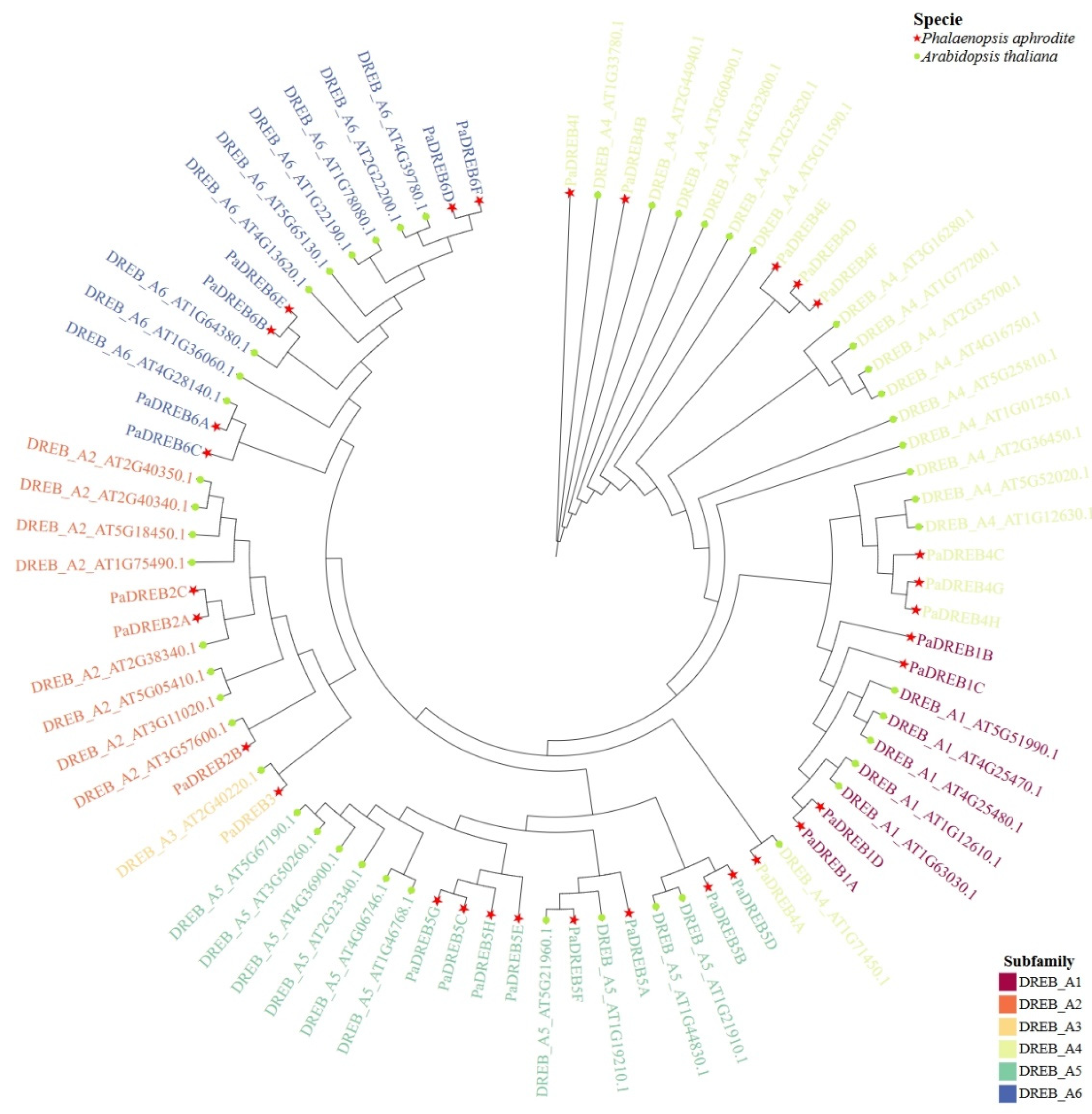
3.1. Identification of DREB Family Members in P. aphrodite and Phylogenetic Tree Construction

To correctly identify PaDREBs, we used two methods: HMMER screening and BLAST sequence comparison. The gene sequences with poor sequence quality and a poorly conserved AP2 structural domain were removed, and 30 members of the PaDREB family were preliminarily identified. To further identify all PaDREBs, the initially matched genes were matched again using the BLAST interface of the P. aphrodite genome database, and unannotated transcript PATC127667 was obtained and categorized as belonging to the PaDREB family. A total of 31 PaDREB family members were identified and used for subsequent analyses in this study.
To study the evolutionary relationships of the DREB family in P. aphrodite, the amino acid sequences of the PaDREB members in Arabidopsis and Phalaenopsis were used to construct a phylogenetic tree (Table S2). They were named according to their subgroup classification and relatives of AtDREB family members (Table S3). The sequence structure information is shown in Figure S1. According to the DREB protein sequence comparison, the 31 PaDREB TFs were classified into six subgroups from A1 to A6 (Figure 1). Among them, subgroup A1, i.e., CBF/DREB1, contained four PaDREBs.

3.2. Analysis of the PaDREB Expression Patterns in Different Tissues

To analyze the PaDREB expression patterns at different tissue sites, transcriptome data were obtained from the P. aphrodite database. The expression patterns of PaDREB genes are shown in Figure 2. The expression of PaDREBs significantly differed among tissues. PaDREB1A, PaDREB4G, and PaDREB4H were highly expressed in short stems, and PaDREB4D, PaDREB5C, and PaDREB6F were specifically highly expressed in flowers. Most members, such as PaDREB1D, were highly expressed in roots. The differential expression of PaDREBs in different tissues indicates that these genes have different biological functions during Phalaenopsis growth and development.

3.3. PaDREB1D Responded to Four Treatments and Was Expressed in Different Tissues

We identified 31 DREB gene family members in the P. aphrodite transcriptome database using both BLAST and Hidden Markov Graph methods. Among them, PaDREB1D was classified as belonging to the A1 group of the DREB family. To further understand its role in protection against external stress, RT-PCR was used to analyze the gene expression levels of PaDREB1D in Phalaenopsis leaves after four treatments. Under low-temperature stress (Figure 3A), PaDREB1D expression was upregulated after the beginning of the treatment, and its expression was higher than that of the control level until the end of the treatment. Under drought treatment (Figure 3B), PaDREB1D was upregulated after the beginning of the treatment, and its expression was highest at 6 h of treatment and then decreased with the extension of treatment time. Under salt treatment (Figure 3C), PaDREB1D was upregulated at the beginning of treatment, and its expression was highest at 6 h of treatment, after which the expression decreased back to the control level. Under ABA treatment (Figure 3D), PaDREB1D expression was highest at 6 h of treatment and then decreased to the control level.

3.4. Gene Isolation and Molecular Characterization Analysis of PaDREB1D

For subsequent functional validation, we isolated the PaDREB1D CDS from the cDNA of P. aphrodite leaves. PaDREB1D only had one exon, and it encoded 195 aa with a predicted molecular weight of 21.58 kDa and an isoelectric point of 7.17. PaDREB1D contained an AP2 domain, which was highly conserved among homologous genes in other plants, as shown by the amino acid sequences in BLAST. The 14th amino acid of the AP2 domain was valine (14V), and the 19th position was glutamic acid (19E) (Figure S2, red point). PaDREB1D had a putative NLS flanking the AP2 domain, a DSAWR sequence, and a LWSY sequence at the C terminus, which are characteristic of the CBF/DREB1 TF (Figure S2).

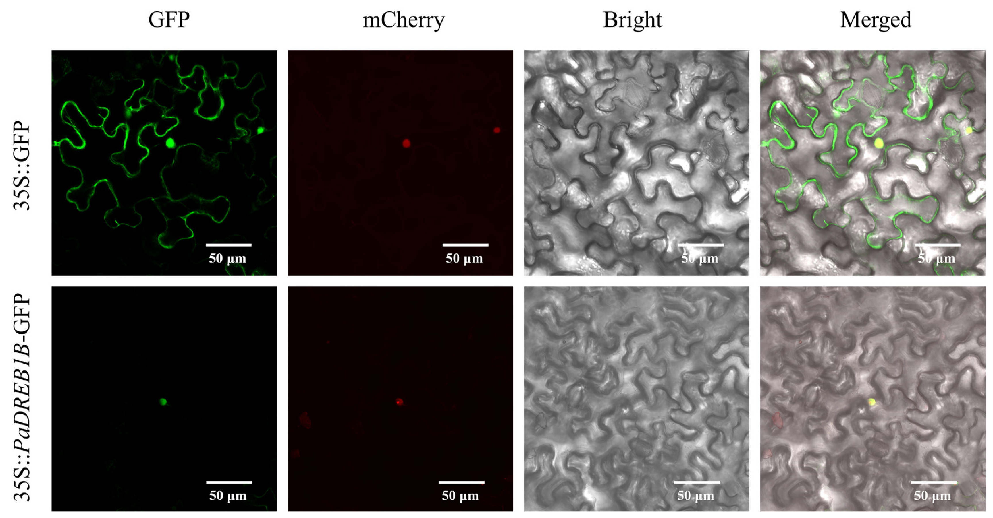
3.5. Subcellular Localization of PaDREB1D

To determine the location of PaDREB1D protein expression in cells, the full-length PaDREB1D CDS without the stop codon was connected to a 35S promoter-driven green fluorescent protein reporter gene, and the pLGFP1301 vector was transferred into Agrobacterium GV3101, using the blank vector as a control. Confocal observation showed (Figure 4) that the fluorescent signals of 35S:GFP were distributed in both the nucleus and cell membrane, while 35S:PaDREB1D-GFP only showed fluorescent signals in the nucleus, indicating that the PaDREB1D protein was localized in the nucleus.

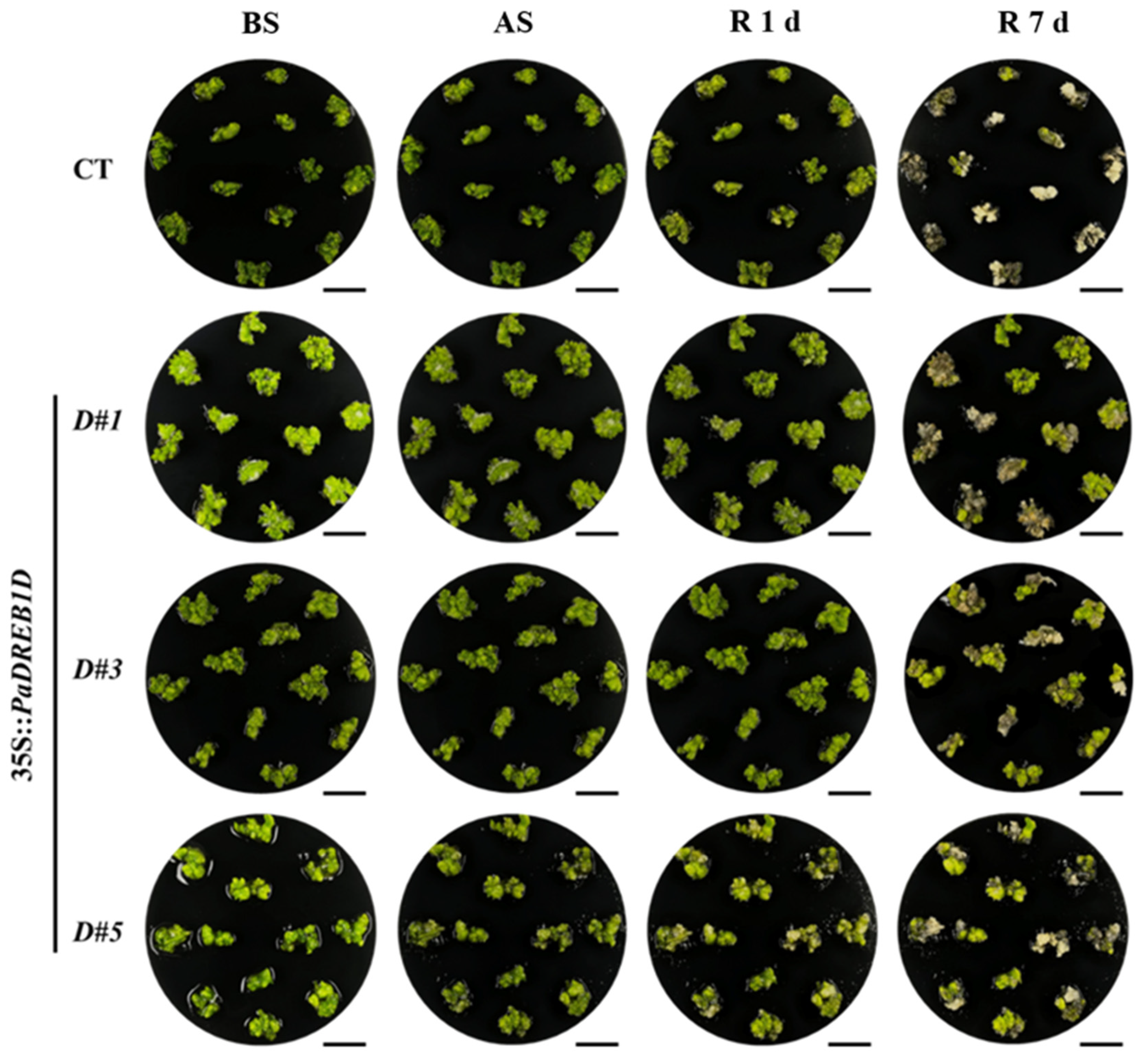
3.6. PaDREB1D Overexpression Enhanced Cold Tolerance in PLBs

To investigate the role of PaDREB1D in the cold resistance of Phalaenopsis ‘1474’ PLBs, the control line (CT) and three PaDREB1D-overexpressing lines (D#1, D#3, and D#5) were stressed by a low temperature. Considering the PLBs that remained green after stress as surviving, all three trans-PaDREB1D lines had a higher survival rate than the control (Figure 5). Before low-temperature stress, all three transgenic lines showed normal growth compared with the control. There were no differences in the survival rate between transgenic and control lines after 4 d of low-temperature stress; most of the transgenic and control PLBs remained green, as shown in Figure 5. After 1 d of recovery, differences in survival rates between transgenic and control lines appeared, while some PLBs turned yellow. After 7 d of recovery, the survival rate of the PLBs remained stable; the survival rate of the transgenic lines (around 50%) was significantly higher than the control line (7.75%) (Figure 6A).
During low-temperature stress, the damaged PLBs changed color from green to white, yellow, brown, and black, and the phenotypic changes were more clearly related to the chlorophyll content (Figure 5). The change in the chlorophyll content of the samples at the four time points was determined using the ethanol-soaking extraction method. The chlorophyll content changes were similar to the phenotypic changes during low-temperature stress (Figure 6B). There was no significant change in the total chlorophyll content between BS and AS. After the start of recovery, the total chlorophyll content decreased; at 7 d of recovery, the total chlorophyll content of the control PLBs was significantly less than that of the transgenic PLBs.

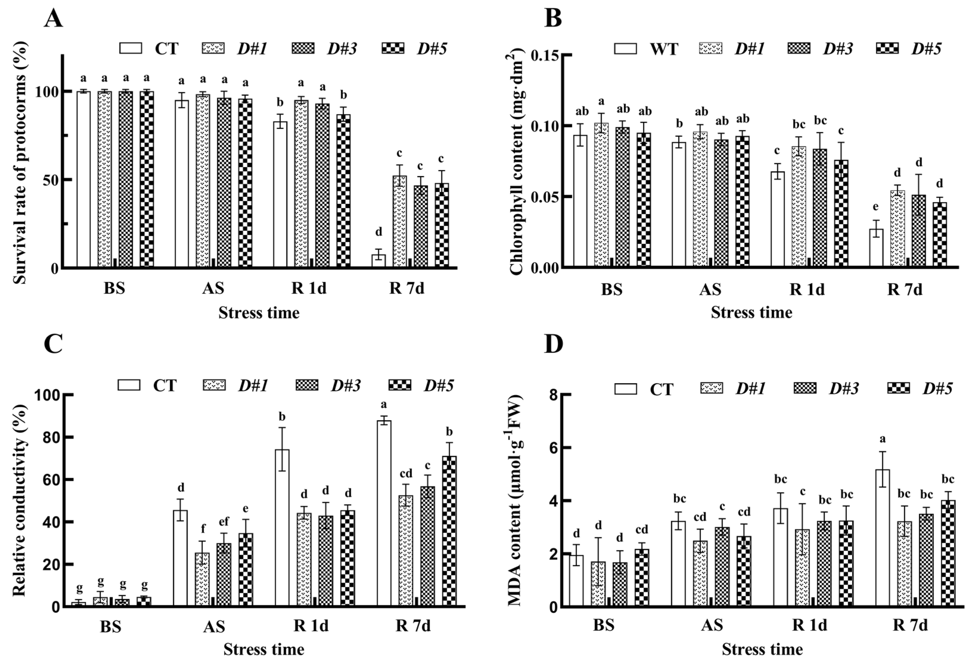
3.7. Overexpression of the PaDREB1D Gene Reduced Cellular Damage to PLBs

To detect cellular damage caused by low-temperature stress in Phalaenopsis PLBs, the REC and MDA content of the collected samples were determined. The REC of both the control and transgenic lines increased with treatment duration (Figure 6C). The average increase in REC between BS and AS was about 40% for the control and about 20% for the three transgenic PLB lines. The REC continued to increase after recovery, with an average REC of 87.92% for the control line and only about 50% for the three transgenic PLB lines at 7 d of recovery. Before low-temperature stress, there was no significant difference in the REC between the control PLBs and the trans-PaDREB1D PLBs; however, at the next three time points, the REC between the lines showed significant variability. Before and after cold treatment, the MDA content of both the control and trans-PaDREB1D lines increased, but the magnitude and amount of the increase were lower in the transgenic lines than in the control line (Figure 6D). At 7 d of recovery, the average MDA content was 5.18 μmol/g FW for the control line and around 3 μmol/g FW for transgenic PaDREB1D lines. The REC and MDA content indicate that overexpression of the PaDREB1D gene reduced cellular damage under low-temperature stress.

3.8. PaDREB1D Overexpression Enhanced the Antioxidant Enzyme Activities of PLBs

To investigate the effects of overexpressing PaDREB1D on the antioxidant enzyme activities of PLBs under low-temperature stress, the changes in the soluble protein content and SOD, POD, and CAT activities in the samples were determined at each time point (Figure 7). The soluble protein content of the transgenic PLBs was not significantly different from the control PLBs under normal growth conditions (Figure 7A). However, at 7 d of recovery, the soluble protein content of the two showed significant differences. Soluble proteins in the control group were more severely decomposed after low-temperature stress, whereas soluble proteins in the PaDREB1D-overexpressing lines were relatively less decomposed after low-temperature stress. There were no significant differences in SOD activity between the control and transgenic lines before low-temperature stress (Figure 7B). After 4 d of low-temperature stress, the SOD activity increased and was significantly higher in the PaDERB1D-overexpressing PLBs than in the control PLBs. After 7 d of recovery, the SOD activity of the two groups showed significant differences. Under normal growth conditions, both POD and CAT activities were higher in the transgenic PaDREB1D PLB lines than in the control line, and both POD and CAT activities decreased gradually over time and were significantly different at all four time points (Figure 7C,D). These results indicate that antioxidant enzyme activities were enhanced in PaDREB1D-overexpressing PLBs to resist the reactive oxygen species produced by the PLBs when exposed to low-temperature stress.

4. Discussion

Phalaenopsis is popular among consumers because of its colors, elegance, and long flowering period. As a variety that blooms in winter in the northern hemisphere, it caters to the timing of the Chinese New Year and is often sold as a New Year’s Eve flower in East Asia. Because it is mostly native to tropical and subtropical regions, temperature is the main environmental factor limiting its development in the middle and high latitudes. In China, Japan, and the Netherlands, Phalaenopsis needs to be grown in greenhouses and protected by heating during low-temperature periods. There are relatively few studies on the molecular mechanisms related to low-temperature responses in Phalaenopsis. The ICE-CBF-COR signaling pathway in plants has been established as the pathway through which plants acclimatize to cold stress [12]. PaCBF1 overexpression in Arabidopsis maintained better membrane integrity after cold stress [2].
Research has determined the whole genome of P. Aphrodite as a model plant of Phalaenopsis spp. [21]. The DREB gene family is subordinate to the AP2/ERF superfamily and plays an important role in plant responses to abiotic stress. In this study, 31 DREB family members were identified from the P. aphrodite genome (Figure 1). This number is lower than that found in Ammopiptanthus nanus (55), Arabidopsis thaliana (56), Oryza sativa subsp. japonica (57), Solanum lycopersicum (58), and Triticum aestivum (210) and similar to that found in Fragaria vesca (32) and Dendrobium catenatum (26) [31,32,33,34,35]. This suggests that the number of DREB family members varies between species. Phylogenetic results showed that PaDREB could be classified into six subgroups, A1 to A6, corresponding to the classification results in Arabidopsis. In addition, differences in the expression of PaDREBs in different tissues indicate that these genes have different biological functions during growth and development (Figure 2). In this study, based on the identification of the PaDREB family, PaDREB1D of CBF/DREB1s (subgroup A1) was selected, and specific primers were designed for gene isolation. RT-PCR showed that PaDREB1D expression was upregulated under low-temperature stress (Figure 3). Its expression was relatively high in the root and short stalk. Protein sequence alignment showed that the amino acid sequence of PaDREB1D contained one AP2-conserved structural domain, which contained the conserved sites 14V and 19E, indicating that it belongs to the CBF/DREB1 gene family [4]. The sequences of PaDREB1D and other homologous plant proteins contain differences between sequence lengths and non-conserved regions, but the conserved structural domains are highly similar. Thus, it is speculated that their gene functions are related and similar.
Most TFs have functional roles in the nucleus. The AP2/ERF superfamily, which the CBF/DREB1 gene family belongs to, is a typical TF family. In recent years, there have also been studies on the subcellular localization of its members [5]. All three CsCBF proteins of cucumber localize to the nucleus under low-temperature stress [36]. In addition, EgDREB1, SsCBF1, and MbDREB1 have been shown to localize in the nucleus [4,37,38]. To further verify the location of PaDREB1D, recombinant plasmids were constructed and transferred into Agrobacterium, and subcellular localization assays using tobacco leaves revealed that the PaDREB1D protein was localized to the nucleus (Figure 4).
Orchid breeding mainly adopts a hybrid breeding method, but this method has many limitations, such as a long breeding cycle, seed source limitations, cross-genus hybrids, and no affinity; thus, molecular breeding of orchid plants has been increasingly valued by breeders and researchers [39,40,41]. As the tissue culture technology for Phalaenopsis has matured, molecular breeding for its genetic transformation has achieved some results in recent years [42,43], and most utilize Agrobacterium-mediated methods. To date, there are no studies on the construction of a stable and efficient genetic transformation system in Phalaenopsis or even in orchids because Agrobacterium-mediated genetic transformation is affected by many conditions, for example, selection of explants, antibiotic species, and infection time with Agrobacterium. In this study, we relied on the pre-constructed genetic transformation system of Phalaenopsis ‘1474’ in our laboratory and successfully obtained PLBs with positive RNA detection. However, there were still problems during the experiment, such as low transformation efficiency and difficulties in the differentiation culture of PLBs. It is important to mention that in the pre-experiment, we tested all four members of PaDREB1 by a PLB transgenic system, but only PaDREB1D was ultimately successful. This is the direct reason for placing only PaDREB1D in our experimental results. The genetic transformation system needs to be further optimized and improved.
Studies on low-temperature-related molecular pathways in Phalaenopsis are relatively rare, and most studies on Phalaenopsis have focused on flower color and flower development [44,45,46,47]. No studies have reported on low-temperature stress treatment of Phalaenopsis PLBs. The minimum growing temperature for mature plants of most Phalaenopsis spp. is about 10 °C, and they may suffer severe injury and even death when exposed to temperatures below 5 °C [2]. Therefore, in the pre-test of low-temperature treatment, the temperature was set at 4 °C and 10 °C, and the stress time was set at 2, 4, and 7 d to find the optimal conditions for low-temperature stress of Phalaenopsis PLBs. Treatment at 4 °C for 4 d was ultimately selected for subsequent experiments (Figure 5).
After 4 d of stress treatment, the control line was nearly completely dead, whereas the transgenic lines maintained about 40% survival (Figure 6). Transgenic lines showed less damage and less chlorophyll decomposition after low-temperature stress compared with the control line. The activities of antioxidant enzymes (SOD, POD, and CAT) were also significantly higher in the transgenic lines than in the control line (Figure 7). Physiological analysis showed that the cold tolerance of the PaDREB1D-overexpressing PLBs was higher than that of the control PLBs. However, upstream and downstream regulation of PaDREB1D in the molecular pathway of cold resistance in Phalaenopsis needs to be further explored.

5. Conclusions

In this study, 31 members of the DREB family, including four members of subgroup A1 (CBF/DREB1 TFs), were identified in P. aphrodite. The gene expression of PaDREB1D increased after low-temperature stress. Compared with the other members in the A1 subgroup, it responded to low-temperature stress faster and had a higher expression. Therefore, PaDREB1D was selected for subsequent gene function verification. PaDREB1D was overexpressed in PLBs, and positive lines were obtained as material for subsequent experiments. PaDREB1D expression increased after low-temperature stress. The survival rate of transgenic PaDREB1D PLBs was significantly higher than that of the control line after low-temperature stress. The REC and MDA content of the transgenic lines were lower than those of the control line. After low-temperature stress, the SOD activity of transgenic lines was higher than that of the control line, and the POD and CAT activities were higher than those of the control line. In summary, this study provides a theoretical and experimental basis for investigating the molecular regulation mechanism of cold resistance in Phalaenopsis and the breeding of new cold-resistant Phalaenopsis varieties.

Supplementary Materials

The following supporting information can be downloaded at: https://www.mdpi.com/article/10.3390/horticulturae10090933/s1, Figure S1: Conserved motifs, conserved structural domains, and gene structure of PaDREB family members; Figure S2: Alignment of conserved AP2 domain sequences between PaDREB1D and its homologous genes in other species; Figure S3: Expression level analysis of PaDREB1D in transgenic PLB lines. Table S1: AtDREB sequences used in constructing the phylogenetic tree; Table S2: Identification of DREB gene family members in Phalaenopsis aphrodite; Table S3: All primer sequences involved in this study; Table S4: All culture media involved in this study.

Author Contributions

Z.H. designed the experiments and wrote the first draft; Z.H., Y.W. and J.L. performed the experiments; S.W. and Y.W. analyzed the data; P.L., J.X. and Y.C. revised the manuscript. All authors have read and agreed to the published version of the manuscript.

Funding

This study was supported by the Zhejiang Science and Technology Major Program on Agricultural New Variety Breeding (2021C02071-5) and the “Pioneer” and “Leading Goose” R&D Programs of Zhejiang (2023C02028).

Data Availability Statement

The data presented in this study are available within the article.

Conflicts of Interest

The authors declare no conflicts of interest.

References

  1. Chen, J.; Chen, C. The effect of temperature on the inflorescence formation model for Phalaenopsis. Plants 2024, 13, 1280. [Google Scholar] [CrossRef]
  2. Peng, P.H.; Lin, C.H.; Tsai, H.W.; Lin, T.Y. Cold response in Phalaenopsis aphrodite and characterization of PaCBF1 and PaICE1. Plant Cell Physiol. 2014, 55, 1623–1635. [Google Scholar] [CrossRef] [PubMed]
  3. Lee, H.C.; Chen, Y.J.; Markhart, A.; Lin, T.Y. Temperature effects on systemic endoreduplication in orchid during floral development. Plant Sci. 2007, 172, 588–595. [Google Scholar] [CrossRef]
  4. Akhtar, M.; Jaiswal, A.; Taj, G.; Jaiswal, J.P.; Qureshi, M.I.; Singh, N.K. DREB1/CBF transcription factors: Their structure, function and role in abiotic stress tolerance in plants. J. Genet. 2012, 91, 385–395. [Google Scholar] [CrossRef]
  5. Feng, K.; Hou, X.L.; Xing, G.M.; Liu, J.X.; Duan, A.Q.; Xu, Z.S.; Li, M.Y.; Zhuang, J.; Xiong, A.S. Advances in AP2/ERF super-family transcription factors in plant. Crit. Rev. Biotechnol. 2020, 40, 750–776. [Google Scholar] [CrossRef]
  6. Sakuma, Y.; Liu, Q.; Dubouzet, J.G.; Abe, H.; Shinozaki, K.; Yamaguchi-Shinozaki, K. DNA-binding specificity of the ERF/AP2 domain of Arabidopsis DREBs, transcription factors involved in dehydration- and cold-inducible gene expression. Biochem. Biophys. Res. Commun. 2002, 290, 998–1009. [Google Scholar] [CrossRef]
  7. Nakano, T.; Suzuki, K.; Fujimura, T.; Shinshi, H. Genome-wide analysis of the ERF gene family in Arabidopsis and rice. Plant Physiol. 2006, 140, 411–432. [Google Scholar] [CrossRef]
  8. Kim, J.S.; Kidokoro, S.; Yamaguchi-Shinozaki, K.; Shinozaki, K. Regulatory networks in plant responses to drought and cold stress. Plant Physiol. 2024, 195, 170–189. [Google Scholar] [CrossRef]
  9. Kidokoro, S.; Shinozaki, K.; Yamaguchi-Shinozaki, K. Transcriptional regulatory network of plant cold-stress responses. Trends Plant Sci. 2022, 27, 922–935. [Google Scholar] [CrossRef] [PubMed]
  10. Jaglo, K.R.; Kleff, S.; Amundsen, K.L.; Zhang, X.; Haake, V.; Zhang, J.Z.; Deits, T.; Thomashow, M.F. Components of the Arabidopsis C-repeat/dehydration-responsive element binding factor cold-response pathway are conserved in Brassica napus and other plant species. Plant Physiol. 2001, 127, 910–917. [Google Scholar] [CrossRef]
  11. Dubouzet, J.G.; Sakuma, Y.; Ito, Y.; Kasuga, M.; Dubouzet, E.G.; Miura, S.; Seki, M.; Shinozaki, K.; Yamaguchi-Shinozaki, K. OsDREB genes in rice, Oryza sativa L., encode transcription activators that function in drought-, high-salt- and cold-responsive gene expression. Plant J. 2003, 33, 751–763. [Google Scholar] [CrossRef] [PubMed]
  12. Hwarari, D.; Guan, Y.; Ahmad, B.; Movahedi, A.; Min, T.; Hao, Z.; Lu, Y.; Chen, J.; Yang, L. ICE-CBF-COR signaling cascade and its regulation in plants responding to cold stress. Int. J. Mol. Sci. 2022, 23, 1549. [Google Scholar] [CrossRef] [PubMed]
  13. Zarka, D.G.; Vogel, J.T.; Cook, D.; Thomashow, M.F. Cold induction of Arabidopsis CBF genes involves multiple ICE (inducer of CBF expression) promoter elements and a cold-regulatory circuit that is desensitized by low temperature. Plant Physiol. 2003, 133, 910–918. [Google Scholar] [CrossRef] [PubMed]
  14. Haake, V.; Cook, D.; Riechmann, J.L.; Pineda, O.; Thomashow, M.F.; Zhang, J.Z. Transcription factor CBF4 is a regulator of drought adaptation in Arabidopsis. Plant Physiol. 2002, 130, 639–648. [Google Scholar] [CrossRef]
  15. Lehti-Shiu, M.D.; Uygun, S.; Moghe, G.D.; Panchy, N.; Fang, L.; Hufnagel, D.E.; Jasicki, H.L.; Feig, M.; Shiu, S.H. Molecular evidence for functional divergence and decay of a transcription factor derived from whole-genome duplication in Arabidopsis thaliana. Plant Physiol. 2015, 168, 1717–1734. [Google Scholar] [CrossRef]
  16. Xie, Z.; Nolan, T.M.; Jiang, H.; Yin, Y. AP2/ERF transcription factor regulatory networks in hormone and abiotic stress responses in Arabidopsis. Front. Plant Sci. 2019, 10, 228. [Google Scholar] [CrossRef]
  17. Rubio, S.; Noriega, X.; Pérez, F.J. Abscisic acid (ABA) and low temperatures synergistically increase the expression of CBF/DREB1 transcription factors and cold-hardiness in grapevine dormant buds. Ann. Bot. 2019, 123, 681–689. [Google Scholar] [CrossRef]
  18. Zhang, S.Y.; Cao, K.F.; Wei, Y.Y.; Jiang, S.; Ye, J.F.; Xu, F.; Chen, Y.; Shao, X.F. PpBZR1, a BES/BZR transcription factor, enhances cold stress tolerance by suppressing sucrose degradation in peach fruit. Plant Physiol. Biochem. 2023, 202, 107972. [Google Scholar] [CrossRef]
  19. Kazan, K. Diverse roles of jasmonates and ethylene in abiotic stress tolerance. Trends Plant Sci. 2015, 20, 219–229. [Google Scholar] [CrossRef]
  20. Livak, K.J.; Schmittgen, T.D. Analysis of relative gene expression data using real-time quantitative PCR and the 2(-Delta Delta C(T)) Method. Methods 2001, 25, 402–408. [Google Scholar] [CrossRef]
  21. Chao, Y.T.; Chen, W.C.; Chen, C.Y.; Ho, H.Y.; Yeh, C.H.; Kuo, Y.T.; Su, C.L.; Yen, S.H.; Hsueh, H.Y.; Yeh, J.H.; et al. Chromosome-level assembly, genetic and physical mapping of Phalaenopsis aphrodite genome provides new insights into species adaptation and resources for orchid breeding. Plant Biotechnol. J. 2018, 16, 2027–2041. [Google Scholar] [CrossRef] [PubMed]
  22. Chao, Y.T.; Yen, S.H.; Yeh, J.H.; Chen, W.C.; Shih, M.C. Orchidstra 2.0-A transcriptomics resource for the Orchid family. Plant Cell Physiol. 2017, 58, e9. [Google Scholar] [CrossRef]
  23. Reiser, L.; Subramaniam, S.; Li, D.; Huala, E. Using the Arabidopsis information resource (TAIR) to find information about Arabidopsis genes. Curr. Protoc. Bioinform. 2017, 60, 1.11.1–1.11.45. [Google Scholar] [CrossRef] [PubMed]
  24. Chen, C.J.; Chen, H.; Zhang, Y.; Thomas, H.R.; Frank, M.H.; He, Y.H.; Xia, R. TBtools: An integrative toolkit developed for interactive analyses of big biological data. Mol. Plant 2020, 13, 1194–1202. [Google Scholar] [CrossRef]
  25. Tamura, K.; Stecher, G.; Kumar, S. MEGA11: Molecular evolutionary genetics analysis version 11. Mol. Biol. Evol. 2021, 38, 3022–3027. [Google Scholar] [CrossRef]
  26. Xie, J.M.; Chen, Y.R.; Cai, G.J.; Cai, R.L.; Hu, Z.; Wang, H. Tree Visualization By One Table (tvBOT): A web application for visualizing, modifying and annotating phylogenetic Trees. Nucleic Acids Res. 2023, 51, W587–W592. [Google Scholar] [CrossRef]
  27. Gong, X.Q.; Zhang, J.Y.; Hu, J.B.; Wang, W.; Wu, H.; Zhang, Q.H.; Liu, J.H. FcWRKY70, a WRKY protein of Fortunella crassifolia, functions in drought tolerance and modulates putrescine synthesis by regulating arginine decarboxylase gene. Plant Cell Environ. 2015, 38, 2248–2262. [Google Scholar] [CrossRef]
  28. Dahro, B.; Wang, F.; Peng, T.; Liu, J.H. PtrA/NINV, an alkaline/neutral invertase gene of Poncirus trifoliata, confers enhanced tolerance to multiple abiotic stresses by modulating ROS levels and maintaining photosynthetic efficiency. BMC Plant Biol. 2016, 16, 76. [Google Scholar] [CrossRef]
  29. Porra, R.J.; Thompson, W.A.; Kriedemann, P.E. Determination of accurate extinction coefficients and simultaneous equations for assaying chlorophylls a and b extracted with four different solvents: Verification of the concentration of chlorophyll standards by atomic absorption spectroscopy. BBA-Bioenerg. 1989, 975, 384–394. [Google Scholar] [CrossRef]
  30. Bradford, M.M. A rapid and sensitive method for the quantitation of microgram quantities of protein utilizing the principle of protein-dye binding. Anal. Biochem. 1976, 72, 248–254. [Google Scholar] [CrossRef]
  31. Maqsood, H.; Munir, F.; Amir, R.; Gul, A. Genome-wide identification, comprehensive characterization of transcription factors, cis-regulatory elements, protein homology, and protein interaction network of DREB gene family in Solanum Lycopersicum. Front. Plant Sci. 2022, 13, 1031679. [Google Scholar] [CrossRef] [PubMed]
  32. Han, Y.L.; Cai, M.H.; Zhang, S.Q.; Chai, J.W.; Sun, M.Z.; Wang, Y.W.; Xie, Q.Y.; Chen, Y.H.; Wang, H.Z.; Chen, T. Genome-wide identification of AP2/ERF transcription factor family and functional analysis of DcAP2/ERF#96 associated with abiotic stress in Dendrobium Catenatum. Int. J. Mol. Sci. 2022, 23, 13603. [Google Scholar] [CrossRef]
  33. Dong, C.; Xi, Y.; Chen, X.L.; Cheng, Z.M. Genome-wide identification of AP2/EREBP in Fragaria Vesca and expression pattern analysis of the FvDREB subfamily under drought stress. BMC Plant Biol. 2021, 21, 295. [Google Scholar] [CrossRef]
  34. Niu, X.; Luo, T.L.; Zhao, H.Y.; Su, Y.L.; Ji, W.Q.; Li, H.F. Identification of wheat DREB genes and functional characterization of TaDREB3 in response to abiotic stresses. Gene 2020, 740, 144514. [Google Scholar] [CrossRef] [PubMed]
  35. Cao, S.L.; Wang, Y.; Li, X.T.; Gao, F.; Feng, J.C.; Zhou, Y.J. Characterization of the AP2/ERF transcription factor family and expression profiling of DREB subfamily under cold and osmotic stresses in Ammopiptanthus nanus. Plants 2020, 9, 455. [Google Scholar] [CrossRef] [PubMed]
  36. Li, J.L.; Li, H.M.; Quan, X.Y.; Shan, Q.; Wang, W.B.; Yin, N.; Wang, S.Q.; Wang, Z.H.; He, W.X. Comprehensive analysis of cucumber C-repeat/dehydration-responsive element binding factor family genes and their potential roles in cold tolerance of Cucumber. BMC Plant Biol. 2022, 22, 270. [Google Scholar] [CrossRef]
  37. Azzeme, A.M.; Abdullah, S.N.A.; Aziz, M.A.; Wahab, P.E.M. Oil palm drought inducible DREB1 induced expression of DRE/CRT- and non-DRE/CRT-containing genes in lowland transgenic tomato under cold and PEG treatments. Plant Physiol. Biochem. 2017, 112, 129–151. [Google Scholar] [CrossRef]
  38. Yang, W.; Liu, X.D.; Chi, X.J.; Wu, C.A.; Li, Y.Z.; Song, L.L.; Liu, X.M.; Wang, Y.F.; Wang, F.W.; Zhang, C.; et al. Dwarf apple MbDREB1 enhances plant tolerance to low temperature, drought, and salt stress via both ABA-dependent and ABA-independent pathways. Planta 2011, 233, 219–229. [Google Scholar] [CrossRef]
  39. Bolaños-Villegas, P.; Chen, F.C. Advances and perspectives for polyploidy breeding in Orchids. Plants 2022, 11, 1421. [Google Scholar] [CrossRef]
  40. Teixeira da Silva, J.A.; Jin, X.; Dobránszki, J.; Lu, J.; Wang, H.; Zotz, G.; Cardoso, J.C.; Zeng, S. Advances in Dendrobium molecular research: Applications in genetic variation, identification and breeding. Mol. Phylogenet. Evol. 2016, 95, 196–216. [Google Scholar] [CrossRef]
  41. Popova, E.; Kim, H.H.; Saxena, P.K.; Engelmann, F.; Pritchard, H.W. Frozen beauty: The cryobiotechnology of orchid diversity. Biotechnol. Adv. 2016, 34, 380–403. [Google Scholar] [CrossRef] [PubMed]
  42. Iiyama, C.M.; Vilcherrez-Atoche, J.A.; Germanà, M.A.; Vendrame, W.A.; Cardoso, J.C. Breeding of ornamental orchids with focus on Phalaenopsis: Current approaches, tools, and challenges for this century. Heredity 2024, 132, 163–178. [Google Scholar] [CrossRef] [PubMed]
  43. Cardoso, J.C.; Zanello, C.A.; Chen, J.T. An overview of orchid protocorm-like bodies: Mass propagation, biotechnology, molecular aspects, and breeding. Int. J. Mol. Sci. 2020, 21, 985. [Google Scholar] [CrossRef]
  44. Lou, Y.X.; Zhang, Q.Y.; Xu, Q.Y.; Xu, X.Y.; Wang, W.X.; Gai, R.N.; Ming, F. PhCHS5 and PhF3’5’H Genes over-expression in Petunia (Petunia hybrida) and Phalaenopsis (Phalaenopsis aphrodite) regulate flower color and branch number. Plants 2023, 12, 2204. [Google Scholar] [CrossRef]
  45. Xu, Q.Y.; Xia, M.; He, G.R.; Zhang, Q.Y.; Meng, Y.; Ming, F. New insights into the influence of NHX-type Cation/H+ antiporter on flower color in Phalaenopsis orchids. J. Plant Physiol. 2022, 279, 153857. [Google Scholar] [CrossRef]
  46. Chen, W.H.; Jiang, Z.Y.; Hsu, H.F.; Yang, C.H. Silencing of FOREVER YOUNG FLOWER-like genes from Phalaenopsis orchids promotes flower senescence and abscission. Plant Cell Physiol. 2021, 62, 111–124. [Google Scholar] [CrossRef] [PubMed]
  47. Liang, C.Y.; Rengasamy, K.P.; Huang, L.H.; Hsu, C.C.; Jeng, M.F.; Chen, W.H.; Chen, H.W. Assessment of violet-blue color formation in Phalaenopsis orchids. BMC Plant Biol. 2020, 20, 212. [Google Scholar] [CrossRef]
Figure 1. Phylogenetic tree of the DREB family in Arabidopsis thaliana and Phalaenopsis aphrodite. The AtDREB sequences used are shown in Table S2.
Horticulturae 10 00933 g001
Figure 2. PaDREB expression patterns in different tissues. F: flower; L: leaf; R: root; SS: short stalk; LS: long stalk; SB: small bud; LB: large bud. The transcript per kilobase per million mapped reads of PaDREBs were screened from the Phalaenopsis aphrodite transcriptome data.
Horticulturae 10 00933 g002
Figure 3. Expression pattern analysis of PaDREB1D under abiotic stress and abscisic acid (ABA) treatments. (A) Low-temperature treatment; (B) drought treatment; (C) salt treatment; (D) ABA treatment. Untreated Phalaenopsis aphrodite was used as a control (0 h). The data represent the mean ± standard deviation (n = 3). Different letters show significant differences (p < 0.05) between samples.
Horticulturae 10 00933 g003
Figure 4. Subcellular localization of PaDREB1D. The 35S:PaDREB1D-GFP and 35S:GFP controls were transiently expressed in tobacco leaves. mCherry was chosen as a nuclear localization marker. Scale bar = 50 μm.
Horticulturae 10 00933 g004
Figure 5. Phenotypic changes in transgenic PaDREB1D protocorm-like bodies after low-temperature stress. BS: before stress, AS: after stress, R 1 d: 1 d of recovery cultivation, R 7 d: 7 d of recovery cultivation. PLBs that remained green at 7 d of recovery were considered alive; those that were whitened, yellowed, or browned were considered dead. Scale bar = 1 cm.
Horticulturae 10 00933 g005
Figure 6. Survival rate and cellular damage in protocorm-like bodies under low-temperature stress. (A) Survival rate, (B) chlorophyll content, (C) relative conductivity, and (D) MDA content of the control and transgenic PaDREB1D PLBs under low-temperature stress. BS: before stress, AS: after stress, R 1 d: 1 d of recovery cultivation, R 7 d: 7 d of recovery cultivation. The data represent the mean ± standard deviation (n = 3). Different letters show significant differences (p < 0.05) between samples.
Horticulturae 10 00933 g006
Figure 7. Changes in the soluble protein content and antioxidant enzyme activity of control and transgenic PaDREB1D protocorm-like bodies before and after 4 d of low-temperature stress. (A) Soluble protein content; (B) SOD activity; (C) POD activity; (D) CAT activity. BS: before stress, AS: after stress, R 1 d: 1 d of recovery cultivation, R 7 d: 7 d of recovery cultivation. The data represent the mean ± standard deviation (n = 3). Different letters show significant differences (p < 0.05) between samples.
Horticulturae 10 00933 g007
Disclaimer/Publisher’s Note: The statements, opinions and data contained in all publications are solely those of the individual author(s) and contributor(s) and not of MDPI and/or the editor(s). MDPI and/or the editor(s) disclaim responsibility for any injury to people or property resulting from any ideas, methods, instructions or products referred to in the content.

