Article

Ultraviolet-C Light Effects in Actinidia spp. Infected by Pseudomonas syringae pv. actinidiae

1
CREA, Consiglio per la Ricerca in Agricoltura e L’analisi dell’Economia Agraria—Centro Olivicoltura, Frutticoltura e Agrumicoltura, 00134 Rome, Italy
2
ENEA Centro Ricerche Frascati, Dipartimento Nucleare, Divisione Studi del Plasma e DTT, Laboratorio Fusione Inerziale, Plasmi ed Esperimenti Interdisciplinari, 00044 Frascati, Italy
3
CREA, Consiglio per la Ricerca in Agricoltura e L’analisi dell’Economia Agraria—Centro Difesa e Certificazione, 00156 Rome, Italy
4
ENEA Centro Ricerche Frascati, Dipartimento Nucleare, Divisione Tecnologie Fisiche e Sicurezza, Laboratorio Diagnostica e Metrologia, 00044 Frascati, Italy
5
ENEA Centro Ricerche Casaccia, Dipartimento Sostenibilità, Circolarità e Adattamento al Cambiamento Climatico dei Sistemi Produttivi e Territoriali, Divisione Sistemi Agroalimentari Sostenibili, Laboratorio Bioeconomia Circolare Rigenerativa, 00123 Rome, Italy
*
Authors to whom correspondence should be addressed.
Horticulturae 2024, 10(9), 944; https://doi.org/10.3390/horticulturae10090944
Submission received: 20 July 2024 / Revised: 27 August 2024 / Accepted: 2 September 2024 / Published: 4 September 2024
(This article belongs to the Section Plant Pathology and Disease Management (PPDM))

Abstract

:
Several studies have demonstrated that ultraviolet-band-C (UV-C) irradiation can enhance plants’ natural resistance to pathogens and diseases. A suitable dose of UV-C radiation induces the production of metabolites that strengthen plant defenses, an effect known as “hormesis”. Hormesis presents a promising alternative that could supplement and reduce the use of pesticides, which pose risks to the environment and human health. This paper investigates the effects of UV-C radiation emitted by an array of Light-Emitting Diodes (LEDs) in generating a hormetic response in three kiwifruit species, namely A. chinensis var. deliciosa cv. Hayward, A. chinensis var. chinensis cv. Soreli®, and A. arguta plantlets, grown in vitro and in pots, exposed to the pathogen Pseudomonas syringae pv. actinidiae (Psa) either before or after UV-C irradiation. Analyses of morpho-physiological parameters and spectrophotometric assays were conducted to evaluate changes in chlorophyll a and b content, carotenoids, total phenols, and antioxidant activity in relation to the UV-C irradiation. Results indicate partial protection against Psa infection and increased levels of chlorophylls, carotenoids, polyphenols and antioxidant activity. The optimal UV-C dose was determined to be 2.2 kJ/m2 for in vitro shoots and 1.3 kJ/m2, for ex vitro plants.

1. Introduction

Exposure to ultraviolet-band-C (UV-C) light can induce metabolite production in plants in response to biotic and abiotic stresses. These metabolites inhibit pathogenic microorganisms and enhance plants’ resistance to pathogens [1,2]. Specifically, antioxidant enzymes, secondary metabolites, and cell wall modifications improve protective responses and mitigate UV-induced oxidative damage by eliminating reactive oxygen species (ROS) produced when the metabolism is compromised by stress [3]. Phenolic compounds, which have antioxidant properties, increase in response to various stresses caused by environmental conditions and pathogens. Their accumulation helps plants inhibit ROS. Carotenoids, pigments that absorb light in the visible spectrum and transfer energy to chlorophyll, also play a crucial photoprotective and antioxidant role. The combination of biochemical events activated by UV-C light and their beneficial effects is referred to as “hormesis”.
In addition to conventional mercury lamps emitting light at 254 nm, an array of Light-Emitting Diodes (LEDs) emitting light at 277 nm has demonstrated a fungicidal/fungistatic effect against infection by Penicillium and Botrytis [4]. LEDs are preferred over mercury lamps due to their robustness, compact size, portability, and stable radiative power over time, making them more suitable for field applications [5]. The single LED is a solid-state device powered at 5–6 V and 0.35 amps, emitting continuous-wave UV-C light of a few tens of mW. LEDs can be organized in many element arrays, resulting in medium-power radiation sources that are compact, robust, and quick to turn on/off. Additionally, LEDs are space-saving, lightweight, mercury-free, and require a low-voltage, compact power supply. However, UV-C LEDs are less efficient at converting electrical power into light (2–3% efficiency) compared to UV-C lamps (up to 40% efficiency). Despite this, LEDs provide constant UV light output over time, while lamps show a gradual decrease in radiated optical power.
Little is known about the precise mechanisms of UV-C light’s interaction with plants, depending on wavelength and dose. For example, the shortest UV-C wavelength (222 nm) has been shown to damage guard cells and epidermal cells at low doses [6]. In this context, UV-C LEDs emitting at 277 nm, in the longer range of UV-C wavelengths, may offer an advantage in reducing unwanted plant damage.
In vitro cultures are a useful tool for the rapid characterization of biotic and abiotic stress responses, particularly in tree species, due to the lengthy process of growing large numbers of woody or semi-woody plants. In vitro cultures provide an experimental model for early screening of genotype-specific responses to stresses [7,8].
According to FAO estimates, the global area of land designated for kiwifruit cultivation comprises 270,475 hectares [9]. Actinidia chinensis var. deliciosa and A. chinensis var. chinensis are the most economically significant varieties [10]. The demand for kiwifruit is largely driven by knowledge of its nutritional benefits.
Since 2008, bacterial canker of kiwifruit caused by Pseudomonas syringae pv. actinidiae (Psa) has spread globally, causing severe damage to crop production and resulting in substantial economic losses. Psa is the most critical disease that affects kiwifruit and is capable of infecting all kiwi and mini-kiwi species. Currently, this disease is managed through chemical and agronomic approaches, with limited success [11,12,13]. The potential for Psa to evolve into more virulent biovars or resistant lineages underscores the need for effective and sustainable solutions to reduce agrochemical use while minimizing damage to kiwifruit plantations.
Among Actinidia spp., A. chinensis is highly susceptible to Psa, while A. arguta has been classified as tolerant [14,15]. The mechanisms underlying this tolerance are not fully understood [16,17].
This study investigated the protective effects of UV-C LED irradiations against Psa in Actinidia. First, in vitro shoots of Actinidia chinensis var. deliciosa ‘Hayward’, A. chinensis var. chinensis ‘Soreli’, and A. arguta A0061-AG4 were treated to assess potential protective responses. To determine whether reduced susceptibility to Psa was due to a hormetic effect of UV-C or a germicidal effect, Psa was inoculated, in two different trials, either before or after UV-C irradiation. The results achieved justified extending the investigation to UV-C protection against Psa in kiwifruit plants.
Additionally, analyses of morpho-physiological parameters and spectrophotometric assays were conducted to evaluate changes in chlorophylls a and b content, carotenoids, total phenolic content (TPC), and antioxidant capacity (AC) resulting from UV-C irradiation.

2. Materials and Methods

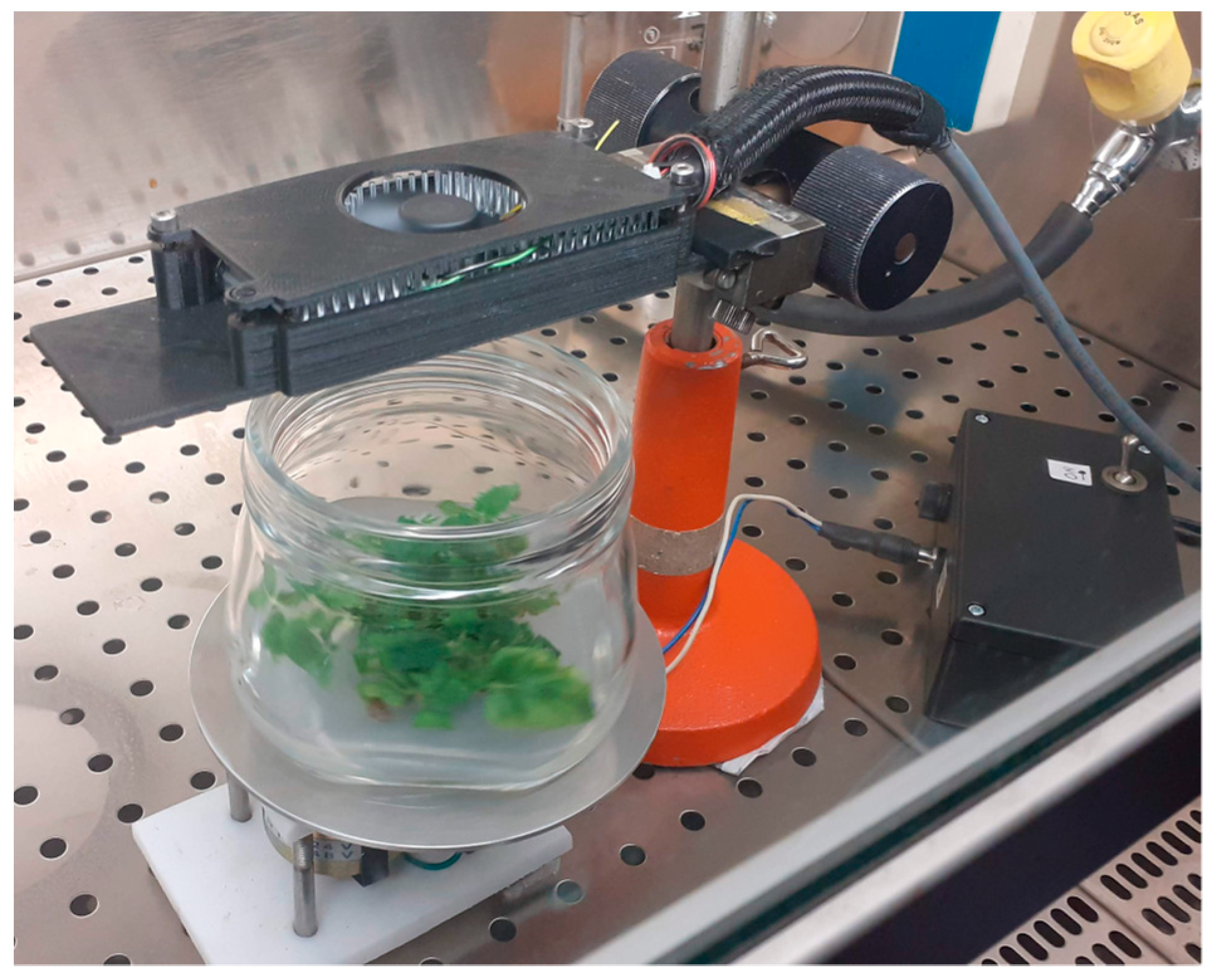
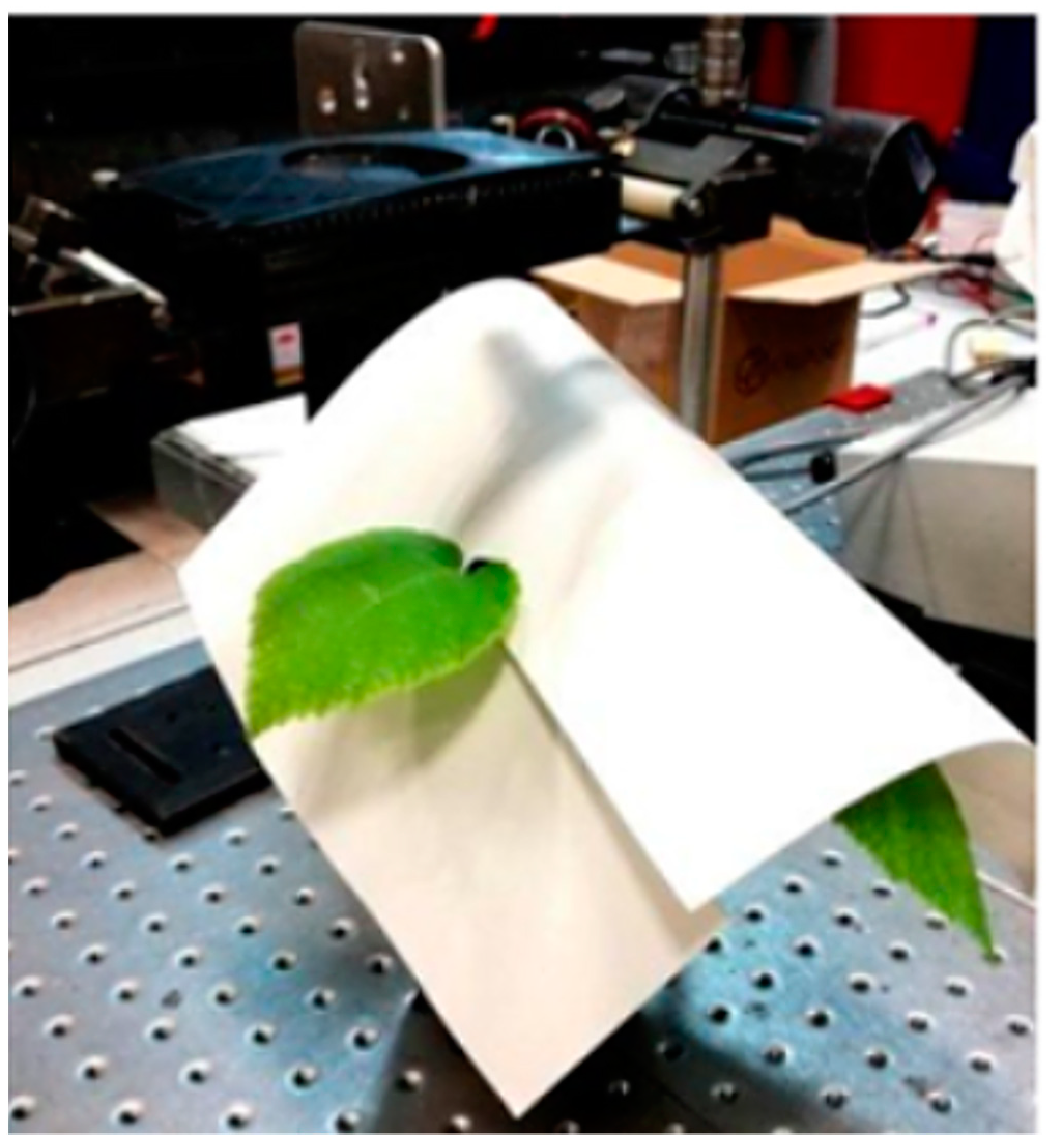
2.1. UV-C Irradiation Equipment

Arrays of Luminus XBT-3535-UV LEDs emitting light (Luminus Devices, Inc., Sunnyvale, CA, USA) centered at 277 nm and bandwidth 12 nm (full width at half height) have been assembled and characterized at the ENEA Frascati Research Center. The device used in this work is an array of 20 LEDs arranged in three rows on a printed circuit board of (9 × 3) cm2 in size and is operated by electronics that allow irradiances of variable power and duration as desired. The device includes a cooling system, which is essential to ensure stability of the irradiated intensity over time, and a constant current power supply that can be set from 0.1 to 0.4 amps [5]. In addition to the device, we have developed optical simulation software [5] that provides both the spatial distribution and the mean value of the intensity of UV-C light as a function of the distance from the LED array. The simulation results are in excellent agreement with the experimental data of 2-D intensity distribution and variation of the average intensity with distance from the array.

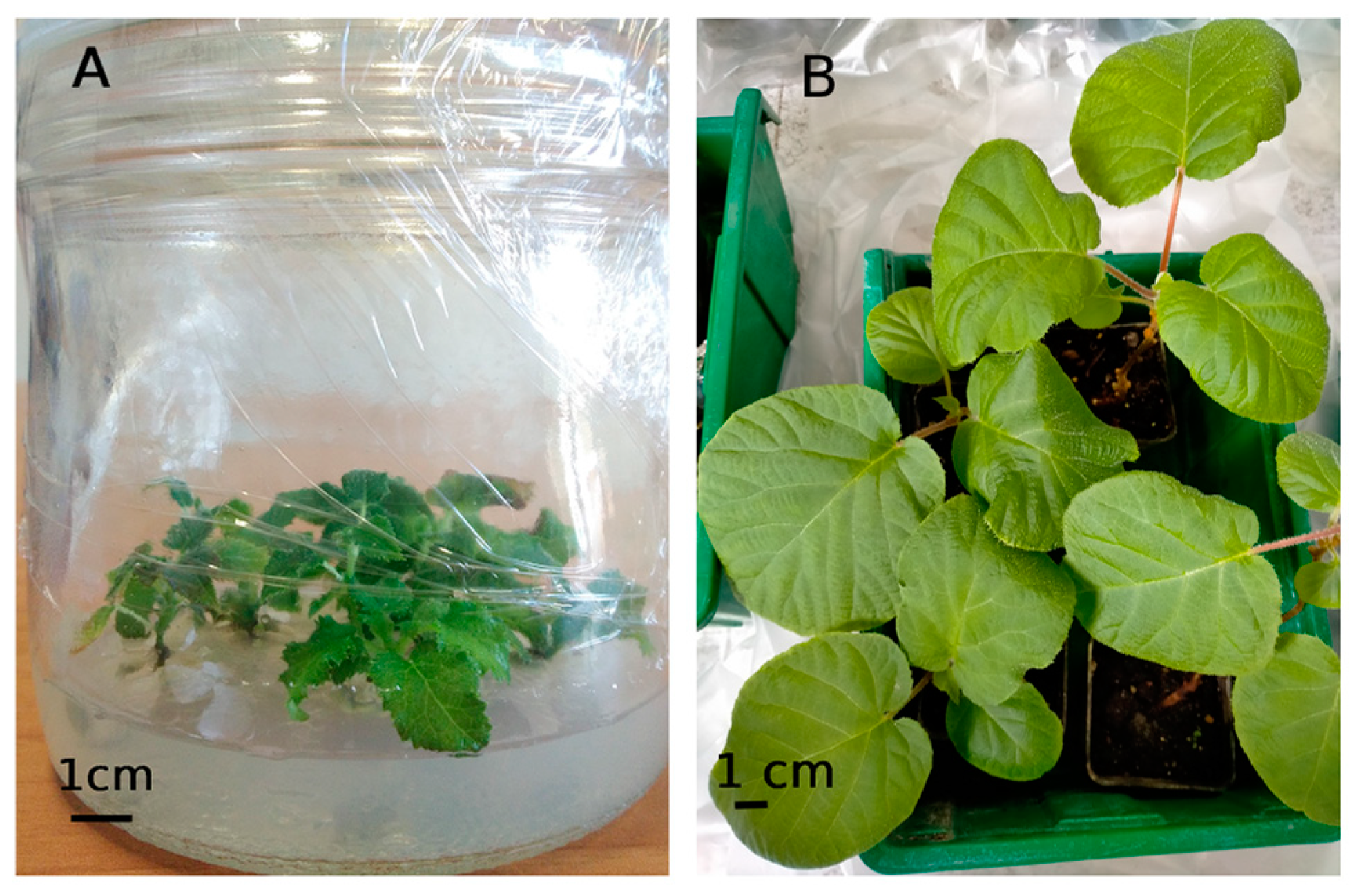
2.2. Plant Material and Growing Conditions
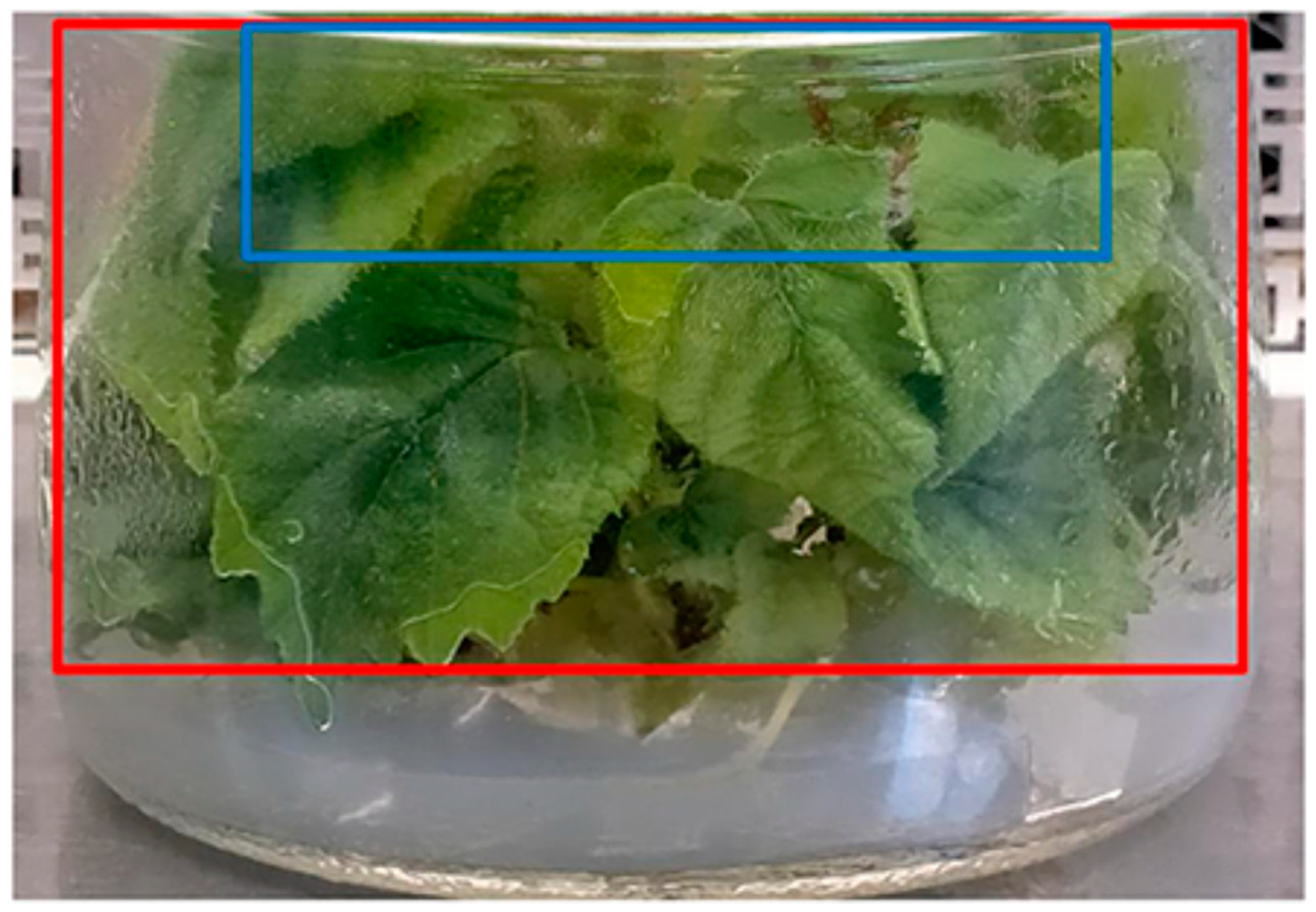
In vitro growing shoots of A. chinensis var. deliciosa cv. Hayward, A. chinensis var. chinensis cv. Soreli® and A. arguta accession A0061-AG4 were obtained from the in vitro germplasm collection of the National Repository Centre for fruit crops species of the CREA in Rome, Italy [7,18]. The shoots were multiplied into Magenta jars (Sigma Aldrich s.r.l., Milan, Italy) containing 50 mL of a multiplication (Mult) medium consisting of Quoirin macrosalts [19], MS microsalts chelates and organic compounds [20], 30 g/L sucrose and 5.8 g/L agar, with the addition of 1 mg/L BA. The pH of the medium was adjusted to 5.80 before autoclaving at 121 °C for 20 min. Cultures were maintained in the growth chamber at 24 ± 1 °C, 70% relative humidity, with a 16 h photoperiod and light intensity of 37.5 µmol m−2s−1 photosynthetic photon flux emitted by Philips TLD 58W/33 fluorescent tubes (Philips S.p.A, Milan, Italy) (standard conditions) and subcultured every four weeks
In vivo acclimated plants of A. chinensis var. chinensis cv. Soreli® were produced from in vitro shoots transferred into glass jars containing 100 mL of a rooting medium comprising MS basal medium containing 30 g/L sucrose and 5.8 g/L agar and supplied with 0.5 mg/L Indole butyric acid; the pH was adjusted to 5.6 before autoclaving. Microcuttings were maintained for 20 days at 24 ± 2 °C under standard culture room conditions, then the rooted explants were transferred to Jiffy-7® Peat Pellets (Jiffy Products International BV, Zwijndrecht, The Netherlands) under a polyethylene tunnel in a greenhouse at 18–25 °C, 30–40% relative humidity, and natural daylight. After an additional 30 days, rooted plantlets were transferred into 4 cm × 8 cm (height × diameter) pots filled with a mixture of 75% peat and 25% vermiculite, which was previously sterilized at 121 °C for 15 min in an autoclave, in the greenhouse in the same controlled conditions.

2.3. Irradiations

The irradiations were carried out on two different types of plant material. Initially, experiments were performed on in vitro shoots (A. chinensis var. deliciosa cv. Hayward, A. chinensis var. chinensis cv. Soreli® and A. arguta). These shoots, at the 4-leaf stage, were cultured on 100 mL of gelled Mult medium in glass jars, with each jar containing 5 shoots (Figure 1A).
Subsequently, experiments were conducted on ex vitro healthy rooted plantlets of cv. Soreli (Figure 1B). These plantlets had been grown for six months in pots following their transplantation.
Preliminarily, the effective intensity of UV-C light (radiated power per unit area, W/m2) was measured by using the absolute power meter Hamamatsu C9536/H9535 (Hamamatsu Photonics s.r.l., Milan, Italy).
The dose released to the target (J/m2) is calculated based on the light intensity on an ideal plane—whose position is chosen a priori—multiplied by the duration of irradiation(s). For ease of use and best reproducibility, the dose was varied by acting on the exposure time once the distance between LED and plant was fixed and measured [4].
In vitro shoots were irradiated under sterile conditions in a BioHazard continuous laminar flow hood (Steril VBH, AS Instruments s.r.l.,Turin, Italy) with a UV-C dose of 2.2 kJ/m2 at the center of the jar and about 1.5 kJ/m2 on the outermost leaves at the edge of the jar (Figure 2). The jars were placed on a rotating base during irradiation to achieve spatially uniform exposure of the shoots. The number of jars (containing 5 shoots each) irradiated for each species in each assay is specified in the dedicated section.
Four groups of potted Soreli plants were irradiated in each assay. In addition to the 2.2 kJ/m2 dose, 3 lower doses (1.3, 0.8, or 0.3 kJ/m2) were tested because the 2.2 kJ/m2 dose caused plant decline. Each leaf was individually irradiated with UV-C to ensure uniform exposure.
Figure 3 illustrates the experimental apparatus for single-leaf irradiation.

2.4. Morphological Observations

On in vitro cultures of A. chinensis var. deliciosa cv. Hayward (4 jars), A. chinensis var. chinensis cv. Soreli® (11 jars) and A. arguta accession A0061-AG4 (4 jars), three weeks (one subculture) after irradiation with a dose of 2.2 kJ/m2, the height and weight of shoots, the rate of multiplication, the number of leaves, and sustained damage (if any) were recorded.
For the pot-grown Soreli plants, any morphological damage was assessed 14 days after irradiation was carried out at the following doses (3 plants each): 2.2 kJ/m2, 1.3 kJ/m2, 0.8 kJ/m2 and 0.3 kJ/m2.

2.5. Artificial Inoculation and Assessment of the Severity of Visible Symptoms

Forty-eight hours after UV-C irradiation (2.2 kJ/m2), 10 shoots from each genotype were individually transferred under sterile conditions into tubes containing 10 mL of a Psa bacterial suspension (1–2 × 10⁸ CFU mL−1), strain CREA-OFA 8.43 [21], in sterile water. As controls, 10 unirradiated shoots were inoculated to confirm the infection; 10 irradiated shoots were inoculated with sterile double-distilled water; 10 were neither irradiated nor inoculated. A second group of 10 shoots was infected 3 days before UV-C irradiation under the same conditions as described above. After a 12 h incubation period, the shoots were transferred aseptically into Mult medium and maintained in the growth chamber under standard conditions. Symptoms were recorded at 5, 10, and 21 days post infection (dpi).
Forty-eight hours after UV-C irradiation (1.3 kJ/m2), 18 potted plants of A. chinensis var. chinensis cv. Soreli® were infected by applying 0.2 mL of Psa bacterial suspension (1–2 × 10⁸ CFU mL−1 to a 4–5 mm wound made on the leaves with a sterile scalpel. A total of 36 leaves were both irradiated and inoculated. As control samples, 18 unirradiated leaves were inoculated to confirm the infection; 18 irradiated shoots were inoculated with sterile double-distilled water; 18 were neither irradiated nor inoculated. Immediately after inoculation, each plant was placed in a clear plastic bag to minimize air circulation. All pots were irrigated every seven days. The presence of symptoms was monitored every 5 days post inoculation. All tests were conducted in an isolated room illuminated by natural light, with temperatures between 15 °C and 25 °C.
The severity of visible symptoms induced by the infection was evaluated using a rating scale, ranking each shoot or leaf into 5 classes according to Table 1, using a modified McKinney MKI index [22]; see Equation (1):
MKI(%) = {[Σ (c·n)]/N}·(100/C)
where c is the value of the infection class, n is the number of samples belonging to the same class, N is the total number of samples, and C is the maximum value of the rating scale.

2.6. Spectrophotometric Analyses

On in vitro cultures of A. chinensis var. deliciosa cv. Hayward (3 jars), A. chinensis var. chinensis cv. Soreli® (3 jars) and of A. arguta (3 jars), 48 h after irradiation with a dose of 2.2 kJ/m2, spectrophotometric assays were performed for the determination of chlorophylls a and b and carotenoids TPC and AC. The sample of shoots exposed to direct irradiation (within the blue frame in Figure 4) was compared with the shaded portions (red frame in Figure 4). This experimental design was employed to further control the variability of sensitive parameters such as TPC and AC.
The same spectrophotometric analyses were conducted on pot-grown cv. Soreli leaves, which were harvested 14 days after irradiation with doses of 1.3 kJ/m2, 0.8 kJ/m2 and 0.3 kJ/m2. Three plants were used for each dose.

2.6.1. Contents of Chlorophyll a and b and Carotenoids

To determine the contents of chlorophyll a and b and carotenoids, leaf samples (100 mg F.W.) were ground in 5 mL acetone and 20 mg calcium carbonate and centrifuged at 8500× g (Allegra 21R Beckman, Brea, CA, USA) for 5 min [23]. This procedure was repeated three times.
Quantification was performed by measuring the absorbance of the supernatant at λ = 661.6, 644.8, and 470 nm, for chlorophylls a and b and carotenoids, respectively. The pigment concentration was determined according to [24]:
Chla = 11.24 A661.6 − 2.04 A644.8
Chlb = 20.13 A644.8 − 4.19 A661.6
Carotenoids = (1000 A470 − 1.90 Chla − 63.14 Chlb)/214
where Aλ is the absorbance at the wavelength λ in nm. The concentration was expressed as µg mg−1 F.W. The total chlorophyll concentration is calculated as the sum of Chla and Chlb.

2.6.2. TPC and AC

To determine the TPC and the AC, leaf samples (200 mg F.W.) were ground in 5 mL of ethanol and centrifuged at 8500× g for 5 min. The procedure was repeated three times. All the procedures were performed under dim light, and glassware containing samples were covered with aluminum foil to minimize photo-oxidation.
The TPC of the ethanol extract solution was determined using the Folin-Ciocalteu method as described by [25], with slight modifications. Briefly, 0.1 mL of the supernatant of each centrifuged sample was added to 0.2 mL of Folin–Ciocalteu reagent in 3 mL of double-distilled water, then 0.6 mL of 20% (w/v) sodium carbonate solution was added to the mixture. After the mixture was kept in total darkness for 120 min, its absorbance was measured at 765 nm. TPC was calculated using a gallic acid (GA) calibration curve with seven calibration points (y = 0.0015x; R2 = 0.999). The results were expressed as micrograms of gallic acid equivalents (GAEs) per milligram of fresh weight of the sample (µg GAEs/mg F.W.).
The AC was measured by the DPPH free radical scavenging effect induced by ethanolic extracts as described by [26], with a few modifications. Each diluted extract (750 μL) was added to 750 mL of ethanol. Then, 25 μM DPPH solution in ethanol was added. The solution was kept in the dark at room temperature for 30 min and the absorbance at 515 nm (Abs515) was measured. For the standard calibration curve (8–24 μM), Trolox (6-hydroxy-2,5,7,8-tetramethylchroman-2-carboxylic acid) was used as standard with five calibration points (R2 > 0.99). The results were expressed as micrograms of Trolox equivalent per milligram of fresh weight (µg Trolox eq/mg F.W.). The DPPH scavenging effect was calculated as follows:
Scavenging effect (%) = (Abs515 (DPPH) Abs515 (sample))/Abs515 (DPPH) × 100
Five technical replicates were performed for each assay.
All quantitative analyses were performed using the Evolution™ 300 UV-Vis spectrophotometer (Thermo Fisher Scientific Inc., Rome, Italy).

2.7. Statistical Analysis

The standard error was calculated for the collected data, which were evaluated for normality distribution using Levene’s test. The homoscedasticity of variance was assessed with Shapiro–Wilk’s test. Significant differences among mean values were determined by one-way analysis of variance (ANOVA). Tukey’s Multiple Comparison Test (p ≤ 0.05) was applied to evaluate differences in means among treatments using Past software, version 2.17c. Each analysis was performed on three samples and repeated five times.

3. Results

3.1. A. chinensis var. deliciosa cv. Hayward, A. chinensis var. chinensis cv. Soreli® and A. arguta In Vitro Cultured Shoots (Irradiation Dose 2.2 kJ/m2)

3.1.1. Macroscopic Morphological Response to Irradiation

The shoots were visually inspected for up to three months (two subcultures) following UV-C irradiation. No damage was observed on irradiated shoots, which exhibited growth comparable to the unirradiated controls and maintained a normal multiplication rate.

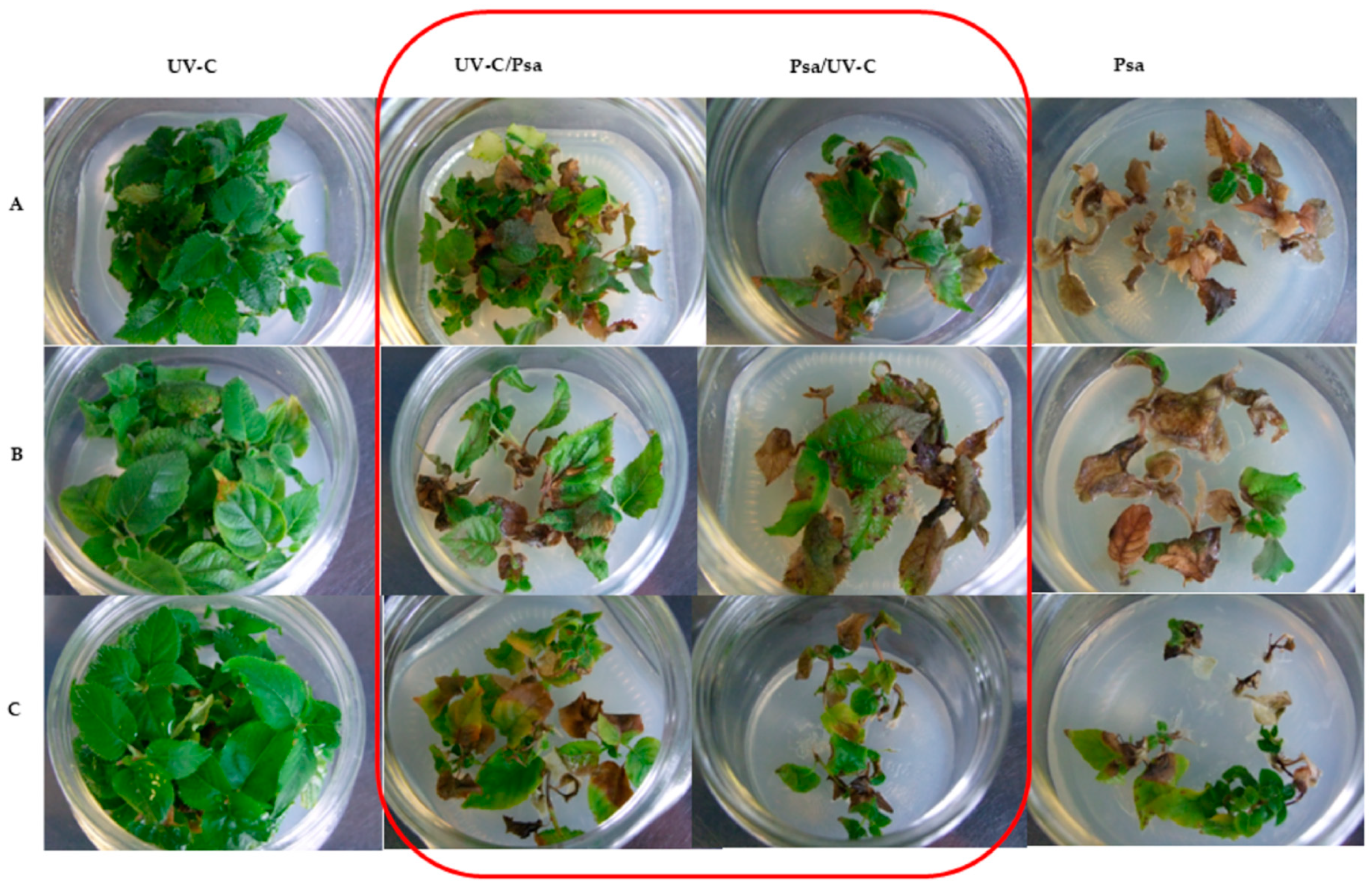
3.1.2. Psa Inoculation

In the in vitro control plant group inoculated with Psa and not treated with UV-C (10 shoots), initial foliar symptoms were observed as early as 5 dpi. In contrast, no symptoms were present in the two irradiated groups (10 shoots each) at this time. By 10 dpi, the irradiated groups (except for the 10 shoots in the non-inoculated control group) also began to exhibit symptoms of infection.
Table 2 presents the MKI at 21 dpi, when all inoculated genotypes displayed the maximum percentage of damage rate. Figure 5 illustrates the condition of the shoots of the three genotypes under the different experimental conditions at 21 dpi.

3.1.3. Photosynthetic Pigments, Total Phenolics Content, and Antioxidant Capacity

As shown in Table 3, UV-C irradiated in vitro shoots of cv. Hayward, cv. Soreli and A. arguta (three jars each) exhibited an increase in carotenoid production across all three genotypes compared with the controls, while chlorophyll levels remained unchanged or were slightly reduced. Table 4 indicates that cv. Soreli and A. arguta demonstrated increased TPC and AC compared to the controls. In contrast, cv. Hayward showed increased TPC and AC in UV-C-treated leaves in vitro (54.7 vs. 33.5 µg GAE eq/mg F.W. and 0.07 vs. 0.04 µg of Trolox eq/mg F.W.), but in the control that was not treated with UV-C, the TPC values were comparable (55.5 µg GAE eq/mg F.W.).

3.2. A. chinensis var. chinensis cv. Soreli® Potted Plants (Irradiation Dose 2.2, 1.3, 0.8, and 0.3 kJ/m2)

3.2.1. Macroscopic Morphological Response to Irradiation

‘Soreli’ plants grown in pots (five plants per treatment) and irradiated at 2.2 kJ/m2 exhibited signs of distress within three days compared to the non-irradiated controls. These signs included leaf browning, which ultimately led to the death of the plants within approximately two weeks. Conversely, plants irradiated at lower doses of 0.3, 0.8, and 1.3 kJ/m2 (three plants per dose) displayed no macroscopic damage and remained still healthy after six months. To optimize the hormesis effect while minimizing plant damage, the highest dose that did not cause harm to the potted plants (1.3 kJ/m2) was selected for subsequent assays.

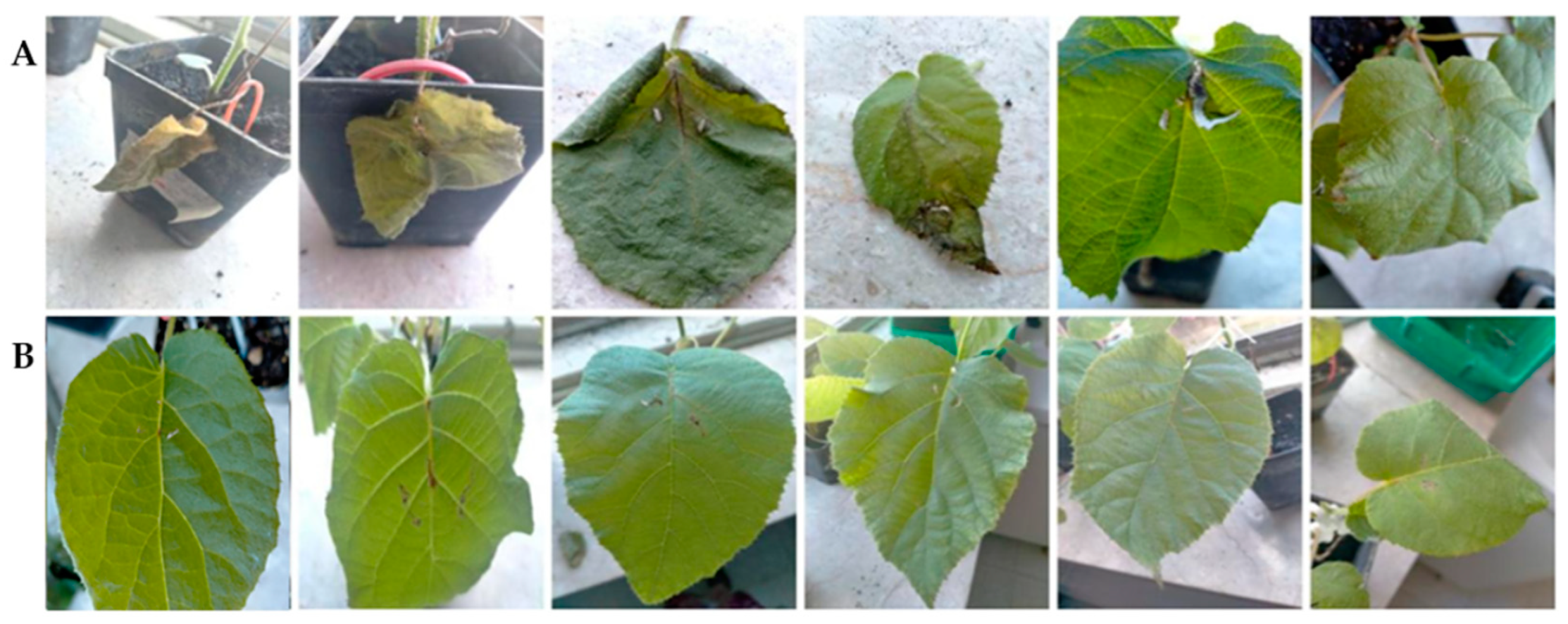
3.2.2. Psa Inoculation

In the leaves of Actinidia spp. acclimated plants inoculated with Psa and not treated with UV-C, initial symptoms appeared at 7 dpi. In contrast, no symptoms were observed in the UV-C irradiated group (1.3 kJ/m2). By 10 dpi, symptoms began to appear in the irradiated and inoculated plants. At 21 dpi, the MKI (%) was 35.8% in the irradiated and inoculated leaves compared to 90% in the inoculated leaves that were not treated with UV-C (Figure 6). The irradiated leaves inoculated with sterile double-distilled water alone exhibited an MKI of 20%; the control group, which was neither irradiated nor infected, showed no symptoms (MKI 0%).

3.2.3. Photosynthetic Pigments, Total Polyphenols, and Antioxidant Activity

The results of the spectrophotometric analysis for plants irradiated at 0.3, 0.8, and 1.3 kJ/m2 (three plants each) are presented in Table 5 and in Table 6. The data indicate an increase in the production of chlorophylls, carotenoids, TPC, and AC in leaves irradiated at 1.3 kJ/m2. In contrast, irradiation at the lower dose of 0.3 kJ/m2 did not result in a significant difference in the production of these compounds or in AC compared to unirradiated control leaves.

4. Discussion

UV-C irradiation has proven its effectiveness in many horticultural species and post-harvest fruits exposed to numerous pathogens, as evidenced by a large body of literature [2,3,27,28,29,30,31,32]. However, there is limited literature on whether UV-C light can inhibit infections in fruit trees. Therefore, studies that explore the agronomic potential of UV-C light in protecting crops against pathogen attacks before harvest are crucial. The hormesis effect induced by UV-C irradiations may enable plants to counteract biotic stresses without sustaining damage.
In this study, the hormesis effect induced by UV-C light was evaluated as a potential means of providing partial protection from Psa infection in Actinidia. Initial results were obtained from three in vitro grown Actinidia genotypes. In vitro culture allows for a rapid assessment for potential pathogenicity and phytotoxicity of pathogen components, with simultaneous testing on multiple homogeneous plantlets. To determine whether the reduced sensitivity to Psa was due to a hormetic effect or a germicidal effect, Psa was inoculated in two separate trials—either before or after UV-C irradiation.
The results showed that UV-C irradiation conferred protection to the shoots both before and after artificial infection. A significant delay in the appearance of infection symptoms was observed in both irradiated groups; as shown in Table 2, at the end of subculture (21 dpi), irradiated plantlets exhibited reduced susceptibility to Psa, particularly when irradiation preceded Psa inoculation, with an MKI of 38% and 58% for cv. Soreli and cv. Hayward, respectively, compared with 96% and 82% for the corresponding infected non-irradiated controls.
A. arguta is known for its tolerance against Psa, which seems to be related to earlier pathogen recognition and activation of the antioxidant system compared to A. chinensis [33]. In our experimental system, A. arguta confirmed limited damage (44%) caused by Psa, and UV–C irradiation showed reduced protection; this phenomenon could be due to the early and redundant activation of the antioxidant system and PSA recognition mechanisms in this species.
Irradiation and infection trials were also conducted on young potted plants. The cv. Soreli, which is more susceptible to Psa and demonstrated the most effective hormesis-induced pathogen reaction in vitro, was chosen. The UV-C dose of 1.3 kJ/m2 was applied, as the 2.2 kJ/m2 dose was found to be harmful to the ‘Soreli’ plants. The results confirmed the beneficial effects of hormesis observed in vitro, showing partial protection from infection, with MKI (%) improving markedly from 90% in infected and unirradiated plants to 35.8% in irradiated and infected plants.
Based on these results, we can conclude that the reduced susceptibility to Psa is due to a hormetic effect of UV-C light rather than a germicidal effect. This finding aligns with the results reported by [1], which highlighted how UV-C can induce a defensive response in plants through the activation of metabolic pathways linked to the accumulation of phenols and other antioxidant compounds.
To our knowledge, this study is among the first to evaluate UV-C-mediated pest control in fruit plants. Furthermore, for the first time, we measured the effective intensity of UV-C radiation using an absolute detector, which is essential for reliable data when determining the optimal hormetic dose. In the future, it will be essential to design targeted experiments to establish the minimum effective UV-C dose, defined as the lowest dose that provides significant protection against Psa infection.
Additional investigations were conducted to assess how the content of chlorophylls and carotenoids responds to UV-C treatment. Furthermore, we evaluated the nonenzymatic antioxidative system in terms of TPC and AC. In the plantlets grown in vitro, the portion under direct irradiation at a dose of 2.2 kJ/m2 showed a marked increase in carotenoids and TPC and AC production in all three genotypes compared to shoots at the periphery of the irradiated zone, or those which were not irradiated (Table 3 and Table 4). Slight degradation of chlorophylls occurred, warranting further investigation. This effect did not seem to impair the shoots’ performance. Under tissue culture conditions, the photosynthesis of the shoots is limited by the low CO2 level in the vessel during the lighting period. To compensate for this, the shoots use the sucrose supplied in the medium as a carbon source [34,35]. In turn, high sucrose levels adversely affect photosynthesis. Although shoots may appear normal, it is unlikely that they are actively photosynthesizing [36]. Despite this, for in vitro plant propagation, photomixotrophic conditions are considered advantageous over photoautotrophic conditions [37].
According to [38], the results of this study also indicate the possibility of enhancing the antioxidant defenses of micropropagated plants and increasing their stress tolerance through physical elicitors. This method could be applied to the micropropagation of species such as woody species, whose yields are significantly impacted by poor in vitro performance. Moreover, UV-C treatments could pave the way for using kiwi shoot cultures as biofactories to generate bioactive substances that could be utilized in agrifood, cosmetics, or pharmaceuticals [39]. However, certain issues still need to be addressed, such as the duration of treatment effects and their influence on the subsequent performance of ex vitro plants.
While in in vitro shoots, chlorophyll degradation did not affect the shoots’ performance, the UV-C dose of 2.2 kJ/m2 was found to be harmful to the fully photoautotrophic ‘Soreli’ plants. In these plants, UV-C treatments with lower doses (0.3, 0.8, and 1.3 kJ/m2) resulted in a dose-dependent improvement in the content of chlorophylls, carotenoids, polyphenols, and antioxidant activity, which appeared to plateau at 0.8 kJ/m2 for chlorophylls and carotenoid contents (Table 5 and Table 6). This is consistent with the finding of [38], who observed a significant increase in phenols and antioxidant capacity in Ceratonia siliqua in response to low-dose UV-C irradiation and cytokinin treatments. Low-dose UV irradiation has also been shown to stimulate chlorophyll and carotenoid concentrations in other plant species, including groundnut, mung bean, and tomato [40,41]. Irradiations at doses of 1.3 and 0.8 kJ/m2 stimulated the production of polyphenols, antioxidant activity, chlorophylls, and carotenoids to a greater extent than the irradiation at a dose of 0.3 kJ/m2, whose values were close to those of the unirradiated leaves. A dose-dependent response to UV-C light mediated by UVR8 photoreceptors protein has recently been demonstrated in the model plant Arabidopsis thaliana [42]. UVR8 absorbs both the UV-B and UV-C region of the spectrum and induces the accumulation of flavonoids, strengthening plants’ defenses against pathogens [43,44,45]. Although reports of a UV-C-specific photoreceptor are currently scarce, some initial hypotheses suggest that UVR8 may be involved.
It cannot be excluded that the effects of UV-C light are due to mechanisms other than those considered in this work. For example, UV-C stress modulates vitamin C biosynthesis in acerola fruit and numerous studies have demonstrated increased salicylic acid production in response to UV-C and UV-B stress, which is important in defending against plant disease [46,47,48,49,50,51,52]. These studies show that UV-C and UV-B light not only induce phenols accumulation but also stimulate salicylic acid production and the consequent activation of genes linked to systemic acquired resistance. According to [53], the response of salicylic acid biosynthesis and other metabolites can vary significantly depending on the plant species and variety under combined stress conditions such as UV-B and water deficit. This suggests that UV-C may act on multiple levels and that further research is required to better understand the mechanisms underlying UV-C light-induced defensive stimuli. Identifying markers of UV-C light perception could be useful for maximizing efficacy and reducing probability of the damage caused by UV-C light treatments.
The growing popularity of sustainable farming practices is significantly influencing the expansion of the kiwifruit market. Consumers are becoming increasingly aware that organically grown kiwifruits have been shown to contain higher levels of beneficial polyphenols, vitamin C, minerals, and antioxidants. Recently, alternative tools for sustainable management of kiwi bacterial canker have been reviewed [13]. Over the past twenty years, there has been progress in developing more environmentally friendly alternatives to copper and antibiotics, such as plant elicitors, beneficial fungi and bacteria, replacing susceptible kiwifruit cultivars with tolerant ones, and precision agriculture technologies. All these methods have obtained partial and unsatisfactory results. In particular, the pandemic spread of Psa cannot be satisfactorily managed through conventional chemical and agronomic approaches.
A growing body of scientific research suggests that UV-C irradiation could be a promising physical treatment for crop pest management in sustainable agriculture. To address consumers’ environmental and health concerns, and in light of recent advances in robotics, UV-induced hormesis should be considered an essential component of integrated pest control systems.

5. Conclusions

The evidence presented in this work suggests that UV-C light emitted by LEDs is a promising and innovative technology that can enhance the defense systems of kiwifruit plants against Psa. The effectiveness of UV-C light-induced hormesis varies depending on the species and the dose applied, necessitating the setting up and optimization of irradiation protocols to maximize benefits and minimize side effects during each phenological phase of the crop.
This work was made possible through the collaboration of specialists from various fields and underscores the added value of multidisciplinary studies in reducing the use of agrochemicals by exploring sustainable alternatives for controlling infectious plant diseases.

Author Contributions

Conceptualization, S.L. and P.D.L.; methodology, S.L., S.B., D.M, L.B., A.L. and P.D.L.; software, S.B. and D.M.; formal analysis, S.L., S.B., D.M. and P.N.; investigation, S.L., S.B., D.M., P.N and P.D.L.; resources, M.S., E.C. and P.D.L.; data curation, S.L., S.B., D.M. and P.N.; writing—original draft preparation, S.L.; writing—review and editing, S.L., S.B., D.M., P.N., M.S., E.C., A.L., L.B. and P.D.L.; supervision, M.S., E.C. and P.D.L. All authors have read and agreed to the published version of the manuscript.

Funding

This research received no external funding.

Data Availability Statement

The original contribution presented in the study are included in the article, further inquiries can be directed to the corresponding author.

Conflicts of Interest

The authors declare no conflict of interest.

References

  1. Loconsole, D.; Santamaria, P. UV Lighting in Horticulture: A Sustainable Tool for Improving Production Quality and Food Safety. Horticulturae 2021, 7, 9. [Google Scholar] [CrossRef]
  2. Sidibé, A.; Charles, M.T.; Lucier, J.F.; Xu, Y.; Beaulieu, C. Preharvest UV-C hormesis induces key genes associated with homeostasis, growth, and defense in lettuce inoculated with Xanthomonas campestris pv. vitians. Front. Plant Sci. 2019, 12, 793989. [Google Scholar] [CrossRef]
  3. Godínez-Mendoza, P.L.; Rico-Chávez, A.K.; Ferrusquía-Jimenez, N.I.; Carbajal-Valenzuela, I.A.; Villagómez-Aranda, A.L.; Torres-Pacheco, I.; Guevara-González, R.G. Plant hormesis: Revisiting the concepts of biostimulation, elicitation, and their application in sustainable agricultural production. Sci. Total Environ. 2023, 894, 164883. [Google Scholar] [CrossRef] [PubMed]
  4. Di Lazzaro, P.; Metelli, G.; Bollanti, S.; Lai, A.; Montecchi, M.; Murra, D.; Bernabei, G.; Bacchetta, L. Pathogen growth inhibition in Ocimum basilicum (L.), Malus domestica (Borkh.), and Citrus limon (L.) by low-dose UV-C LED exposure. Ann. Agric. Crop Sci. 2024, 9, 1144. [Google Scholar] [CrossRef]
  5. Bollanti, S.; Di Lazzaro, P.; Flora, F.; Gallerano, G.P.; Mezi, L.; Murra, D.; Aquilini, M. Design, realization, and test of ultraviolet-C LED arrays suitable for long-lasting irradiation of biological samples. Machines 2023, 11, 792. [Google Scholar] [CrossRef]
  6. Otake, M.; Yoshiyama, K.O.; Yamaguchi, H.; Hidema, J. 222 nm ultraviolet radiation C causes more severe damage to guard cells and epidermal cells of Arabidopsis plants than does 254 nm ultraviolet radiation. Photochem. Photobiol. Sci. 2021, 20, 1675–1683. [Google Scholar] [CrossRef]
  7. Spigaglia, P.; Barbanti, F.; Marocchi, F.; Mastroleo, M.; Baretta, M.; Ferrante, P.; Caboni, E.; Lucioli, S.; Scortichini, M. Clostridium bifermentans and C. subterminale are associated with kiwifruit vine decline, known as moria, in Italy. Plant Pathol. 2020, 69, 765–774. [Google Scholar] [CrossRef]
  8. Manici, L.M.; Caboni, E.; Caputo, F.; Frattarelli, A.; Lucioli, S. Phytotoxins from Dactylonectria torresensis involved in replant disease of fruit trees. Rhizosphere 2021, 17, 100300. [Google Scholar] [CrossRef]
  9. Fideghelli, C. Panorama della actinidicoltura mondiale, situazione e prospettive. Resoconto del Convegno italo-francese. Kiwinforma 2022, 10–12, 13–20. [Google Scholar]
  10. Huang, H. Domestication and Commercialization of Actinidia. In Kiwifruit; Huang, H., Ed.; Academic Press: Cambridge, MA, USA, 2016; pp. 191–210. ISBN 9780128030660. [Google Scholar] [CrossRef]
  11. Scortichini, M.; Marcelletti, S.; Ferrante, P.; Petriccione, M.; Firrao, G. Pseudomonas syringae pv. actinidiae: A re-emerging, multi-faceted, pandemic pathogen. Mol. Plant Pathol. 2012, 13, 631–640. [Google Scholar] [CrossRef]
  12. Nunes da Silva, M.; Santos, M.G.; Vasconcelos, M.W.; Carvalho, S.M.P. Mitigation of emergent bacterial pathogens using Pseudomonas syringae pv. actinidiae as a case study—From orchard to gene and everything in between. Crops 2022, 2, 351–377. [Google Scholar] [CrossRef]
  13. Santos, M.G.; Nunes da Silva, M.; Vasconcelos, M.W.; Carvalho, S.M.P. Scientific and technological advances in the development of sustainable disease management tools: A case study on kiwifruit bacterial canker. Front. Plant Sci. 2024, 14, 1306420. [Google Scholar] [CrossRef] [PubMed]
  14. Song, Y.L.; Lin, M.M.; Zhong, Y.P.; Chen, J.Y.; Qi, X.J.; Sun, L.M.; Fang, J.B. Evaluation of resistance of kiwifruit varieties (strains) against bacterial canker disease and correlation analysis among evaluation indexes. J. Fruit Sci. 2020, 37, 900–908. [Google Scholar]
  15. Michelotti, V.; Lamontanara, A.; Buriani, G.; Orrù, L.; Cellini, A.; Donati, I.; Spinelli, F. Comparative transcriptome analysis of the interaction between Actinidia chinensis var. chinensis and Pseudomonas syringae pv. actinidiae in absence and presence of acibenzolar-S-methyl. BMC Genom. 2018, 19, 585. [Google Scholar] [CrossRef] [PubMed]
  16. Wang, T.; Wang, G.; Jia, Z.; Pan, D.; Zhang, J.; Guo, Z. Transcriptome analysis of kiwifruit in response to Pseudomonas syringae pv. actinidiae infection. Int. J. Mol. Sci. 2018, 19, 373. [Google Scholar] [CrossRef]
  17. Nunes da Silva, M.; Machado, J.; Balestra, G.M.; Mazzaglia, A.; Vasconcelos, M.W.; Carvalho, S.M.P. Exploring the expression of defence-related genes in Actinidia spp. after infection with Pseudomonas syringae pv. actinidiae and pv. actinidifoliorum: First steps. Eur. J. Hortic. Sci. 2019, 84, 206–212. [Google Scholar] [CrossRef]
  18. Vaccino, P.; Antonetti, M.; Balconi, C.; Brandolini, A.; Cappellozza, S.; Caputo, A.R.; Carboni, A.; Caruso, M.; Copetta, A.; de Dato, G.; et al. Plant Genetic Resources for Food and Agriculture: The Role and Contribution of CREA (Italy) within the National Program RGV-FAO. Agronomy 2024, 14, 1263. [Google Scholar] [CrossRef]
  19. Quoirin, M.; Lepoivre, P.H. Improved media for in vitro culture of Prunus sp. Acta Hortic. 1977, 78, 437–442. [Google Scholar] [CrossRef]
  20. Murashige, T.; Skoog, F. A revised medium for rapid growth and bioassays with tobacco tissue cultures. Physiol. Plant. 1962, 15, 473–497. [Google Scholar] [CrossRef]
  21. Ferrante, P.; Scortichini, M. Molecular and phenotypic features of Pseudomonas syringae pv. actinidiae isolated during recent epidemics of bacterial canker on yellow kiwifruit (Actinidia chinensis) in central Italy. Plant Pathol. 2010, 59, 954–962. [Google Scholar] [CrossRef]
  22. McKinney, H.H. Influence of soil temperature and moisture on infection of wheat plantlets by Helminthosporium sativum. J. Agric. Res. 1923, 26, 195–217. [Google Scholar]
  23. Gentile, A.; Nota, P.; Urbinati, G.; Frattarelli, A.; Forni, C.; Caboni, E.; Lucioli, S. Morpho-physiological effects of irrigation with saline water in ex vitro plants of Juglans regia ‘Sorrento’. Plant Biosyst. 2023, 157, 984–991. [Google Scholar] [CrossRef]
  24. Lichtenthaler, H.K. [34] Chlorophylls and carotenoids: Pigments of photosynthetic biomembranes. Methods Enzymol. 1987, 148, 350–382. [Google Scholar] [CrossRef]
  25. Di Bari, C.; Forni, C.; Di Carlo, A.; Barrajón-Catalán, E.; Micol, V.; Teoli, F.; Nota, P.; Matteocci, F.; Frattarelli, A.; Caboni, E.; et al. Pigments for natural dye-sensitized solar cells from in vitro grown shoot cultures. J. Photon. Energy 2017, 7, 025503. [Google Scholar] [CrossRef]
  26. Lucioli, S.; Di Bari, C.; Forni, C.; Di Carlo, A.; Barrajón-Catalán, E.; Micol, V.; Nota, P.; Teoli, F.; Matteocci, F.; Frattarelli, A.; et al. Anthocyanic pigments from elicited in vitro grown shoot cultures of Vaccinium corymbosum L., cv. Brigitta Blue, as photosensitizer in natural dye-sensitized solar cells (NDSSC). J. Photochem. Photobiol. B Biol. 2018, 188, 69–76. [Google Scholar] [CrossRef]
  27. D’Hallewin, G.; Schirra, M.; Manueddu, E.; Piga, A.; Ben-Yehoshua, S. Scoparone and scopoletin accumulation and ultraviolet-C induced resistance to postharvest decay in oranges as influenced by harvest date. J. Am. Soc. Hortic. Sci. 1999, 124, 702–707. [Google Scholar] [CrossRef]
  28. Calabrese, E.J. Hormesis: Changing view of the dose-response, a personal account of the history and current status. Mutat. Res. 2002, 511, 181–189. [Google Scholar] [CrossRef]
  29. Turtoi, M. Ultraviolet light treatment of fresh fruits and vegetables surface: A review. J. Agroaliment. Process. Technol. 2013, 19, 325–337. [Google Scholar]
  30. Duarte-Sierra, A.; Nadeau, F.; Angers, P.; Michaud, D.; Arul, J. UV-C hormesis in broccoli florets: Preservation, phyto-compounds and gene expression. Postharvest Biol. Technol. 2019, 157, 110965. [Google Scholar] [CrossRef]
  31. Scott, G.; Almasrahi, A.; Malekpoor Mansoorkhani, F.; Rupar, M.; Dickinson, M.; Shama, G. Hormetic UV-C seed treatments for the control of tomato diseases. Plant Pathol. 2019, 68, 700–707. [Google Scholar] [CrossRef]
  32. Scott, G.; Dickinson, M.; Shama, G. Preharvest high-intensity, pulsed polychromatic light and low-intensity UV-C treatments control Botrytis cinerea on lettuce (Lactuca sativa). Eur. J. Plant Pathol. 2021, 159, 449–454. [Google Scholar] [CrossRef]
  33. Nunes da Silva, M.; Vasconcelos, M.W.; Gaspar, M.; Balestra, G.M.; Mazzaglia, A.; Carvalho, S.M.P. Early pathogen recognition and antioxidant system activation contributes to Actinidia arguta tolerance against Pseudomonas syringae pathovars actinidiae and actinidifoliorum. Front. Plant Sci. 2020, 11, 1022. [Google Scholar] [CrossRef] [PubMed]
  34. Hazarika, B.N. Morpho-physiological disorders in in vitro culture of plants. Sci. Hortic. 2006, 108, 105–120. [Google Scholar] [CrossRef]
  35. Wainwright, H.; Scrace, J. Influence of in vitro conditioning with carbohydrates during rooting of microcuttings on in vivo establishment. Sci. Hortic. 1989, 38, 261–267. [Google Scholar] [CrossRef]
  36. Gago, J.; Martínez-Núñez, L.; Landin, M.; Flexas, J.; Gallego, P.P. Modeling the effects of light and sucrose on in vitro propagated plants: A multiscale system analysis using artificial intelligence technology. PLoS ONE 2014, 9, e85989. [Google Scholar] [CrossRef]
  37. Tichá, I.; Čáp, F.; Pacovská, D.; Hofman, P.; Haisel, D.; Čapková, V.; Schäfer, C. Culture on sugar medium enhances photosynthetic capacity and high light resistance of plantlets grown in vitro. Physiol. Plant. 1998, 102, 155–162. [Google Scholar] [CrossRef]
  38. Costa-Pérez, A.; Ferrer, M.A.; Calderón, A.A. Combined Effects of Cytokinin and UV-C Light on Phenolic Pattern in Ceratonia siliqua Shoot Cultures. Agronomy 2023, 13, 621. [Google Scholar] [CrossRef]
  39. Jalota, K.; Sharma, V.; Agarwal, C.; Jindal, S. Eco-friendly approaches to phytochemical production: Elicitation and beyond. Nat. Prod. Bioprospect. 2024, 14, 5. [Google Scholar] [CrossRef]
  40. Siddiqui, A.; Dawar, S.; Hamid, N.; Zaki, M.J.; Hamid, N. Role of ultraviolet (UV-C) radiation in the control of root infecting fungi on groundnut and mung bean. Pak. J. Bot. 2011, 43, 2221–2224. [Google Scholar]
  41. Bravo, S.; García-Alonso, J.; Martín-Pozuelo, G.; Gómez, V.; García-Valverde, V.; Navarro-González, I.; Periago, M.J. Effects of postharvest UV-C treatment on carotenoids and phenolic compounds of vine-ripe tomatoes. Food Chem. 2013, 138, 1006–1012. [Google Scholar] [CrossRef]
  42. Jawad, A.; Ben, H.D.; Alice, D.; Isabelle, B.; Lauri, F.; Laurent, U. Flashes of UV-C light are perceived by the protein UVR8, the photoreceptor of UV-B light. J. Plant Sci. Phytopathol. 2022, 6, 151–153. [Google Scholar] [CrossRef]
  43. Kaiserli, E.; Jenkins, G.I. UV-B promotes rapid nuclear translocation of the Arabidopsis UV-B specific signaling component UVR8 and activates its function in the nucleus. Plant Cell 2007, 19, 2662–2673. [Google Scholar] [CrossRef] [PubMed]
  44. Yin, J.; Yi, H.; Chen, X.; Wang, J. Post-translational modifications of proteins have versatile roles in regulating plant immune responses. Int. J. Mol. Sci. 2019, 20, 2807. [Google Scholar] [CrossRef] [PubMed]
  45. Hideg, E.; Jansen, M.A.; Strid, A. UV-B exposure, ROS, and stress: Inseparable companions or loosely linked associates? Trends Plant Sci. 2013, 18, 107–115. [Google Scholar] [CrossRef] [PubMed]
  46. Rabelo, M.C.; Bang, W.Y.; Nair, V.; de Miranda, M.R.A.; Cisneros-Zevallos, L. UVC light modulates vitamin C and phenolic biosynthesis in acerola fruit: Role of increased mitochondria activity and ROS production. Sci. Rep. 2020, 10, 21972. [Google Scholar] [CrossRef]
  47. Herrera-Vásquez, A.; Carvallo, L.; Blanco, F.; Tobar, M.; Villarroel-Candia, E.; Vicente-Carbajosa, J.; Salinas, P.; Holuigue, L. Transcriptional control of glutaredoxin GRXC9 expression by a salicylic acid-dependent and NPR1-independent pathway in Arabidopsis. Plant Mol. Biol. Rep. 2015, 33, 624–637. [Google Scholar] [CrossRef]
  48. Vanhaelewyn, L.; Van Der Straeten, D.; De Coninck, B.; Vandenbussche, F. Ultraviolet radiation from a plant perspective: The plant-microorganism context. Front. Plant Sci. 2020, 11, 597642. [Google Scholar] [CrossRef]
  49. Mintoff, S.J.L.; Rookes, J.E.; Cahill, D.M. Sub-lethal UV-C radiation induces callose, hydrogen peroxide and defence-related gene expression in Arabidopsis thaliana. Plant Biol. 2015, 17, 703–711. [Google Scholar] [CrossRef]
  50. Mahdavian, K.; Ghorbanli, M.; Kalantari, K.M. Role of salicylic acid in regulating ultraviolet radiation-induced oxidative stress in pepper leaves. Russ. J. Plant Physiol. 2008, 55, 560–563. [Google Scholar] [CrossRef]
  51. Yalpani, N.; Enyedi, A.J.; León, J.; Raskin, I. Ultraviolet light and ozone stimulate accumulation of salicylic acid, pathogenesis-related proteins and virus resistance in tobacco. Planta 1994, 193, 372–376. [Google Scholar] [CrossRef]
  52. Kovacs, V.; Gondor, O.K.; Szalai, G.; Majlath, I.; Janda, T.; Pal, M. UV-B radiation modifies the acclimation processes to drought or cadmium in wheat. Environ. Exp. Bot. 2014, 100, 122–131. [Google Scholar] [CrossRef]
  53. Bandurska, H.; Cieslak, M. The interactive effect of water deficit and UV-B radiation on salicylic acid accumulation in barley roots and leaves. Environ. Exp. Bot. 2013, 94, 9–18. [Google Scholar] [CrossRef]
Figure 1. (A) A. chinensis var. chinensis, cv. Soreli in vitro shoots growing in glass jar on multiplication medium; (B) acclimated plants growing in pots.
Horticulturae 10 00944 g001
Figure 2. UV-C treatment of cv. Soreli in vitro. Above the glass jar containing in vitro shoots is the black box containing the LED array which emits UV-C light (invisible to the human eye) directed toward the shoots.
Horticulturae 10 00944 g002
Figure 3. UV-C treatment of a single cv. Soreli leaf. The light emitted by the LED array is directed toward the plant. The paper sheet totally absorbs the UV-C light, ensuring that only the exposed leaf surface receives the scheduled UV-C dose.
Horticulturae 10 00944 g003
Figure 4. Sampling of in vitro material for spectrophotometric assays: shoot portions exposed to direct irradiation (blue frame) and shoot portions shaded by upper shoots (red frame).
Horticulturae 10 00944 g004
Figure 5. (A) In vitro A. chinensis var. chinensis cv. Soreli®; (B) A. chinensis var. deliciosa cv. Hayward; (C) A. arguta under the different experimental conditions (vertical columns) at 21 dpi. UV-C dose of 2.2 kJ/m2. The red rectangle highlights the effect of UV-C light, before and after pathogen inoculation, on the development of Psa disease.
Horticulturae 10 00944 g005
Figure 6. Response of cv. Soreli plants at 21 dpi. (A) unirradiated controls; (B) irradiated leaves (1.3 kJ/m2).
Horticulturae 10 00944 g006
Table 1. McKinney index (MKI): description of the infection scale for in vitro shoots and leaves of potted plants.
Infection Scale (c)Symptoms Detected on In Vitro ShootsSymptoms Detected on Plant Leaves
0Absence of symptomsAbsence of symptoms
1≤5% infected shootsVery mild damage
25–50% infected shootsMild damage
3>50% infected shootsIntermediate damage
4100% infected shootsSevere infection
5100% dead shootsLeaf abscission
Table 2. MKI (%) at 21 dpi for in vitro cv. Hayward, cv. Soreli®, and A. arguta. The first row of the table shows damage detected on the non-inoculated plant treated with UV-C at a dose of 2.2 kJ/m2.
MKI (%)
Treatmentcv. Soreli®cv. HaywardA. arguta
UV-C000
UV-C/Psa385842
Psa/UV-C426640
Psa968244
Controls000
Table 3. Total chlorophylls and total carotenoid content, ug/mg fresh weight (F.W.), in cv. Soreli, Hayward, and A. arguta grown in vitro and irradiated with UV-C. Non irradiated shoots are used as controls. Samples are referenced in Figure 4. On the row for each compound, means with different letters indicate statistically significant differences (one way ANOVA, Tukey’s post hoc test, p < 0.05; n = 15).
Total Chlorophylls (ug/mg F.W.)Total Carotenoids (ug/mg F.W.)
Control Shoots, (Not Irradiated)Partial
Irradiation (Red Box)
Direct
Irradiation (2.2 kJ/m2,(Blue Box)
Control Shoots (Not Irradiated)Partial
Irradiation (Red Box)
Direct
Irradiation (2.2 kJ/m2, Blue Box)
Genotype
Soreli4.79 a3.41 c4.20 b0.71 b0.61 c0.78 a
Hayward5.35 a4.04 b5.04 a0.53 b0.57 b0.79 a
A. arguta9.88 a7.68 c8.75 b1.59 b1.40 c1.89 a
Table 4. Total phenolics content (TPC) and antioxidant activity (AC), in cv. Soreli, cv. Hayward, and A. arguta grown in vitro and irradiated with UV-C. Non-irradiated shoots are used as controls. Samples are referenced in Figure 4. On the row for TPC and AC, means with different letters indicate statistically significant differences (one-way ANOVA, Tukey’s post hoc test, p < 0.05; n = 15).
TPC (ug GAEeq/mg F.W.)AC (ug Trolox eq/mg F.W.)
Control Shoots, (Not Irradiated)Partial Irradiation (Red Box)Direct Irradiation (2.2 kJ/m2, Blue Box)Control Shoots (Not Irradiated)Partial Irradiation (Red Box)Direct Irradiation (2.2 kJ/m2, Blue Box)
Genotype
Soreli34.4 c45.8 b54.3 a4.3 b6.6 a7.4 a
Hayward55.4 a33.5 b54.7 a3.0 c4.9 b7.4 a
A. arguta20.2 b21.5 b25.1 a1.0 b1.2 b1.5 a
Table 5. Content of chlorophyll a, chlorophyll b, total chlorophylls, and total carotenoids measured in leaves of cv. Soreli plants irradiated with UV-C doses of 1.3 kJ/m2, 0.8 kJ/m2, or 0.3 kJ/m2, and in non-irradiated leaves (0.0 kJ/m2). Means within each column with different letters are statistically significantly different (ANOVA, Tukey’s post hoc test, p < 0.05; n = 15; standard error ≤ 10%).
Irradiated Dose (kJ/m2)Chlorophylls a (ug/mg F.W.)Chlorophylls b (ug/mg F.W)Total Chlorophylls (ug/mg F.W) Total Carotenoids (ug/mg F.W.)
0.01.90 b0.77 b2.67 b0.70 b
0.32.11 b0.77 b2.78 b0.81 b
0.83.96 a1.50 a5.46 a1.39 a
1.33.44 a1.27 a4.71 a1.19 a
Table 6. Content of total polyphenols (TPCs) and antioxidant activity (AC) measured in leaves of cv. Soreli plants irradiated with UV-C doses of 1.3 kJ/m2, 0.8 kJ/m2, or 0.3 kJ/m2 and in non-irradiated leaves (0.0 kJ/m2). Means within each column with different letters are statistically significantly different (ANOVA, Tukey’s post hoc test, p < 0.05; n = 15; standard error ≤ 10%).
Irradiated Dose (kJ/m2)TPC
(ug GAE/mg F.W.)
AC
(ug Trolox eq/mg F.W.)
0.034.38 c4.33 c
0.333.96 c4.99 c
0.834.88 b6.56 b
1.338.63 a7.47 a
Disclaimer/Publisher’s Note: The statements, opinions and data contained in all publications are solely those of the individual author(s) and contributor(s) and not of MDPI and/or the editor(s). MDPI and/or the editor(s) disclaim responsibility for any injury to people or property resulting from any ideas, methods, instructions or products referred to in the content.

