Article

Identification of the Oleosin Genes and Functional Analysis of CeOle4 Gene in Cyperus esculentus L.

1
College of Life Sciences, Jilin Agricultural University, Changchun 130118, China
2
Epigenetics Laboratory, Max Planck Institute for Heart and Lung Research, 61231 Bad Nauheim, Germany
3
Department of Agriculture and Forestry Technology, Weinan Vocational and Technical College, Weinan 714000, China
*
Author to whom correspondence should be addressed.
Horticulturae 2024, 10(9), 945; https://doi.org/10.3390/horticulturae10090945
Submission received: 10 July 2024 / Revised: 31 August 2024 / Accepted: 2 September 2024 / Published: 4 September 2024
(This article belongs to the Section Genetics, Genomics, Breeding, and Biotechnology (G2B2))

Abstract

:
Tiger nut is the tuber of the perennial herbaceous plant Cyperus esculentus L., whose unique underground tubers are not only the main reproductive organ but also an important oil storage site. Oleosin is the most abundant structural protein in the oil body, which is an important membrane structural protein, playing a role in the formation and stability of lipid droplets in oilseed crops. Most studies have focused on the oleosin in oilseeds, but rarely on the oil containing tuber. In this study, nine oleosin genes from the Cyperus esculentus transcriptome were identified and divided into two groups via phylogenetic analysis. The expression patterns of the nine oleosins were examined through quantitative real-time PCR (qRT-PCR) in various development stages of stem tissue (35 d, 50 d, 75 d, 90 d, and 120 d after sowing). The subcellular localization of CeOle4 indicated that this protein was localized exclusively to membrane, indicating that it functioned in the plasma membrane. The highly expressed gene CeOle4 within the CeOleosin gene family was further transformed into yeast cells and plant materials. The results demonstrate that CeOle4 can promote lipid synthesis, enhancing the stability of oil lipids at low temperature and changing seed phenotypic traits. This discovery addresses and enriches the research on the function of CeOleosin genes and lays the groundwork for future studies on novel and superior transgenic crops related to tiger nut.

1. Introduction

Cyperus esculentus L. is a perennial herbaceous plant belonging to the Cyperaceae family and Cyperus genus, propagated through tubers. It is widespread in tropical and temperate zones and is also present in cooler regions [1]. Tiger nut is the tuber of Cyperus esculentus L. and contains abundant nutrients such as lipids, protein, starch, fiber, vitamins, minerals, and various bioactive compounds, which have been found to play a role in preventing liver damage [2]. The predominant and highest content component in tiger nut is oil, accounting for approximately 20% to 35% of the total volume in its tubers [3]. Oil extracted from tiger nut tubers is particularly abundant in oleic acid and linoleic acid, possessing nutritional value comparable to olive oil while exhibiting strong antioxidant properties [4,5]. It has been shown to promote blood circulation, protect cardiovascular and cerebrovascular health, and prevent related diseases [6,7]. The drought-resistant, salt-alkali tolerant and infertile soil adaptive characteristics of tiger nut make it an attractive candidate for the development of novel oil crops.
Lipids serve as a crucial means of energy storage in plants, with triacylglycerols (TAGs) being a pivotal component. Within plant cells, TAGs primarily exist in the form of subcellular droplets known as oil bodies [8,9,10]. Oleosins, alkaline hydrophobic structural proteins, are found across a spectrum of organisms from algae to higher plants, acting on the surface of plant oil bodies [9]. Oleosins consist of three structural domains: a hydrophilic N-terminus, a central hydrophobic domain, and a hydrophilic C-terminus. Near the C-terminus, adjacent to the central hydrophobic domain, there exist amphipathic α-helical structures capable of interacting with the phospholipid bilayer horizontally [10]. However, due to the lower conservation of amino acid sequences, the N- and C-termini are present only on the surface of oil bodies, primarily functioning as binding receptors for lipases during TAG degradation [11,12]. The central hydrophobic region, composed of 70 amino acids arranged in two inversely oriented β-fold structures, features a proline knot consisting of one serine and three proline residues at the apex [13]. Research suggests that this proline knot is crucial for the functional role of oleosin in oil bodies, adopting a hairpin configuration within the oil phase [8,14], Oleosins maintain oil body stability primarily due to their electronegativity and the presence of this structure, which impedes fusion between oil bodies, thus ensuring their stable dispersion within the cytoplasm [12,15]. Studies indicate that even under the action of strong detergents like SDS, oleosin molecules retain considerable stability [16]. Oleosins can stabilize oil bodies and enhance plant tolerance to low temperatures. Under low-temperature conditions, plant fatty acid chains transition from a normal disordered state to an ordered state, causing normally dispersed oil bodies in the cytoplasm to aggregate and fuse, forming large and unstable oil bodies. This severely reduces the fluidity, morphology, and thickness of plant lipid membranes, thereby impeding normal physiological functions and, in severe cases, affecting plant growth and development [17,18].
In previous research, we studied the molecular mechanism of lipid biosynthesis during the development of tiger nut [19]. In this study, we identified nine oleosin genes to analyze the different expression levels in tubers at different stages, and further transform yeast cells and plant materials to validate the function of oil body protein genes.

2. Materials and Methods

2.1. Plant Material and Treatment

In field experiments, Cyperus esculentus plants were grown for 120 days from May to September, 43°05′ N 124°18′ W, 24–32 °C, Jilin, Changchun. Regular planting was performed with the planting density of three rows with a row length of 20 m, a distance of 40 cm between rows, and a 17 cm interval distance between two plants within a row. Each sample was planted in 24 m2, with triplicate plots, 72 m2 in total. Based on observations on Cyperus esculentus tuber development, five developmental stages were classified in this study. We arranged with names these five developmental stages as 35 days after sowing (35 DAS), 50 DAS, 70 DAS, 90 DAS, and finally 120 DAS. Tubers were collected in three replicates for each developmental stage (five developmental stages), then immediately the collected materials were frozen in liquid nitrogen and stored at −80 °C until further use.

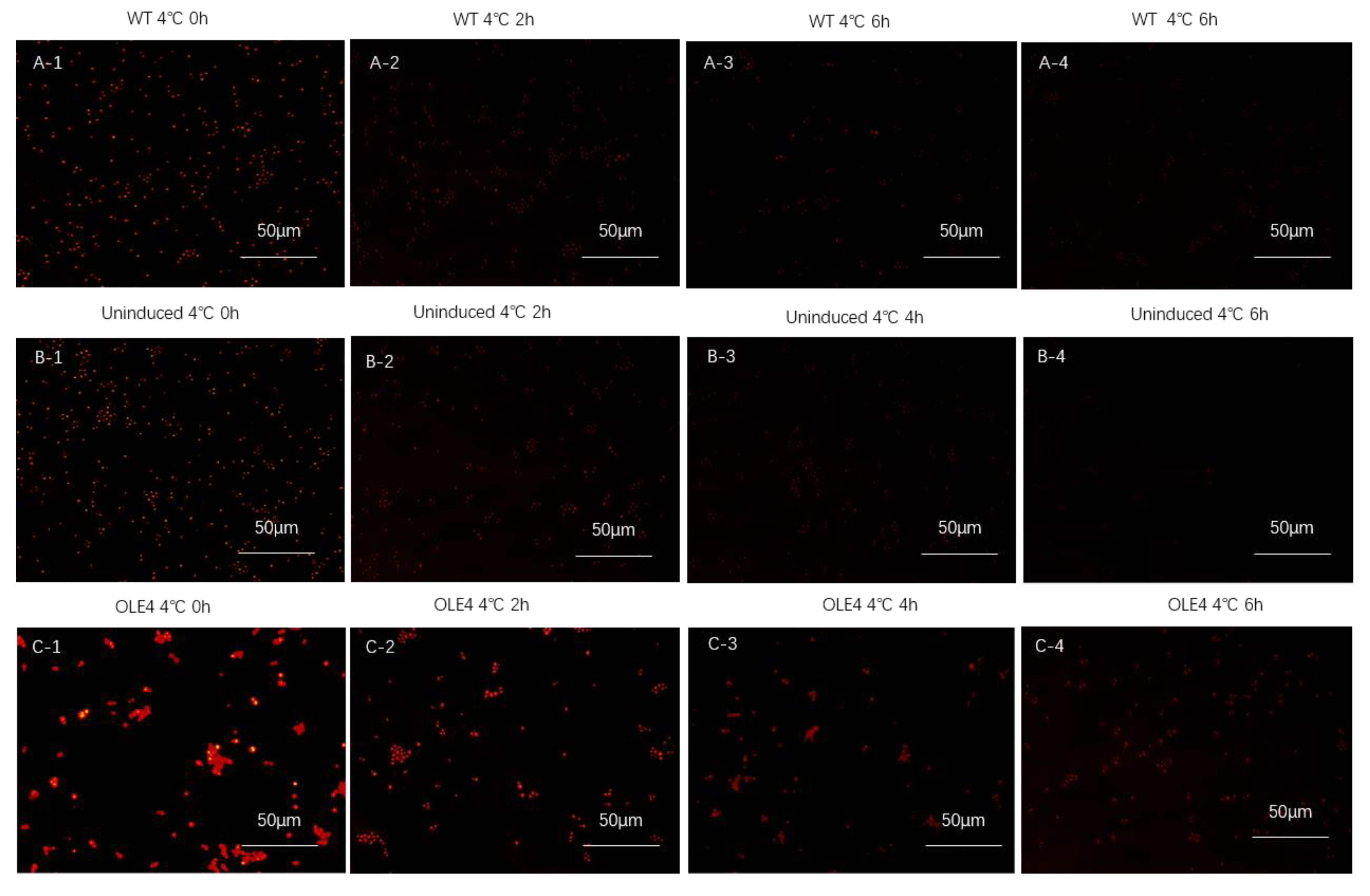
2.2. Nile Red Staining

The Nile red staining experimental method was performed as described previously [20]. Five development tubers were collected, peeled, cut into small pieces of 0.5 × 0.5 cm, and then transferred into a fix solution of 4% tissues for 24 h at 4 °C. Then, the tuber tissues were embedded into paraffin by using embedding cassettes, blocked, held at 4 °C until complete solidification, and cut to 5–10 µm, dewaxing with an Electro Thermostatic Blast Oven (Henan Lanphan Technology Co., Ltd., Zhengzhou, China) at 60 °C. The sections were deparaffinized, rehydrated, and stained with Nile red (Invitrogen, Waltham, MA, USA) for a period of 2 min.
Yeast cells were centrifuged at 12,000 r/min for 1 min and 500 μL PBS was used to resuspend the cells, 1 μL Nile red solution added (1 mg Nile red powder with 1 mL dimethyl sulfoxide), vortex oscillation carried out for 2 min, staining in the dark for 10 min, and finally, rinsing twice with PBS solution.
Nile red fluorescence was imaged by confocal microscopy (TCS-SP8; Leica, Weztlar, Germany), with 488 nm excitation and 562–611 nm emission. Sections were then rinsed in running tap water, dehydrated, mounted, and captured; the sheet was sealed with neutral gum at the end.

2.3. RNA Isolation and qRT-PCR Analysis

The total RNA content was extracted using the FastPure Plant Total RNA Isolation Kit (Vazyme, Nanjing, China), and the extracted RNA was measured for purity and concentration by a NanoDrop 2000 ultraviolet spectrophotometer. Then, the first-strand cDNA was synthesized using MonScript™ RTIII All-in-One Mix with dsDNase (Monad, Suzhou, China) kit and preserved at −20 °C. A set of gene-specific primers of CeOleosin genes was synthesized based on the information obtained from the coding sequences in Premier version 6 (Premier Biosoft, Palo Alto, CA, USA). Primer sequences are to be found in Supplementary Table S2. To verify the integrity of primer specificity, each primer pair was generated away from the conserved domain of the genes. The internal reference genes of tiger nut used in the qRT-PCR analysis included the 18S ribosomal RNA gene [21]. All qRT-PCR reactions were performed with MonAmp™ ChemoHS qPCR Mix from Monad Biotechnology Co., Ltd. (Suzhou, China) using the Stratagene MX3000P real-time PCR machine. Following the manufacturer’s instructions, a 20 µL reaction mixture was prepared with 2.0 µL cDNA template, 0.2 µM primers (F/R), 0.2 µL ROX dye (100×), 10 µL master mix, and 7 µL RNase-free water. PCR conditions were set according to the manufacturer’s protocol. The relative transcript abundance values were calculated using the 2−∆Ct method.

2.4. Identification and Characterization of Oleosin Genes

DNAMAN 6.0 software was used to align the amino acid sequences of tiger nut with model plants including C. esculentus, Oryza sativa, Hordeum vulgare, Brassica napus, grain sorghum, Ananas comosus, and Arabidopsis thaliana. For the evolutionary analysis of the CeOles, we employed the neighbor-joining method to construct a phylogenetic tree with MEGA 7.0 software [22] (1000 bootstraps) and EvolVIEW 2.0 (China National Center for Bioinformation, Beijing, China) [23] was used for a better image. The Expasy Protparam tool website was used to analyze the amino acid sequence. The NovoPro online website was used to analyze and predict the transmembrane region, using SWISS-MODLE online analysis software to predict the three-dimensional structure of the oleosin protein family.

2.5. Cloning and Vector Construction of CeOle4 Genes in Tiger Nut

RNA was extracted from tiger nut at 120 days after sowing, and the RNA was reverse transcribed into cDNA templates. Primers were designed for CeOle4 as target genes (Table S3) for PCR amplification. The cloned CeOle4 gene band was ligated to the pMD-18T cloning vector and transformed into the DH5α E. coli competent state. They were sent to a biotech company for sequencing and aligned with the correct sequence using DNAMAN software. The correctly sequenced genes were cloned into the pCAMBIA1302 plant expression vector and transformed into EHA105 Agrobacterium for protein subcellular localization research.
CeOle4 was constructed into the pYES2/NTC yeast recombinant expression vector, the successfully sequenced pYES2/NTC-CeOle4 plasmid was transformed into INVSC1 yeast cells, while the pYES2/NTC empty vector was transformed as a negative control. INVSc1 yeast strain was purchased from AngYuBio Company, and pYES2/NT C carrier was purchased from Wuhan Miaoling Biotechnology Co., Ltd. (Wuhan, China), while 2 × Taq PCR premix was purchased from the Beijing Solarbio Science & Technology Co., Ltd. (Beijing, China). The seamless cloning kit was purchased from Monad Biotechnology Co., Ltd. (Suzhou, China), the restriction endonuclease from TAKARA, and the purification and recovery kit, plasmid extraction kit, and Escherichia coli DH5α competent state from Qingke Biotechnology Co., Ltd. (Beijing, China).
The CeOle4 gene was constructed using seamless cloning technology onto the pCAMBIA3301 plant expression recombinant vector to obtain the pCAMBIA3301- CeOle4 recombinant expression vector, which was then transformed into Agrobacterium EHA105 for transgenic plant expression research. The pCAMBIA3301 vector was provided by the Bioreactor and Drug Development Engineering Research Center of the Ministry of Education in Jilin Agricultural University.

2.6. Transformation and Observation of Tobacco Leaf Cells

This was carried out as follows. Extract the plasmids pCAMBIA1302-CeOle4, and perform EHA105 Agrobacterium competent transformation. Incubate in liquid YEP medium containing kanamycin (50 μg/mL) and rifampicin (100 µg/mL) resistance, shake at 28 °C for 20 h on a shaker. Inoculate the cultured bacterial solution into 50 mL of liquid YEP medium containing the above-mentioned resistance, and add 100 µL MES and 4 µL AS (acetyl eugenol) to every 10 mL medium. Shake the medium at 28 °C on a shaker until the OD600 value of Agrobacterium is between 1.0–1.2. Centrifuge at 4000 r/min for 10 min in a high-speed centrifuge. Resuspend the bacterial pellet in 10 mM MgCl2 solution containing 20 µL AS per 10 mL until the OD600 value is 1.0, and let it stand for at least 2 h. Select healthy and disease-free tobacco plants at around three weeks of age, use a syringe to extract the resuspended bacterial solution, and inject it from the back of the tobacco leaves. The injected tobacco plants were cultured in a dark place for 60 h. Take the injected leaf slices and observe them under a confocal laser microscope. EHA105 Agrobacterium competent cells were purchased from Shanghai Angyu Biotechnology Co., Ltd. (Shanghai, China), MES, AS, kanamycin and rifampicin were purchased from Beijing Solarbio Science & Technology Co., Ltd. (Beijing, China).

2.7. Induced Expression of Target Protein in Yeast Cell

This was carried out as follows. Inoculate the yeast cells successfully transformed with pYES2/NTC-CeOle4 recombinant plasmids into 50 mL Cultivate in SD-URA liquid medium for 24 h. Centrifuge the cultured bacterial solution at 4 °C and 5000 r/min for 10 min. Discard the supernatant, resuspend the bacterial pellet in 0.9% NaCl solution, and centrifuge at 4 °C, 5000 r/min for 10 min. Repeat the above steps. Inoculate the obtained bacterial pellet into SC-URA medium containing 2% lactose until the OD600 value reaches 0.4. Take 5 mL samples of the inoculated bacterial solution at time points of 4 h, 6 h, 8 h, 12 h, 24 h, 36 h, and 48 h. The total protein extraction of yeast liquid is carried out according to the fungal total protein extraction kit.

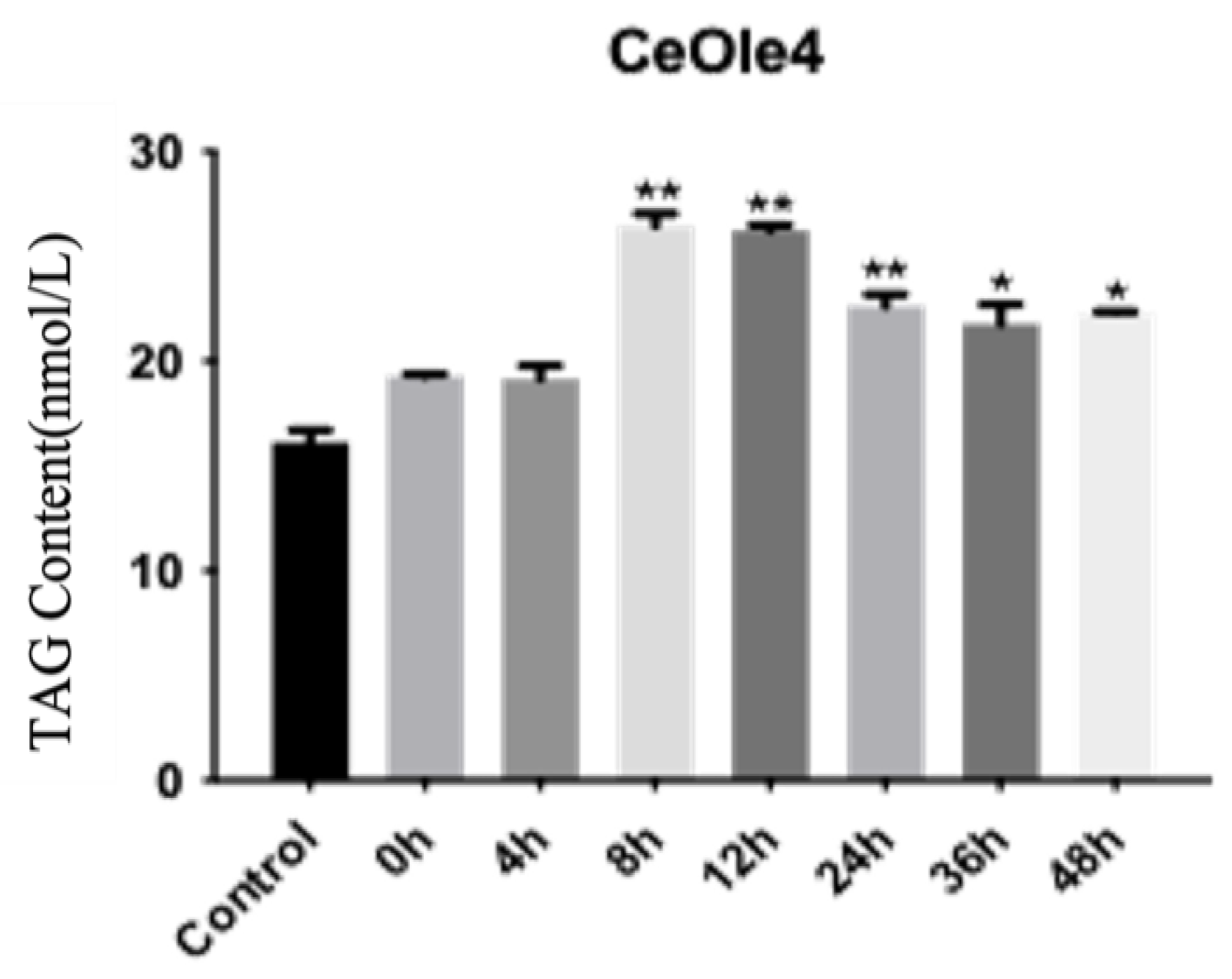
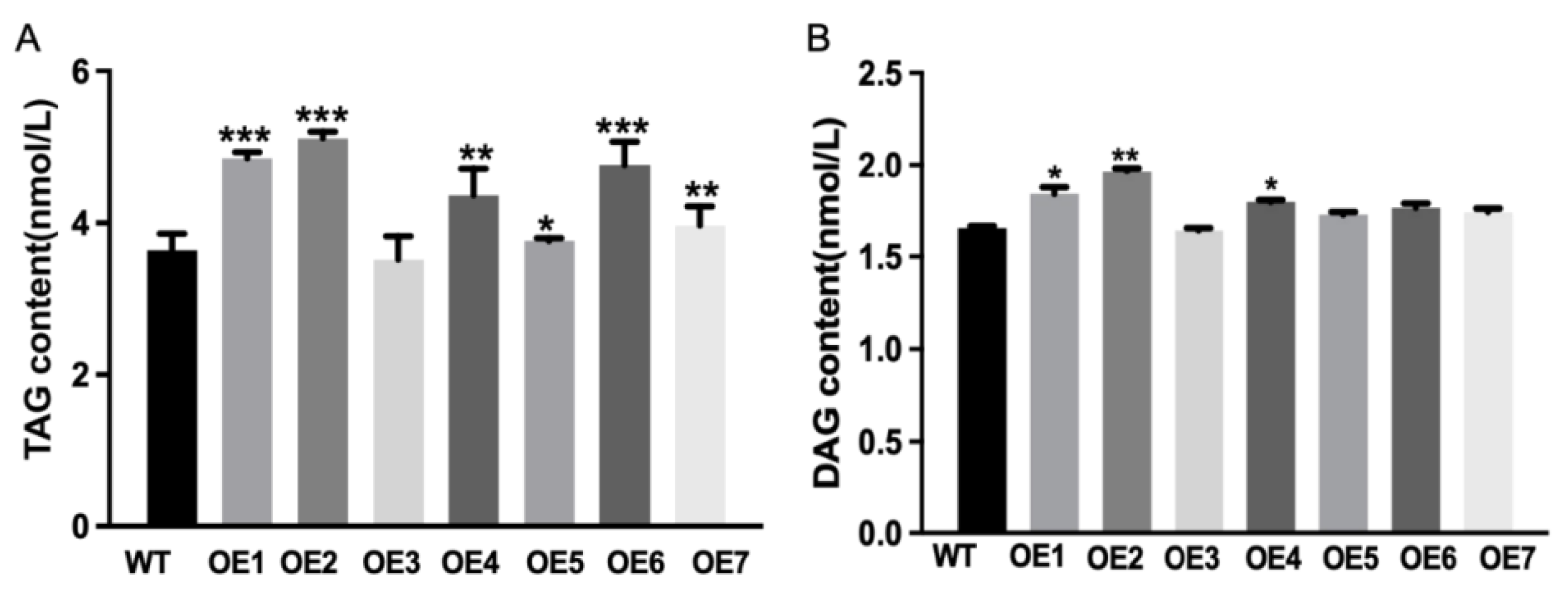
2.8. TAG and DAG Content Detection

This was carried out as follows. Take 2 mL of the induced cell solution, uninduced cell solution, and empty cell solution from each time period, centrifuge at 3000 r/min for 10 min in a centrifuge, resuspend the cells in 1mL PBS solution for precipitation, boil in water for 10 min, place in liquid nitrogen for 5 min, and then boil in water for 5 min. The treated cell solution is centrifuged at 3000 r/min for 20 min, and the supernatant is taken for TAG content detection. The determination of TAG and DAG content in transgenenic Camelina sativa is as follows. Take 0.1 g of T3 generation seeds and grind them thoroughly in liquid nitrogen. After grinding, transfer them into a 2 mL centrifuge tube, add 900 µL PBS solution, centrifuge at 4 °C for 20 min, collect the supernatant for TAG content detection, and treat the wild-type seeds in the same way as the control group. The operational procedure for determining the DAG content is the same as TAG. The TAG content detection kits (Triglyceride (TG) Content Assay kit) were purchased from Beijing Boxbio Science & Technology Co., Ltd. (Beijing, China).

2.9. Western Blotting Analysis Target Protein Expression

Yeast cells were lysed with yeast total protein extraction kit (Solarbio, Beijing, China) for protein extraction [24]. The protein concentration was determined by bicinchoninic acid protein kit. Extracted proteins were separated by SDS-PAGE assays, transferred onto nitro-cellulose membranes, followed by blocking with 10% skimmed milk in the TBST. PVDF membranes were treated with primary antibodies targeted to anti-his taq (1:5000) at room temperature for 1.5 h. Subsequently the membranes were incubated with secondary antibody which was HRP-conjugate (1:7000) at room temperature for 1 h. Signals were detected using an ECL kit (GE Healthcare, Chicago, IL, USA). The primary antibody for anti-his taq and the secondary antibody were from Solarbio, Beijing, China.

2.10. CeOle4-Overexpressed Transgenic Camelina Sativa and Expression Analysis

For the construction of overexpression vectors, the CDS of CeOle4 was cloned into the pCAMBIA3301 vector and transformed into Agrobacterium tumefaciens EHA105 competent cells (liquid nitrogen for 5 min, then 37 °C water bath for 5 min). The transformation was carried out by the floral dip method and Agrobacterium-mediated inoculation of plants at the early flowering stage along with a vacuum infiltration procedure [25]. The gene-specific primers (F:TATGACCATGATTACGAATTCATGGCG GACCGCGGGCAG; R:CAGGTCGACTCTAGAGGATCCCTACTTGTCGGTCTTCCCG CC) were used to screen out transgenic plants until T3 generation was obtained harboring the CeOle4 gene. The phenotype of the T3 transgenic plants [26,27] was observed for morphological differences, and the expression profiling was carried out simultaneously using qRT-PCR assay.

2.11. Observation of Phenotype of Transgenic Camelina Sativa

This was carried out as follows. Select transgenic Camelina sativa seeds with relatively high expression levels, TAG content, and DAG content, comparing them with wild-type Camelina sativa seeds to observe differences in seed size and seed coat color, and then measure the hundred-seed weight, further demonstrating the function of CeOle4 in plants.

2.12. Data Analysis

All experiments mentioned were conducted in triplicate, and the resulting data were subjected to statistical analysis using the t-test. A significant difference was defined as * p < 0.05, ** p < 0.01, and *** p < 0.001.

3. Results

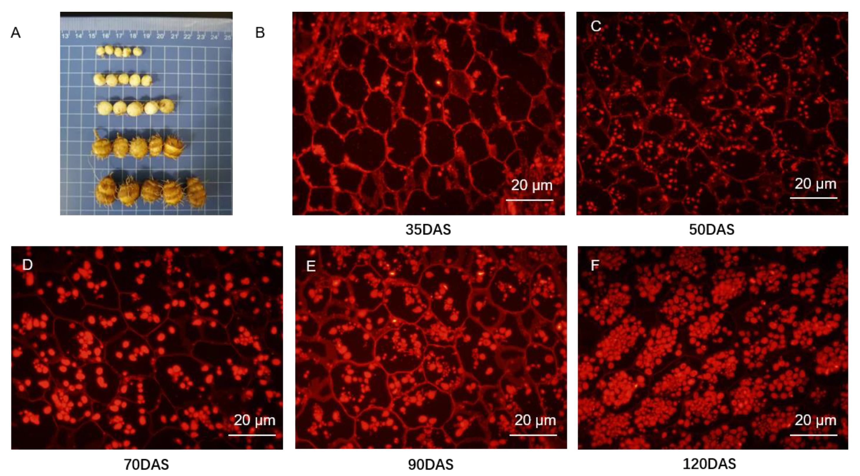
3.1. Lipid Droplets Accumulation during Tuber Development

As the days of tuber cultivation increased, its diameter also continued to increase (Figure 1A). The different development stages of tubers were stained by Nile red. Nile red fluorescence was imaged by confocal microscopy with 488 nm excitation and 562–611 nm emission. There was a significant increase in lipid accumulation within the tuber (Figure 1B–F), accompanied by a rising trend in structural abundance and density, gradually transitioning from sporadic occurrences to clustering into dense cell-like structures. The maximum density was reached at the 120 DAS developing stage.

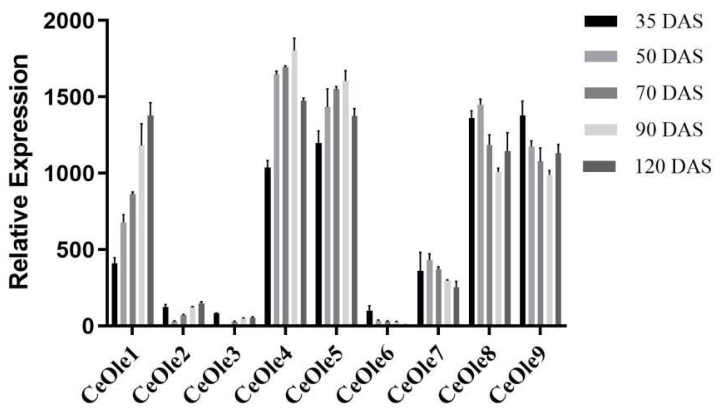
3.2. Gene Expression Analysis of CeOleosin Family

The qRT-PCR expression analysis of the CeOleosin gene family is illustrated in Figure 2. The expression levels of different oleosin genes vary significantly across different developing stages. CeOle1, CeOle4, CeOle5, CeOle8, and CeOle9 maintain relatively high expression levels throughout the entire growth period. Specifically, CeOle1 shows a gradual increase in expression as the growth period extends, peaking at 120 DAS (days after sowing). CeOle4 and CeOle5 also exhibit increasing expression levels with prolonged growth, reaching their highest levels at 90 DAS. In contrast, ole8 and ole9 initially display high expression levels in the early growth stages but decrease thereafter, reaching their lowest point at 90 DAS. CeOle7 demonstrates relatively stable expression, peaking at 50 DAS, and gradually declining thereafter. On the other hand, CeOle2, CeOle3, and CeOle6 consistently exhibit lower expression levels throughout the developing period.

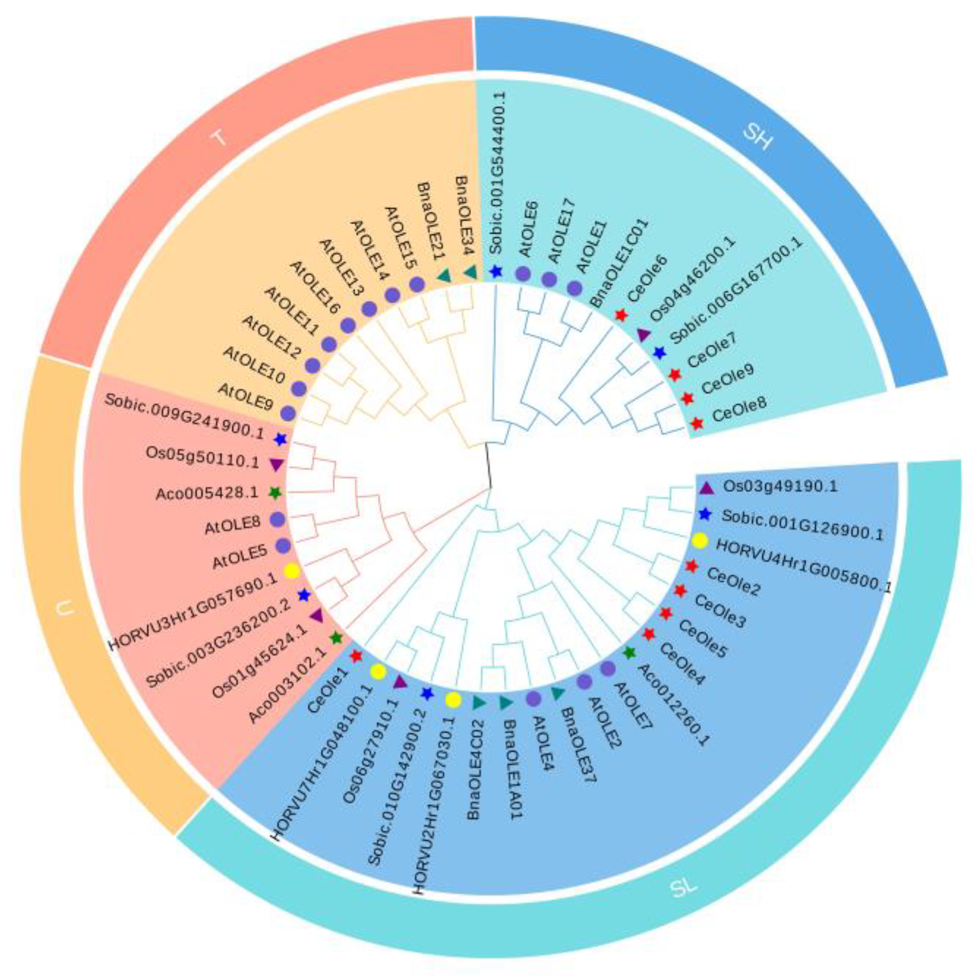
3.3. Phylogenetic Analysis of Oleosin Family Members

Using MEGA 7.0 software, a phylogenetic tree was constructed to validate the evolutionary status of the oleosin family in C. esculentus (shown in Figure 3). The resulting phylogenetic chart indicates a high degree of conservation of oleosin across various species. The oleosin family members of these species are categorized into four lineages, T, U, SL, and SH [28]. The nine oleosin sequences from the C. esculentus transcriptome data were identified and divided into two lineages. CeOle6, CeOle7, CeOle8, and CeOle9 were divided into SH lineage, while CeOle1, CeOle2, CeOle3, CeOle4, and CeOle5 were divided into SL. CeOle6-9 likely plays a role in the synthesis of oleic acid, total fatty acids (FAs), and phosphatidic acid (PA) [29]. CeOle1–5 are likely associated with neutral lipid synthesis [30].

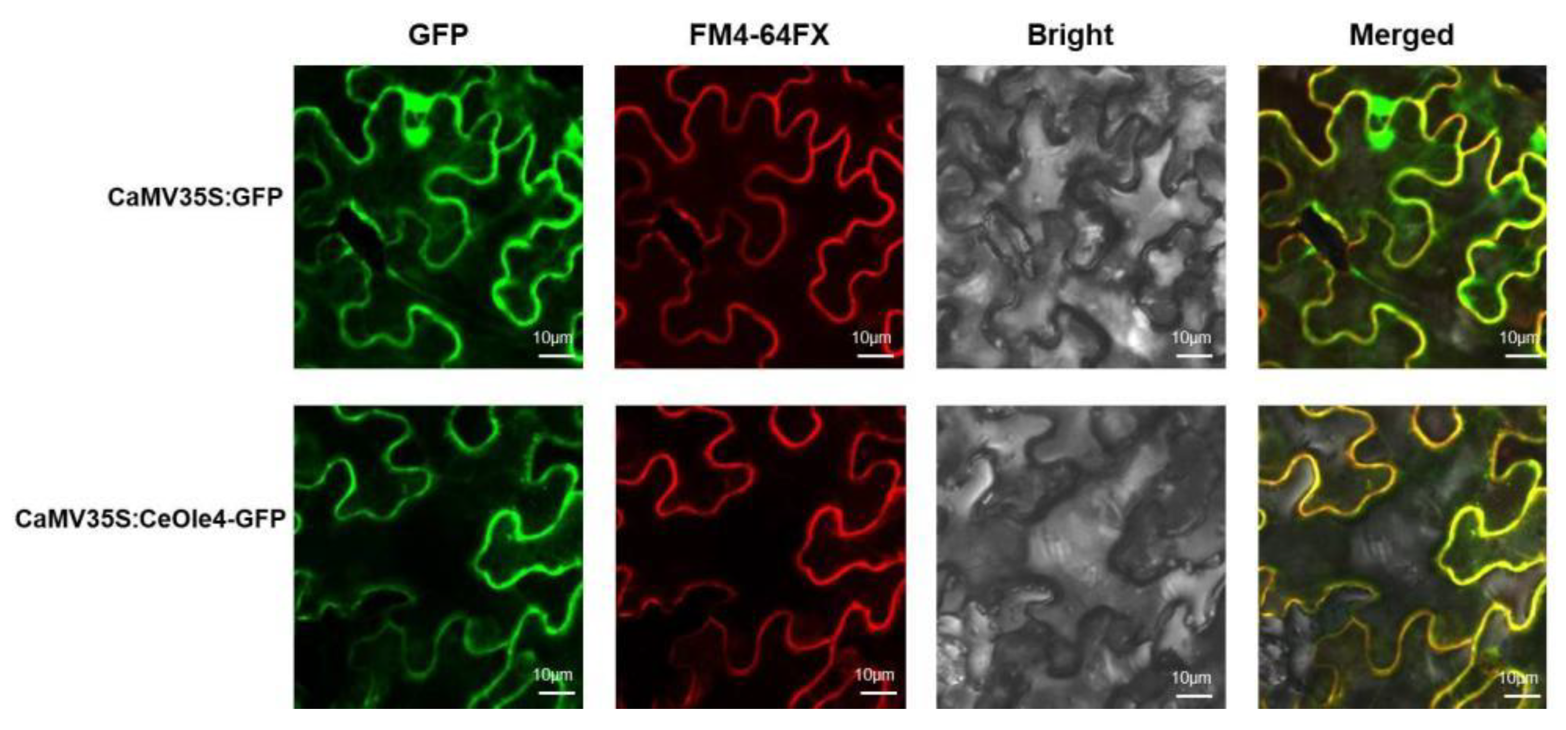
3.4. Subcellular Location of CeOle4

Using Agrobacterium transformed with the empty vector pCAMBIA1302 as a positive control, and selecting FM4-64FX as a specific marker for cell membranes, the localization of the pCAMBIA1302-CeOle4 recombinant vector in tobacco leaf cells was observed. Tobacco plants with consistent growth were chosen for injection. Under the same culture conditions, tobacco leaf cells injected with Agrobacterium harboring the pCAMBIA1302 empty vector exhibited fluorescence excitation at both the cell membrane and the cell nucleus. In contrast, tobacco leaf cells injected with the pCAMBIA1302-CeOle4 recombinant vector showed fluorescence only at the cell membrane under the same conditions (Figure 4). This indicates that CeOle4 is localized to the cell membrane and classified as a membrane-targeted protein.

3.5. Exogenous Expression of CeOleosin4 and Function Analysis

To investigate the functionality of the CeOle4 gene on affecting the size and stability of oil bodies, formation and degradation of oil bodies, etc., yeast cells were employed for the exogenous induction of CeOle4 expression. The target protein was induced using SC-URA liquid medium containing 2% galactose, and yeast cells were harvested at 4 h, 6 h, 8 h, 12 h, 24 h, 36 h, and 48 h, respectively. Western blot analysis was conducted, with empty vector-containing yeast and uninduced yeast cells serving as negative controls. Based on the Western blot results, the optimal induction time for CeOle4 gene-transformed yeast cells was determined to be 8 h (as shown in Figure 5 and Figure 6).
To further investigate the function of CeOle4, yeast cells were subjected to low-temperature (4 °C) cultivation during the optimal induction period, and the growth status and Nile red staining results of the cells were continuously monitored. The results revealed that after 2 h of low-temperature treatment, yeast cells overexpressing the CeOle4 gene still exhibited a high accumulation of lipids. However, with increasing treatment duration, the lipid accumulation noticeably decreased (Figure 7). When the low-temperature treatment duration reached 6 h, the accumulation of lipid substances in yeast cells overexpressing the CeOle4 gene showed no significant difference compared to the wild type and uninduced yeast cells. This suggests that CeOle4 not only promotes lipid synthesis in eukaryotes, but also enhances the stability of the oil body and protects lipid droplets from degradation under low-temperature stress.

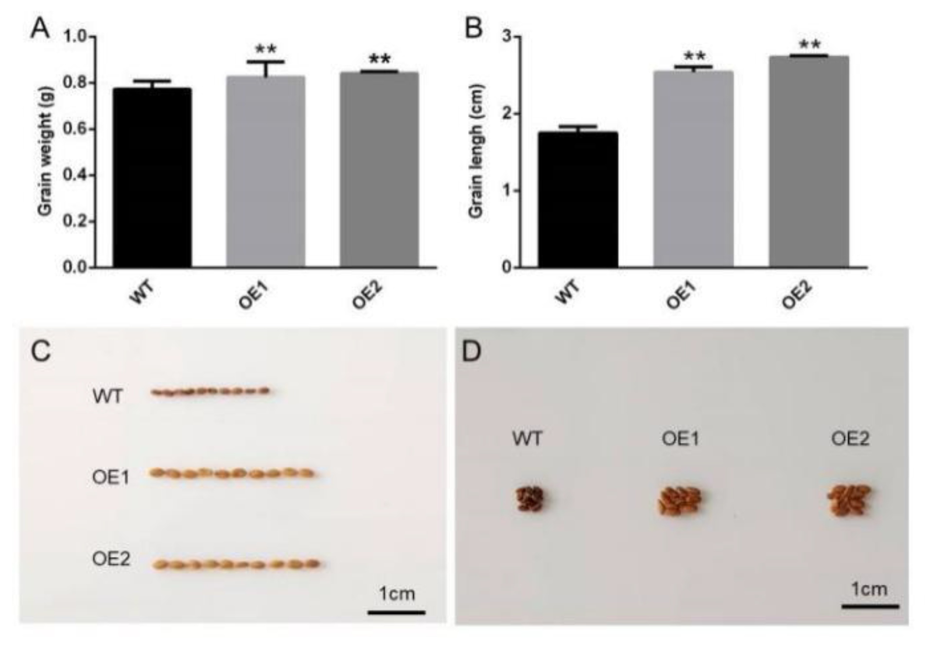
3.6. Analysis of Agronomic Traits in Transgenic Camelina Sativa Seeds

To further elucidate the function of the CeOle4 gene, the triacylglycerol (TAG) content in transgenic Camelina sativa seeds was determined. Seven transgenic lines were compared to the wild type, and the TAG and diacylglycerol (DAG) contents were measured. TAG analysis revealed that the TAG content in lines OE1, OE2, OE4, and OE6 increased by 33.2%, 40.52%, 20.02%, and 30.82%, respectively, compared to the wild type, while lines OE3, OE5, and OE7 showed marginal increases of 0.25%, 3.34%, and 9.00%, respectively (Figure 8A). DAG analysis indicated that compared to the wild type, lines OE1, OE2, and OE4 exhibited significantly elevated DAG levels, whereas lines OE5, OE6, and OE7 showed only slight increases, and OE3 line exhibited a slight decrease in DAG content (Figure 8B).
Based on the expression levels of CeOle4 and the content of TAG and DAG in transgenic Camelina sativa seeds, OE1 and OE2 lines were selected for further experimental measurements. By comparing the seed length of WT Camelina sativa seeds with those of the selected transgenic lines, it was observed that the seed length of the OE1 line increased by 41% compared to the wild type, while the OE2 line increased by 53% (Figure 9A). Additionally, during the observation of seed size, it was noticed that the seed coat color of the wild-type lines was darker than that of the transgenic lines (Figure 9C). Subsequently, hundred-seed weight analysis was conducted on both wild-type and transgenic lines. The results revealed that the weight of seeds in the OE1 group increased by 12.6% compared to the wild type, while the OE2 group increased by 11.3% (Figure 9B). The presence of CeOle4 not only increased the length and weight of seeds but also had a certain influence on the color of the seed coat (Figure 9D).

4. Discussion

Lipids are essential components of eukaryotic cells, typically categorized into two subgroups: polar and neutral lipids. Neutral lipids, including sterol esters (SE) and TAG, have garnered considerable interest in recent years for their formation and accumulation within neutral lipid bodies, driving extensive research across various fields. As a high-quality oil crop, tiger nut primarily metabolizes and stores lipids in its tubers [31].
Previous studies indicated that the primitive type oleosin (P) is distributed in mosses and ferns, the ubiquitous type (U) is found in all land plants, the low molecular weight type (SL) is prevalent in seed plants, while the high molecular weight type (SH) is present in angiosperms. Additionally, the woolly type (T) is distributed in Brassicaceae plants [32]. A systematic phylogenetic analysis was conducted on nine oleosin proteins within the tiger nut gene family, along with gene expression profiling across different developmental stages of stem tubers. The highly expressed proteins in tiger nut tubers belong to the SL type. It is speculated that this type of oleosin exhibits a highly conserved structure, with maximum expression levels observed during the mature stage of stem tuber development, suggesting a close association between the expression level of SL-type oleosin and lipid accumulation and synthesis in tiger nut stem tubers.
To gain preliminary insights into the gene function of CeOle4 in tiger nut, the gene was cloned, and a subcellular localization vector, pCAMBIA1302-CeOle4, was constructed to observe the subcellular localization of CeOle4. CeOle4 was found to localize on the cell membrane. Its functional domain shares high similarity with GmOle5 and AtOle7, suggesting involvement in cell membrane morphogenesis, assembly, or transport. Previous studies have also suggested its association with plant immune responses and intracellular and extracellular signal transduction [33].
To further investigate the function of CeOloe4, this study constructed a yeast recombinant expression vector and introduced it into yeast cells. Verification was conducted using precise SDS-PAGE and Western blot analysis. Subsequently, different types of yeast cells were subjected to low-temperature stress and Nile red staining to observe differences in size, density, and brightness, verifying their cold tolerance characteristics. In yeast cells, the primary focus of neutral lipid research lies in the production of biofuels from oleaginous yeast species [34]. Studies have shown that TAG within eukaryotic cells serves not only as a storage form of fatty acids but also plays a crucial role in maintaining cell membrane structure and fluidity [35,36]. This is consistent with the localization of CeOle4 on the cytoplasmic membrane in this study, implying that the homeostasis of TAG is closely associated with the process of membrane lipid renewal and remodeling, which are crucial for maintaining the structure and fluidity of cell membranes. However, the traditional view of TAG as a storage form for cellular carbon and energy is being challenged by recent research, which suggests that TAG also plays an important role in maintaining membrane function [37]. The results of this study also indicate that while CeOle4 localizes to the cytoplasmic membrane, increasing the content of intracellular TAG enhances cell cold tolerance. Since the cell membrane is the primary site of cold stress, the elevation of unsaturated fatty acids on the membrane increases its fluidity, thereby significantly improving cold tolerance [38]. Due to the stabilizing effect of the oleosin on cell membranes, plants with high levels of oleosin expression also exhibit strong cold tolerance [39].
Oil body proteins are related to lipid synthesis and content, with a decrease in oil body protein content in rice seeds resulting in reduced oil content and increased oil body size [40]. Expression of the oleosin gene from Brassica napus in Arabidopsis thaliana leads to larger oil bodies in the seeds of all transgenic lines except OLE1 compared to the wild type. In Arabidopsis thaliana, removal of the oleosin gene results in decreased C18:1 fatty acid content and increased C20:1 fatty acid content in seeds, and studies have shown that overexpression of OLEs can increase oil and linoleic acid production [41]. Camelina sativa is a cruciferous oil crop that contains a greater variety of unsaturated fatty acids in its seeds compared to other oil crops, and it has low cultivation costs, rapid growth, and many other excellent characteristics. In this study, Camelina sativa was used as the gene transformation recipient, and the content of TAG and DAG in the seeds was detected to validate the function of the CeOle4 gene. It was found that Ceole4 is related to the lipid content in Camelina sativa seeds and is associated with seed weight [42], and there is also a certain correlation between lipid accumulation and seed coat color [43]. Overexpression of Ceole4 in Camelina sativa seeds resulted in lighter seed coat color compared to the WT, with significantly higher TAG content, suggesting a negative correlation between TAG and seed coat color. Recent studies showed that seed coats participate in plant cold tolerance by increasing embryo cutinization [44]. However, other studies showed that plant lipid substances accumulate in the seed coat rather than primarily in the embryo under low temperature treatment, indicating that the accumulation of lipid substances in seeds may be related to embryo cutinization, ultimately promoting plant seed tolerance to low temperatures [45,46]. Based on a comprehensive analysis of the results of this study, it is speculated that CeOle4 may be involved in the regulation of seed coat color changes and plant cold tolerance. This study provides a preliminary exploration of the function of the CeOle4 gene in the oleosin family of Camelina sativa and has achieved promising results, laying the foundation for further in-depth research into the molecular mechanisms of oilseed crops.

Supplementary Materials

The following supporting information can be downloaded at: https://www.mdpi.com/article/10.3390/horticulturae10090945/s1, Table S1: Analysis of physical and chemical properties of Oleosin family; Table S2: Primer sequences; Table S3: CeOle4 Coding DNA Sequence; Figure S1: Amino acid sequence alignment of Oleosin family; Figure S2:The transmembrane structure and analysis of Oleosin family; Figure S3: Three-dimensional structure prediction of Oleosin family.

Author Contributions

Conceptualization, Y.D. and P.X.; Methodology, Y.C., Y.W. and Q.Y.; Software, Y.W., S.L. and W.L.; Validation, Y.C., Y.W., X.L. (Xinyi Liu), X.L. (Xiaowei Li) and N.W.; Formal analysis, N.W.; Investigation, Y.C., S.L., X.L. (Xinyi Liu), W.L. and X.L. (Xiaowei Li); Resources, X.L. (Xiaowei Li); Data curation, F.W.; Writing—original draft, Y.D.; Writing—review & editing, P.X., Y.D. and L.G.; Visualization, F.W.; Funding acquisition, P.X. All authors have read and agreed to the published version of the manuscript.

Funding

This research was funded by the Science and Technology Development Project of Jilin province (20220101354JC), the Jilin Provincial College Students’ Innovative Training Project, Shaanxi Vocational and Technical Education Association’s 2024 Vocational Education Teaching Reform Research Project (2024SZX539), Shaanxi Provincial Administration for Market Regulation Science and Technology Plan Project (2023KY20), Key R & D projects in Shaanxi Province (2018 NY-113), and the project of Weinan City Science and Technology Plan Project (2017JCYJ-4-1) (2019ZDYF-JCYJ-150).

Data Availability Statement

The original contributions presented in the study are included in the article/Supplementary Material, further inquiries can be directed to the corresponding author.

Conflicts of Interest

The funders had no role in the design of the study; in the collection, analyses, or interpretation of data; in the writing of the manuscript; or in the decision to publish the results.

References

  1. De Castro, O.; Gargiulo, R.; Del Guacchio, E.; Caputo, P.; De Luca, P. A molecular survey concerning the origin of Cyperus esculentus (Cyperaceae, Poales): Two sides of the same coin (weed vs. crop). Ann. Bot. 2015, 115, 733–745. [Google Scholar] [CrossRef] [PubMed]
  2. Onuoha, N.O.; Ogbusua, N.O.; Okorie, A.N.; Ejike, C. Tigernut (Cyperus esculentus L.) “milk” as a potent “nutri-drink” for the prevention of acetaminophen-induced hepatotoxicity in a murine model. J. Intercult. Ethnopharmacol. 2017, 6, 290–295. [Google Scholar] [CrossRef]
  3. Avicor, S.W.; Wajidi, M.F.F.; Achoribo, E.S.; Ong, M.T.; Hamzah, S.N. Tiger nut (Cyperus esculentus) as a potential phytoinsecticide: Larvicidal activity of crude extracts on Aedes aegypti and Culex quinquefasciatus (Diptera: Culicidae). Trop. Biomed. 2021, 38, 186–191. [Google Scholar] [CrossRef]
  4. Zhang, Y.; Sun, S. Tiger nut (Cyperus esculentus L.) oil: A review of bioactive compounds, extraction technologies, potential hazards and applications. Food Chem. X 2023, 19, 100868. [Google Scholar] [CrossRef] [PubMed]
  5. Yu, Y.; Lu, X.; Zhang, T.; Zhao, C.; Guan, S.; Pu, Y.; Gao, F. Tiger Nut (Cyperus esculentus L.): Nutrition, Processing, Function and Applications. Foods 2022, 11, 601. [Google Scholar] [CrossRef]
  6. Ibitoye, O.B.; Aliyu, N.O.; Ajiboye, T.O. Tiger nut oil-based diet improves the lipid profile and antioxidant status of male Wistar rats. J. Food Biochem. 2018, 42, e12587. [Google Scholar] [CrossRef]
  7. Adenowo, A.F.; Kazeem, M.I. Tiger Nut as A Functional Food, Pharmacological and Industrial Agent: A Mini Review. Ann. Sci. Technol. 2020, 5, 31–38. [Google Scholar] [CrossRef]
  8. Abell, B.M.; Holbrook, L.A.; Abenes, M.; Murphy, D.J.; Hills, M.J.; Moloney, M.M. Role of the proline knot motif in oleosin endoplasmic reticulum topology and oil body targeting. Plant Cell 1997, 9, 1481–1493. [Google Scholar] [CrossRef] [PubMed]
  9. Huang, A.H.C. Plant Lipid Droplets and Their Associated Proteins: Potential for Rapid Advances. Plant Physiol. 2018, 176, 1894–1918. [Google Scholar] [CrossRef]
  10. Murphy, D.J. Storage lipid bodies in plants and other organisms. Prog. Lipid Res. 1990, 29, 299–324. [Google Scholar]
  11. Jolivet, P.; Acevedo, F.; Boulard, C.; d’Andréa, S.; Faure, J.D.; Kohli, A.; Nesi, N.; Valot, B.; Chardot, T. Crop seed oil bodies: From challenges in protein identification to an emerging picture of the oil body proteome. Proteomics 2013, 13, 1836–1849. [Google Scholar] [CrossRef]
  12. Tzen, J.; Cao, Y.; Laurent, P.; Ratnayake, C.; Huang, A. Lipids, Proteins, and Structure of Seed Oil Bodies from Diverse Species. Plant Physiol. 1993, 101, 267–276. [Google Scholar] [CrossRef] [PubMed]
  13. Roux, E.; Baumberger, S.; Axelos, M.A.; Chardot, T. Oleosins of Arabidopsis thaliana: Expression in Escherichia coli, purification, and functional properties. J. Agric. Food Chem. 2004, 52, 5245–5249. [Google Scholar] [CrossRef] [PubMed]
  14. Huang, A.H. Oleosins and oil bodies in seeds and other organs. Plant Physiol. 1996, 110, 1055–1061. [Google Scholar] [CrossRef] [PubMed]
  15. Zhao, H.Q.; Wang, X.F.; Gao, S.P. Progress on the functional role of oleosin gene family in plants. Yi Chuan 2022, 44, 1128–1140. [Google Scholar] [CrossRef]
  16. Li, H.; Yang, J.; Chen, Y.; Guan, L.; Du, L.; Guo, Y.; Wang, W.; Wang, L.; Li, H.; Jiang, C.; et al. Expression of a functional recombinant oleosin-human hyaluronidase hPH-20 fusion in Arabidopsis thaliana. Protein Expr. Purif. 2014, 103, 23–27. [Google Scholar] [CrossRef]
  17. Kargiotidou, A.; Deli, D.; Galanopoulou, D.; Tsaftaris, A.; Farmaki, T. Low temperature and light regulate delta 12 fatty acid desaturases (FAD2) at a transcriptional level in cotton (Gossypium hirsutum). J. Exp. Bot. 2008, 59, 2043–2056. [Google Scholar] [CrossRef]
  18. Parthibane, V.; Rajakumari, S.; Venkateshwari, V.; Iyappan, R.; Rajasekharan, R. Oleosin is bifunctional enzyme that has both monoacylglycerol acyltransferase and phospholipase activities. J. Biol. Chem. 2012, 287, 1946–1954. [Google Scholar] [CrossRef]
  19. Wang, L.; Jing, M.; Ahmad, N.; Wang, Y.; Wang, Y.; Li, J.; Li, X.; Liu, W.; Wang, N.; Wang, F.; et al. Tracing Key Molecular Regulators of Lipid Biosynthesis in Tuber Development of Cyperus esculentus Using Transcriptomics and Lipidomics Profiling. Genes 2021, 12, 1492. [Google Scholar] [CrossRef]
  20. Escorcia, W.; Ruter, D.L.; Nhan, J.; Curran, S.P. Quantification of Lipid Abundance and Evaluation of Lipid Distribution in Caenorhabditis elegans by Nile Red and Oil Red O Staining. JoVE 2018, 133, e57352. [Google Scholar] [CrossRef]
  21. Sun, D.; Quan, W.; Wang, D.; Cui, J.; Wang, T.; Lin, M.; Wang, Y.; Wang, N.; Dong, Y.; Li, X.; et al. Genome-Wide Identification and Expression Analysis of Fatty Acid Desaturase (FAD) Genes in Camelina sativa (L.) Crantz. Int. J. Mol. Sci. 2022, 23, 14550. [Google Scholar] [CrossRef]
  22. Kumar, S.; Stecher, G.; Li, M.; Knyaz, C.; Tamura, K. MEGA X: Molecular Evolutionary Genetics Analysis across Computing Platforms. Mol. Biol. Evol. 2018, 35, 1547–1549. [Google Scholar] [CrossRef] [PubMed]
  23. He, Z.; Zhang, H.; Gao, S.; Lercher, M.J.; Chen, W.H.; Hu, S. Evolview v2: An online visualization and management tool for customized and annotated phylogenetic trees. Nucleic Acids Res. 2016, 44, W236–W241. [Google Scholar] [CrossRef] [PubMed]
  24. Tang, J.; Yang, X.; Xiao, C.; Li, J.; Chen, Y.; Li, R.; Li, S.; Lü, S.; Hu, H. GDSL lipase occluded stomatal pore 1 is required for wax biosynthesis and stomatal cuticular ledge formation. New Phytol. 2020, 228, 1880–1896. [Google Scholar] [CrossRef]
  25. Lu, C.; Kang, J. Generation of transgenic plants of a potential oilseed crop Camelina sativa by Agrobacterium-mediated transformation. Plant Cell Rep. 2008, 27, 273–278. [Google Scholar] [CrossRef]
  26. Noman, M.; Jameel, A.; Qiang, W.D.; Ahmad, N.; Liu, W.C.; Wang, F.W.; Li, H.Y. Overexpression of GmCAMTA12 Enhanced Drought Tolerance in Arabidopsis and Soybean. Int. J. Mol. Sci. 2019, 20, 4849. [Google Scholar] [CrossRef] [PubMed]
  27. Raffan, S.; Sparks, C.; Huttly, A.; Hyde, L.; Martignago, D.; Mead, A.; Hanley, S.J.; Wilkinson, P.A.; Barker, G.; Edwards, K.J.; et al. Wheat with greatly reduced accumulation of free asparagine in the grain, produced by CRISPR/Cas9 editing of asparagine synthetase gene TaASN2. Plant Biotechnol. J. 2021, 19, 1602–1613. [Google Scholar] [CrossRef] [PubMed]
  28. Chen, K.; Yin, Y.; Liu, S.; Guo, Z.; Zhang, K.; Liang, Y.; Zhang, L.; Zhao, W.; Chao, H.; Li, M. Genome-wide identification and functional analysis of oleosin genes in Brassica napus L. BMC Plant Biol. 2019, 19, 294. [Google Scholar] [CrossRef]
  29. Parthibane, V.; Iyappan, R.; Vijayakumar, A.; Venkateshwari, V.; Rajasekharan, R. Serine/threonine/tyrosine protein kinase phosphorylates oleosin, a regulator of lipid metabolic functions. Plant Physiol. 2012, 159, 95–104. [Google Scholar] [CrossRef]
  30. Niu, R.; Chen, F.; Liu, C.; Duan, X. Composition and Rheological Properties of Peanut Oil Bodies from Aqueous Enzymatic Extraction. J. Oleo Sci. 2021, 70, 375–383. [Google Scholar] [CrossRef]
  31. Kilaru, A.; Cao, X.; Dabbs, P.B.; Sung, H.J.; Rahman, M.M.; Thrower, N.; Zynda, G.; Podicheti, R.; Ibarra-Laclette, E.; Herrera-Estrella, L.; et al. Oil biosynthesis in a basal angiosperm: Transcriptome analysis of Persea Americana mesocarp. BMC Plant Biol. 2015, 15, 203. [Google Scholar] [CrossRef] [PubMed]
  32. Huang, M.D.; Huang, A.H. Bioinformatics Reveal Five Lineages of Oleosins and the Mechanism of Lineage Evolution Related to Structure/Function from Green Algae to Seed Plants. Plant Physiol. 2015, 169, 453–470. [Google Scholar] [CrossRef] [PubMed]
  33. Wei, Q.; Liu, J.; Guo, F.; Wang, Z.; Zhang, X.; Yuan, L.; Ali, K.; Qiang, F.; Wen, Y.; Li, W.; et al. Kinase regulators evolved into two families by gain and loss of ability to bind plant steroid receptors. Plant Physiol. 2023, 191, 1167–1185. [Google Scholar] [CrossRef] [PubMed]
  34. Rostron, K.A.; Lawrence, C.L. Nile Red Staining of Neutral Lipids in Yeast. Methods Mol. Biol. 2017, 1560, 219–229. [Google Scholar] [CrossRef] [PubMed]
  35. Rajakumari, S.; Daum, G. Multiple functions as lipase, steryl ester hydrolase, phospholipase, and acyltransferase of Tgl4p from the yeast Saccharomyces cerevisiae. J. Biol. Chem. 2010, 285, 15769–15776. [Google Scholar] [CrossRef]
  36. Arisz, S.A.; Heo, J.Y.; Koevoets, I.T.; Zhao, T.; van Egmond, P.; Meyer, A.J.; Zeng, W.; Niu, X.; Wang, B.; Mitchell-Olds, T.; et al. DIACYLGLYCEROL ACYLTRANSFERASE1 Contributes to Freezing Tolerance. Plant Physiol. 2018, 177, 1410–1424. [Google Scholar] [CrossRef]
  37. Yoon, K.; Han, D.; Li, Y.; Sommerfeld, M.; Hu, Q. Phospholipid:diacylglycerol acyltransferase is a multifunctional enzyme involved in membrane lipid turnover and degradation while synthesizing triacylglycerol in the unicellular green microalga Chlamydomonas reinhardtii. Plant Cell 2012, 24, 3708–3724. [Google Scholar] [CrossRef]
  38. Vigh, L.; Maresca, B.; Harwood, J.L. Does the membrane’s physical state control the expression of heat shock and other genes? Trends Biochem. Sci. 1998, 23, 369–374. [Google Scholar] [CrossRef]
  39. Huo, C.; Zhang, B.; Wang, R. Research progress on plant noncoding RNAs in response to low-temperature stress. Plant Signal Behav. 2022, 17, 2004035. [Google Scholar] [CrossRef]
  40. Wu, L.S.; Wang, L.D.; Chen, P.W.; Chen, L.J.; Tzen, J.T. Genomic cloning of 18 kDa oleosin and detection of triacylglycerols and oleosin isoforms in maturing rice and postgerminative seedlings. J. Biochem. 1998, 123, 386–391. [Google Scholar] [CrossRef]
  41. Jia, Y.; Yao, M.; He, X.; Xiong, X.; Guan, M.; Liu, Z.; Guan, C.; Qian, L. Transcriptome and Regional Association Analyses Reveal the Effects of Oleosin Genes on the Accumulation of Oil Content in Brassica napus. Plants 2022, 11, 3140. [Google Scholar] [CrossRef] [PubMed]
  42. Hu, Y.; Liu, Y.; Tao, J.J.; Lu, L.; Jiang, Z.H.; Wei, J.J.; Wu, C.M.; Yin, C.C.; Li, W.; Bi, Y.D.; et al. GmJAZ3 interacts with GmRR18a and GmMYC2a to regulate seed traits in soybean. J. Integr. Plant Biol. 2023, 65, 1983–2000. [Google Scholar] [CrossRef] [PubMed]
  43. Niñoles, R.; Arjona, P.; Azad, S.M.; Hashim, A.; Casañ, J.; Bueso, E.; Serrano, R.; Espinosa, A.; Molina, I.; Gadea, J. Kaempferol-3-rhamnoside overaccumulation in flavonoid 3′-hydroxylase tt7 mutants compromises seed coat outer integument differentiation and seed longevity. New Phytol. 2023, 238, 1461–1478. [Google Scholar] [CrossRef] [PubMed]
  44. Luzarowska, U.; Ruß, A.K.; Joubès, J.; Batsale, M.; Szymański, J.P.; Thirumalaikumar, V.; Luzarowski, M.; Wu, S.; Zhu, F.; Endres, N.; et al. Hello darkness, my old friend: 3-KETOACYL-COENZYME A SYNTHASE4 is a branch point in the regulation of triacylglycerol synthesis in Arabidopsis thaliana. Plant Cell 2023, 35, 1984–2005. [Google Scholar] [CrossRef] [PubMed]
  45. D’Angeli, S.; Falasca, G.; Matteucci, M.; Altamura, M.M. Cold perception and gene expression differ in Olea europaea seed coat and embryo during drupe cold acclimation. New Phytol. 2013, 197, 123–138. [Google Scholar] [CrossRef] [PubMed]
  46. Yamaguchi, N.; Suzuki, C.; Yamashita, Y.; Senda, M. A pubescence color gene enhances tolerance to cold-induced seed cracking in yellow soybean. Breed. Sci. 2021, 71, 467–473. [Google Scholar] [CrossRef] [PubMed]
Figure 1. Changes in lipid droplet accumulation during tuber development in C. esculentus. (A) Changes in appearance and growth status of C. esculentus tubers. (BF) Lipid droplet accumulation inside the tiger nut tubers. Scale bar = 20 µm.
Horticulturae 10 00945 g001
Figure 2. Relative expression of CeOleosin genes under different developing stages of tuber nut. DAS (days after sowing).
Horticulturae 10 00945 g002
Figure 3. Phylogenetic analysis of oleosin family proteins. The red start refers to CeOles of C. esculentus; teal triangle refers to Oryza sativa oleosins; yellow circle refers to Hordeum vulgare; green trangle refers to Brassica napus; blue star refers to grain sorghum; green star refers to Ananas comosus; and purple circle refers to Arabidopsis thaliana. SH: seed high-molecular-weight; SL: seed low-molecular-weight; T: tapetum; U: universal.
Horticulturae 10 00945 g003
Figure 4. Subcellular localization of CeOle4 using transient expression system in tobacco leaves. The CeOle4-GFP (green fluorescent protein) fusion construct was localized to the plasmalemma. Fluorescence signal was detected with a laser scanning confocal microscope. GFP indicates fluorescence of green fluorescent protein, and the red color shows the fluorescence of FM4-64FX specially localized in plasmalemma. Scale bar = 10 µm.
Horticulturae 10 00945 g004
Figure 5. Western blot analysis of CeOle4 protein at different induction time. Line1: yeast cells with pYES2 empty vector. Line 2–Line 8: yeast cells were induced at 0 h, 4 h, 8 h, 12 h, 24 h, 36 h, and 48 h with CeOle4 expression vector, respectively.
Horticulturae 10 00945 g005
Figure 6. Detection of TAG content of CeOle4 in yeast cells, * p < 0.05, ** p < 0.01.
Horticulturae 10 00945 g006
Figure 7. CeOle4 transgenic yeast cold tolerance analysis. (A-1A-4) were wide type yeast cells cultured under 4 °C, stained with Nile red under fluorescence. (B-1B-4) were transgenic yeast cells with CeOle4 but uninduced, (C-1C-4) were transgenic with CeOle4 and induced expression, Scale bar = 50 µm.
Horticulturae 10 00945 g007
Figure 8. Analysis of gyceride content in Camelina sativa seeds. (A) Determination of TAG content in transgenic Camelina sativa seeds; (B) Determination of DAG content in transgenic Camelina sativa seeds. OE1–OE7 lines were transformed with CDS of CeOle4. * p < 0.05, ** p < 0.01, and *** p < 0.001.
Horticulturae 10 00945 g008
Figure 9. Phenotypic observation of transgenic Camelina sativa seeds. (A) Analysis of seed weight between transgenic and WT Camelina sativa. (B) Statistical analysis of seed length between transgenic and WT Camelina sativa. (C) Comparison of seed length traits between transgenic and WT Camelina sativa. (D) Comparison of seed coat color between transgenic and WT Camelina sativa, ** p < 0.01.
Horticulturae 10 00945 g009
Disclaimer/Publisher’s Note: The statements, opinions and data contained in all publications are solely those of the individual author(s) and contributor(s) and not of MDPI and/or the editor(s). MDPI and/or the editor(s) disclaim responsibility for any injury to people or property resulting from any ideas, methods, instructions or products referred to in the content.

