Article

Construction of an Editing System for Forest Tree Genomes Based on an Efficient Visual Screening Marker in Eucalyptus urophylla × Eucalyptus grandis

College of Biological and Food Engineering, Guangdong University of Petrochemical Technology, Maoming 525000, China
*
Author to whom correspondence should be addressed.
Horticulturae 2025, 11(4), 406; https://doi.org/10.3390/horticulturae11040406
Submission received: 16 February 2025 / Revised: 4 April 2025 / Accepted: 7 April 2025 / Published: 11 April 2025
(This article belongs to the Section Genetics, Genomics, Breeding, and Biotechnology (G2B2))

Abstract

:
Herein, the clustered regularly interspaced short palindromic repeats (CRISPRs)/CRISPRs-associated protein 9 (Cas9) technology for genome editing was used to develop an efficient gene editing system for Eucalyptus urophylla × Eucalyptus grandis and generate a new Eucalyptus germplasm with reduced lignin content in the pulp for environmental sustainability in papermaking. By targeting the cinnamate-4-hydroxylase (C4H) gene in E. urophylla × E. grandis, the recombinant plasmid pHEE401E-35S-RUBY-EuC4H was constructed through homologous recombination. This plasmid was then transformed into E. urophylla × E. grandis callus tissue. Using the RUBY gene as a marker, positive transformants were screened based on the callus tissue phenotype. Subsequent PCR and sequencing confirmed the successful creation of mutants with a significantly edited EuC4H gene. This method offers a valuable framework and guidance for genetically improving and establishing an efficient gene editing system in Eucalyptus.

1. Introduction

Eucalyptus is a genus within the Myrtle family (Myrtaceae) [1] that comprises the three fastest-growing tree globally, serving as a key raw material for the pulp and paper industry [2]. The wide applications of the Eucalyptus species are attributed to a variety of advantages, including short growth periods, high wood density, long fibers, and high yield. The genus has been introduced in >100 countries [3]. In China, the history of Eucalyptus dates back >130 years, with substantial progress made in its cultivation, particularly in the southeast coastal regions of the Guangxi and Guangdong provinces [4]. It is one of the most important commercial forest species in China and is extensively used in the production of pulp for paper and in wood-based panel manufacturing [5]. This genus plays an instrumental role in upholding China’s timber security.
Lignin predominantly comprises three types: syringyl lignin (S lignin), guaiacyl lignin (G lignin), and hydroxy-phenyl lignin (H lignin). It is the second most abundant amorphous natural polymer in plants after cellulose. In Eucalyptus, a dicotyledonous angiosperm, lignin primarily comprises an approximately equal amount of G and S lignins, with a minor amount of H lignin [6,7]. The lignin biosynthetic pathway is currently believed to include the shikimic acid, phenylpropanoid metabolic, and other specific pathways [8]. Phenylalanine is the substrate for lignin biosynthesis, and the phenylpropanoid pathway is critical in generating plant secondary metabolites, with all components of known lignin polymers produced or derived from this pathway [9]. Cinnamate-4-hydroxylase (C4H), an essential enzyme in this pathway, is a conserved cytochrome P450 (CYP) monooxygenase in the CYP73 family [10]. Regulating C4H activity affects the overall lignin content of plants. Yan et al. showed an increase in lignin accumulation and enhanced defense against pests and diseases in Nicotiana benthamiana plants overexpressing the C4H gene [11]. In contrast, suppressing C4H expression led to a significant decrease in the lignin content of tobacco stems [12]. Simmons et al. reported that Arabidopsis species with C4H gene mutations displayed phenotypes such as pollen sterility, dwarfism, and a significant reduction in lignin content compared with those of wild-type plants [13]. Sykes et al. used RNA interference to inhibit C4H activity in Eucalyptus grandis and observed an approximate 30% decrease in the total lignin content and a reduced S/G ratio [14]. Franke et al. found that using the C4H promoter to drive ferulate 5-hydroxylase (F5H) gene expression increased S lignin monomer synthesis in tobacco stems, whereas the 35S promoter further enhanced S lignin levels in petioles. Similar phenotypes were observed in Populus tremula × alba containing the C4H-F5H transgene, suggesting stem-specific expression of C4H [15]. Cellulose serves as the primary raw material for pulp production. However, the separation of lignin from cellulose in Eucalyptus wood currently requires substantial chemical inputs, which not only increases production costs but also constitutes the primary source of pollution in papermaking industries [8].
The RUBY reporter system is a novel, convenient system that allows for the detection of reporter gene expression with the naked eye under natural light. Independently developed by our group, this system is based on the principle that the conversion of tyrosine into betanin, providing a vivid red color that is visible under natural light, requires as few as three enzymatic reactions [16]. This approach offers considerable advantages over traditional reporter genes, with in situ observations eliminating the necessity for additional instruments and sample preprocessing for screening, rendering the process fast and convenient. The color change in the transformed callus tissue allows for the rapid screening of positive transformants, which substantially enhances the efficiency of transformant identification. While the system of RUBY has been validated in Arabidopsis and rice, its global impact on cellular metabolism remains unexplored [17,18]. To date, no studies have investigated whether the RUBY system might directly influence the biosynthesis pathways of plant secondary metabolites. Using RUBY as a marker for visual screening of positive transformants mitigates the issues of low screening efficiency and the high false-positive rate that is associated with the antibiotic screening of Eucalyptus chimeric transformants.
Herein, E. urophylla × E. grandis served as the experimental subject. Using clustered regularly interspaced short palindromic repeats (CRISPRs)/CRISPRs-associated protein 9 (Cas9) gene editing technology, a site-specific editing vector targeting the EuC4H gene was constructed. After transformation, the expression of RUBY was used to identify positive transformants, which were further confirmed through polymerase chain reaction (PCR) and sequencing to verify the editing of the C4H gene. Consequently, the development of low-lignin Eucalyptus mutants through genetic engineering approaches has emerged as an international research focus. These efforts aim to create novel germplasms with either reduced lignin content or improved lignin extractability, which could significantly decrease environmental contamination while generating substantial economic value for sustainable forestry practices.

2. Materials and Methods

2.1. Test Materials

32-29-day-old aseptic E. urophylla × E. grandis seedlings were purchased from the Zhanjiang Academy of Forestry in Guangdong Province, China. Competent cells of the Escherichia coli strain DH5α and Agrobacterium tumefaciens strain GV3101 were acquired from BM Biomed Co., Ltd. (Beijing, China). The plasmids pHDE, pCBC, and pHEE401E-EC2-Cas9 were stored in an ultra-low-temperature freezer in the laboratory [19]. The plasmid template containing the RUBY reporter gene was kindly provided by Professor Yu Bing of Nanjing Agricultural University, Nanjing, China.

2.2. Main Test Reagents

The Easy Pure Quick Gel Extraction Kit, TransZol Up Plus Kit, TransStart Tip Green Quantitative PCR (qPCR) SuperMix, and TransScript One-Step gDNA Removal and cDNA Synthesis Supermix were purchased from TransGen Biotech Co., Ltd. (Beijing, China). The Ezup Spin Column Super Plant Genomic DNA Extraction Kit was purchased from Sangon Biotech Co., Ltd. (Shanghai, China).

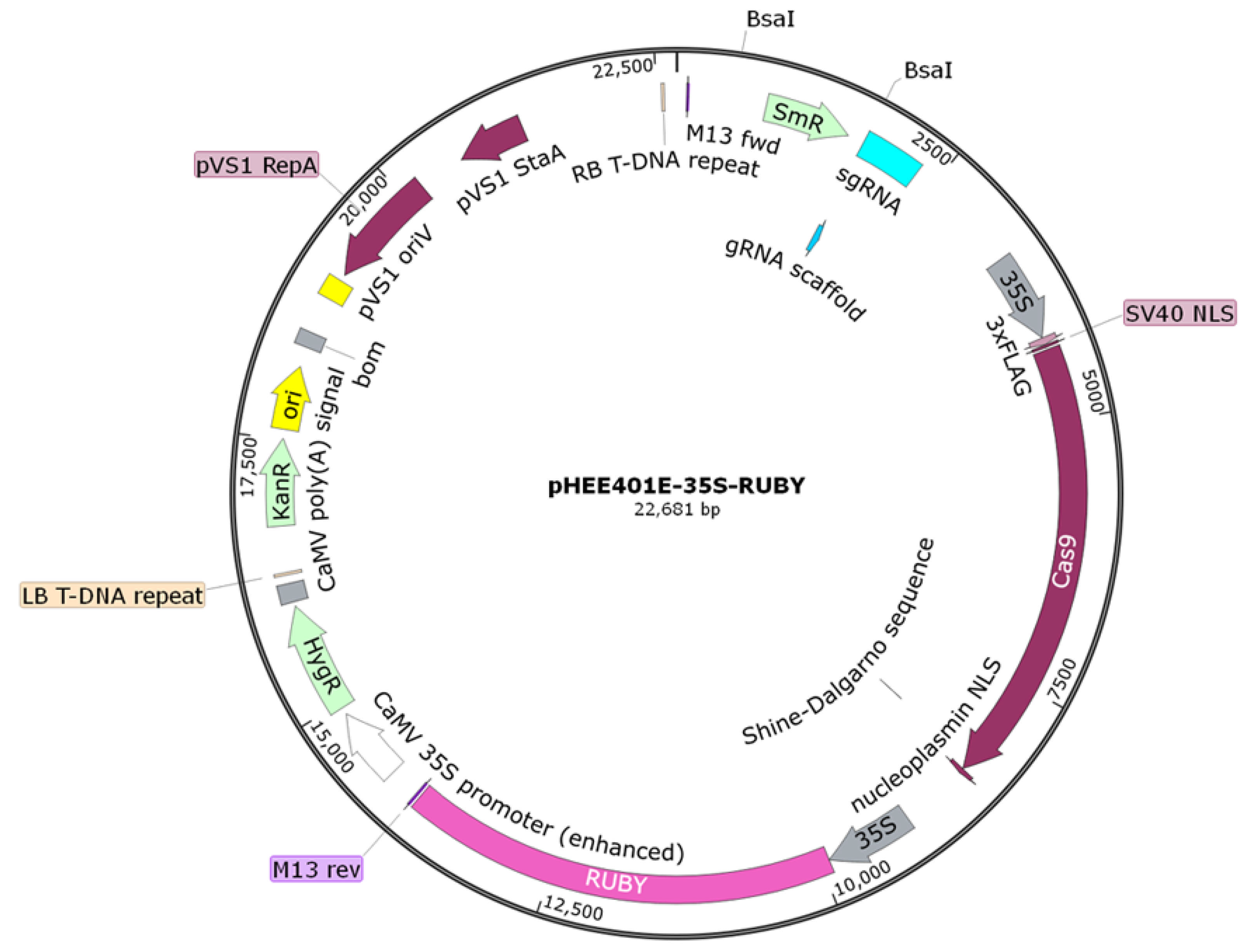
2.3. The Construction, Transformation, and Verification of the pHEE401E-35S-RUBY-EuC4H Vector

2.3.1. Primer Design

The C4H sequence of E. grandis (XM_010034641) was retrieved from the National Center for Biotechnology Information (https://www.ncbi.nlm.nih.gov/ (accessed on 12 April 2021)). BLAST alignment was performed against the whole genome of E. urophylla × E. grandis to identify the EuC4H (Eug10G0172600.1) sequence from TBtools version:1.120. For plasmid construction, primers containing target sequences were designed using the online tool CRISPR Direct (http://crispr.dbcls.jp/ (accessed on 20 April 2021)). Other primers were designed using the online tool, Primer3 (Table 1), and were synthesized by Sangon Biotech Co., Ltd. (Shanghai, China).

2.3.2. Construction of the pHEE401E-35S-RUBY Vector

(1)
Amplification of the 35S promoter and RUBY target fragment
The 35S promoter fragment was amplified from the pHDE plasmid using R35S-F and R35S-R as the upstream and downstream primers, respectively. The RUBY target fragment was amplified from the DR5-RUBY plasmid using RRUBY-F and RRUBY-R primers. The PCR program used is shown in Table 2. Following electrophoresis, the corresponding PCR products were purified and recovered.
(2)
Fusion PCR amplification of the 35S-RUBY fragment
The purified 35S promoter and RUBY fragments were used as templates for overlap extension PCR with R35S-F and RRUBY-R as the primers. The PCR program was the same as described previously (Table 2). After electrophoresis, the PCR products were purified and recovered.
(3)
Restriction digestion and homologous recombination of the pHEE401E-35S-Cas9 plasmid
The pHEE401E-35S-Cas9 plasmid (stored in our laboratory) was digested with the restriction enzyme MfeI. The purified 35S-RUBY target fragment underwent homologous recombination with the digestion product via Gibson Assembly to construct the pHEE401E-35S-RUBY plasmid, using homologous overlaps designed at the fragment termini.
(4)
Transformation of the recombinant plasmid into E. coli DH5α competent cells and the identification of transformants
The recombinant plasmid was transformed into E. coli DH5α competent cells via heat shock. Transformants were identified using bacterial and plasmid PCR. Positive clones were sequenced to confirm the successful construction of the pHEE401E-35S-RUBY plasmid.

2.3.3. Construction of the pHEE401E-35S-RUBY-EuC4H Vector

(1)
Amplification of small guide RNA (sgRNA) fragments containing two specific target sites
Using the pCBC plasmid stored in our laboratory as a template, PCR amplification of the corresponding target sites was performed using primers EuC4H-gF and EuC4H-gR. The PCR program is shown in Table 3. Following electrophoresis, the corresponding PCR products were purified and recovered.
(2)
Restriction digestion and homologous recombination of the pHEE401E-35S-RUBY plasmid
The pHEE401E-35S-RUBY plasmid was digested with the restriction enzyme BsaI, and recombinational cloning with the guide RNA (gRNA) fragment was performed, using homologous overlaps designed at the fragment termini. The reaction mixture contained 5 µL of recombinase, 2.5 µL of linear vector, and 2.5 µL of the insert fragment, and the reaction was performed by incubation in a 55 °C water bath for 15 min.
(3)
Transformation of the recombinant plasmid into E. coli DH5α competent cells and the identification of transformants
The recombinant plasmid was introduced into E. coli DH5α competent cells using heat shock. Positive clones were identified using bacterial PCR, followed by culture expansion and extraction of the recombinant plasmid. Plasmid PCR was conducted to identify transformants, and the recombinant plasmid was then sequenced.
(4)
Preparing the engineered bacterial culture
Positive recombinant plasmid clones confirmed using sequencing were transformed into A. tumefaciens competent cells. Single colonies resistant to rifampicin and kanamycin were selected for culturing. These bacterial cultures were then used as templates for PCR identification using EuC4H-F and EuC4H-R as primers. A. tumefaciens colonies with the desired band size were selected for further culturing at 28 °C and 200 rpm for 20 h to prepare the engineered bacterial cultures.

2.3.4. Transformation and Identification of the pHEE401E-35S-RUBY-EuC4H Plasmid

Stem segments without buds were cut into 3–6 mm pieces from the aseptic seedlings of E. urophylla × E. grandis and used as explants. These explants were cultured on a callus-induction medium at 24 °C in the dark for 1 week. They were then immersed in 100 mL of transformation solution and subjected to 40 kHz ultrasonication for 1 min. A fresh bacterial culture of A. tumefaciens carrying the pHEE401E-35S-RUBY-C4H plasmid was centrifuged at 2315× g for 1 min, and the resulting pellet was resuspended in transformation solution with the callus tissue. This mixture was incubated on a shaker at 50 rpm and 28 °C in the dark for 3 h. Subsequently, the treated callus tissue was transferred back to the callus-induction medium and co-cultured in the dark at 22 °C for 1 week. The callus tissue was then transferred to a bud-induction medium containing 300 mg/L cefotaxime sodium and further cultured in the dark for another week before being cultured in the light. The medium was changed biweekly, and the growth of the callus tissue and RUBY expression were monitored.
Transformed callus tissue exhibiting red coloration was selected and cut, and the DNA was extracted to serve as a template. PCR amplification was performed using RUBY-GT1 and RUBY-GT2 primers to confirm the integration of the marker gene RUBY into the callus tissue. Then, using EuC4H-GT1 and EuC4H-GT2 primers, the PCR products were analyzed by electrophoresis to verify the expected band sizes. If a smaller band was observed, it was excised and sequenced, and the sequencing results were compared with the expected sequence to confirm the successful editing of the EuC4H gene.

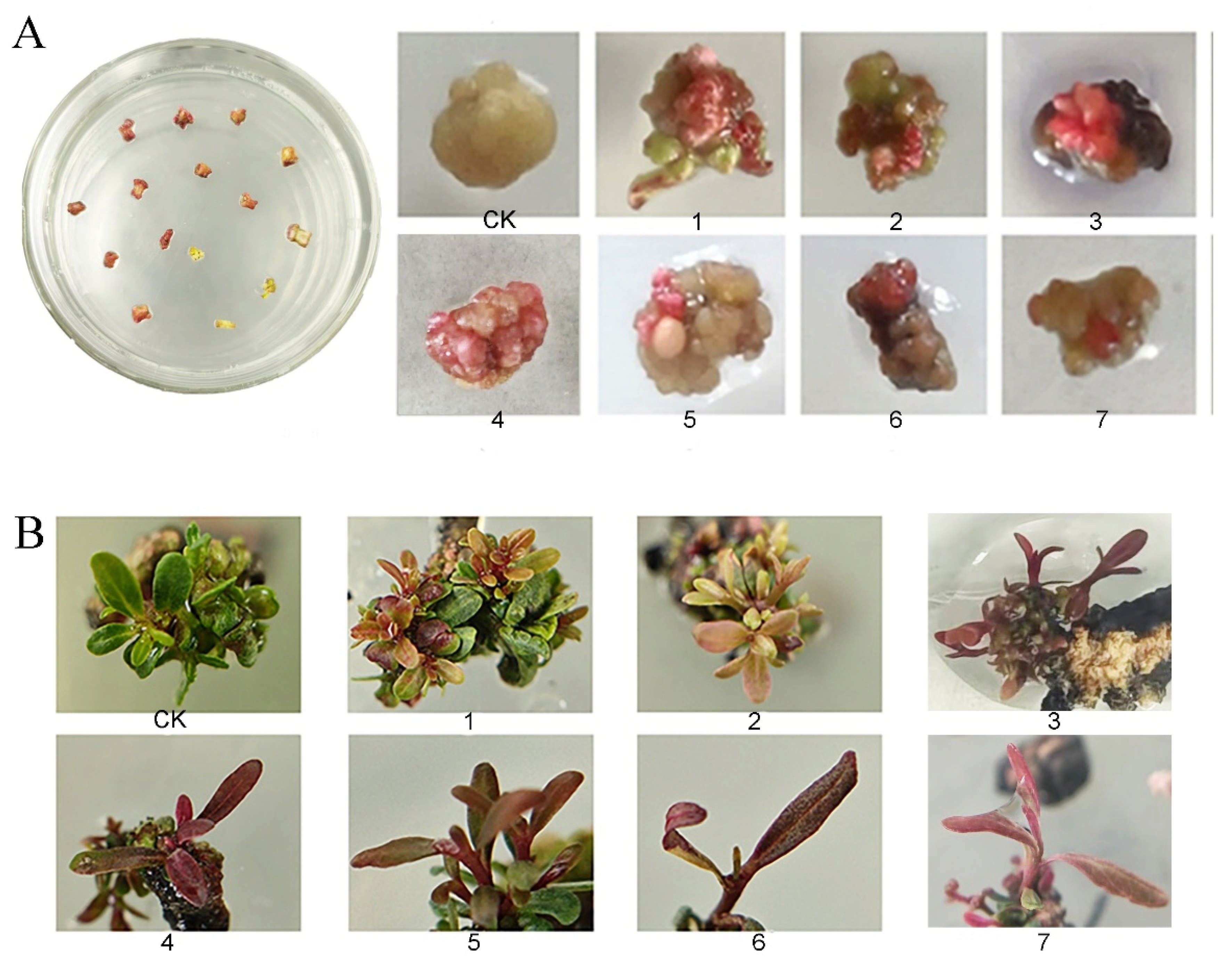
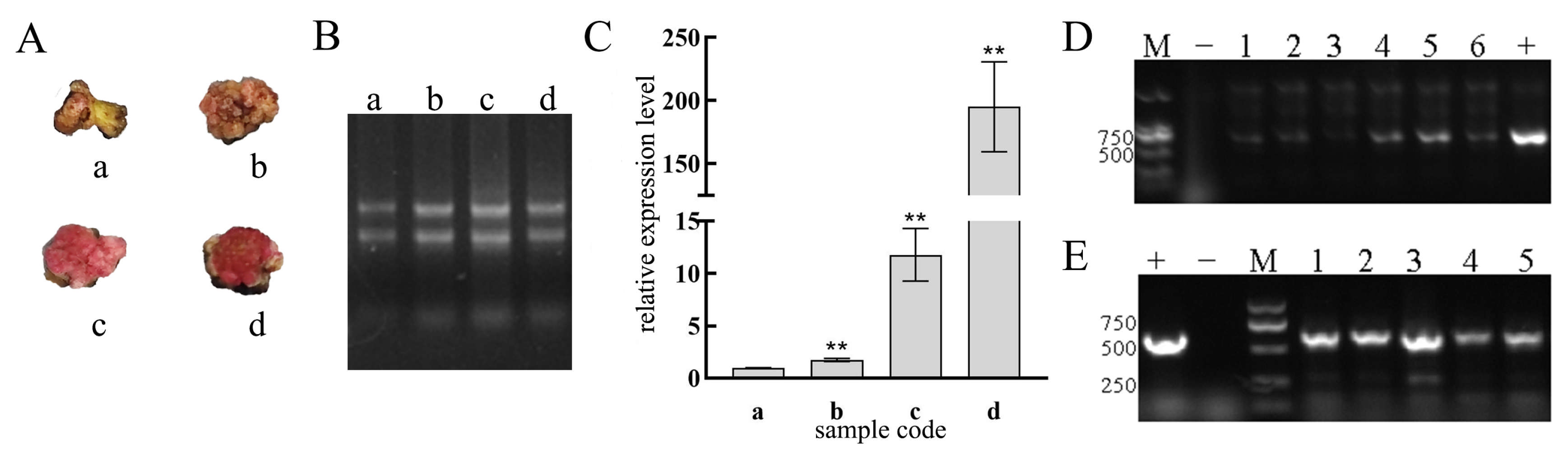
2.3.5. Correlation Between the Degree of Red Coloration and RUBY Gene Expression Levels in the Callus Tissue of E. urophylla × E. grandis

One hundred milligram samples from four different types of callus tissues, each showing varying degrees of red coloration indicative of the various expression levels of the RUBY gene, were weighed and used for RNA extraction. RNA was extracted using the TransZol Up Plus RNA Kit and Total RNA Extraction Kit (TransGen Biotech Co., Ltd., Beijing, China). The TransScript One-Step gDNA Removal and cDNA Synthesis SuperMix kits (TransGen Biotech Co., Ltd., Beijing, China) were used for reverse transcription.
The actin gene of E. urophylla × E. grandis (XM_010038187.3) served as an internal control for the qPCR experiments. The relationship between the varying degrees of red coloration in the callus tissues and RUBY gene expression levels was assessed through fluorescence qPCR. The primer sequences are listed in Table 1.
The TransStart Tip Green qPCR SuperMix Kit was used for qPCR experiments. The total qPCR reaction volume was 20 µL and comprised 0.4 µL of each of the upstream (F) and downstream (R) primers at a concentration of 20 µM after dilution, 10 µL of the super mix, 0.4 µL of dye, 1 µL of the sample, and 7.8 µL of nuclease-free water. Reaction conditions were as follows: initial denaturation at 94 °C for 4 min 30 s, followed by 40 cycles of (1) 94 °C for 5 s, (2) 56 °C for 15 s, and (3) 72 °C for 10 s. Each sample in the qPCR reaction was set up in quadruplicate. Relative gene expression was calculated using the 2−ΔΔCt method. Once normality and lognormality tests were used to confirm the data’s normal distribution, we performed the f-test to compare the variance. Samples with variance homogeneity were analyzed using unpaired Student’s t-tests; otherwise, an unpaired Student’s t-test with Welch’s correction was used. GraphPad Prism trial version 8.3.0 (GrpahPad, San Diego, CA, USA) was used to analyze and plot the exported data containing Ct values.

3. Results

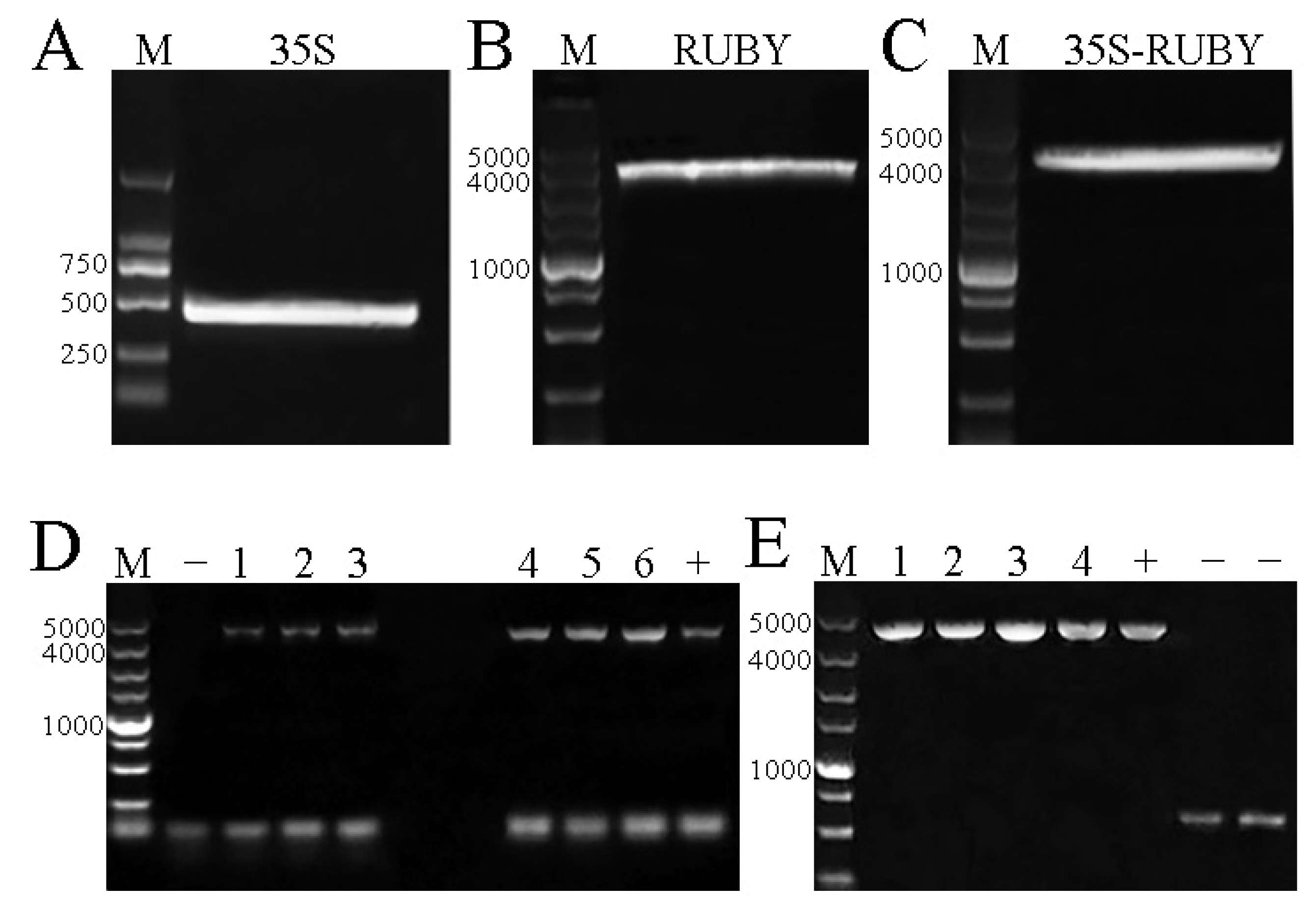
3.1. The Construction Results of the pHEE401E-35S-RUBY Vector

Electrophoresis after amplification showed that the 35S promoter (Figure 1A) and RUBY target bands (Figure 1B) were consistent with the expected size. The 35S promoter and RUBY bands were then excised from the gel for recovery.
The size of the 35S-RUBY band t was consistent with the expected band size (Figure 1C). The recovered 35S-RUBY target band was recombined with the MfeI-digested pHEE401E-35S-Cas9 plasmid. After transformation into E. coli DH5α competent cells, verification by bacterial and plasmid PCR (Figure 1E) indicated successful construction of the pHEE401E-35S-RUBY vector. The vector structure is shown in Figure 2.

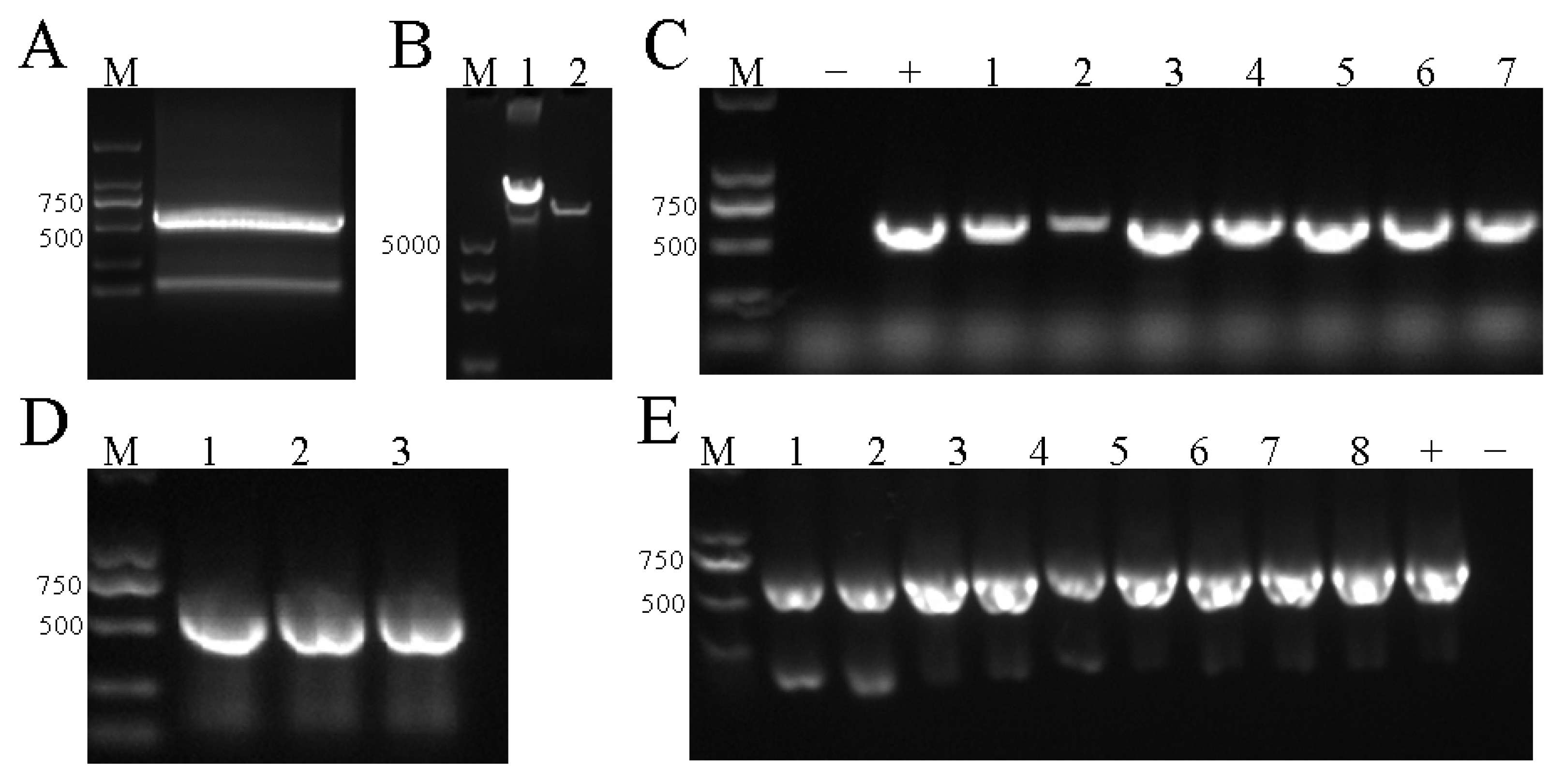
3.2. The Construction Results of the pHEE401E-35S-RUBY-EuC4H Vector

sgRNA target fragment was recovered and the size of the product was between 500 and 750 bp, consistent with the expected size (Figure 3A). BsaI digestion of the pHEE401E-35S-RUBY plasmid resulted in a notable difference in the band size pre- and post-digestion, yielding linear plasmids with bright bands, indicating complete digestion (Figure 3B).
The recombinant plasmid was transformed into E. coli via heat shock, followed by resistance screening. Bacterial PCR results revealed band sizes between 500 and 750 bp (Figure 3C). The bands matched the expected size and indicated a high ratio of positive clones. Plasmid extraction and PCR verification of the plasmids were performed, and the band sizes matched the expected sizes (Figure 3D). Sequencing analysis confirmed the successful construction of the pHEE401E-35S-RUBY-EuC4H vector; the structure of the recombinant plasmid is shown in Figure 4. It was then transformed into A. tumefaciens competent cells using the heat shock method to prepare the engineered bacterial culture.

3.3. Transformation and Identification of the pHEE401E-35S-RUBY-EuC4H Vector

After culturing in the light post-transformation, most of the E. urophylla × E. grandis callus tissue exhibited red coloration on the surface (Figure 5A). Upon culture continuation until budding (Figure 5B), parts of the leaves turned red, indicating partial expression of RUBY in the plant tissues.
Following callus tissue culturing in the light, no significant differences in external characteristics, aside from varying degrees of redness, were observed (Figure 5A). The callus tissues that turned red post-transformation were divided into four groups (Figure 6A). RNA was extracted from each group for quality assessment via electrophoresis. Slight trailing in the bands, indicative of diffusion, was observed. The brightness of the 28S rRNA band was approximately twice that of the 18S rRNA band, demonstrating good sample integrity and that they met the requirements for subsequent experiments (Figure 6B).
Using Sample “a” as the reference, Student’s t-test was conducted to analyze differential gene expression levels. The results are presented using a histogram (Figure 6C). A positive correlation between the expression level of RUBY and degree of redness of the E. urophylla × E. grandis callus tissue was observed, confirming RUBY as an effective marker gene for screening.
DNA from red callus tissue was used as a template to identify the marker gene RUBY. The PCR products were approximately 750 bp (Figure 6D; Table 4), consistent with the expected size and suggesting successful integration of RUBY into the callus tissue at a high transformation efficiency (the average transgenic efficiency was 63.3%). Gene editing results were then verified using EuC4H-specific primers. The average gene-editing efficiency was 0.0876% (Table 4). As shown in Figure 6E, Lane 3 exhibited two bands, with the smaller band between 250 and 500 bp, consistent with the expected size post-editing. This smaller band was recovered for sequencing, and the sequencing results were compared with the expected sequence (Figure 7A). The analysis revealed overlapping peaks starting from 125 bp. Using the Sanger sequencing check advanced plugin from TBtools version 1.120, with the Gap Extension parameter set to 0 and other parameters at default settings, two allelic sequences, S1 and S2, were derived after separating the peaks. A comparison of these two allelic sequences with the reference sequence (Figure 7B) revealed insertions, deletions, and partial base mutations in the EuC4H gene between and beyond the two target sites, preliminarily confirming the successful editing of the target gene.

4. Discussion

Currently, traditional Agrobacterium-mediated transformation and biolistic (gene gun) methods are primarily used for gene delivery into plant cells. The efficiency of these transformation methods generally depends on the genotype and species of the plant, limiting their applicability to a select number of species and genotypes, and an extended period of tissue culture is often required to cultivate complete plants [20]. A robust and efficient genetic transformation system remains elusive for most woody plants. In most systems, callus tissues serve as the transformation recipient, and selecting the appropriate type and dosage of antibiotics is crucial when developing these systems. Antibiotics, such as kanamycin and hygromycin B, are commonly used for selecting positive transformants, yet their toxicity to most plants is relatively high. The reliance on antibiotic selection for transformed chimeras poses significant challenges in screening and identification owing to low efficiency and a high rate of false positives.
E. urophylla × E. grandis is a fast-growing hybrid. Its large and complex genome complicates the acquisition of homozygous transgenic and gene-edited plants. A significant challenge in using callus tissue for genetic transformation in this species is the identification of positive transformants, which is also crucial for enhancing screening efficiency. In this study, the traditional Agrobacterium-mediated approach was used to deliver recombinant plasmids. Despite a brief experimental cycle, straightforward approach, and relatively high transformation rate, the transformed explants obtained by tissue culture frequently showed chimerism, which made subsequent genotyping highly laborious and inefficient. Additionally, the probability of obtaining homozygous gene-edited plants was also low. Therefore, it is crucial to focus on developing and applying novel methods for rapidly acquiring positive transformants. This will promote basic and applied plant research and fulfill the increasing public demand for new germplasms with superior traits.
The reporter gene RUBY enables visual screening through direct in situ observation of transgenic plants, offering the benefits of cost-effectiveness, convenience, and high conversion efficiency. In our study, we identified several causes for false positives in the actual tissue culturing of E. urophylla × E. grandis. First, the inherent characteristics of the species contributed to the issue. As a woody plant abundant in phenolic substances, E. urophylla × E. grandis is prone to browning during tissue culture [21]. Furthermore, the accumulation of certain secondary metabolites, such as anthocyanins [22], can mimic the color of RUBY. Additionally, our observations revealed that certain explants displayed a reddish-brown color before inoculation with A. tumefaciens. Upon reviewing the literature, we speculated that false positives could have resulted from the integration of coniferol into lignin polymers, which can cause red coloration of the stems of E. urophylla × E. grandis [23]. These factors could all potentially disrupt the visual screening process. Moreover, we observed issues inherent to RUBY itself. In our extended tissue culture of E. urophylla × E. grandis, we noted that callus tissues exhibited widespread reddening during the initial light exposure after Agrobacterium transformation, with this red coloration fading over time during the middle and later stages of tissue culture. Therefore, we speculated that the stability of RUBY was compromised under prolonged light exposure, resulting in a decline in the red phenotype. Additionally, callus tissues that initially turned red after bud formation showed a specific pattern: red buds with larger areas of redness and deeper coloration on the leaves correlated with a higher likelihood of these buds ceasing growth and eventually dying. We thus hypothesized that RUBY overexpression in regenerated E. urophylla × E. grandis buds could have a negative impact on the buds owing to secondary metabolites. Alternatively, betanin expression could have interfered with chloroplast growth and development in the leaves, thereby affecting chlorophyll synthesis, which ultimately hindered plant growth. The RUBY system produces betanin through the endogenous tyrosine metabolic pathway. Notably, tyrosine serves not only as the precursor for betanin biosynthesis but also as the starting substrate for multiple critical secondary metabolites, including tocopherols (vitamin E) and ubiquinones (coenzyme Q). The targeted regulation of tyrosine metabolism by the RUBY system may indirectly affect the biosynthesis of plant secondary metabolites through mechanisms such as precursor competition, enzyme activity inhibition, energy resource allocation, and transcriptional regulation interference [24,25]. However, these proposed mechanisms still require experimental validation through metabolomic analyses and functional studies.
This study offers valuable insights and guidance for the future development of an efficient and precise site-specific editing system in Eucalyptus, as well as for the targeted breeding of new, environmentally friendly E. urophylla × E. grandis germplasms suitable for pulp production.

Author Contributions

Formal analysis, L.L.; Data curation, M.S. and X.W.; Writing—original draft, M.S.; Writing—review & editing, Z.W.; Funding acquisition, L.O. All authors have read and agreed to the published version of the manuscript.

Funding

This research was funded by the Natural Science Foundation of Guangdong Province, grant number 2025A1515012337; The National Natural Science Foundation of China, grant number 32071780; Guangdong Science and Technology Program, grant number 2021S0074 & 2022DZXHT072 & 2023S018087.

Data Availability Statement

Data is contained within the article.

Conflicts of Interest

The authors declare no conflict of interest.

References

  1. Hong, W.; Huang, J.-H.; Li, J.; Lin, H.; Chen, C.; Wu, C.-Z. Study on the δ13C Value in Different Eucalyptus Species. J. Fujian Coll. For. 2008, 28, 289–293. [Google Scholar] [CrossRef]
  2. Liu, T.; Xie, Y.-J. Studies on the Causes of Rapid Development of Eucalyptus Plantations in China. Eucalypt Sci. Technol. 2020, 37, 38–47. [Google Scholar] [CrossRef]
  3. Tang, Q.-H.; Chen, Y.-P. Status Quo and Prospect of Wood Processing and Utilization of Eucalyptus in China and Abroad. China Wood-Based Panels 2020, 27, 18–21. [Google Scholar]
  4. Huang, G.-Q.; Zhao, Q.-G. The History, Status Quo, Ecological Problems and Countermeasures of Eucalyptus Plantations in Guangxi. Acta Ecol. Sin. 2014, 34, 5142–5152. [Google Scholar] [CrossRef]
  5. Xie, Y.-J. Analysis of China’s Wood Resource Security and the Contributions of Eucalyptus Plantation. Eucalypt Sci. Technol. 2018, 35, 3–6. [Google Scholar] [CrossRef]
  6. Liu, Y.-T. Analysis of Enzymatic Properties of Ferulic Aid Esterase from Clostridium thermocellum and Construction of Its High-Efficiency Plant Expression Vector. Master’s Thesis, Jiangsu University, Zhenjiang, China, 2017. [Google Scholar]
  7. Feng, H.-X.; Tu, Y. Research on Lignin Biosynthesis. J. Qingdao Univ. Nat. Sci. Ed. 2018, 31, 46–54. [Google Scholar]
  8. Chen, B.-W. Cloning of Key Genes Involved in Lignin Monomer Biosynthesis from Eucalyptus urophylla and Transformation in Tobacco. Ph.D. Thesis, Beijing Forestry University, Beijing, China, 2017. [Google Scholar]
  9. Vanholme, R.; De Meester, B.; Ralph, J.; Boerjan, W. Lignin Biosynthesis and Its Integration into Metabolism. Curr. Opin. Biotechnol. 2019, 56, 230–239. [Google Scholar] [CrossRef]
  10. Li, W.; Yang, L.-X.; Jiang, L.-Z.; Zhang, G.-L.; Luo, Y.-G. Molecular Cloning and Functional Characterization of a Cinnamate 4-Hydroxylase-Encoding Gene from Camptotheca Acuminata. Acta Physiol. Plant. 2016, 38, 1–9. [Google Scholar] [CrossRef]
  11. Yan, Q.; Si, J.-R.; Cui, X.-X.; Peng, H.; Chen, X.; Xing, H.; Dou, D.-L. The Soybean Cinnamate 4-Hydroxylase Gene GmC4H1 Contributes Positively to Plant Defense via Increasing Lignin Content. Plant Growth Regul. 2019, 88, 139–141. [Google Scholar] [CrossRef]
  12. Blount, J.W.; Korth, K.L.; Masoud, S.A.; Rasmussen, S.; Lamb, C.; Dixon, R.A. Altering Expression of Cinnamic Acid 4-Hydroxylase in Transgenic Plants Provides Evidence for a Feedback Loop at the Entry Point into the Phenylpropanoid Pathway. Plant Physiol. Bethesda 2000, 122, 107–116. [Google Scholar] [CrossRef]
  13. Simmons, C.R.; Grant, S.; Altier, D.J.; Dowd, P.F.; Crasta, O.; Folkerts, O.; Yalpani, N. Maize rhm1 Resistance to Bipolaris Maydis Is Associated with Few Differences in Pathogenesis-Related Proteins and Global mRNA Profiles. Mol. Plant-Microbe Interact. 2001, 14, 947–954. [Google Scholar] [CrossRef] [PubMed]
  14. Sykes, R.W.; Gjersing, E.L.; Foutz, K.; Rottmann, W.H.; Kuhn, S.A.; Foster, C.E.; Ziebell, A.; Turner, G.B.; Decker, S.R.; Hinchee, M.A.W.; et al. Down-Regulation of p-Coumaroyl Quinate/Shikimate 3′-Hydroxylase (C3′H) and Cinnamate 4-Hydroxylase (C4H) Genes in the Lignin Biosynthetic Pathway of Eucalyptus urophylla × E. grandis Leads to Improved Sugar Release. Biotechnol. Biofuels 2015, 8, 128. [Google Scholar] [CrossRef] [PubMed]
  15. Franke, R.; McMichael, C.M.; Meyer, K.; Shirley, A.M.; Cusumano, J.C.; Chapple, C. Modified Lignin in Tobacco and Poplar Plants Over-expressing the Arabidopsis Gene Encoding Ferulate 5-hydroxylase. Plant J. Cell Mol. Biol. 2000, 22, 223–234. [Google Scholar] [CrossRef]
  16. Polturak, G.; Aharoni, A. Advances and Future Directions in Betalain Metabolic Engineering. New Phytol. 2019, 224, 1472–1478. [Google Scholar] [CrossRef]
  17. He, Y.-B.; Zhang, T.; Sun, H.; Zhan, H.-D.; Zhao, Y.-D. A Reporter for Noninvasively Monitoring Gene Expression and Plant Transformation. Hortic. Res. 2020, 7, 152. [Google Scholar] [CrossRef]
  18. Tanaka, Y.; Kojima, M.; Uritani, I. Properties, Development and Cellular-Localization of Cinnamic Acid 4-Hydroxylase in Cut-Injured Sweet Potato. Plant Cell Physiol. 1974, 15, 843–854. [Google Scholar] [CrossRef]
  19. Wang, Z.-C.; Liu, Y.-M.; Ouyang, L.-J.; Li, L.-M.; Liang, C.-Y.; Pan, J.-Y. Construction and Verification of an Eucalyptus Gene Editing Vector with Visual Selection Markers. Bull. Bot. Res. 2021, 41, 816–823. [Google Scholar] [CrossRef]
  20. Ji, X.; Yang, B.; Wang, D.-W. Achieving Plant Genome Editing While Bypassing Tissue Culture. Trends Plant Sci. 2020, 25, 427–429. [Google Scholar] [CrossRef]
  21. Chen, G. Common Problems and Countermeasures in Rapid Propagation Technology of Forest Tissue Culture. Prot. For. Sci. Technol. 2017, 4, 100–102. [Google Scholar] [CrossRef]
  22. Su, M.; Liu, Y.-M.; Li, L.-M.; Zhang, Y.-X.; Huang, H.; Guo, K.-X. Metabolomics Analysis and Differential Expression of Related Genes in Different Callus of Eucalyptus urophylla × E. grandis. Plant Physiol. J. 2022, 59, 943–953. [Google Scholar] [CrossRef]
  23. Stewart, D.; Yahiaoui, N.; McDougall, G.J.; Myton, K.; Marque, C.; Boudet, A.M.; Haigh, J. Fourier-Transform Infrared and Raman Spectroscopic Evidence for the Incorporation of Cinnamaldehydes into the Lignin of Transgenic Tobacco (Nicotiana tabacum L.) Plants with Reduced Expression of Cinnamyl Alcohol Dehydrogenase. Planta 1997, 201, 311–318. [Google Scholar] [CrossRef] [PubMed]
  24. Yang, J.-Q.; Han, G.-Y.; Liu, N.-X.; Zhou, Q. A Bioinformatics Study on Key Enzymes Involved in Tyrosine Metabolism in Beet. Chin. Agric. Sci. Bull. 2024, 40, 124–131. [Google Scholar] [CrossRef]
  25. Xu, J.-J.; Fang, X.; Li, C.-Y.; Yang, L.; Chen, X.-Y. General and Specialized Tyrosine Metabolism Pathways in Plants. aBIOTECH 2019, 1, 97–105. [Google Scholar] [CrossRef] [PubMed]
Figure 1. Electrophoretograms of Eucalyptus urophylla × Eucalyptus grandis pHEE401E-35S-RUBY vectors. Note: (A): M. DL 2000 DNA Marker. 35S promoter amplification. (B): M. DL 5000 DNA Marker. RUBY target band amplification. (C): M. DL 5000 DNA Marker. 35S-RUBY target band amplification. (D): M. DL 5000 DNA Marker. 1–6: pHEE401-35S-RUBY transformed Escherichia coli DH5α bacteria liquid PCR. (E): M. DL 5000 DNA Marker. 1–4: pHEE401E-35S-RUBY plasmid PCR. +: positive reference, −: negative reference.
Horticulturae 11 00406 g001
Figure 2. Schematic diagram of the structure of the vector pHEE401E-35S-RUBY. Note: bom: basis of mobility region from pBR322. CaMV 35S promoter (enhanced): cauliflower mosaic virus 35S promoter with a duplicated enhancer region. CaMV poly(A) signal: cauliflower mosaic virus polyadenylation signal. CAP binding site: CAP binding activates transcription in the presence of cAMP. gRNA scaffold: guide RNA scaffold for the Streptococcus pyogenes CRISPR/Cas9 system. HygR: aminoglycoside phosphotransferase from E. coli. KanR aminoglycoside phosphotransferase.
Horticulturae 11 00406 g002
Figure 3. Electrophoretograms of Eucalyptus urophylla × Eucalyptus grandis pHEE401E-35s-RUBY-EuC4H vectors. Note: (A): M. DL 2000 DNA Marker. sgRNA target fragment recovery; (B): M. DL 5000 DNA Marker. 1, undigested plasmid; 2, digested plasmid; (C): M. DL 2000 DNA Marker. 1–7: PCR results of Agrobacterium transformed with pHEE401E-35s-RUBY-EuC4H recombinant plasmid; (D): M. DL 2000 DNA Marker. 1–3: the results of the PCR of plasmid pHEE401E-35s-RUBY-EuC4H; (E): M. DL 2000 DNA Marker. 1–8: PCR results of pHEE401E-35s-RUBY-EuC4H recombinant plasmid transformed into A. tumefaciens. +: positive reference, −: negative reference.
Horticulturae 11 00406 g003
Figure 4. Diagram of the structure of the recombinant plasmid sgRNA of Eucalyptus urophylla × Eucalyptus grandis.
Horticulturae 11 00406 g004
Figure 5. Transformed calli and buds of Eucalyptus urophylla × Eucalyptus grandis. Note: (A): left panel displays the overall status of callus in the culture dish; CK: the control group; 1–7: which represent transformed callus. (B): CK: the control group; 1–7: which represent transformed buds.
Horticulturae 11 00406 g005
Figure 6. Combined diagram. Note: (A): representative images of calli with different degrees of red coloration; a–d: the calli exhibit a progressive increase in redness (B,C): RNA bands and qPCR results, ** p = 0.01; (D): M. DL 2000 DNA Marker. 1–6: RUBY identification in red calli after EuC4H transformation; (E): M. DL 2000 DNA Marker. 1–5: gene editing identification of EuC4H. +: positive reference, −: negative reference.
Horticulturae 11 00406 g006
Figure 7. Sequencing confirmation of EuC4H gene editing in Eucalyptus urophylla × Eucalyptus grandis. Note: (A) is the result of the original sequencing comparison; (B) is the result of haplotype comparison.
Horticulturae 11 00406 g007
Table 1. Primer sequences related to Eucalyptus urophylla × Eucalyptus grandis.
Primer NamePrimer Sequence (5′→3′)PurposeLength/bp
EuC4H-gFctagagtcgaagtagtgattgCATATATGGGCTCGAGAAG
gttttagagctagaaatagc
Constructed plasmids592
EuC4H-gRtgctatttctagctctaaaacACCTATGAATCGAACTCGT
caatcactacttcgactcta
R35S-FTAATGCATTTTATGACTTGCAACATGGTGGA
GCACGACAC
462
R35S-RGCGAGGGTCGCATGATCCATCGTGTCCTCT
CCAAATGAAA
RRUBY-FTTTCATTTGGAGAGGACACGATGGATCAT
GCGACCCTCG
3957
RRUBY-FGAATTCGTTGTCAATCAATTTCACTATCACTG
GAGGCTTG
EuC4H-GT1TATCATCGAAAATCGCGGCGGIdentification primers736
EuC4H-GT2ACCTCTCGAAAAATTGCTTGCC
RUBY-GT1CTCACAACTCCGCTCAACGC725
RUBY-GT2GAGTCCGGCTCTTTGAGGCT
RUBY-qFCGCCACACTCCTCCAGTTCTTCqPCR primers_
RUBY-qRCGTCCTCGCCGTTCATCATCTT
EuActin-FGCACCGCCAGAGAGGAAATA
EuActin-RGAAGCACTTCCTGTGGACGA
Table 2. Procedure for PCR amplification of the pHEE401E-35S-RUBY vector.
TemperatureStepTime
98 °CInitialization2 min
98 °CDenaturation10 s
58 °CAnnealing15 s
72 °CElongation5 s/kb
Table 3. Procedure for PCR amplification of the pHEE401E-35S-RUBY-EuC4H vector.
TemperatureStepTime
94 °CInitialization5 min
94 °CDenaturation30 s
56 °CAnnealing30 s
72 °CExtension60 s/kb
72 °CComplete extension7 min
Table 4. Data on transformation and gene-editing efficiency.
ExperimentExplantsTransgenic BudsEdited BudsTransformation EfficiencyGene-Editing Efficiency
I600413168.8%0.0876%
II600357059.5%
III600371061.8%
Disclaimer/Publisher’s Note: The statements, opinions and data contained in all publications are solely those of the individual author(s) and contributor(s) and not of MDPI and/or the editor(s). MDPI and/or the editor(s) disclaim responsibility for any injury to people or property resulting from any ideas, methods, instructions or products referred to in the content.

