Article

Genomic and Transcriptomic Analysis to Explore the Biological Characteristics of Cyclocybe chaxingu

1
Bioengineering and Technological Research Center for Edible and Medicinal Fungi, Jiangxi Agricultural University, Nanchang 330045, China
2
Nanchang Key Laboratory of Edible and Medicinal Fungi, Jiangxi Agricultural University, Nanchang 330045, China
3
Jiangxi Provincial Key Laboratory of Excavation and Utilization of Agricultural Microorganisms, Jiangxi Agricultural University, Nanchang 330045, China
4
Key Laboratory of Crop Physiology, Ecology and Genetic Breeding, Jiangxi Agricultural University, Ministry of Education of the People’s Republic of China, Nanchang 330045, China
5
Beijing Key Laboratory of Environment Friendly Management on Fruit Diseases and Pests in North China, Institute of Plant Protection, Beijing 100097, China
6
Academy of Agriculture and Forestry Sciences, Beijing 100097, China
*
Authors to whom correspondence should be addressed.
These authors contributed equally to this work.
Horticulturae 2025, 11(4), 409; https://doi.org/10.3390/horticulturae11040409
Submission received: 23 February 2025 / Revised: 4 April 2025 / Accepted: 7 April 2025 / Published: 11 April 2025
(This article belongs to the Special Issue Edible Mushrooms: Genetics, Genomics, and Breeding)

Abstract

:
Cyclocybe chaxingu is an edible wood-decaying fungus widely cultivated in China, valued for its nutritional and economic significance. Despite its importance, molecular and genetic breeding studies on C. chaxingu have been limited by the lack of comprehensive genomic information. In this study, we performed whole-genome sequencing of the type strain JAUCC1847 of C. chaxingu for the first time and conducted extensive genomic and transcriptomic analyses. We assembled a high-quality genome of the C. chaxingu strain C27, with a total length of 50.79 Mb and a GC content of 50.90%. Comparative genomic analysis revealed a close evolutionary relationship with species from the genera Agrocybe and Stropharia, suggesting a recent common ancestor. The high ANI values between C. chaxingu, Agrocybe chaxingu, and Agrocybe cylindracea indicate a close phylogenetic relationship, raising the possibility of synonymy among these strains. Genome annotation identified a rich array of 573 carbohydrate-active enzymes, highlighting the metabolic diversity of C. chaxingu, particularly in lignocellulose degradation. Comprehensive analysis of the A and B mating-type locus in C. chaxingu revealed the distribution and structural characteristics of mating-type genes, providing crucial genetic information for further studies on the reproductive biology of this species. Transcriptomic analysis revealed distinct gene expression patterns in mycelia, stipe, and cap, reflecting their functional specialization. GO and KEGG enrichment analyses demonstrated the stipe’s association with structural integrity and transport, while the cap is linked to metabolic activity, gene regulation, stress responses, and DNA repair. These insights clarify the taxonomic status of C. chaxingu, supporting its recognition within the genus Cyclocybe and providing a valuable resource for future research and breeding programs.

1. Introduction

Cyclocybe chaxingu (synonym Agrocybe chaxingu) is an edible wood-decaying fungus extensively cultivated in China [1]. The species is easily confused with C. aegerita, C. cylindracea, and C. salicaceicola due to their similar morphology. Commercially, they are collectively referred to as Cyclocybe cylindracea or Agrocybe cylindracea (Chashugu) [2,3]. Recently, Liu et al. revisited the taxonomy of C. chaxingu using a combination of morphological and multi-gene phylogenetic analyses, proposing the new combination Cyclocybe chaxingu, under Basidiomycota, Agaricales, Tubariaceae, and the genus Cyclocybe [1].
The Cyclocybe species are known for their high nutritional value and delightful taste, with a crisp and refreshing stem and a smooth and tender cap, giving it a unique flavor. For instance, the C. cylindracea extracts contain 29.5% mass fractions of protein, 0.16% crude fat, and 43.6% polysaccharides accounted. It is also rich in elements such as calcium (3.607 mg/g), magnesium (3.021 mg/g), zinc (79.3 µg/g), and iron (79.2 µg/g) [4]. Lee et al. reported that Agrocybe chaxingu contains 17 distinct amino acids. Among these, essential amino acids constitute 2.70 g/100 g of edible weight, while non-essential amino acids amount to 5.31 g/100 g. Leucine was identified as the most abundant essential amino acid, and glutamate was the predominant non-essential amino acid [5]. In addition, studies have shown that polysaccharides and mycelial polysaccharides from C. cylindracea possess antitumor, hypoglycemic, lipid peroxidation inhibitory, immunomodulatory, anti-aging, and in vitro antioxidant activities, making them effective in treating various human diseases [6,7].
Within the order Agaricales, species such as Agaricus bisporus [8], Laccaria bicolor [9], Coprinopsis cinerea [10], Schizophyllum commune [11], Pleurotus giganteus [12], Flammulina velutipes [13], Lentinula edodes [14], Oudemansiella raphanipes [15], Lyophyllum decastes [16], and Stropharia rugosoannulata [17] have been sequenced and analyzed at the whole-genome level. Sequencing analysis reveals that the genome sizes of Agaricales species range from 22.12 Mb [18] to over 100 Mb, with predicted gene counts ranging from 10,000 to 20,000. In studies of species closely related to C. chaxingu, researchers have sequenced the genome of Agrocybe aegerita, revealing a set of conserved genes related to fruit body development, as well as a high content of carbohydrate-active enzymes, indicating its lignocellulose-degrading capacity [19]. Genome sequencing of Agrocybe cylindracea has unveiled complex pathways involved in the biosynthesis of polysaccharides and other bioactive compounds, and RNA-seq has highlighted the differential functions of the stipe and cap anatomical structures [20].
Mating-type recognition plays a crucial role in the genetic breeding and species identification of edible fungi, influencing the reproductive system, fruiting bodies, and gamete quality [21]. Cyclocybe chaxingu has a tetrapolar mating system with homogenic incompatibility [22], controlled by two unlinked mating-type locus (MAT), A and B. In typical basidiomycetes, the HD genes flanking the MatA are the mitochondrial intermediate peptidase (MIP) gene and the beta-flanking (β-fg) gene. The β-fg gene is a conserved gene that flanks the opposite side of the HD loci in most species [23]. The MatA includes HD1 and HD2 genes, which are transcribed in opposite directions and encode two different proteins with homeodomain motifs, primarily controlling clamp connection formation and nuclear pairing [24]. In most species (e.g., Pleurotus ostreatus, Pleurotus eryngii, Schizophyllum commune, Coprinopsis cinerea, Agaricus bisporus, and Volvariella volvacea), the MIP gene is closely linked to the HD1 gene [25], and the upstream region of MIP typically contains Sec61 protein and glycine dehydrogenase (GLYDH) genes [26]. The MatB can encode multiple pheromone receptors (Pr) and pheromone precursors (Pp), which mainly regulate nuclear migration and clamp connection fusion. These genes often appear in close proximity and are linked to form the MatB [25]. In the study of mating-type genes in Cyclocybe and Agrocybe species, Chen et al. used populations of C. aegerita and C. salicacola strains as research materials, through genome sequencing and sequence analysis, they cloned the B mating loci of C. aegerita and C. salicacola strains, including the pheromone receptors rcb1, rcb2, and part of the non-coding region [27]. Additionally, they found that the Asrcb1 and Asrcb2 genes in C. salicacola are conserved pheromone receptors in monokaryotic strains [28]. Yang et al. identified the A and B mating-type gene loci based on the genome sequences of the dikaryotic strain YSG and its monokaryotic strain of Agrocybe aegerita [29]. Using the conserved regions of the MIP gene sequence at the MatA and the B2-rcb3a and B2-rcb3b sequences at the MatB, they designed specific primers as molecular markers for mating-type molecular identification [29,30,31]. Chen et al. performed telomere-to-telomere assembly of the genomes of the two sexually compatible haploid strains (named as CchA and CchB) of Agrocybe chaxingu [32], analyzing the genetic differences between the two monokaryons and their expression levels during growth and development. They discovered a highly conserved glycosyltransferase family 8 gene (Glgen) on the other side of the HD2 gene at the MatA, while the β-fg gene was located on different chromosomes in the genomes of CchA and CchB, no longer directly associated with the MatA locus of A. chaxingu. In the study of the MatB, they identified and characterized 5 Pr genes and 3 Pp genes in the genomes of CchA and CchB.
In this study, we performed de novo whole-genome sequencing on haploids of the type strain JAUCC 1847 of C. chaxingu with three different mating types for the first time. Additionally, we conducted transcriptome sequencing on the dikaryotic mycelia as well as the caps and stipes of the fruiting bodies through various bioinformatics to explore the biological characteristics of C. chaxingu.

2. Materials and Methods

2.1. Species Information

The new species name Cyclocybe chaxingu was proposed by Liu et al. [1], but it was not validly published because it lacked a registration number [33]. To correct this error, the species name is validated below:
Cyclocybe chaxingu (N.L. Huang) Q.M. Liu, Yang Gao & D.M. Hu, comb. nov.
MycoBank MB840094
Agrocybe chaxingu N.L. Huang, in Edible Fungal Flora of China, ed. by Institute of Edible Fungi, Shanghai Academy of Agricultural Sciences: 182 (1991); Agrocybe chaxingu N.L. Huang, Exp. Mycol. 10: 24 (1972), nom. inval.

2.2. Strains and Culture Conditions of Cyclocybe chaxingu

The dikaryotic strain JAUCC1847 of C. chaxingu used in this study is a neotype strain, which is preserved at the Jiangxi Agricultural University Culture Collection (JAUCC). In order to obtain monokaryotic strains, strain JAUCC1847 was cultivated in liquid potato dextrose broth (PDB) under dark conditions at 25 °C for 10 d, and protoplasts were prepared following the methods established by Li et al. and Wang et al. [34,35]. The protoplasts were diluted and spread. Single colonies were isolated for individual cultivation after 3–5 d. Following the methods of Zhu et al. and Qian et al. [30,31], specific conserved regions were amplified via PCR to identify the mating type of obtained monokaryotic strains. Consequently, three C. chaxingu monokaryotic strains with different mating types, strain C5 (AyBx), C8 (AyBy), and C27 (AxBx), were selected for genome sequencing.
For fruiting body production, the strain JAUCC1847 was inoculated into a sterile solid medium contained in polypropylene bags. The medium composition was as follows: 15% (w/w) cottonseed hulls, 20% (w/w) coarse sawdust, 20% (w/w) fine sawdust, 20% (w/w) bagasse, 15% (w/w) wheat bran, 5% (w/w) soybean meal, 3% (w/w) cornmeal, 1% (w/w) gypsum, and 1% (w/w) lime. The cultures were incubated in the dark at 25 °C with 65% humidity for approximately 50 days until the mycelia fully established on the solid medium. After full colonization, the bags were opened, and primordia differentiation and fruiting body formation were induced under conditions of 22 °C, 95% humidity, and 600 lx light intensity.

2.3. Genome Sequencing and Assembly

The three monokaryotic strains were inoculated into PDB and cultured on a shaker. The mycelial pellets were filtered and dried using sterile filter paper. Total genomic DNA was extracted using a modified CTAB method [36]. The genome of three monokaryotic strains was sequenced using the Illumina NovaSeq 6000 platform (paired-end, 2 × 150 bp) and the PacBio Sequel platform (CLR) by Personalbio Technology (Nanjing, China). The sequencing depth was approximately 150× for the Illumina platform and 200× for the PacBio platform.
Raw sequencing data were quality-trimmed using Fastp v0.23.1 to obtain high-quality data [37]. Third-generation single-molecule sequencing data were assembled using NextDenovo v2.5.0 to generate contig sequences [38]. High-quality second-generation data were used with the Pilon v1.24 to correct the third-generation contig results [39], ultimately producing a complete sequence. The completeness of the genome assembly was assessed using the BUSCO (Benchmarking Universal Single-Copy Orthologs) basidiomycota_odb10 database (v5.4.5) [40].

2.4. Gene Prediction and Annotation

Repeat sequences in the genome were identified using both homology prediction and ab initio gene prediction methods [41,42]. Tandem repeat sequences were identified using TRF software (version 4.10.0) [43], and de novo repeat sequence prediction was performed using RepeatModeler software (version v2.0.4) [44]. Homologous repeat sequences were annotated using RepeatMasker software (version 4.1.4) [45]. tRNA was predicted in the whole genome using tRNAscan-SE (version 2.0) [46], and rRNA genes were predicted using Barrnap 0.9 [47]. Other non-coding RNA predictions were mainly obtained through comparison with Rfam [48].
Gene models of the genome were de novo predicted using three software tools: Augustus (version 2.5.5) [49], glimmerHMM (version 3.0.4) [50], and GeneMark-ES (version 4.71) [51], resulting in corresponding gene prediction outcomes. Homologous prediction for related species was conducted using exonerate software (version 2.2.0) [52], using protein sequences of related species to obtain gene prediction results. EVidenceModeler (version 2.0.0) was used to integrate de novo and homology prediction results [53].
Functional annotation of predicted protein-coding sequences (CDS) was based on NCBI Nr (release 10 October 2017), eggNOG (v4.5, 28 November 2017) [54], Swiss-Prot (release 22 November 2017) [55], Pfam (http://pfam.xfam.org/) [56], and Carbohydrate Active Enzymes (CAZymes) database (release 14 August 2024, https://bcb.unl.edu/dbCAN2/) [57]. GO annotation was completed using InterPro (version 66.0, release 23 November 2017) [58]. KO and pathway annotations were performed using the KEGG KAAS automatic annotation system [59].

2.5. Comparative Genomic Analysis

Gene families and single-copy orthologs were analyzed using OrthoFinder v2.5.4 software [60]. Single-copy orthologous protein sequences were used to infer the chronogram and estimate species divergence times with the MCMCtree program in the PAML package [61]. Species trees were constructed using IQ-TREE v2.2.0 [62]. Collinearity analyses were performed with MCScanX v1.0.0 [63], based on positional information from GFF3 files of C. chaxingu, S. rugosoannulata, L. decastes, A. pediades, and L. edodes. Pairwise average nucleotide identity (ANI) analysis was conducted using fastANI v1.34 [64].

2.6. Analysis of Mating Genes

Following the method of Chen et al. [32], the identification of the MatA locus was performed using genome annotation results, along with homologous searches for the Mip gene associated with MatA in closely related and model species. The selected closely related species include C. aegerita [19] and C. cylindracea [20], while the chosen model species are Coprinopsis cinerea [10] and Schizophyllum commune [11]. The Pr genes of the MatB locus were identified through genome annotation results. Additionally, homologous searches were conducted for the two highly conserved Pr genes reported in the closely related species C. salicacola [28] to determine the location of MatB.

2.7. Transcriptomic Analysis of Different Tissues of Cyclocybe chaxingu

For transcriptomic analysis, at the stage when the fruiting bodies mature and begin to produce spores, samples were collected. Total RNA was extracted from three biological replicates of mycelia, stipe, and cap of strain JAUCC1847 using the Qiagen RNeasy extraction kit (Qiagen, Valencia, CA, USA) and measured on a Qubit fluorometer (Invitrogen, Eugene, OR, USA). RNA-Seq library was constructed, and RNA sequencing following the deoxyuridine triphosphate method on the NovaSeq 6000 platform (paired-end, 2 × 150 bp, carried out by Personalbio Technology (Nanjing, China). The sequencing data were processed using Fastp to remove 3′ end adapter sequences and reads with an average quality score below Q20. Subsequently, the cleaned reads were aligned to the reference genome using HISAT2 (v2.2.1) [65], and gene expression quantification was performed using FeatureCounts (v2.0.3) [66].
The correlation coefficients between and within sample groups were calculated based on the expression data of all annotated genes to assess inter-group differences and intra-group sample repeatability. Differential expression analysis of the gene expression matrix was conducted using the R package DESeq2 v1.42.1 [67], with criteria set to |log2(FoldChange)| ≥ 1 and padj ≤ 0.05 to identify significantly differentially expressed genes (DEGs) between different tissues. Functional and pathway enrichment analyses of the DEGs were performed using the GO and KEGG annotation results of the genome.

3. Results and Discussion

3.1. Cultivation of Cyclocybe chaxingu and Monokaryotic Strain Isolation

Cyclocybe chaxingu strain JAUCC1847 shows typical clamp connections when cultured on a PDA plate (Figure 1A), while in isolated monokaryotic strains, the clamp connections are lacking (Figure 1B). After inoculating the JAUCC1847 strain onto a solid substrate in polypropylene bags, the mycelia fully colonized the bags in approximately 50 days. Once the bags were fully colonized, they were opened, and primordia began to emerge after around 15 days. The fruiting bodies matured within 2–3 days and began to produce spores (Figure 1H). The morphological characteristics of C. chaxingu fruiting bodies are shown in Figure 1C–G. The fruiting bodies can grow solitarily or in clusters. When mature, the pileus is convex-lens shaped. Initially, it is dark brown, transitioning to brown or light brown, with the central part darker and the margin lighter. The surface is smooth, and the flesh is white, thicker in the center and thinner at the edges. The gills are initially white and transition to brown upon maturity, varying in length. The stipe is centrally attached, nearly cylindrical, grayish-white in color, and marked with longitudinal fibrous striations. The annulus is located on the upper part of the stipe and is membranous, thin, and single-layered. Before the fruiting body matures, the annulus appears white; as the fruiting body matures, spores fall onto the upper surface of the annulus, causing it to turn brown. Once the fruiting body is fully mature, the annulus becomes fragile and is prone to detachment. The spore print is brown. These characteristics align with the descriptions provided by Liu et al. [2].

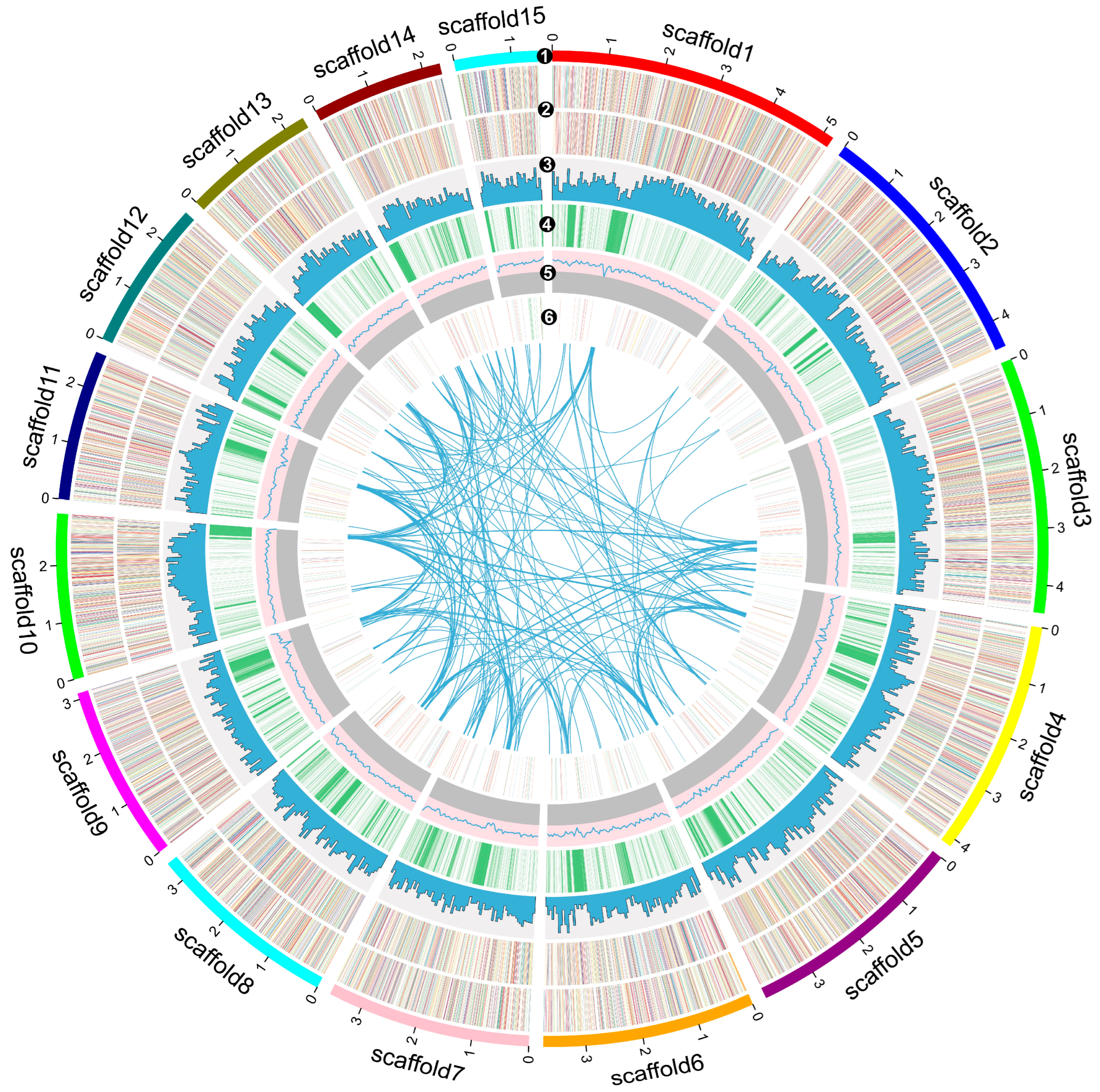
3.2. Genome Assembly, Prediction, and Annotation of Cyclocybe chaxingu

Considering all statistical indicators (Table S1), the assembly quality of C27 is superior, and it was ultimately chosen as the reference genome for this study. In the genome sequencing and assembly of C27, Illumina sequencing yielded 48,783,366 clean reads (~145× coverage), which were used for k-mer analysis and genome polishing. A sequencing depth distribution histogram (k = 19) was generated using GenomeScope (Figure S1). A single k-mer coverage peak was observed, with a heterozygosity rate of 0.015%, confirming that strain C27 is a monokaryotic strain.
Approximately 544,901 clean PacBio reads (~200× coverage) were reassembled into 20 scaffolds, with an N50 of 3.75 Mbp and an N90 of 2.37 Mbp. The total sequence length was 50,786,201 bp, and the maximum scaffold length was 5.19 Mbp (Figure 2). The GC content of the Cyclocybe chaxingu genome is 50.90%. Genome completeness was assessed using BUSCO, which determined a completeness of 98.5%. The scaffold lengths range from 0.04 Mb to 5.19 Mb. The genome size of C. chaxingu is similar to other species in the Cyclocybe genus and related genera Agrocybe and Stropharia. Its genome size is slightly larger than Cyclocybe aegerita [19], Agrocybe pediades (GCA_013053245.1), and Stropharia rugosoannulata [17], but smaller than Agrocybe cylindracea [20] and Agrocybe praecox (GCA_964017145.1). Compared to more distantly related species, its genome size is slightly larger than Lyophyllum decastes [16] and Lentinula edodes [14], but smaller than Flammulina velutipes [13] and Schizophyllum commune [11]. Compared to the two T2T chromosome-level haploid genomes of Agrocybe chaxingu assembled by Chen et al. [32], CchA (50.60 Mb, N50: 3.95 Mb, GC content: 50.99%) and CchB (51.66 Mb, N50: 3.97 Mb, GC content: 50.98%), each containing 13 chromosomes, our results align well in terms of total sequence length and GC content, confirming the accuracy of our sequencing and assembly.
Based on homology comparison and ab initio gene prediction, RepeatMasker identified 8,195,452 bp of repetitive sequences, accounting for 16.14% of the C27 genome. Most repetitive sequences were retroelements (1.88%), including 1.78% LTR retrotransposons, 0.01% SINEs, and 0.09% LINEs. Additionally, DNA transposons accounted for 0.22%. Furthermore, 18 satellite sequences totaling 1980 bp were predicted in the C. chaxingu genome (Table S2).
Genome annotation and comparison analysis predicted a total of 253 copies of various non-coding RNAs, primarily tRNAs, with 201 copies accounting for 0.0328% of the genome. The C27 genome predicted 15,925 genes with an average length of 1742.5 bp, with coding genes totaling 27.75 Mb, representing 54.64% of the entire genome. On average, each gene contained 5.2 exons (Table S2). Of the identified genes, 14,924 (93.7%), 10,691 (67.1%), 7092 (44.5%), 8586 (53.9%), 6896 (43.3%), and 3555 (22.3%) were annotated in the NCBI Nr (Table S3, Figure S2), EggNOG (Table S4, Figure S3), SwissProt (Table S5, Figure S5), Pfam (Table S6), GO (Table S7, Figure S4), and KEGG (Table S8, Figure S5) databases, respectively. The BUSCO v5.4.5 assessed the completeness of the C27 genome at 98.5% (Table S1). These results and assembly parameters indicate that we obtained a high-quality C. chaxingu genome. Based on the gene prediction and annotation information of the C27, the Circos v0.69.9 [68] was used to perform circular visualization of the genome information (Figure 2).

3.3. Genome Evolutionary and Comparative Analysis

To further understand the evolutionary relationships between C. chaxingu and other fungal species, we conducted a phylogenetic analysis based on single-copy orthologous genes from the genomes of C. chaxingu and 27 other fungi (24 Basidiomycetes and 4 Ascomycetes, Table S10). Phylogenetic reconstruction and species divergence time estimation were performed using 229 single-copy orthologous genes (Figure 3A). In the phylogenetic tree of fungi, species from the phylum Ascomycota and Basidiomycota exhibit distinct branching patterns, with an estimated divergence time of approximately 6.457 million years, indicating significant evolutionary distances. Within the branches of the Basidiomycota phylogenetic tree, most edible fungi are positioned in the order Agaricales, clustering together in a major branch that highlights their close evolutionary relationships. Orders closely related to Agaricales include Boletales, Polyporales, Auriculariales, and Tremellomycetes, arranged sequentially according to phylogenetic proximity. Each species’ taxonomic position is clearly represented by the branching structure of the phylogenetic tree, revealing their evolutionary relationships and genetic divergence. Among these species, Cyclocybe chaxingu strain C27 and A. cylindracea strain AC9 are grouped within the smallest branch, with an estimated divergence time of 3.1 million years, indicating the closest phylogenetic relationship between the two. In contrast, other strains from the genus Agrocybe, such as A. pediades CBS 102.39 and A. praecox GCA_964017115, form a separate clade, distinct from A. cylindracea strain AC9. Although A. cylindracea was historically classified outside the genus Cyclocybe, its strain AC9 appears to be phylogenetically closer to Cyclocybe species within the tree. This suggests potential synonymy between A. cylindracea strain AC9 and C. chaxingu, warranting further taxonomic examination. Moreover, C. chaxingu and C. aegerita exhibit the closest phylogenetic relationship, with species divergence estimated to have occurred approximately 13.6 million years ago. Among species closely related to C. chaxingu, strains such as A. pediades CBS 102.39, A. praecox GCA_964017115, S. rugosoannulata strain A15, and Pholiota microspora strain MG134 all belong to the family Strophariaceae. These strains cluster within a single branch, reflecting close evolutionary relationships. In comparison, strains related to the genus Cyclocybe are located adjacent to the Strophariaceae branch, suggesting a degree of phylogenetic proximity among these taxa. In summary, species of the genus Cyclocybe are distinctly separated from the family Strophariaceae, forming their own distinct cluster. These findings align with previous phylogenetic studies based on ribosomal (ITS, LSU) and protein-coding (TEF1α, RPB2) DNA sequences [69].
Collinearity analysis (Figure 3B) indicates a high degree of collinearity between C. chaxingu and both S. rugosoannulata and A. pediades, suggesting that they have retained a large portion of their similar genome structures during evolution, indicating a recent common ancestor. Conversely, a more complex collinearity network exists between C. chaxingu and both L. decastes and L. edodes, implying more frequent gene rearrangement events during their evolutionary processes, thus further influencing their evolutionary trajectories. Additionally, the connections among species suggest that scaffolds 10 and 14, as well as scaffolds 13 and 15 of C. chaxingu, are likely located on the same chromosome. The connections of these scaffolds can be further validated using PCR or Hi-C techniques [70], indicating that the genome sequencing and assembly of strain C27 require further precision improvement.
ANI values are commonly used for species identification at the genome level in prokaryotes [64], with ANI values greater than 95% identifying the same species [71]. Here, we used ANI values to reflect the genetic relationship between Cyclocybe and Agrocybe strains. We extracted genomes of four Agrocybe strains and one Cyclocybe strain from the NCBI genome database for comparison with the two haploid genomes of C. chaxingu assembled in this study. The ANI value between C. chaxingu strain C27 and C. chaxingu strain C5 was 99.22%, showing the highest similarity. ANI values among C. chaxingu, Agrocybe chaxingu strain MP-N11, and A. cylindracea strain AC9 were all greater than 96%, indicating close intra-species taxonomic relationships. This suggests that A. chaxingu strain MP-N11, A. cylindracea strain AC9, and C. chaxingu strain C27 are synonyms (Figure 3C). Based on the phylogenetic analysis of single-copy orthologous genes, we think that A. chaxingu strain MP-N11 and A. cylindracea strain AC9 in this study should be classified under the genus Cyclocybe.

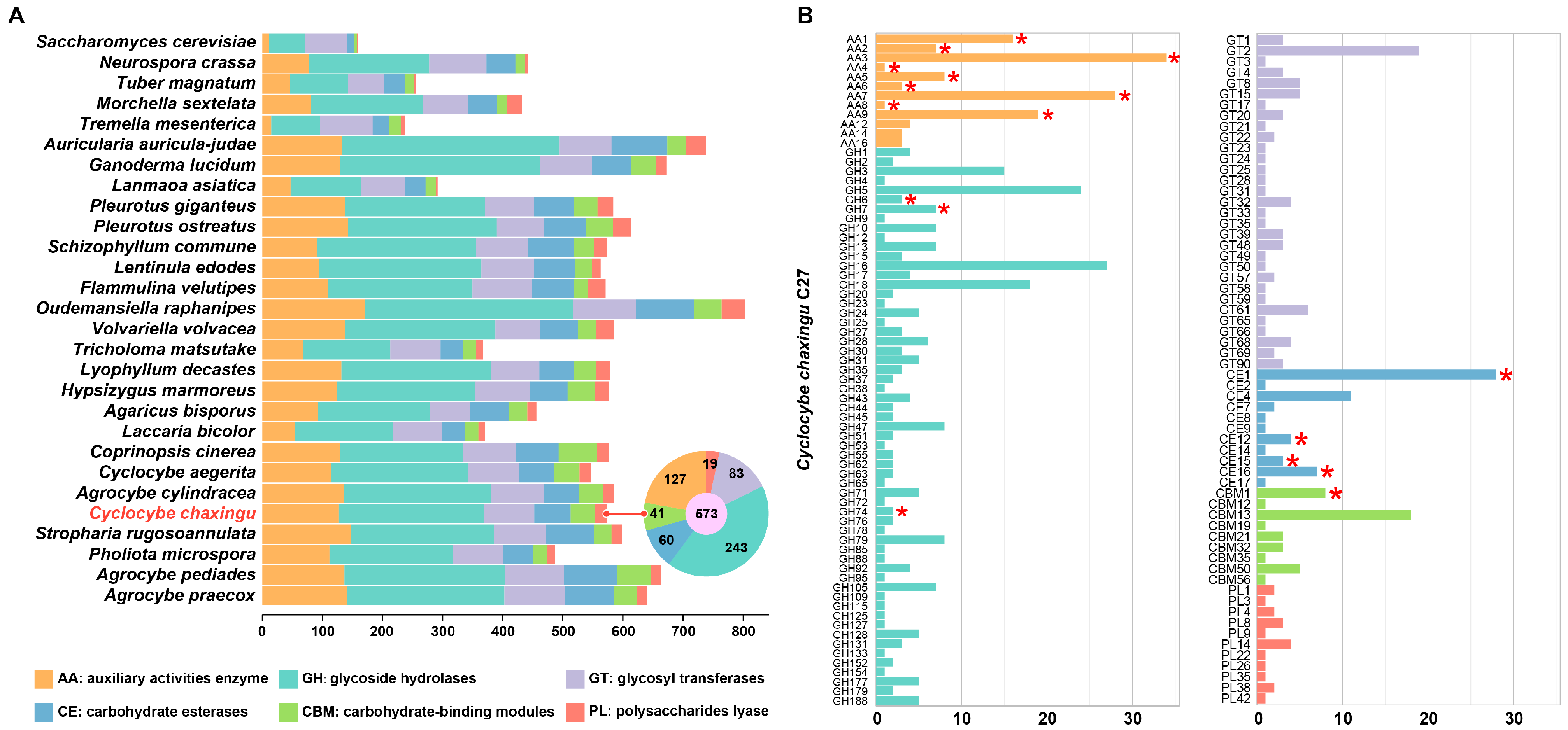
3.4. Carbohydrate Active Enzymes in Cyclocybe chaxingu Genome

CAZymes represent one of the most significant gene families in fungal genomes, encompassing a variety of functions [72]. They are involved not only in carbohydrate metabolism and lignocellulose degradation but also in numerous cellular biological processes such as development and stress response [73]. According to the dbCAN2 database, CAZymes are classified into six categories: glycosyltransferases (GTs), glycoside hydrolases (GHs), polysaccharide lyases (PLs), carbohydrate esterases (CEs), auxiliary activities (AAs), and carbohydrate-binding modules (CBMs) [57]. Utilizing the online annotation platform dbCAN, a total of 407 enzymes were identified in the C. chaxingu genome, comprising 573 CAZyme entries, including 127 AAs (22.2%), 243 GHs (42.4%), 83 GTs (14.5%), 60 CEs (10.5%), 41 CBMs (7.2%), and 19 PLs (3.3%) (Figure 4B, Table S9).
In the context of wood degradation-related genes, Floudas et al. [74] identified 17 CAZyme families, which are all present in C. chaxingu strain C27, encompassing a total of 179 genes (Figure 4A). Within these families, auxiliary activity (AA) families are predominantly distributed across AA3, AA7, AA9, AA1, AA5, and AA2 (Figure 4A). Specifically, proteins within families AA3 and AA9 are involved in the degradation of cellulose and hemicellulose, while laccases (AA1 family) and class II peroxidases (AA2 family) play key roles in lignin degradation within lignocellulose [75]. The class II lignin-degrading peroxidases are also considered markers for distinguishing between white-rot and brown-rot fungi. AA2 genes were possessed by more than half of the brown-rot fungi, mainly Polyporales, but the number of copies varied greatly, with white-rot fungi possessing on average about 10 times as many AA2 genes as brown-rot fungi. [76]. These enzymes play critical roles in the degradation of plant cell wall components [77]. The results indicate that this strain possesses a robust capacity for lignocellulose degradation, consistent with the characteristic features of C. chaxingu as a typical wood-decaying fungus.
Genes from the GH families are crucial for the degradation of cellulose and hemicellulose [78]. Studies have shown that they also play a significant role in the fruiting body development and senescence of Volvariella volvacea [79]. The top four GH families (27 GH16 genes, 24 GH5 genes, 18 GH18 genes, and 15 GH3 genes) have been identified as involved in the degradation of cellulose and hemicellulose, demonstrating the efficiency of this process in C. chaxingu. Among GT families, the GT2 family has the largest number of enzymes (19). GT2 family enzymes can catalyze the formation of glycosidic bonds, transferring sugars from activated donor molecules to acceptor substrates, which may include polysaccharides, peptides, lipids, or small molecules. This process is integral to the biosynthesis of complex carbohydrate compounds and contributes to the structural diversity of polysaccharides [80]. GT2 family enzymes, such as glucosyltransferase, mannosyltransferase, cellulose synthase, and chitin synthase, are involved in the polymerization and secretion of polysaccharides. These enzymes play a crucial role in the formation of glycosidic bonds and the biosynthesis of carbohydrate compounds, enhancing the complexity and diversity of polysaccharides [81], which likely reflects its significant role in polysaccharide biosynthesis and structural diversity in fungal systems.
The CAZymes of 29 fungi were analyzed using the same parameters with the dbCAN2 database. The number of CAZymes in C. chaxingu is comparable to those in related genera such as Schizophyllum and Flammulina, ranking in the upper-middle range among the listed species. Oudemansiella raphanipes (803), Auricularia auricula-judae (738), and Ganoderma lucidum (673) exhibit higher CAZyme numbers, whereas Saccharomyces cerevisiae (159), Tuber magnatum (256), and Tremella mesenterica (237) had relatively fewer (Figure 4B, Table S11).
Based on the conclusions from comparative genomic analysis, we consider A. cylindracea strain AC9 and A. chaxingu strain MP-N11 to belong to the Cyclocybe genus. Whether comparing the four strains within the genus (C. aegerita strain AAE3, A. cylindracea strain AC9, A. chaxingu strain MP-N11, and C. chaxingu strain C27) or the same species (A. chaxingu strain MP-N11 and C. chaxingu strain C27), their CAZyme family numbers and distributions are roughly similar. Considering the varying assembly quality and completeness levels of the genomes, we think that the four Cyclocybe strains in this study show no significant differences in CAZyme composition. When comparing with closely related genera, the two Agrocybe strains (Agrocybe pediades strain CBS_102.39 and Agrocybe praecox strain GCA_964017145.1) have relatively higher gene counts in the AA, GH, GT, and CE families and also a higher total number of CAZyme genes compared to Cyclocybe strains. This indicates that the two Agrocybe strains possess higher metabolic potential in terms of CAZyme gene diversity and quantity. These results reflect the genomic differences between Cyclocybe and Agrocybe genera and further support the classification results from comparative genomic analysis.

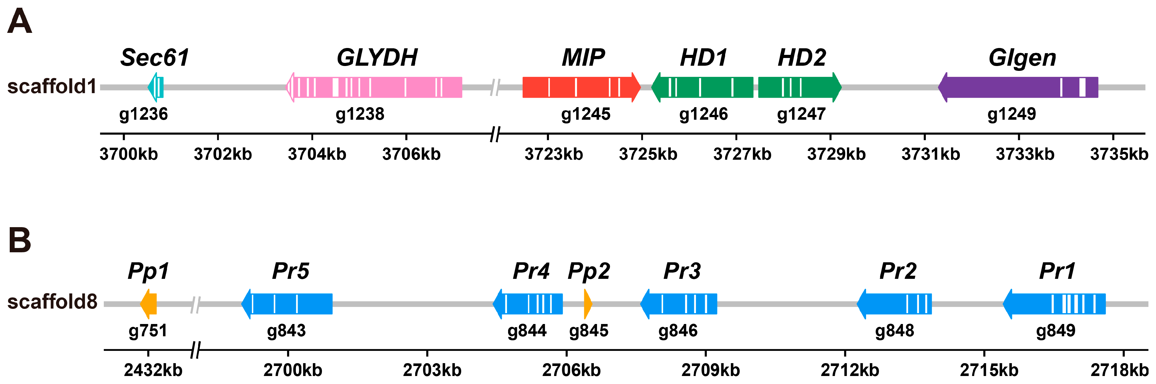
3.5. Identification of the Mating Locus

We identified two mating-type genes, HD1 and HD2, in MatA, each with homeobox domains, spanning a physical distance of 4207 bp in the genome. HD1 is located 239 bp downstream of the MIP gene, with HD1 and HD2 transcribed in opposite directions and separated by 130 bp (Figure 5A). HD1 consists of 641 amino acids with a genomic length of 2148 bp, including 5 exons and 4 introns, while HD2 consists of 617 amino acids with a genomic length of 2029 bp, including 4 exons and 3 introns (Figure 5A). Upstream of the MIP gene, 2 kb away, we identified the Sec61 gene and GLYDH gene. The Sec61 protein complex forms transmembrane channels that transport proteins into the endoplasmic reticulum membrane for further modification and localization, playing a crucial role in protein transport and secretion [82]. GLYDH catalyzes the oxidation of glycine to pyruvate, converting NAD+ to NADH [83]. These genes are likely essential components for the function of mating-type genes. Additionally, we identified a Glgen gene located 2 kb downstream of the HD2 gene. Unlike the typical MIP-HD-β-fg collinearity structure in basidiomycetes, the β-fg gene in C. chaxingu C27 is located on scaffold 9.g357 and is not linked to the MatA.
MatB contains 5 Pr and 2 Pp2, all clustered within an approximately 2 kb gene cluster with a total sequence length of 18,596 bp. This is identical to the sequence of Cyclocybe aegerita YSG-M2 monokaryotic strain (GenBank ID: MG737348) studied by Chen et al. [27]. Additionally, a pheromone precursor gene (Pp1) was identified 38 kb upstream of the 2 kb gene cluster (Figure 5B). Despite the long distance, it may interact with the downstream 2 kb gene cluster through unknown regulatory factors.
The MatA of C. chaxingu C27 closely aligns with that of A. chaxingu AS-5, as previously described by Chen et al. [32], indicating conserved genetic features within these strains. However, differences were observed in the number and distribution of Pp genes within the MatB, which may reflect unique genetic characteristics of the strains. These discrepancies could be attributed to the genome of C. chaxingu C27 not being assembled at the chromosomal level, potentially limiting the precision of gene predictions. Alternatively, they may be the result of genetic variation between the two strains, highlighting evolutionary diversification within the species. This result is significant as it underscores the potential for strain-specific adaptations and provides a foundation for further exploration of mating-type gene architecture in Cyclocybe species.

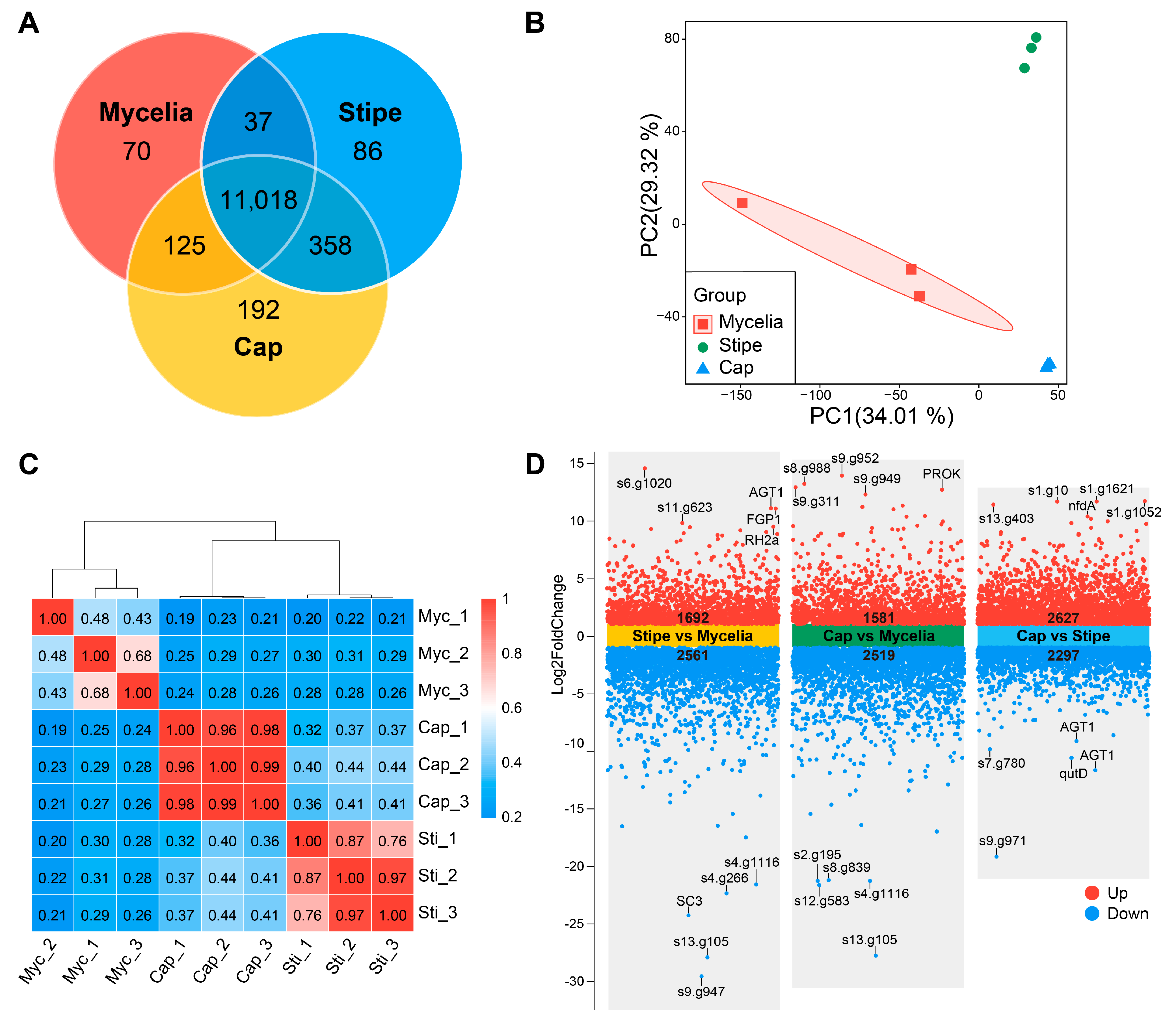
3.6. Transcriptome Data Analysis

Transcriptome sequencing of the 9 samples generates 424,331,642 raw reads, with Q30 scores ranging from 94.84% to 96.82%, indicating high sequencing quality. After filtering, the total number of clean reads is 416,400,884 (97.79%). The mapping rates to the reference genome ranged from 72.40% to 77.19%. The GC content is approximately 54%, which is consistent across all samples (Table S12). These data indicate that the transcriptome sequencing data obtained are of high quality, providing a reliable basis for subsequent differential gene expression analysis and functional annotation.

3.7. Differential Expression and Gene Enrichment Analysis

After aligning the clean reads to the reference genome, a gene expression matrix was obtained (Table S13). The Venn diagram shows differences in gene expression among the mycelia, stipe, and cap of C. chaxingu (Figure 6A). The cap has the highest number (192) of uniquely expressed genes, followed by stipe (82) and mycelia (70). Additionally, 11,018 genes are commonly expressed across all three parts, suggesting strong conservation and importance of these genes in the life cycle of C. chaxingu. Using the normalized gene expression matrix, principal component analysis (PCA) and correlation analyses were performed to assess inter-group sample differences and intra-group sample replicates. The PCA indicate significant gene expression differences between different tissues (Figure 6B). The results of the Pearson correlation and hierarchical clustering further show low inter-group sample correlation and high intra-group replicate correlation, with the three sample groups being well distinguished (Figure 6C). These findings reveal the complexity and diversity of gene expression in different tissues of the mushroom, providing a foundation for further understanding its developmental mechanisms. It is noteworthy that the correlation between the three mycelia samples is slightly lower than that of the stipe and cap groups. The sequencing depth (i.e., the number of raw reads and clean reads) of the mycelia samples varies more compared to the stipe and cap samples, with mycelia_3 having significantly higher sequencing depth than the other two mycelia samples. Additionally, the GC content of the mycelia samples (54.52% to 54.88%) varies more compared to the stipe samples (53.85% to 54.01%) and cap samples (53.68% to 53.82%) (Table S12). The differences in sequencing depth among the mycelia samples may lead to varying sensitivity and accuracy in detecting gene expression levels, affecting the PCA and correlation analysis results.
To study the differential gene expression patterns in different tissues of C. chaxingu, pairwise comparisons were conducted between mycelia, stipe, and cap (Table S14). The stipe vs. mycelia comparison group identified 4253 DEGs, with 1692 upregulated and 2561 downregulated. The cap vs. mycelia group identified 4100 DEGs, including 1581 upregulated and 2519 downregulated genes. The cap vs. stipe group identified 4924 DEGs, including 2627 upregulated and 2297 downregulated genes (Figure 6D).
The volcano plot (Figure 6D) illustrates the differential gene expression across three comparison groups: stipe vs. mycelia, cap vs. mycelia, and cap vs. stipe. Notably, the AGT1 gene is significantly upregulated in the stipe vs. mycelia comparison and significantly downregulated in the cap vs. stipe comparison, indicating its pronounced expression in the stipe. AGT1 gene encodes the Ageritin protein, a specific ribonuclease from the edible mushroom Cyclocybe aegerita (synonym Agrocybe aegerita). Ageritin functions by cleaving a single phosphodiester bond within the universally conserved alpha-sarcin loop (SRL) of 23–28S rRNAs. This cleavage inhibits protein biosynthesis, leading to cell apoptosis and subsequent cell death. The structural and enzymatic properties of Ageritin reveal that it is the prototype of a novel family of specific ribonucleases known as ‘ribotoxin-like proteins’ [84]. This protein has also been found in other edible mushrooms such as Pleurotus ostreatus [85], Boletus edulis [86], and Calocybe gambosa [84], where its strong antifungal and insecticidal activities robustly support its defensive role. The specific expression pattern of Ageritin protein in the stipe likely plays a crucial role in protecting this part from pathogens and pests. The SC3 gene is significantly downregulated in the stipe vs. mycelia group, indicating its prominent expression in the mycelia. SC3 encodes a hydrophobin protein involved in the formation of aerial hyphae [87]. The primary task of mycelia is nutrient absorption and transport, necessitating abundant aerial hyphae to increase surface area. The upregulation of SC3 in the mycelia supports this function, whereas the stipe, which primarily serves structural support and nutrient distribution, relies less on SC3 expression. Furthermore, the s4.g1116 and s13.g105 genes are significantly upregulated expression in the mycelia, but their specific functions require further exploration.
To investigate the biological functions of the relevant DEGs, GO enrichment and KEGG enrichment analysis was performed for the DEGs in different comparison groups. As shown in Figure 7 and Figure 8, in the GO enrichment analysis, the stipe vs. mycelia and cap vs. mycelia comparison groups were predominantly enriched in cellular component (CC) and molecular function (MF) categories, while the cap vs. stipe comparison group was mainly enriched in biological process (BP) categories. In the KEGG enrichment analysis, the stipe vs. mycelia and cap vs. mycelia comparison groups were predominantly enriched in metabolism, while the cap vs. stipe comparison group was mainly enriched in metabolism and genetic information processing.
DEGs in the stipe vs. mycelia comparison group were significantly enriched in several key areas such as membrane part, ribosome, extracellular region, external encapsulating structure, and cell wall. As a supporting structure of the fruiting body, the stipe often requires a sturdy cell wall and membrane structure to provide structural support [88]. Additionally, the high expression of oxidoreductase activity, cofactor binding, iron ion binding, and transmembrane transport indicate active metabolic processes and structural maintenance essential for stipe development [89]. The KEGG enrichment analysis further supported these findings, showing significant enrichment in metabolism pathways such as amino sugar and nucleotide sugar metabolism, pyruvate metabolism, tryptophan metabolism, glycolysis/gluconeogenesis, and purine metabolism. In genetic information processing, the enrichment of DEGs in ribosome, base excision repair, and mismatch repair pathways highlights the stipe’s need for robust protein synthesis and DNA repair mechanisms, ensuring proper cellular function and resilience against genetic damage [90].
In the cap vs. mycelia comparison group, DEGs were enriched in similar cellular component and molecular function categories in GO enrichment, reflecting the cap’s complexity in membrane and cell wall structures essential for protecting and releasing spores. The active catalytic and oxidoreductase functions suggest intensive metabolic activities in the cap. The KEGG enrichment analysis revealed that DEGs were enriched in metabolic pathways such as pyruvate metabolism, tryptophan metabolism, glycolysis/gluconeogenesis, and starch and sucrose metabolism. Enrichment in pathways such as methane metabolism, tyro-sine metabolism, and penicillin and cephalosporin biosynthesis indicate the cap’s role in secondary metabolite production, which may contribute to defense mechanism [91]. In genetic information processing, the enrichment in mismatch repair and homologous recombination pathways suggests active DNA repair processes in the cap to ensure genomic stability during spore formation [90]. The involvement of the cell cycle and meiosis pathways in cellular processes further supports the cap’s role in reproductive activities [92]. The enrichment of these metabolic pathways demonstrates the diverse functions of the cap in energy generation, gene expression regulation, and maintenance of cell function. These functions play a crucial role in the cap’s protection and release of spores.
For the cap vs. stipe comparison group, DEGs were significantly enriched in multiple GO and KEGG pathways related to DNA replication and repair. Specifically, GO enrichment analysis revealed that differential genes were predominantly enriched in biological processes such as DNA repair, DNA packaging, and protein–DNA complex assembly. These processes are crucial for ensuring the accuracy of DNA replication and repairing damage. Concurrently, KEGG enrichment analysis indicated that differential genes were significantly enriched in pathways including DNA replication, nucleotide excision repair, base excision repair, mismatch repair, homologous recombination, and non-homologous end-joining. These enriched pathways underscore the cap’s critical requirement for precise and efficient DNA replication and repair mechanisms during spore generation and release, thereby maintaining genomic integrity and ensuring successful reproduction [93].

4. Conclusions

In this study, we used the type strain JAUCC 1847 to conduct the first comprehensive genomic and transcriptomic analyses of C. chaxingu. Our findings provide valuable insights into the biological characteristics and genetic composition of this species, aiding its classification and potential applications in biotechnology. The genome assembly of the haploid strain C27 exhibited high completeness and continuity, with a total length of 50.79 Mb and a GC content of 50.90%. Comparative genomic analysis revealed a close evolutionary relationship between C. chaxingu and species from the genera Agrocybe and Stropharia, suggesting a recent common ancestor.
The identification of 573 CAZyme genes underscores the metabolic diversity of C. chaxingu, particularly in biodegradation and biotransformation processes, especially in lignocellulose degradation. This extensive library of carbohydrate-active enzymes not only highlights its substantial potential for industrial applications but also provides new perspectives and possibilities for biotechnology and environmental science. Furthermore, the identification of MatA and MatB characterization of mating genes deepened our understanding of the genetic traits of C. chaxingu. The presence of MatA and MatB with specific gene arrangements and sequences indicates potential evolutionary conservation and provides insights into the genetic basis of mating compatibility and sexual reproduction in this species.
Transcriptomic analysis uncovered distinct gene expression patterns across the mycelia, stipe, and cap of C. chaxingu, reflecting the functional specialization of these tissues. The mycelia showed upregulation of genes related to aerial hyphae formation, highlighting its role in vegetative growth and adaptation to environmental conditions by facilitating nutrient absorption and surface colonization. The stipe is primarily associated with structural integrity and material transport, while the cap exhibits metabolic activity and gene regulation. Additionally, the differential stress responses and DNA repair mechanisms underscore the adaptive features of the cap. These insights provide valuable clues for understanding the growth and development mechanisms of edible fungi, showcasing the complex interplay of structural, metabolic, and regulatory processes across different fungal tissues.
Importantly, our study addressed the previous lack of comprehensive genomic data for C. chaxingu, which had limited its acceptance as a distinct species. By providing detailed genomic and transcriptomic information, we clarified the taxonomic status of C. chaxingu, supporting its recognition and classification within the genus Cyclocybe. This comprehensive dataset serves as a valuable resource for future research and breeding programs aimed at improving the yield and quality of this commercially significant fungus.
Additionally, the high similarity between C. chaxingu and other species within the genus warrants further research. Future studies may focus on comparative genomics and functional analysis to unravel these potential processes. Our research not only provides a detailed genetic and functional blueprint for C. chaxingu but also lays the groundwork for further exploration of its biological, taxonomic, and industrial potential. These insights can pave the way for innovative applications in biotechnology and a deeper understanding of fungal evolution and diversity. By supplementing previously incomplete genomic data, our study establishes C. chaxingu as a well-defined species within the genus Cyclocybe, providing a solid foundation for its acceptance and further research. This work not only advances our understanding of C. chaxingu but also contributes to the broader field of fungal genomics and biotechnology.

Supplementary Materials

The following supporting information can be downloaded at: https://www.mdpi.com/article/10.3390/horticulturae11040409/s1, Figure S1: 19 k-mer depth distribution; Figure S2: NR.species of C. chaxingu C27 genome; Figure S3: EggNOG function classification of C. chaxingu C27 genome; Figure S4: GO function classification of C. chaxingu C27 genome; Figure S5: KEGG function classification of C. chaxingu C27 genome; Table S1: De novo genome assembly features of monokaryotic C. chaxingu; Table S2: Characteristics of genome prediction and annotation of C. chaxingu C27; Tables S3–S9: Genome annotation of C. chaxingu C27 by NR, EggNOG, Swiss-Prot, Pfam, GO, KEGG and CAZymes databases; Table S10: Genomic information of fungal species used in the study; Table S11: Distribution and number of CAZymes genes in C. chaxingu C27 and 28 other fungi. Table S12: Statistical analysis of transcriptome sequencing data; Table S13: Gene counts in different tissues of C. chaxingu; Table S14: Differential gene expression analysis of C. chaxingu in various comparison groups.

Author Contributions

Conceptualization, D.H. and Y.G.; Methodology, D.H., Y.G., H.S. and X.W.; Investigation, Q.Y., H.S. and G.S.; Resources, X.W., H.H., Z.Z., M.C., J.Z. and H.Y.; Formal analysis, Q.Y.; Data curation, H.S.; Visualization, Q.Y., Validation, H.S.; Writing—original draft preparation, Q.Y. and H.S.; Writing—review and editing, Y.G.; Supervision, D.H., Y.G. and X.W.; Funding acquisition, D.H. and Y.G. All authors have read and agreed to the published version of the manuscript.

Funding

This work was supported by the National Science Foundation of China (32160010, 32160673), the Talents Support Project of Jiangxi Province-Training Program for Academic and Technical Leaders in Major Disciplines (20232BCJ22019), the Key Research and Development Program Project of Jiangxi Province (20243BBH81011), and the Natural Science Foundation of Jiangxi Province, China (20242BAB25378).

Data Availability Statement

The raw sequences of genome assembly and transcriptomes were submitted to NGDC (https://ngdc.cncb.ac.cn/, accessed on 6 February 2025) BioProject under accession number PRJCA035577. The annotated genome was submitted to CNCB GWH under accession number GWHFJIP00000000.1.

Conflicts of Interest

The authors declare no conflicts of interest.

References

  1. Liu, Q.; Song, H.; Chen, R.; Chen, M.; Zhai, Z.; Zhou, J.; Gao, Y.; Hu, D. Species concept of Cyclocybe chaxingu, an edible mushroom cultivated in China. Mycosystema 2021, 40, 981–991. [Google Scholar] [CrossRef]
  2. Liu, Q.; Hua, R.; Zhang, J.; Li, J.; Shang, L.; Luo, X.; Liu, S. Biological characteristics, genetic breeding and cultivation situation of Cyclocybe chaxingu. Edible Med. Mushrooms 2021, 29, 501–505. [Google Scholar]
  3. Liu, Q.; Liu, Y.; Chen, M.; Zhai, Z.; Zhou, J.; Hu, D. Study on the Systematics of the Genus Agrocybe. Edible Fungi China 2020, 39, 1–7+16. [Google Scholar] [CrossRef]
  4. Xu, J.; Wang, S.; Gong, X.; Yu, R.; Wang, L. Composition analysis of Agrocybe cylindracea extract. Sci. Technol. Food Ind. 2006, 27, 165–167. [Google Scholar] [CrossRef]
  5. Lee, K.J.; Yun, I.J.; Kim, K.H.; Lim, S.H.; Ham, H.J.; Eum, W.S.; Joo, J.H. Amino acid and fatty acid compositions of Agrocybe chaxingu, an edible mushroom. J. Food Compos. Anal. 2011, 24, 175–178. [Google Scholar] [CrossRef]
  6. Zhao, H.; Li, J.; Zhang, J.; Wang, X.; Hao, L.; Jia, L. Purification, in vitro antioxidant and in vivo anti-aging activities of exopolysaccharides by Agrocybe cylindracea. Int. J. Biol. Macromol. 2017, 102, 351–357. [Google Scholar] [CrossRef] [PubMed]
  7. Kiho, T.; Yoshida, I.; Nagai, K.; Ukai, S.; Hara, C. (1→3)-α-d-glucan from an alkaline extract of Agrocybe cylindracea, and antitumor activity of its O-(carboxymethyl)ated derivatives. Carbohydr. Res. 1989, 189, 273–279. [Google Scholar] [CrossRef]
  8. Morin, E.; Kohler, A.; Baker, A.R.; Foulongne-Oriol, M.; Lombard, V.; Nagy, L.G.; Ohm, R.A.; Patyshakuliyeva, A.; Brun, A.; Aerts, A.L.; et al. Genome sequence of the button mushroom Agaricus bisporus reveals mechanisms governing adaptation to a humic-rich ecological niche. Proc. Natl. Acad. Sci. USA 2012, 109, 17501–17506. [Google Scholar] [CrossRef]
  9. Martin, F.; Aerts, A.; Ahren, D.; Brun, A.; Danchin, E.G.J.; Duchaussoy, F.; Gibon, J.; Kohler, A.; Lindquist, E.; Pereda, V.; et al. The genome of Laccaria bicolor provides insights into mycorrhizal symbiosis. Nature 2008, 452, 88–92. [Google Scholar] [CrossRef]
  10. Stajich, J.E.; Wilke, S.K.; Ahrén, D.; Au, C.H.; Birren, B.W.; Borodovsky, M.; Burns, C.; Canbäck, B.; Casselton, L.A.; Cheng, C.K.; et al. Insights into evolution of multicellular fungi from the assembled chromosomes of the mushroom Coprinopsis cinerea (Coprinus cinereus). Proc. Natl. Acad. Sci. USA 2010, 107, 11889–11894. [Google Scholar] [CrossRef]
  11. Ohm, R.A.; de Jong, J.F.; Lugones, L.G.; Aerts, A.; Kothe, E.; Stajich, J.E.; de Vries, R.P.; Record, E.; Levasseur, A.; Baker, S.E.; et al. Genome sequence of the model mushroom Schizophyllum commune. Nat. Biotechnol. 2010, 28, 957–963. [Google Scholar] [CrossRef]
  12. Yu, H.; Zhang, M.; Sun, Y.; Li, Q.; Liu, J.; Song, C.; Shang, X.; Tan, Q.; Zhang, L.; Yu, H. Whole-genome sequence of a high-temperature edible mushroom Pleurotus giganteus (zhudugu). Front. Microbiol. 2022, 13, 941889. [Google Scholar] [CrossRef] [PubMed]
  13. Park, Y.; Baek, J.H.; Lee, S.; Kim, C.; Rhee, H.; Kim, H.; Seo, J.; Park, H.; Yoon, D.; Nam, J.; et al. Whole genome and global gene expression analyses of the model mushroom Flammulina velutipes reveal a high capacity for lignocellulose degradation. PLoS ONE 2014, 9, e93560. [Google Scholar] [CrossRef] [PubMed]
  14. Chen, L.; Gong, Y.; Cai, Y.; Liu, W.; Zhou, Y.; Xiao, Y.; Xu, Z.; Liu, Y.; Lei, X.; Wang, G.; et al. Genome Sequence of the Edible Cultivated Mushroom Lentinula edodes (Shiitake) Reveals Insights into Lignocellulose Degradation. PLoS ONE 2016, 11, e0160336. [Google Scholar] [CrossRef]
  15. Zhu, L.; Gao, X.; Zhang, M.; Hu, C.; Yang, W.; Guo, L.; Yang, S.; Yu, H.; Yu, H. Whole Genome Sequence of an Edible Mushroom Oudemansiella raphanipes (Changgengu). J. Fungi 2023, 9, 266. [Google Scholar] [CrossRef] [PubMed]
  16. Xu, L.; Yang, W.; Qiu, T.; Gao, X.; Zhang, H.; Zhang, S.; Cui, H.; Guo, L.; Yu, H.; Yu, H. Complete genome sequences and comparative secretomic analysis for the industrially cultivated edible mushroom Lyophyllum decastes reveals insights on evolution and lignocellulose degradation potential. Front. Microbiol. 2023, 14, 1137162. [Google Scholar] [CrossRef]
  17. Li, S.; Zhao, S.; Hu, C.; Mao, C.; Guo, L.; Yu, H.; Yu, H. Whole Genome Sequence of an Edible Mushroom Stropharia rugosoannulata (Daqiugaigu). J. Fungi 2022, 8, 99. [Google Scholar] [CrossRef]
  18. Hess, J.; Skrede, I.; Wolfe, B.E.; Labutti, K.; Ohm, R.A.; Grigoriev, I.V.; Pringle, A. Transposable element dynamics among asymbiotic and ectomycorrhizal Amanita fungi. Genome Biol. Evol. 2014, 6, 1564–1578. [Google Scholar] [CrossRef]
  19. Gupta, D.K.; Rühl, M.; Mishra, B.; Kleofas, V.; Hofrichter, M.; Herzog, R.; Pecyna, M.J.; Sharma, R.; Kellner, H.; Hennicke, F.; et al. The genome sequence of the commercially cultivated mushroom Agrocybe aegerita reveals a conserved repertoire of fruiting-related genes and a versatile suite of biopolymer-degrading enzymes. BMC Genom. 2018, 19, 48. [Google Scholar] [CrossRef]
  20. Liang, Y.; Lu, D.; Wang, S.; Zhao, Y.; Gao, S.; Han, R.; Yu, J.; Zheng, W.; Geng, J.; Hu, S. Genome Assembly and Pathway Analysis of Edible Mushroom Agrocybe cylindracea. Genom. Proteom. Bioinform. 2020, 18, 341–351. [Google Scholar] [CrossRef]
  21. Duan, Y.; Han, H.; Qi, J.; Gao, J.; Xu, Z.; Wang, P.; Zhang, J.; Liu, C. Genome sequencing of Inonotus obliquus reveals insights into candidate genes involved in secondary metabolite biosynthesis. BMC Genom. 2022, 23, 314. [Google Scholar] [CrossRef] [PubMed]
  22. Heitman, J.; Kronstad, J.W.; Taylor, J.W.; Casselton, L.A. Sex in Fungi: Molecular Determination and Evolutionary Implications; American Association for Microbiology Press: Washington, DC, USA, 2007; ISBN 1555814212. [Google Scholar]
  23. Wang, G.; Wang, Y.; Chen, L.; Wang, H.; Guo, L.; Zhou, X.; Dou, M.; Wang, B.; Lin, J.; Liu, L.; et al. Genetic structure and evolutionary diversity of mating-type (MAT) loci in Hypsizygus marmoreus. IMA Fungus 2021, 12, 35. [Google Scholar] [CrossRef] [PubMed]
  24. Kües, U.; Casselton, L.A. Homeodomains and regulation of sexual development in basidiomycetes. Trends Genet. 1992, 8, 154. [Google Scholar] [CrossRef]
  25. Bao, D. Research progress on the mating-typing locus structures of basidiomycete mushrooms. Mycosystema 2019, 38, 2061–2077. [Google Scholar] [CrossRef]
  26. Shang, J.; Hou, D.; Li, Y.; Zhou, C.; Guo, T.; Tang, L.; Mao, W.; Chen, Q.; Bao, D.; Yang, R. Analyses of mating systems in Stropharia rugosoannulata based on genomic data. Mycosystema 2020, 39, 1152–1161. [Google Scholar] [CrossRef]
  27. Chen, W.; Chai, H.; Yang, W.; Ma, Y.; Zhao, Y. Characterization of B Mating Loci Identification and Application in Cyclocybe aegerita and C. salicacola. Biotechnol. Bull. 2021, 37, 57–64. [Google Scholar] [CrossRef]
  28. Chen, W.; Chai, H.; Yang, W.; Zhang, X.; Chen, Y.; Zhao, Y. Characterization of Non-coding Regions in B Mating Loci of Agrocybe salicacola Groups: Target Sites for B Mating Type Identification. Curr. Microbiol. 2017, 74, 772–778. [Google Scholar] [CrossRef]
  29. Yang, W.; Zhao, Y.; Chen, Y.; Chen, W. Cloning Analysis of Mitochondrial Intermediate Peptidase Encoding Gene and Its Application on A Mating-locus Identification in Agrocybe aegerita. North. Hortic. 2018, 12, 150–156. [Google Scholar]
  30. Zhu, L.; Qian, R.; Zhao, Y.; Chen, W.; Chen, Y. The Singleness Analysis of Monokaryon from Agrocybe aegerita. Chin. Agric. Sci. Bull. 2019, 35, 68–72. [Google Scholar]
  31. Qian, R.; Chen, W.; Chai, H.; Tao, N.; Zhao, Y.; Chen, Y. B Mating Locus from Agrocybe aegerita: Structural Analysis. Chin. Agric. Sci. Bull. 2021, 37, 14–19. [Google Scholar]
  32. Chen, X.; Wei, Y.; Meng, G.; Wang, M.; Peng, X.; Dai, J.; Dong, C.; Huo, G. Telomere-to-Telomere Haplotype-Resolved Genomes of Agrocybe chaxingu Reveals Unique Genetic Features and Developmental Insights. J. Fungi 2024, 10, 602. [Google Scholar] [CrossRef] [PubMed]
  33. May, T.W. Procedures and timetable for proposals to amend Chapter F of the International Code of Nomenclature for algae, fungi, and plants. IMA Fungus 2020, 11, 21. [Google Scholar] [CrossRef] [PubMed]
  34. Li, L.; Zhang, L.; Shang, X.; Song, C.; Yu, H.; Zhang, M.; Tan, Q. A New Method for Protoplast Preparation in Edible Fungi. Acta Edulis Fungi 2020, 27, 108–114. [Google Scholar] [CrossRef]
  35. Wang, H.; Zhang, L.; Sun, P.; Hu, J.; Chen, X.; Wei, Y. Protoplast regeneration and monokaryotation characteristics of Cyclocybe aegerita. Microbiology+ 2022, 49, 2076–2087. [Google Scholar] [CrossRef]
  36. Porebski, S.; Bailey, L.G.; Baum, B.R. Modification of a CTAB DNA extraction protocol for plants containing high polysaccharide and polyphenol components. Plant Mol. Biol. Rep. 1997, 15, 8–15. [Google Scholar] [CrossRef]
  37. Chen, S.; Zhou, Y.; Chen, Y.; Gu, J. fastp: An ultra-fast all-in-one FASTQ preprocessor. Bioinformatics 2018, 34, i884–i890. [Google Scholar] [CrossRef]
  38. Hu, J.; Wang, Z.; Sun, Z.; Hu, B.; Ayoola, A.O.; Liang, F.; Li, J.; Sandoval, J.R.; Cooper, D.N.; Ye, K.; et al. NextDenovo: An efficient error correction and accurate assembly tool for noisy long reads. Genome Biol. 2024, 25, 107. [Google Scholar] [CrossRef]
  39. Walker, B.J.; Abeel, T.; Shea, T.; Priest, M.; Abouelliel, A.; Sakthikumar, S.; Cuomo, C.A.; Zeng, Q.; Wortman, J.; Young, S.K.; et al. Pilon: An integrated tool for comprehensive microbial variant detection and genome assembly improvement. PLoS ONE 2014, 9, e112963. [Google Scholar] [CrossRef]
  40. Manni, M.; Berkeley, M.R.; Seppey, M.; Simao, F.A.; Zdobnov, E.M. BUSCO Update: Novel and Streamlined Workflows along with Broader and Deeper Phylogenetic Coverage for Scoring of Eukaryotic, Prokaryotic, and Viral Genomes. Mol. Biol. Evol. 2021, 38, 4647–4654. [Google Scholar] [CrossRef]
  41. Scalzitti, N.; Jeannin-Girardon, A.; Collet, P.; Poch, O.; Thompson, J.D. A benchmark study of ab initio gene prediction methods in diverse eukaryotic organisms. BMC Genom. 2020, 21, 293. [Google Scholar] [CrossRef]
  42. Baker, L.; David, C.; Jacobs, D.J.; Mulder, N. Ab initio gene prediction for protein-coding regions. Bioinform. Adv. 2023, 3, vbad105. [Google Scholar] [CrossRef]
  43. Benson, G. Tandem repeats finder: A program to analyze DNA sequences. Nucleic Acids. Res. 1999, 27, 573–580. [Google Scholar] [CrossRef]
  44. Flynn, J.M.; Hubley, R.; Goubert, C.; Rosen, J.; Clark, A.G.; Feschotte, C.; Smit, A.F. RepeatModeler2 for automated genomic discovery of transposable element families. Proc. Natl. Acad. Sci. USA 2020, 117, 9451–9457. [Google Scholar] [CrossRef]
  45. Tarailo-Graovac, M.; Chen, N. Using RepeatMasker to identify repetitive elements in genomic sequences. Curr. Protoc. Bioinform. 2009, 5, 4–10. [Google Scholar] [CrossRef] [PubMed]
  46. Lowe, T.M.; Eddy, S.R. tRNAscan-SE: A program for improved detection of transfer RNA genes in genomic sequence. Nucleic Acids. Res. 1997, 25, 955–964. [Google Scholar] [CrossRef] [PubMed]
  47. Lagesen, K.; Hallin, P.; Rodland, E.A.; Staerfeldt, H.; Rognes, T.; Ussery, D.W. RNAmmer: Consistent and rapid annotation of ribosomal RNA genes. Nucleic Acids. Res. 2007, 35, 3100–3108. [Google Scholar] [CrossRef] [PubMed]
  48. Griffiths-Jones, S.; Moxon, S.; Marshall, M.; Khanna, A.; Eddy, S.R.; Bateman, A. Rfam: Annotating non-coding RNAs in complete genomes. Nucleic Acids. Res. 2005, 33, D121–D124. [Google Scholar] [CrossRef]
  49. Stanke, M.; Keller, O.; Gunduz, I.; Hayes, A.; Waack, S.; Morgenstern, B. AUGUSTUS: Ab initio prediction of alternative transcripts. Nucleic Acids. Res. 2006, 34, W435–W439. [Google Scholar] [CrossRef]
  50. Majoros, W.H.; Pertea, M.; Salzberg, S.L. TigrScan and GlimmerHMM: Two open source ab initio eukaryotic gene-finders. Bioinformatics 2004, 20, 2878–2879. [Google Scholar] [CrossRef]
  51. Ter-Hovhannisyan, V.; Lomsadze, A.; Chernoff, Y.O.; Borodovsky, M. Gene prediction in novel fungal genomes using an ab initio algorithm with unsupervised training. Genome Res. 2008, 18, 1979–1990. [Google Scholar] [CrossRef]
  52. Slater, G.S.C.; Birney, E. Automated generation of heuristics for biological sequence comparison. BMC Bioinform. 2005, 6, 31. [Google Scholar] [CrossRef]
  53. Haas, B.J.; Salzberg, S.L.; Zhu, W.; Pertea, M.; Allen, J.E.; Orvis, J.; White, O.; Buell, C.R.; Wortman, J.R. Automated eukaryotic gene structure annotation using EVidenceModeler and the Program to Assemble Spliced Alignments. Genome Biol. 2008, 9, R7. [Google Scholar] [CrossRef] [PubMed]
  54. Cantalapiedra, C.P.; Hernandez-Plaza, A.; Letunic, I.; Bork, P.; Huerta-Cepas, J. eggNOG-mapper v2: Functional Annotation, Orthology Assignments, and Domain Prediction at the Metagenomic Scale. Mol. Biol. Evol. 2021, 38, 5825–5829. [Google Scholar] [CrossRef] [PubMed]
  55. The UniProt Consortium. UniProt: The universal protein knowledgebase. Nucleic Acids. Res. 2018, 46, 2699. [Google Scholar] [CrossRef]
  56. Mistry, J.; Chuguransky, S.; Williams, L.; Qureshi, M.; Salazar, G.A.; Sonnhammer, E.L.L.; Tosatto, S.C.E.; Paladin, L.; Raj, S.; Richardson, L.J.; et al. Pfam: The protein families database in 2021. Nucleic Acids. Res. 2021, 49, D412–D419. [Google Scholar] [CrossRef] [PubMed]
  57. Zhang, H.; Yohe, T.; Huang, L.; Entwistle, S.; Wu, P.; Yang, Z.; Busk, P.K.; Xu, Y.; Yin, Y. dbCAN2: A meta server for automated carbohydrate-active enzyme annotation. Nucleic Acids. Res. 2018, 46, W95–W101. [Google Scholar] [CrossRef]
  58. Paysan-Lafosse, T.; Blum, M.; Chuguransky, S.; Grego, T.; Pinto, B.L.; Salazar, G.A.; Bileschi, M.L.; Bork, P.; Bridge, A.; Colwell, L.; et al. InterPro in 2022. Nucleic Acids. Res. 2023, 51, D418–D427. [Google Scholar] [CrossRef]
  59. Moriya, Y.; Itoh, M.; Okuda, S.; Yoshizawa, A.C.; Kanehisa, M. KAAS: An automatic genome annotation and pathway reconstruction server. Nucleic Acids. Res. 2007, 35, W182–W185. [Google Scholar] [CrossRef]
  60. Emms, D.M.; Kelly, S. OrthoFinder: Phylogenetic orthology inference for comparative genomics. Genome Biol. 2019, 20, 238. [Google Scholar] [CrossRef]
  61. Yang, Z. PAML 4: Phylogenetic analysis by maximum likelihood. Mol. Biol. Evol. 2007, 24, 1586–1591. [Google Scholar] [CrossRef]
  62. Minh, B.Q.; Schmidt, H.A.; Chernomor, O.; Schrempf, D.; Woodhams, M.D.; Von Haeseler, A.; Lanfear, R. IQ-TREE 2: New Models and Efficient Methods for Phylogenetic Inference in the Genomic Era. Mol. Biol. Evol. 2020, 37, 1530–1534. [Google Scholar] [CrossRef] [PubMed]
  63. Wang, Y.; Tang, H.; Debarry, J.D.; Tan, X.; Li, J.; Wang, X.; Lee, T.; Jin, H.; Marler, B.; Guo, H.; et al. MCScanX: A toolkit for detection and evolutionary analysis of gene synteny and collinearity. Nucleic Acids. Res. 2012, 40, e49. [Google Scholar] [CrossRef] [PubMed]
  64. Jain, C.; Rodriguez-R, L.M.; Phillippy, A.M.; Konstantinidis, K.T.; Aluru, S. High throughput ANI analysis of 90K prokaryotic genomes reveals clear species boundaries. Nat. Commun. 2018, 9, 5114. [Google Scholar] [CrossRef] [PubMed]
  65. Kim, D.; Langmead, B.; Salzberg, S.L. HISAT: A fast spliced aligner with low memory requirements. Nat. Methods 2015, 12, 357–360. [Google Scholar] [CrossRef]
  66. Liao, Y.; Smyth, G.K.; Shi, W. featureCounts: An efficient general purpose program for assigning sequence reads to genomic features. Bioinformatics 2014, 30, 923–930. [Google Scholar] [CrossRef]
  67. Love, M.I.; Huber, W.; Anders, S. Moderated estimation of fold change and dispersion for RNA-seq data with DESeq2. Genome Biol. 2014, 15, 550. [Google Scholar] [CrossRef]
  68. Krzywinski, M.; Schein, J.; Birol, I.; Connors, J.; Gascoyne, R.; Horsman, D.; Jones, S.J.; Marra, M.A. Circos: An information aesthetic for comparative genomics. Genome Res. 2009, 19, 1639–1645. [Google Scholar] [CrossRef]
  69. Frings, R.A.; Macia-Vicente, J.G.; Busse, S.; Cmokova, A.; Kellner, H.; Hofrichter, M.; Hennicke, F. Multilocus phylogeny- and fruiting feature-assisted delimitation of European Cyclocybe aegerita from a new Asian species complex and related species. Mycol. Prog. 2020, 19, 1001–1016. [Google Scholar] [CrossRef]
  70. Teh, B.T.; Lim, K.; Yong, C.H.; Ng, C.C.Y.; Rao, S.R.; Rajasegaran, V.; Lim, W.K.; Ong, C.K.; Chan, K.; Cheng, V.K.Y.; et al. The draft genome of tropical fruit durian (Durio zibethinus). Nat. Genet. 2017, 49, 1633–1641. [Google Scholar] [CrossRef]
  71. Kim, M.; Oh, H.; Park, S.; Chun, J. Towards a taxonomic coherence between average nucleotide identity and 16S rRNA gene sequence similarity for species demarcation of prokaryotes. Int. J. Syst. Evol. Microbiol. 2014, 64, 346–351. [Google Scholar] [CrossRef]
  72. Garron, M.; Henrissat, B. The continuing expansion of CAZymes and their families. Curr. Opin. Chem. Biol. 2019, 53, 82–87. [Google Scholar] [CrossRef] [PubMed]
  73. Pallister, E.; Gray, C.J.; Flitsch, S.L. Enzyme promiscuity of carbohydrate active enzymes and their applications in biocatalysis. Curr. Opin. Struct. Biol. 2020, 65, 184–192. [Google Scholar] [CrossRef] [PubMed]
  74. Floudas, D.; Binder, M.; Riley, R.; Barry, K.; Blanchette, R.A.; Henrissat, B.; Martinez, A.T.; Otillar, R.; Spatafora, J.W.; Yadav, J.S.; et al. The Paleozoic origin of enzymatic lignin decomposition reconstructed from 31 fungal genomes. Science 2012, 336, 1715–1719. [Google Scholar] [CrossRef]
  75. Manavalan, T.; Manavalan, A.; Heese, K. Characterization of lignocellulolytic enzymes from white-rot fungi. Curr. Microbiol. 2015, 70, 485–498. [Google Scholar] [CrossRef] [PubMed]
  76. Hasegawa, N.; Sugiyama, M.; Igarashi, K. Random forest machine-learning algorithm classifies white-and brown-rot fungi according to the number of the genes encoding Carbohydrate-Active enZyme families. Appl. Environ. Microbiol. 2024, 90, e00424–e00482. [Google Scholar] [CrossRef]
  77. Suryadi, H.; Judono, J.J.; Putri, M.R.; Eclessia, A.D.; Ulhaq, J.M.; Agustina, D.N.; Sumiati, T. Biodelignification of lignocellulose using ligninolytic enzymes from white-rot fungi. Heliyon 2022, 8, e08865. [Google Scholar] [CrossRef]
  78. Ding, S.; Ge, W.; Buswell, J.A. Molecular cloning and transcriptional expression analysis of an intracellular beta-glucosidase, a family 3 glycosyl hydrolase, from the edible straw mushroom, Volvariella volvacea. FEMS Microbiol. Lett. 2007, 267, 221–229. [Google Scholar] [CrossRef]
  79. Tao, Y.; Xie, B.; Yang, Z.; Chen, Z.; Chen, B.; Deng, Y.; Jiang, Y.; van Peer, A.F. Identification and expression analysis of a new glycoside hydrolase family 55 exo-beta-1,3-glucanase-encoding gene in Volvariella volvacea suggests a role in fruiting body development. Gene 2013, 527, 154–160. [Google Scholar] [CrossRef]
  80. Lairson, L.L.; Henrissat, B.; Davies, G.J.; Withers, S.G. Glycosyltransferases: Structures, functions, and mechanisms. Annu. Rev. Biochem. 2008, 77, 521–555. [Google Scholar] [CrossRef]
  81. Bi, Y.; Hubbard, C.; Purushotham, P.; Zimmer, J. Insights into the structure and function of membrane-integrated processive glycosyltransferases. Curr. Opin. Struct. Biol. 2015, 34, 78–86. [Google Scholar] [CrossRef]
  82. Greenfield, J.J.; High, S. The Sec61 complex is located in both the ER and the ER-Golgi intermediate compartment. J. Cell Sci. 1999, 112 Pt 10, 1477–1486. [Google Scholar] [CrossRef]
  83. Wang, J.; Chitsaz, F.; Derbyshire, M.K.; Gonzales, N.R.; Gwadz, M.; Lu, S.; Marchler, G.H.; Song, J.S.; Thanki, N.; Yamashita, R.A.; et al. The conserved domain database in 2023. Nucleic Acids. Res. 2023, 51, D384–D388. [Google Scholar] [CrossRef] [PubMed]
  84. Ragucci, S.; Landi, N.; Russo, R.; Valletta, M.; Pedone, P.V.; Chambery, A.; Di Maro, A. Ageritin from Pioppino Mushroom: The Prototype of Ribotoxin-Like Proteins, a Novel Family of Specific Ribonucleases in Edible Mushrooms. Toxins 2021, 13, 263. [Google Scholar] [CrossRef] [PubMed]
  85. Landi, N.; Ragucci, S.; Russo, R.; Valletta, M.; Pizzo, E.; Ferreras, J.M.; Di Maro, A. The ribotoxin-like protein Ostreatin from Pleurotus ostreatus fruiting bodies: Confirmation of a novel ribonuclease family expressed in basidiomycetes. Int. J. Biol. Macromol. 2020, 161, 1329–1336. [Google Scholar] [CrossRef]
  86. Landi, N.; Ragucci, S.; Culurciello, R.; Russo, R.; Valletta, M.; Pedone, P.V.; Pizzo, E.; Di Maro, A. Ribotoxin-like proteins from Boletus edulis: Structural properties, cytotoxicity and in vitro digestibility. Food Chem. 2021, 359, 129931. [Google Scholar] [CrossRef] [PubMed]
  87. van Wetter, M.A.; Schuren, F.H.; Schuurs, T.A.; Wessels, J.G. Targeted mutation of the SC3 hydrophobin gene of Schizophyllum commune affects formation of aerial hyphae. FEMS Microbiol. Lett. 1996, 140, 265–269. [Google Scholar] [CrossRef]
  88. Duan, M.; Long, S.; Wu, X.; Feng, B.; Qin, S.; Li, Y.; Li, X.; Li, C.; Zhao, C.; Wang, L. Genome, transcriptome, and metabolome analyses provide new insights into the resource development in an edible fungus Dictyophora indusiata. Front. Microbiol. 2023, 14, 1137159. [Google Scholar] [CrossRef]
  89. Liu, C.; Bi, J.; Kang, L.; Zhou, J.; Liu, X.; Liu, Z.; Yuan, S. The molecular mechanism of stipe cell wall extension for mushroom stipe elongation growth. Fungal Biol. Rev. 2021, 35, 14–26. [Google Scholar] [CrossRef]
  90. Gartner, A.; Engebrecht, J. DNA repair, recombination, and damage signaling. Genetics 2022, 220, iyab178. [Google Scholar] [CrossRef]
  91. Ozcengiz, G.; Demain, A.L. Recent advances in the biosynthesis of penicillins, cephalosporins and clavams and its regulation. Biotechnol. Adv. 2013, 31, 287–311. [Google Scholar]
  92. Dörter, I.; Momany, M. Fungal cell cycle: A unicellular versus multicellular comparison. Microbiol. Spectr. 2016, 4, 10–1128. [Google Scholar] [CrossRef] [PubMed]
  93. Savelkoul, E.J. Molecular Evolution of Meiosis Genes in Fungi. Ph.D. Thesis, The University of Iowa, Iowa City, IA, USA, 2013. [Google Scholar]
Figure 1. Morphological characteristics of C. chaxingu JAUCC1847. (A) Colony of JAUCC1847 on PDA plate and the mycelia showing clamp connection (red arrow); (B) Colony of monokaryotic strain on PDA plate with microscopic characteristics of mycelia; (C,D) Fruiting bodies of JAUCC1847 growing on the substrate; (E) Cap and stipe of the fruiting body; (F) Underside of the cap showing the gill structure; (G) Cross-section of the cap; (H) Spores dropped from the cap.
Horticulturae 11 00409 g001
Figure 2. Overview of the Cyclocybe chaxingu C27 genome assembly and gene prediction. The outermost layer 1 of colored blocks is a circular representation of the 15 scaffolds, with a scale mark labeling each 1 Mb; Layer 2 predicted genes in forward and reverse strand of genome; Layer 3, gene density; Layer 4, repeat sequences; Layer 5, GC content; Layer 6, CAZymes-coding genes. Links within and between scaffolds represent collinear blocks generated from MCScanX. The window size is 50 kb, scaffolds larger than 1 Mb were selected for visualization.
Horticulturae 11 00409 g002
Figure 3. Genome evolutionary and comparative analysis on C. chaxingu and related species. (A) Phylogenetic tree constructed using OrthoFinder based on 229 single-copy orthologs from C. chaxingu and 27 related fungal species. Divergence times are indicated at the nodes, in units of 100 million years ago. The red font represents the target species of this study. (B) Genome collinearity analysis. Each line connects a pair of syntenic blocks between two genomes. (C) Average nucleotide identity (ANI) values generated using the fastANI algorithm. Clustering was constructed using the Euclidean distance matrix.
Horticulturae 11 00409 g003
Figure 4. Distribution and number of CAZymes genes in C. chaxingu C27 and 27 other fungi. (A) Distribution of CAZymes families in C. chaxingu C27 and other 28 fungi. (B) Gene numbers of CAZyme families in C. chaxingu C27. The red font represents the target species of this study. The asterisks indicate the 17 gene families involved in the wood degradation.
Horticulturae 11 00409 g004
Figure 5. Gene structure of the MAT of C. chaxingu. (A) Structure of the MatA locus, showing HD1 and HD2 genes, and surrounding genes including MIP, Sec61, GLYDH, and Glgen. (B) Structure of the MatB locus, displaying Pr and Pp2 genes in a 2 kb cluster, with Pp1 gene located 38 kb upstream.
Horticulturae 11 00409 g005
Figure 6. Comparative visualization and gene expression analysis in mycelia, stipe, and cap. (A) Venn diagram showing the overlap of DEGs among the three tissues. (B) Principal component analysis (PCA) showing gene expression differences among mycelia, stipe, and cap. (C) Pearson correlation heatmap displaying gene expression correlations and clustering relationships between samples. (D) Volcano plot showing the upregulation and downregulation of DEGs in the three comparison groups, with the top 5 significantly upregulated and downregulated genes labeled for each group.
Horticulturae 11 00409 g006
Figure 7. GO bubble plot showing the top 20 significantly enriched terms in different biological processes, cellular components, and molecular functions in different sample comparisons (stipe vs. mycelia, cap vs. mycelia, cap vs. stipe). Each bubble represents a GO term, with bubble size indicating the number of genes associated with the term and color indicating the adjusted p-value (Padj), representing the significance level of enrichment.
Horticulturae 11 00409 g007
Figure 8. KEGG bubble plot showing the top 20 significantly enriched terms in different biological processes, cellular components, and molecular functions in different sample comparisons (stipe vs. mycelia, cap vs. mycelia, cap vs. stipe).
Horticulturae 11 00409 g008
Disclaimer/Publisher’s Note: The statements, opinions and data contained in all publications are solely those of the individual author(s) and contributor(s) and not of MDPI and/or the editor(s). MDPI and/or the editor(s) disclaim responsibility for any injury to people or property resulting from any ideas, methods, instructions or products referred to in the content.

