Article

A Novel Transcription Factor of Regulating Ag-8 Biocontrol to Grapevine Crown Gall

1
College of Horticulture and Landscape, Tianjin Agricultural University, Tianjin 300384, China
2
Gansu Academy of Agri-Engineering Technology, Lanzhou 730030, China
*
Authors to whom correspondence should be addressed.
These authors contributed equally to this work.
Horticulturae 2025, 11(5), 465; https://doi.org/10.3390/horticulturae11050465 (registering DOI)
Submission received: 6 March 2025 / Revised: 4 April 2025 / Accepted: 22 April 2025 / Published: 26 April 2025
(This article belongs to the Section Plant Pathology and Disease Management (PPDM))

Abstract

:
Grapevine crown gall (GCG) is a major disease caused by the Allorhizobium vitis that causes significant losses in the grape industry. In this study, a strain of Agrobacterium tumefaciens (Ag-8) from soil was found to have a significant effect on grapevine crown gall. The present study was conducted to investigate the potential impact of the AraC family transcription factor ata (Agrobacterium tumefaciens AraC) on the biocontrol capacity of Ag-8 and to elucidate the potential mechanisms of its action. To this end, an ata deletion mutant (Δata) was constructed. It was found that the biofilm, motility, and colonization of the ata deletion mutant strain were significantly lower than those of the wild strain Ag-8. By analyzing the Δata transcriptome data, we focused our attention on the ABC transporter protein araH, and RT-qPCR showed that it was significantly down-regulated in the Δata strain. In a follow-up study, the candidate gene araH deletion strain (ΔaraH) was found to have significantly lower biofilm formation, motility, and biocontrol ability than the wild-type strain Ag-8. Therefore, araH may be implicated as a pivotal target gene of ata in modulating the biocontrol capability of Ag-8. This study supports developing biocontrol strategies targeting ata-regulated colonization in Agrobacterium to enhance the sustainable management of grapevine crown gall.

1. Introduction

Grapevine crown gall (GCG) is caused by infection with Allorhizobium vitis (syn. Agrobacterium vitis), and it has had a serious negative impact on the grape industry, with the incidence of grapevine root cancer reaching more than 80 percent in some plantations in China, resulting in serious reductions in grape yields and even crop failure [1]. The pathogens infect plants through wounding and induces plant cells to produce excessive amounts of phytochemical growth hormones, thereby generating rhizomes or other secondary growths [2]. Since Agrobacterium grows in the xylem, to date, there is no particularly effective chemical control method. Biological control is currently the main method of controlling grapevine crown gall, using non-pathogenic bacterial strains inoculated into the grapevine plant and the soil in which it grows to mitigate or prevent crown gall disease [3]. Research has shown that the earliest strain of Agrobacterium used to control crown gall is A. radiobacter K84, whose mechanism of action is to seize fruit tree wounds to colonize and produce agrobacteriocin, preventing the invasion and growth of pathogenic bacteria [4]. E26 (A. vitis), a non-pathogenic grapevine soil bacterium, produces bacteriocins and exhibits significant control over grapevine crown gall in both greenhouse and field settings [5]. VAR03-1 (A. vitis) controls crown gall caused by pathogenic A. vitis and Agrobacterium tumefaciens strains [6].
The biocontrol strains have the ability to colonize on the host plant and compete with the pathogen [7]. Pseudomonas fluorescens 2-79 can be effective against wheat take-all by colonizing wheat plants, and, when the root colonization increased to 107–108 CFU/cm3, the number of lesions produced decreased significantly [8]. Paenibacillus polymyxa B1 and B2 have the capacity to colonize the interstitial spaces of Arabidopsis roots and establish a bioprotective barrier, effectively preventing pathogen invasion [9]. It is known that Agrobacterium is able to colonize plant tissues. However, there is still much to learn about how the colonization ability of non-pathogenic Agrobacterium affects its biocontrol function.
For beneficial bacteria, biofilm and motility influence their ability to adhere and colonize. The ability to attach to plant surfaces is the crucial step towards the successful colonization and occupation of microbial ecological niches [10]. Swarming motility was found to be a major factor in the colonization of tomato by Bacillus subtilis SWR01, with flagella playing a decisive role in this process [11]. Biofilm is a membrane-like biological community formed by bacteria secreted, adhered to the surface of the organism’s mucous membrane or biological materials, and wrapped around the bacterium by a matrix composed of extracellular polysaccharides (EPSs), which are used to protect against external environmental stimuli and stresses [12]. Bacillus pumilus has been observed to influence its colonization in the host by forming biofilms, thereby affecting its effectiveness in controlling Pine Seedling Damping-Off Diseases [13].
Transcription factors of the AraC family are frequently observed to be involved in the regulation of carbon metabolism, resistance, and virulence genes [14], and these transcription factors contain 99 conserved amino acid fragments [15]. AraC, the first protein of this family, is a regulator of the L-arabinose manipulator in Escherichia coli, which significantly affected the growth rate, biofilm formation, motility, and cyclic di-guanosine monophosphate (c-di-GMP) [16,17]. It has been reported that AraC family transcription factors in the plant commensal Pseudomonas regulate the formation of biofilms [18]. The araC family transcription factors in Xanthomonas campestris influences the strain’s resistance, which affects its colonization capacity [19].
In our previous studies, the non-pathogenic A. tumefaciens Ag-8 was demonstrated to compete for niches through colonization, thereby preventing grapevine crown gall [20]. Because of the extensive regulation of bacterial growth and metabolism by AraC family transcription factors, it is believed that it is highly likely that AraC family transcription factors play an important role in regulating the biocontrol ability of A. tamefaciens Ag-8. Therefore, in this study, the AraC family transcription factors in the genomic data of Ag-8 were the focus, and the gene ata (Agrobacterium tumefaciens AraC) encoding the AraC family transcription factors was selected as the key biocontrol regulator of Ag-8. Consequently, a deletion mutant strain of the gene ata, designated Δata, was constructed, and its biocontrol-related abilities were measured. Meanwhile, the effect of the ata gene on the transcript level of strain Ag-8 was analyzed. Through this study of ata, the mechanism of Ag-8 in the control of grapevine crown gall can be more profoundly analyzed, and this study provides a new way of thinking for the control of grapevine crown gall. It helps to understand the biocontrol mechanism of Agrobacterium and provides reference for its future development and application.

2. Materials and Methods

2.1. Plant, Bacterial, and Pathogenic Culture Conditions

The tomato (Lycopersicum esculentum Mill, Jindun seed, Xi’an, Shaanxi, China) seeds were sterilized with a 3% sodium hypochlorite solution and subsequently activated by soaking in sterile warm water. They were then scattered on the soil surface and incubated for one week at a temperature of 25 °C. Seeds exhibiting consistent germination were selected and sown in seedling pots (6.5 cm × 6.5 cm × 7.5 cm) containing substrate (charcoal soil: vermiculite = 3:1, sterilized), with one plant per pot. The seedlings were incubated at 25 ± 3 °C, a relative humidity of 70% to 75%, and a photoperiod of 16 L:8 D, and the seedlings were used when they grew up to four true leaves.
The Ag-8 and A. vitis strains were kindly provided by the Laboratory for Plants and Beneficial Microorganisms Interaction at Tianjin Agricultural University in China. And these strains were cultured in shaking culture in LB liquid medium at 28 °C.

2.2. Construction of Deletion Strain, Complemented Strain

The upstream and downstream fragments of the gene to be deleted were amplified using Ag-8 genomic DNA as a template. The primers utilized for the amplification of the above fragments are presented in Table A1. The two fragments were then ligated together by overlap extension PCR and ligated into the pEX18 Gm linearization vector using homologous recombination. Subsequently, the vector was transferred into Ag-8 using helper strain pRK2013 (S17-1), and the transformants were screened in ABM medium containing gentamicin at a concentration of 50 μg/mL. The target fragment was introduced into the genome of the wild-type Ag-8 strain with a homologous arm swap, and the resulting strain was further screened on PSA medium containing 10% sucrose to obtain the deletion strain [21].
In the construction of complemented strains, the complemented fragment was amplified and recombined into the vector pRK415 Tc. The primers used for the complemented fragments are shown in Table A2. The constructed vector was then imported into the gene deletion strain using triple parental crosses. Transformants were screened on ABM medium containing 15 μg/mL of tetracycline.

2.3. Determination of Motility and Biofilm Production

The wild strain Ag-8, the deletion mutant strain, and the complemented strain were cultured at 28 °C with shaking until they reached the logarithmic growth phase. Each strain was subjected to centrifugation at 4000 revolutions per minute (rpm). Subsequently, the supernatant was carefully decanted and discarded. The strains were then resuspended in sterile water and centrifuged again. This resuspension–centrifugation cycle was repeated two additional times to remove any remaining medium thoroughly. Finally, the resulting bacterial solution was resuspended in sterile water and adjusted to an optical density at 600 nm (OD600) of 1.0. Subsequently, 3 μL of the bacterial suspension was added dropwise to the center of the semisolid medium with an agar content of 0.2% dilution of each strain that was prepared and incubated in an incubator at 28 °C for 72 h. Five replications were set up for each group. The diameter of the bacterial halo was measured using vernier calipers [22].
To determine the amount of biofilm production by 0.1% crystal violet staining, Ag-8 and the mutant strains were inoculated into LB liquid medium and cultured overnight until the stable growth period, and, then, the strains were diluted to OD600 = 0.2 in LB liquid medium. Finally, 1 mL of LB liquid culture was added to a 24-well plate and incubated at 28 °C for 72 h. After staining, the biofilm was eluted from the dried wall of the tubes with the addition of 1.4 mL of 95% ethanol, and the absorbance at OD575 nm was measured in an enzyme counter [23]. Five replications were set up for each of the groups.

2.4. Determination of the Ability of Mutant Strains to Colonize Tomato Rhizosphere

To ascertain the impact of the target gene on the colonization capacity of Ag-8, the mutant was evaluated for its ability to colonize the root system of tomato plants. The wild strain Ag-8, the deletion strain, and the complemented strain were cultured. The bacterial suspension was then transferred to a centrifuge and spun at 4000× g for five minutes. After centrifugation, the strain was rinsed twice with sterile water to remove the medium, and the final concentration of the strain was adjusted to OD600 = 0.1 with sterile water. Subsequently, 5 mL of the suspension was inoculated into the roots of four-leaf tomato plants by injection, with Ag-8 serving as the control. Each treatment was repeated five times. The tomato plants were incubated at room temperature for varying periods (3, 5, 10, 15, and 20 days) and, subsequently, 1 g of rhizosphere soil was weighed and suspended in 5 mL of sterile water. The suspension was left to dilute in a gradient to four concentrations of 10−2, 10−3, 10−4, and 10−5. Subsequently, the gradient dilutions of the suspension were uniformly spread on a double antibiotic LB medium containing kanamycin (100 µg/mL) and lincomycin (100 µg/mL) using a sterile cell spreader. Colony counting was then performed in order to test the amount of different strains of bacteria that colonized the tomato rhizosphere [20].

2.5. Experiments on the Efficacy of Mutant Strains Against Grapevine Crown Gall on Tomato

To assess the impact of the target gene on the biocontrol capacity of Ag-8, the wild type, mutant strain, and A. vitis were cultivated separately in a shaker flask to reach the logarithmic phase. The strains were centrifuged at 4000 rpm, followed by resuspension of the organisms in sterile water. This step was repeated twice to remove the medium, and the final concentration of the suspension was adjusted to OD600 = 0.1 with sterile water. The mutant strain was combined with A. vitis at a 1:1 ratio, and 300 μL of the resulting mixture was introduced to tomato plants at the four-leaf stage. The negative control consisted of A. vitis alone, while the positive control was Ag-8 mixed with A. vitis. For each treatment, we set up five replicates. The disease onset was observed, recorded, and photographed after 30 days of incubation.
Biological control effectiveness (%) was calculated as follows:
Biocontrol effect (%) = (average tumor weight of negative control − average tumor weight of
treatment)/average tumor weight of negative control × 100%

2.6. Construction of cDNA Libraries

To examine the impact of ata deletion on the biocontrol mechanism of Ag-8, transcriptome sequencing was conducted on both the ∆ata and Ag-8 strains. The Ag-8 and ∆ata strains were cultivated in a shaking incubator until they reached the logarithmic growth phase. The bacterial cells were washed following the previously described methodology; they were resuspended in 30% sterilized glycerol. Thereafter, they were immediately frozen and stored in liquid nitrogen. Three biological replicates were set up for each strain. They were subsequently sent to Shanghai Meiji Biomedical Technology Co., Ltd. (Shanghai, China) for transcriptome sequencing. RNA sequencing libraries were constructed using the TruSeq™ RNA Sample Preparation Kit from Illumina (San Diego, CA, USA). Following this, 15 cycles of PCR amplification were performed using Phusion DNA polymerase (NEB). After quantification, the transcriptome was sequenced bipartite with Illumina HiSeq X Ten (2 × 150 bp) (Illumina, CA, USA).

2.7. Real-Time Fluorescence Quantitative PCR (RT-qPCR) Validation

The extraction method for strain RNA was carried out in accordance with the instructions provided with the Omega Bacterial RNA Kit (San Diego, CA, USA). Total RNA was utilized as a template, and RNA was reverse transcribed to cDNA using a reverse transcription kit (TAKARA, Kyoto, Japan). The cDNA concentration was then adjusted to a consistent level (cDNA concentration of 150 ng/μL) using ddH2O. The expression of the target gene was verified by adding primers, a fluorescence quantification reagent (TB Green), a cDNA template, and ddH2O, in accordance with the instructions provided with the real-time fluorescence quantitative PCR kit (Vazyme, Nanjing, China). In addition, five replicates were set in each group. The primers used for the RT-qPCR test are shown in Table A3.

2.8. Data Analysis

The bar graphs were generated with GraphPad Prism 6.01 software (GraphPad Software, San Diego, CA, USA), subjected to statistical analysis with IBM SPSS Statistics 26.0 (IBM SPSS, Armonk, NY, USA), and presented as images with Adobe Photoshop CS5 (Adobe Systems Inc., San Jose, CA, USA). Genes that demonstrated differential expression (i.e., DEGS) were identified based on the following criteria: Log2 |Fold Change| ≥ 1 and p-value < 0.05.

3. Results

3.1. ata Plays a Key Role in Ag-8 Control of Grapevine Crown Gall Disease in Tomato

In the present study, the genome sequence of Ag-8 was screened for a gene encoding a transcription factor of the AraC family that had not been previously reported. This gene was named ata (Agrobacterium tumefaciens AraC). To verify the biocontrol function of ata in Ag-8, a deletion mutant ∆ata and a complemented mutant ∆ata-comp were constructed. The diseased tumors of tomato plants inoculated with ∆ata and A. vitis mixed suspension were more pronounced than inoculated with Ag-8 and A. vitis mixed suspension. However, following the ata gene-complemented procedure, the size of the tomato disease tumor was observed to be consistent with that of the wild type (Figure 1A). The weighing of diseased tomato tumors revealed that ata gene deletion increased tumor weight by 60.6% (Figure 1B). Subsequent analysis of the biocontrol ability of the wild and mutant strains revealed that Δata was found to be significantly less effective than Ag-8 against A.vitis, with a decrease of about 55.51%. However, this discrepancy diminished following the complementation of the ata gene (Figure 1C).

3.2. ata Gene Affects the Motility and Biofilm Formation of Ag-8

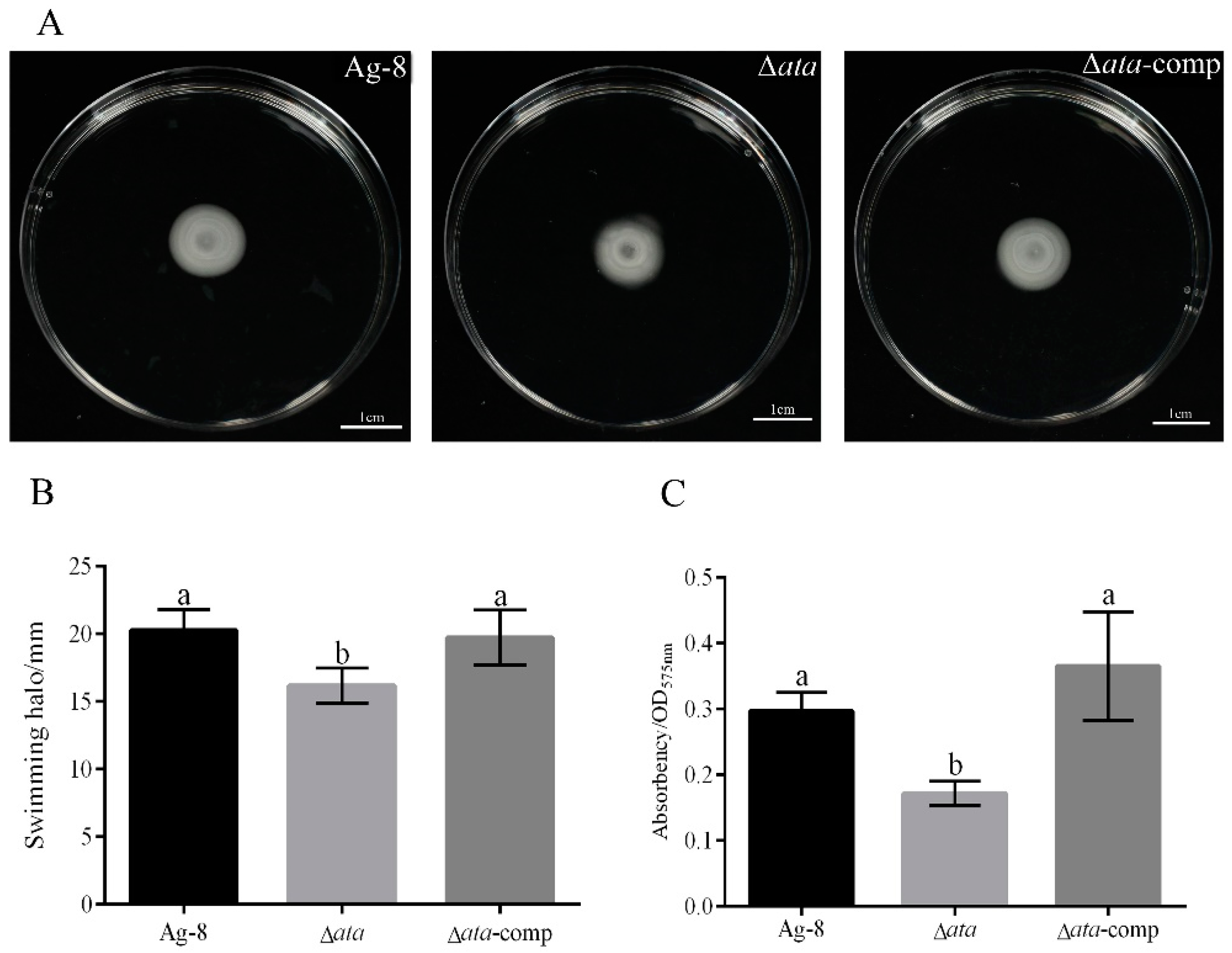
To ascertain the impact of ata deletion on the physiological attributes of Ag-8, the motility and biofilm formation of strains Ag-8, Δata, and Δata-comp were evaluated. The findings indicated that the motility of Δata strains was markedly diminished, exhibiting a reduction of approximately 20.13% in comparison to Ag-8. And there was no significant difference in the motility between the Δata-comp and wild type strain (Figure 2A,B). In addition, it was further found that the biofilm of the deletion strain Δata was significantly reduced by 42.15% compared to Ag-8, whereas the biofilm of the complemented strain Δata-comp exhibited no significant difference from Ag-8 (Figure 2C).

3.3. ata Gene Affects Ag-8 Colonization in Tomato Rhizosphere

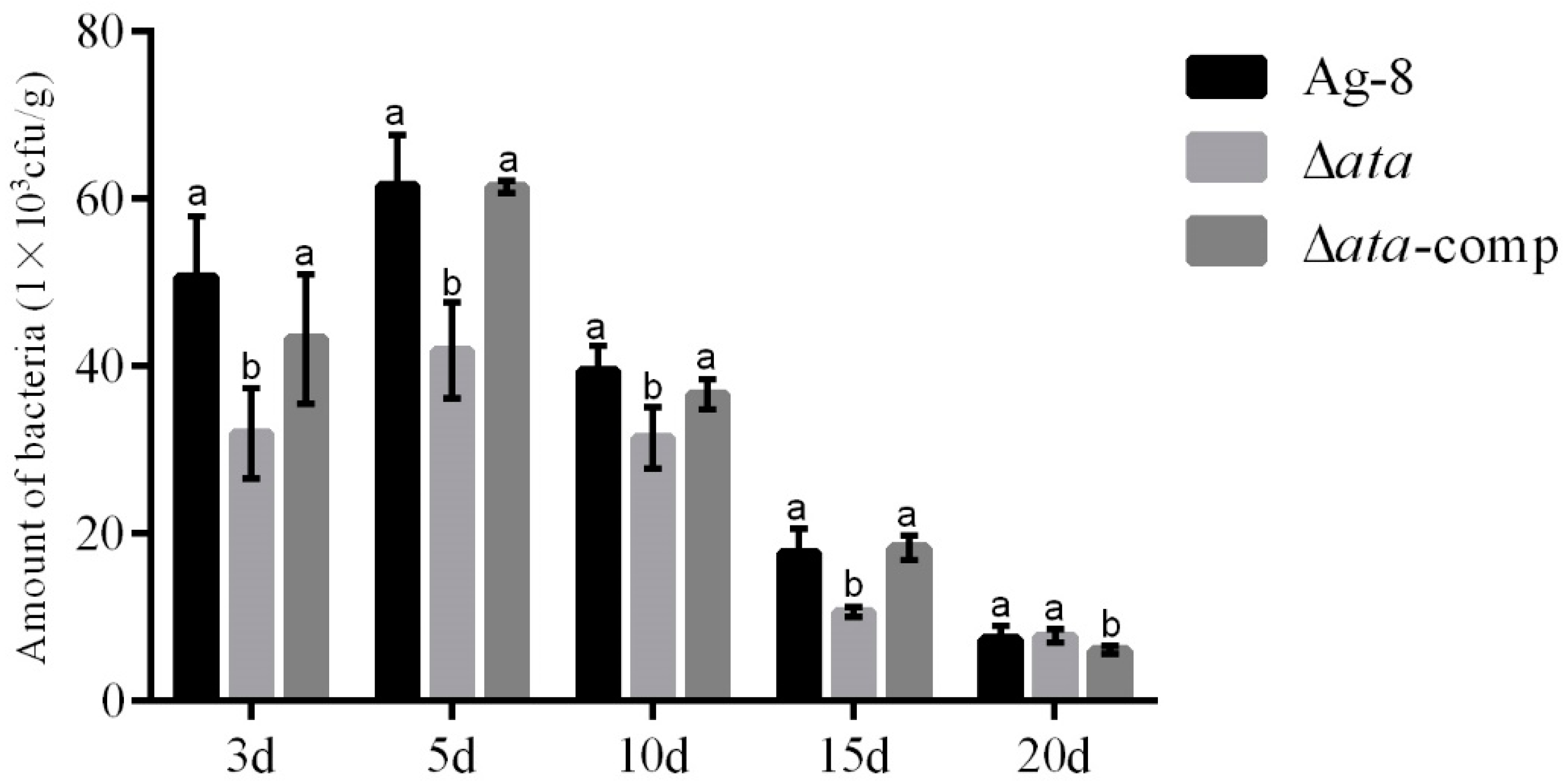
Given the established link between ata and biofilm formation and motility, we postulated that ata may also influence colonization. To verify the effect of ata deletion on the ability of Ag-8 to colonize tomato rhizosphere, the colonization at different stages of the tomato rhizosphere by the biocontrol bacteria Ag-8, Δata, and Δata-comp was examined. The results showed that wild-type Ag-8, Δata, and Δata-comp could colonize the tomato rhizosphere, and the colonization of Δata was significantly lower than that of Ag-8 and Δata-comp. The colonization of the tomato rhizosphere by Δata was significantly lower than that of the wild-type strain Ag-8 at 3 d, 5 d, 10 d, and 15 d, with a reduction of 36.84%, 31.95%, 20.28%, and 40.00%, respectively, compared to the wild strain. In addition, the amount of Ag-8, Δata, and Δata-comp colonization all decreased over time (Figure 3).

3.4. ata Deletion Reduces the Expression of Ag-8 and Biofilm-Related Genes

Based on the fact that theAg-8 deletion of ata reduced the effectiveness of Ag-8 control grapevine crown gall, to investigate the intrinsic molecular mechanism by which the gene ata affects the colonization ability of strain Ag-8, a transcriptome analysis of Ag-8 and Δata was performed. A total of 596 differentially expressed genes (DEGs) were detected in Δata compared to Ag-8 (Figure 4A), of which 270 DEGs were expressed up-regulated and 326 DEGs were expressed down-regulated (Figure 4B). The functional annotation of 596 genes, GO functional annotation revealed that most of the genes were enriched in catalytic activity; cellular anatomical entity; the integral component of the membrane; and intrinsic component of the membrane (Figure 4C). KEGG functional annotation revealed that DEGs were predominantly concentrated on the membrane transport pathway (Figure 4D). ABC transporter proteins, as a pivotal component of membrane transport, garnered our attention, prompting us to observe that the araH gene exhibited significant down-regulation in the transcriptome data as the gene encoding the ABC transporter protein. To identify key target genes regulated by ata, we employed RT-qPCR to validate the screened genes. The results showed that the araH gene was significantly down-regulated in Δata, suggesting that araH may be a key downstream target gene regulated by ata (Figure 4C–F).

3.5. The araH Gene Has Been Demonstrated to Influence the Motility and Biofilm Formation of Ag-8

In order to verify the effect of araH deletion on the physiological traits of Ag-8, the motility and biofilm production of the ∆araH strain were determined using the wild-type Ag-8 as the control. The findings indicated that the motility and biofilm production of the mutant strain ∆araH were markedly diminished in comparison to Ag-8, exhibiting a 15.10% and 42.48% decrease, whereas ∆araH-comp exhibited no statistically significant divergence from the wild type (Figure 5A–C).

3.6. araH Gene Affects the Effectiveness of Ag-8 Against Grapevine Crown Gall Disease in Tomato

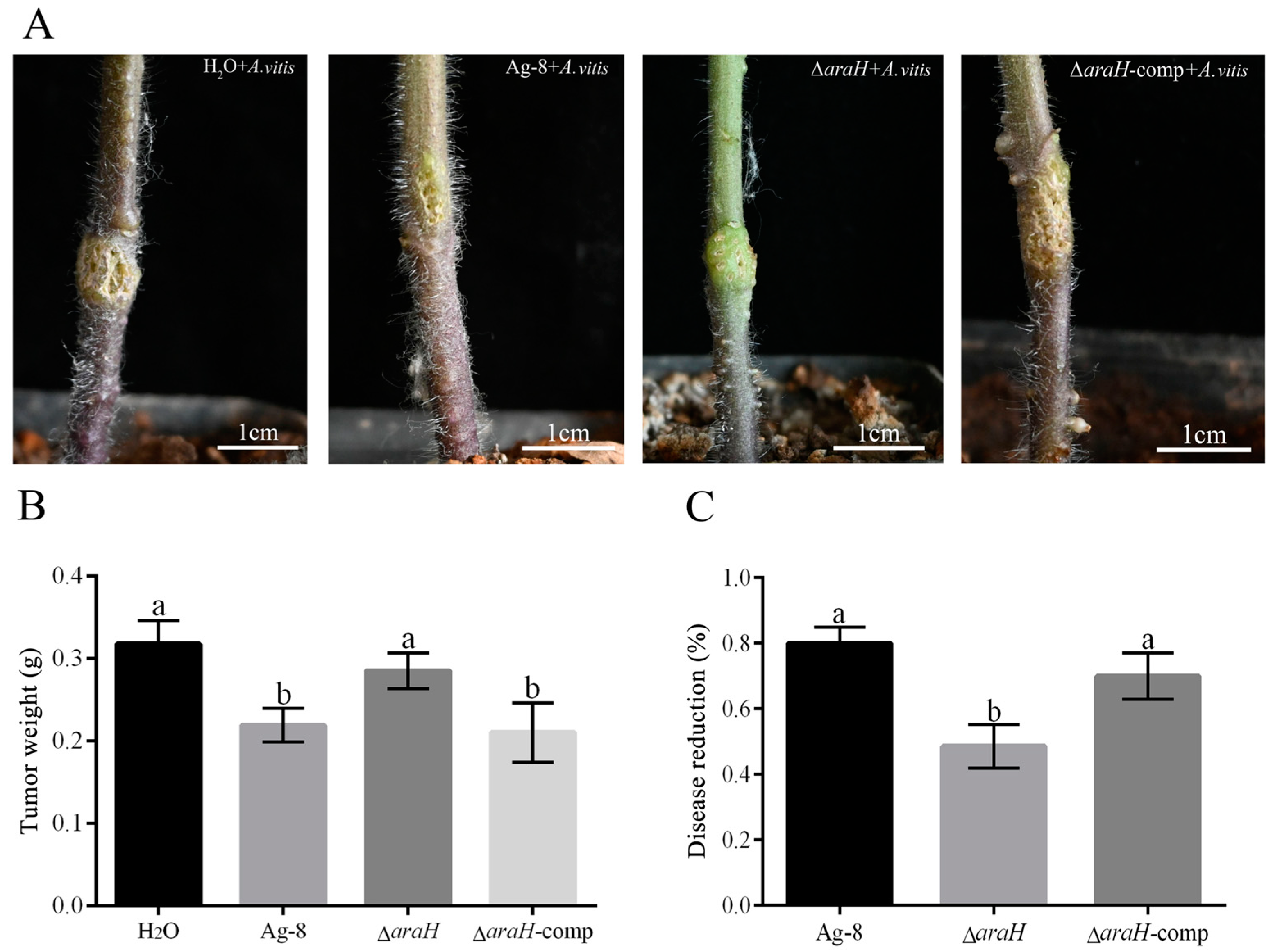
To verify the biocontrol ability of araH in Ag-8, we constructed deletion and complemented strains of araH gene. Then, the effectiveness of the araH deletion strain and complemented strain was investigated. The results indicated that the tomato inoculated with the ΔaraH and A. vitis mixed suspension exhibited more pronounced symptoms than the tomato inoculated with the Ag-8 and A. vitis mixed suspension, but tomatoes inoculated with the ΔaraH-comp and A. vitis mixed suspension had similar symptoms as when inoculated with the Ag-8 and A. vitis mixed suspension (Figure 6A). We investigated the efficacy of the mutant against A. vitis and compared it to Ag-8. Our results showed that the weight of tomato tumors was significantly up-regulated, and the biocontrol capacity of ΔaraH was significantly reduced in the presence of mixed injections of ΔaraH. The weight of tomato tumors increased by 27.07% in the case of the mixed injection of ΔaraH. Furthermore, the biocontrol capacity of ΔaraH was found to be significantly reduced by 39.25%. Following the back-complementation of the araH gene, the biocontrol ability was restored to the wild level (Figure 6B,C).

4. Discussion

To investigate the mechanism of control of grapevine crown gall disease by the biocontrol bacterium Ag-8, the function of the gene ata was investigated. In this study, an ata deletion-complemented strain was constructed, and the gene ata was found to positively regulate the efficacy of Ag-8 in the control of grapevine crown gall (Figure 1). This indicates that ata plays an important role in the control of grapevine crown gall by Ag-8. The AraC family comprises a number of proteins that are closely related to the formation of biofilms and the process of motility. The ArgR protein of this family was demonstrated to affect biofilm formation in the bacterium Pseudomonas putida through the regulation of arginine metabolism [24]. Additionally, YqhC has been shown to affect the pathogenicity of Erwinia amylovora by regulating its motility [25]. Interestingly, the motility and biofilm production of Ag-8 were significantly reduced when we deleted the ata gene, and the ability was recovered to the wild-type level after being complemented (Figure 2).
The motility and biofilm of strains are closely related to their colonization on the host surface. Exogenous biocontrol strains applied to the root perimeter achieve effective colonization by forming biofilms [26]. The first step for biocontrol strain to perform the function is to establish a certain population density, which involves a competition and inhibition relationship between biocontrol strain and other microorganisms [27]. However, there are fewer reports of AraC family proteins in biocontrol bacteria affecting the colonization ability on plants. In the present study, the colonization ability of ∆ata on tomato decreased significantly compared to the wild type (Figure 3). This further suggests that Ag-8 may regulate its motility and biofilm formation through ata, affecting its colonization on plant roots and thus preventing grapevine crown gall through competition.
In previous reports, AraC family transcription factors have been shown to regulate microbial growth and metabolic processes, including biofilm formation and colonization ability, among others [28,29,30]. In order to identify genes that affect Ag-8 colonization and are subject to regulation by ata, an analysis of the transcriptomics data of ∆ata was conducted. This analysis revealed that the differentially expressed genes were predominantly associated with energy metabolism, carbohydrate metabolism, and membrane transport pathways, as determined by KEGG functional annotation (Figure 4D). In the present study, the emphasis was placed on genes implicated in membrane transport, with a particular focus on ABC transporter proteins. ABC transporter proteins constitute a pivotal class of transmembrane transporters, playing a crucial role in the uptake and secretion of a diverse range of substrates. They are involved in a multitude of cellular processes, including the uptake of nutrients, the cytosolization of metabolites, and the formation of the bacterial periplasm [31,32]. Furthermore, some of the ABC transporter proteins have been reported to be functionally related to AraC family transcription factors. The ABC transporter protein AraH is involved in the metabolism of L-arabinose and influences biofilm formation together with AraC [33,34,35]. In Escherichia coli, during the metabolism of glucose and xylose, the transcription factor AraC exerts negative regulation on the xylose transporter protein AraFGH [36]. In addition, ABC transporter proteins are directly linked to bacterial swimming ability, biofilm formation, and colonization. It was demonstrated that some ABC transporter proteins in Delftia acidovorans RAY209 will play a role in the colonization of oilseed rape by the strain [37]; the ABC transporter proteins YtrBCDEF is involved in biofilm formation in Bacillus subtilis [38]. It was demonstrated that certain ABC transporter proteins within the genus Escherichia coli have the capacity to influence strain motility and chemotaxis by means of activating the flagellum [39]. Consequently, the hypothesis was formulated such that araH, which encodes an ABC transporter protein, may be a downstream target gene regulated by ata. This hypothesis was validated by the finding that araH expression was significantly reduced in Δata (Figure 4F).
In order to investigate the role of the araH gene in the biocontrol process of Ag-8, the biological function and biocontrol effect of araH were studied. The results showed that the expression of araH showed a positive correlation with biofilm formation, motility, and biocontrol ability (Figure 5 and Figure 6). The experimental results showed that Agrobacterium tumefaciens Ag-8 controls grapevine crown gall disease by regulating its biofilm formation and motility through ata, which affects its colonization, and araH, as an ABC transporter regulated by ata, may be involved in the transport of nutrients and metabolites of Ag-8, thereby participating in the regulation of colonization ability. There are few reports on the control of grape crown gall by Agrobacterium biocontrol only through colonization. Previous research demonstrated that Ag-8 lacks antagonistic and induced resistance capabilities, suggesting that its efficacy in combating grapevine crown gall disease may be contingent on its ability to colonize the host. The present experiment demonstrated the biocontrol mechanism of the biocontrol bacterium Ag-8, and this finding offers a novel perspective for future research on plant-beneficial agrobacteria, with a particular focus on the colonization process.
In addition, based on the function of the gene data and araH, it is possible that Ag-8 can be genetically engineered in the future to remold the biocontrol effect of the strain. The modification of Ag-8 will also facilitate its outdoor application, and ata may be able to help Ag-8 withstand various stresses in the natural environment due to its ability to regulate biofilm formation. The present study therefore provides theoretical guidance for later genetic modification of Ag-8.

Author Contributions

Conceptualization, Y.W. and Z.L. (Zhuoran Li); methodology, Z.L. (Zhenxing Liu); software, Y.G. and W.Y.; validation, Y.B. and J.Y.; data curation, S.L. and Y.Z.; writing—original draft preparation, S.L.; writing—review and editing, Y.W. All authors have read and agreed to the published version of the manuscript.

Funding

This research was funded by the Science and Technology Projects of Gansu (23CXNH0014).

Data Availability Statement

Due to the policies of the laboratory and the confidentiality agreements, we are unable to disclose the original data.

Acknowledgments

We would like to express our gratitude to the Science and Technology Projects of Gansu (23CXNH0014) for their invaluable support in enabling us to undertake this research.

Conflicts of Interest

The authors declare no conflicts of interest.

Appendix A

Table A1. Cloning primers for upstream and downstream fragments of the deletion strain.
Primer IDSequence
Δata-1FGTCGACTCTAGAGGATCCCGAGGTCGGGCGCGTTGA
Δata-1RAAATACATGACTGAACCATGAGGCAGAGGATAT
Δata-2FTCCTCTGCCTCATGGTTCAGTCATGTATTTCCT
Δata-2RGACCATGATTACGAATTCCGGGGTTCGGGCTGACAATTAG
ΔaraH-1FACCGAGCTCGAATTCACAGGAAGCGTATCATCG
ΔaraH-1RATGTCAGCGAATTGATCACCACACTGACGATCTTC
ΔaraH-2FTCGTCAGTGTGGTGATCAATTCGCTGACATGCCGG
ΔaraH-2RCAGGTCGACTCTAGAAATCGCTGGACATCCGCC
Table A2. Cloning primers for gene fragments of complemented strains.
Primer IDSequence
Δata-comp-FACGACGGCCAGTGAATTCCCCAAGGTCTCGTGTG
Δata-comp-RCTGCAGGTCGACTCTAGAGTGCTTCGCTCAAGAAGGATAC
ΔaraH-comp-FCGGCCAGTGAATTCGAGCTCTCATCGCGCGGTACCTTC
ΔaraH-comp-RGCCTGCAGGTCGACTCTAGAATGATGGTGGCTGTGACA
Table A3. Primers for RT-qPCR test.
Primer IDSequence
araH QP-FTTTGACAAAGCCCAGAACCG
araH QP-RTCATCCAGACCATCAATACCG

References

  1. Yuan, L.; Yin, X.; Wang, P.; Li, T.; Jiang, X.; Tang, X.; Hna, J.; Wei, Y. Research progress and control measures of grapevine crown gall. Chin. Foreign Grapes Wines 2022, 93–99. [Google Scholar] [CrossRef]
  2. Kawaguchi, A.; Kirino, N.; Inoue, K.; Noutoshi, Y. Biological control for grapevine crown gall through soil injection with Allorhizobium vitis strain ark-1. Eur. J. Plant Pathol. 2024, 170, 479–489. [Google Scholar] [CrossRef]
  3. Brown, P.J.B.; Chang, J.H.; Fuqua, C. Agrobacterium tumefaciens: A transformative agent for fundamental insights into host-microbe interactions, genome biology, chemical signaling, and cell biology. J. Bacteriol. 2023, 205, e0000523. [Google Scholar] [CrossRef]
  4. Stockwell, V.O.; Moore, L.W.; Loper, J.E. Fate of Agrobacterium radiobacter K84 in the environment. Appl. Environ. Microbiol. 1993, 59, 2112–2120. [Google Scholar] [CrossRef]
  5. Yang, Y.L.; Li, J.Y.; Wang, J.H.; Wang, H.M. Mutations affecting chemotaxis of Agrobacterium vitis strain e26 also alter attachment to grapevine roots and biocontrol of crown gall disease. Plant Pathol. 2009, 58, 594–605. [Google Scholar] [CrossRef]
  6. Kawaguchi, A.; Inoue, K.; Ichinose, Y. Biological control of crown gall of grapevine, rose, and tomato by nonpathogenic Agrobacterium vitis strain var03-1. Phytopathology 2008, 98, 1218–1225. [Google Scholar] [CrossRef] [PubMed]
  7. Gohlke, J.; Deeken, R. Plant responses to Agrobacterium tumefaciens and crown gall development. Front. Plant Sci. 2014, 5, 155. [Google Scholar] [CrossRef]
  8. Bull, C.T.; Weller, D.M.; Thomashow, L.S.J.P. Relationship between root colonization and suppression of gaeumannomyces graminis var. Tritici by pseudomonas fluorescens strain 2-79. Phytopathology 1991, 81, 954–959. [Google Scholar] [CrossRef]
  9. Timmusk, S.; Grantcharova, N.; Wagner, E.G.H. paenibacillus polymyxa invades plant roots and forms biofilms. Appl. Environ. Microbiol. 2005, 71, 7292–7300. [Google Scholar] [CrossRef]
  10. Huang, R.; Feng, H.; Xu, Z.; Zhang, N.; Liu, Y.; Shao, J.; Shen, Q.; Zhang, R. Identification of adhesins in plant beneficial rhizobacteria bacillus velezensis sqr9 and their effect on root colonization. Mol. Plant-Microbe Interact. 2022, 35, 64–72. [Google Scholar] [CrossRef]
  11. Gao, S.; Wu, H.; Yu, X.; Qian, L.; Gao, X. Swarming motility plays the major role in migration during tomato root colonization by bacillus subtilis swr01. Biol. Control 2016, 98, 11–17. [Google Scholar] [CrossRef]
  12. Zhao, A.; Sun, J.; Liu, Y. Understanding bacterial biofilms: From definition to treatment strategies. Front. Cell. Infect. Microbiol. 2023, 13, 1137947. [Google Scholar] [CrossRef]
  13. Zhu, M.-L.; Wu, X.-Q.; Wang, Y.-H.; Dai, Y. Role of biofilm formation bybacillus pumilushr10 in biocontrol against pine seedling damping-off disease caused byrhizoctonia solani. Forests 2020, 11, 652. [Google Scholar] [CrossRef]
  14. Gallegos, M.-T.; Schleif, R.; Bairoch, A.; Hofmann, K.; Ramos, J.L.J.M.; Reviews, M.B. Arac/xyls family of transcriptional regulators. Microbiol. Mol. Biol. Rev. 1997, 61, 393–410. [Google Scholar]
  15. Gribskov, M.; McLachlan, A.D.; Eisenberg, D. Profile analysis: Detection of distantly related proteins. Proc. Natl. Acad. Sci. USA 1987, 84, 4355–4358. [Google Scholar] [CrossRef] [PubMed]
  16. Greenblatt, J.; Schleif, R. Arabinose c protein: Regulation of the arabinose operon in vitro. Nat. New Biol. 1971, 233, 166–170. [Google Scholar] [CrossRef] [PubMed]
  17. Zhang, L.; Sun, L.; Srinivasan, R.; Lin, M.; Gong, L.; Lin, X. Unveiling a virulence-regulating mechanism in aeromonas hydrophila: A quantitative exoproteomic analysis of an arac-like protein. Front. Immunol. 2023, 14, 1191209. [Google Scholar] [CrossRef]
  18. Ueda, A.; Ogasawara, S.; Horiuchi, K. Identification of the genes controlling biofilm formation in the plant commensalpseudomonas protegenspf-5. Arch. Microbiol. 2020, 202, 2453–2459. [Google Scholar] [CrossRef]
  19. Chen, B.; Li, R.-F.; Zhou, L.; Qiu, J.-H.; Song, K.; Tang, J.-L.; He, Y.-W. The phytopathogenxanthomonas campestrisutilizes the divergently transcribedpoba/pobrlocus for 4-hydroxybenzoic acid recognition and degradation to promote virulence. Mol. Microbiol. 2020, 114, 870–886. [Google Scholar] [CrossRef]
  20. Ni, X.; Li, S.; Yuan, Y.; Chang, R.; Liu, Q.; Liu, Z.; Li, Z.; Wang, Y. Effect of siad on ag-8 to improve resistance to crown gall in grapes and related mechanisms. Plant Physiol. Biochem. 2024, 215, 108869. [Google Scholar] [CrossRef]
  21. Hmelo, L.R.; Borlee, B.R.; Almblad, H.; Love, M.E.; Randall, T.E.; Tseng, B.S.; Lin, C.; Irie, Y.; Storek, K.M.; Yang, J.J.; et al. Precision-engineering the Pseudomonas aeruginosa genome with two-step allelic exchange. Nat. Protoc. 2015, 10, 1820–1841. [Google Scholar] [CrossRef] [PubMed]
  22. Jailani, A.; Ahmed, B.; Lee, J.-H.; Lee, J. Inhibition of Agrobacterium tumefaciens growth and biofilm formation by tannic acid. Biomedicines 2022, 10, 1619. [Google Scholar] [CrossRef] [PubMed]
  23. Merritt, J.H.; Kadouri, D.E.; O’Toole, G.A. Growing and analyzing static biofilms. Curr. Protoc. Microbiol. 2011, 22, 1B-1. [Google Scholar] [CrossRef]
  24. Molina-Henares, M.A.; Ramos-Gonzalez, M.I.; Rinaldo, S.; Espinosa-Urgel, M. Gene expression reprogramming of pseudomonas alloputida in response to arginine through the transcriptional regulator argr. Microbiol.-Sgm 2024, 170, 001449. [Google Scholar] [CrossRef]
  25. Sahebi, M.; Tarighi, S.; Taheri, P. The arac-like transcriptional regulator yqhc is involved in pathogenicity of Erwinia amylovora. J. Appl. Microbiol. 2022, 132, 1319–1329. [Google Scholar] [CrossRef]
  26. Cai, Y.-m.; Hutchin, A.; Craddock, J.; Walsh, M.A.; Webb, J.S.; Tews, I. Differential impact on motility and biofilm dispersal of closely related phosphodiesterases in Pseudomonas aeruginosa. Sci. Rep. 2020, 10, 6232. [Google Scholar] [CrossRef] [PubMed]
  27. Haas, D.; Défago, G. Biological control of soil-borne pathogens by fluorescent pseudomonads. Nat. Rev. Microbiol. 2005, 3, 307–319. [Google Scholar] [CrossRef]
  28. Lv, J.; Zhu, J.; Wang, T.; Xie, X.; Wang, T.; Zhu, Z.; Chen, L.; Zhong, F.; Du, H. The role of the two-component qsebc signaling system in biofilm formation and virulence of hypervirulent klebsiella pneumoniae atcc43816. Front. Microbiol. 2022, 13, 817494. [Google Scholar] [CrossRef]
  29. Midgett, C.R.; Talbot, K.M.; Day, J.L.; Munson, G.P.; Kull, F.J. Structure of the master regulator rns reveals an inhibitor of enterotoxigenic Escherichia coli virulence regulons. Sci. Rep. 2021, 11, 15663. [Google Scholar] [CrossRef]
  30. Rodriguez-Valverde, D.; Giron, J.A.; Hu, Y.; Nataro, J.P.; Ruiz-Perez, F.; Santiago, A.E. Highly-conserved regulatory activity of the anr family in the virulence of diarrheagenic bacteria through interaction with master and global regulators. Sci. Rep. 2023, 13, 7024. [Google Scholar] [CrossRef]
  31. Rees, D.C.; Johnson, E.; Lewinson, O. Abc transporters: The power to change. Nat. Rev. Mol. Cell Biol. 2009, 10, 218–227. [Google Scholar] [CrossRef]
  32. Tanaka, K.J.; Song, S.; Mason, K.; Pinkett, H.W. Selective substrate uptake: The role of atp-binding cassette (abc) importers in pathogenesis. Biochim. Biophys. Acta-Biomembr. 2018, 1860, 868–877. [Google Scholar] [CrossRef]
  33. Horazdovsky, B.F. The High Affinity l-Arabinose Transport Operon of Escherichia coli. Ph.D. Thesis, Case Western Reserve University, Cleveland, OH, USA, 1988. [Google Scholar]
  34. Sheppard, D.E.; Englesberg, E. Further evidence for positive control of the l-arabinose system by gene arac. J. Mol. Biol. 1967, 25, 443–454. [Google Scholar] [CrossRef] [PubMed]
  35. Vasicek, E.M.; O’Neal, L.; Parsek, M.R.; Fitch, J.; White, P.; Gunn, J.S. L-arabinose transport and metabolism in salmonella influences biofilm formation. Front. Cell. Infect. Microbiol. 2021, 11, 698146. [Google Scholar] [CrossRef] [PubMed]
  36. Zhu, X.; Fan, F.; Qiu, H.; Shao, M.; Li, D.; Yu, Y.; Bi, C.; Zhang, X. New xylose transporters support the simultaneous consumption of glucose and xylose in Escherichia coli. Mlife 2022, 1, 156–170. [Google Scholar] [CrossRef] [PubMed]
  37. Suchan, D.M.; Bergsveinson, J.; Manzon, L.; Pierce, A.; Kryachko, Y.; Korber, D.; Tan, Y.; Tambalo, D.D.; Khan, N.H.; Whiting, M.; et al. Transcriptomics reveal core activities of the plant growth-promoting bacterium delftia acidovorans ray209 during interaction with canola and soybean roots. Microb. Genom. 2020, 6, e000462. [Google Scholar] [CrossRef]
  38. Benda, M.; Schulz, L.M.; Stuelke, J.; Rismondo, J. Influence of the abc transporter ytrbcdef of Bacillus subtilis on competence, biofilm formation and cell wall thickness. Front. Microbiol. 2021, 12, 587035. [Google Scholar] [CrossRef]
  39. Zeng, Y.; Charkowski, A.O. The role of atp-binding cassette transporters in bacterial phytopathogenesis. Phytopathology 2021, 111, 600–610. [Google Scholar] [CrossRef]
Figure 1. Effectiveness of ata against A. vitis on tomato plants: (A) H2O + A. vitis: Co-inoculation with mixed suspension of H2O and A. vitis. Ag-8 + A. vitis: Co-inoculation with mixed suspension of Ag-8 and A. vitis. ∆ata + A. vitis: Co-inoculation with mixed suspension of ∆ata and A. vitis. Δata-comp + A. vitis: Co-inoculation with mixed suspension of Δata-comp and A. vitis. (B) Statistical plot of tomato tumor weights, with the horizontal coordinates from left to right showing the tumor weights of each strain after mixed inoculation with A. vitis. (C) Effectiveness of ata against A. vitis. Five replications are set up for each treatment. Different letters represent significant differences between groups after multiple comparisons using SPSS (p < 0.05).
Horticulturae 11 00465 g001
Figure 2. Effect of ata on Ag-8 motility and biofilm: (A) Motility of Ag-8, Δata, and Δata-comp strains. (B) Diameters of the motility of Ag-8, Δata, and Δata-comp strains on semisolid medium. (C) The absorbance of biofilms of Ag-8, Δata, and Δata-comp strains. Five replications are set up for each treatment. Different letters represent significant differences between groups after multiple comparisons using SPSS (p < 0.05).
Horticulturae 11 00465 g002
Figure 3. Dynamics of Ag-8, Δata, and Δata-comp colonization in root systems: Horizontal coordinates represent the customization of strains at different days. Lowercase letters represent the significance of differences between treatments at one time.
Horticulturae 11 00465 g003
Figure 4. Transcriptome data analysis and downstream target gene screening: (A) Ag-8 vs. Δata volcano plot; (B) statistical number of up and down-regulated genes in Ag-8 vs. Δata; (C) GO functional annotation; (D) KEGG functional analysis; (E) relative expression of araH in RNA-seq; and (F) relative expression of araH in RT-qPCR. The number of * is used to indicate different significance levels: **: p < 0.01; ***: p < 0.001.
Horticulturae 11 00465 g004
Figure 5. Effect of araH on Ag-8 motility and biofilm: (A) Motility of Ag-8, ΔaraH, and ΔaraH-comp strains; (B) diameters of the motility of Ag-8, ΔaraH, and ΔaraH-comp strains on semisolid medium; (C) the absorbance of biofilms of Ag-8, ΔaraH, and ΔaraH-comp strains. Five replications were set up for each treatment. Different letters represent significant differences between groups after multiple comparisons using SPSS (p < 0.05).
Horticulturae 11 00465 g005
Figure 6. Effectiveness of araH against A. vitis on tomato plants: (A) Tumor on tomato plants H2O + A. vitis: co-inoculation with mixed suspension of H2O and A. vitis, Ag-8 + A. vitis: co-inoculation with mixed suspension of Ag-8 and A. vitis, ∆araH +A. vitis: co-inoculation with mixed suspension of ∆araH and A. vitis, and ∆araH-comp + A. vitis: co-inoculation with mixed suspension of ∆araH-comp and A. vitis; (B) Statistical plot of tomato tumor weights, with the horizontal coordinates from left to right showing the tumor weights of each strain after mixed inoculation with A. vitis. (C) The effect of Ag-8, ∆araH, and ∆araH-comp on the control of A. vitis. Five replications are set up for each treatment. Different letters represent significant differences between groups after multiple comparisons using SPSS (p < 0.05).
Horticulturae 11 00465 g006
Disclaimer/Publisher’s Note: The statements, opinions and data contained in all publications are solely those of the individual author(s) and contributor(s) and not of MDPI and/or the editor(s). MDPI and/or the editor(s) disclaim responsibility for any injury to people or property resulting from any ideas, methods, instructions or products referred to in the content.