Article

Biocontrol of Large Patch Disease in Zoysiagrass (Zoysia japonica) by Bacillus subtilis SA-15: Identification of Active Compounds and Synergism with a Fungicide

1
Division of Life and Environmental Science (Horticulture), Daegu University, Gyeongsan 38453, Korea
2
Institute of Natural Sciences, Daegu University, Gyeongsan 38453, Korea
3
Department of Bio-Environmental Chemistry, Chungnam National University, Daejeon 34134, Korea
4
Department of Applied Biology, Chungnam National University, Daejeon 34134, Korea
5
Department of Horticulture, Chungnam National University, Daejeon 34134, Korea
6
Department of Smart Agriculture Systems, Chungnam National University, Daejeon 34134, Korea
*
Author to whom correspondence should be addressed.
These authors contributed equally to this work.
Horticulturae 2022, 8(1), 34; https://doi.org/10.3390/horticulturae8010034
Submission received: 14 November 2021 / Revised: 20 December 2021 / Accepted: 25 December 2021 / Published: 29 December 2021

Abstract

:
Bacillus subtilis SA-15 is a plant growth-promoting bacterium isolated from non-farming soil. We aimed to identify lipopeptides produced by B. subtilis SA-15 and evaluate the control efficacy of B. subtilis SA-15 against large patch disease caused by Rhizoctonia solani AG 2-2 (IV) in zoysiagrass (Zoysia japonica). Bacillus subtilis SA-15 inhibited mycelial growth of R. solani AG 2-2 (IV) in vitro and produced fengycin A and dehydroxyfengycin A, which are antifungal compounds. Fengycin A and deghydroxyfengycin A inhibited R. solani mycelial growth by 30.4 and 63.2%, respectively. We formulated B. subtilis SA-15 into a wettable powder and determined its control efficiency against large patch in a field trial. The control efficacy was 51.2–92.0%. Moreover, when B. subtilis SA-15 powder was applied together with half the regular dose of the fungicide pecycuron, the control efficacy was 88.5–100.0%. These results indicate that B. subtilis SA-15 can be used to control soil-borne diseases, including large patch caused by R. solani, because of lipopeptide production. The use of this bacterium can also reduce the amount of fungicide needed, providing an eco-friendly management option for turfgrass.

1. Introduction

Large patch caused by Rhizoctonia solani AG 2-2 (IV) is a well-known disease of zoysiagrass (Zoysia japonica), which is planted in 70% of golf course fairways in Korea [1,2]. Large patch decreases the growth, shoot density, and visual quality of zoysiagrass. Rhizoctonia solani AG 2-2 (IV) can infect zoysiagrass areas of 0.6–6.0 m and can develop disease patches more seriously during the season of cool temperatures (10 to 21 °C) when turfgrass leaves or enters dormancy in May or September, respectively [3]. Foliar symptoms are roughly circular patches with yellow, tan, straw-brown, or red discoloration [3]. Generally, large patch on zoysiagrass is controlled by applying fungicides, such as pencycuron, or by decreasing organic matter in the root zone via soil renovations in golf courses.
Rhizoctonia solani inhabits the thatch layer of golf courses [4]. The thatch layer of zoysiagrass is where the leaves accumulate after mowing; the layer consists of cellulose, hemicellulose, and lignin material [5]. Various management practices can be used to reduce the amount of organic matter in the thatch layer, including topdressing, aeration, and vertical mowing [3]. These management practices help to increase microbial activity in the root zone, and considering that some beneficial endophytes such as Bacillus and Pseudomonas have cellulase activity, this can result in higher decomposition rates [6].
Microorganisms with cellulase activity not only mineralize organic matter in the root zone but also inhibit the mycelial growth of Oomyetes because of degradation of fungal mycelia [7]. Cell wall lytic (CWL) enzymes include cellulase, chitinase, and β-glucanase, and may degrade or cleave main constituents of mycelial cell walls [7]. In addition, lipopeptides such as bio-surfactant, inturins and fengycin are important antifungal products produced from microbes [7,8,9,10]. Many researchers have screened potential bacteria producing CWLs or antifungal compounds for biocontrol of plant diseases [7,8,9,10]. As an endophytic bacterium, B. subtilis has been reported to have CWL activities and produce antifungal metabolites [7,11].
Bacillus subtilis SA-15, which has been isolated from non-farming soil in Korea, is a plant growth-promoting rhizobacterium (PGPR) [12]. In this study, we aimed (i) to identify the antifungal compounds produced by B. subtilis SA-15 and (ii) to evaluate the suppressive effects of SA-15 application with or without fungicide on large patch caused by R. solani AG 2-2 (IV) in zoysiagrass.

2. Materials and Methods

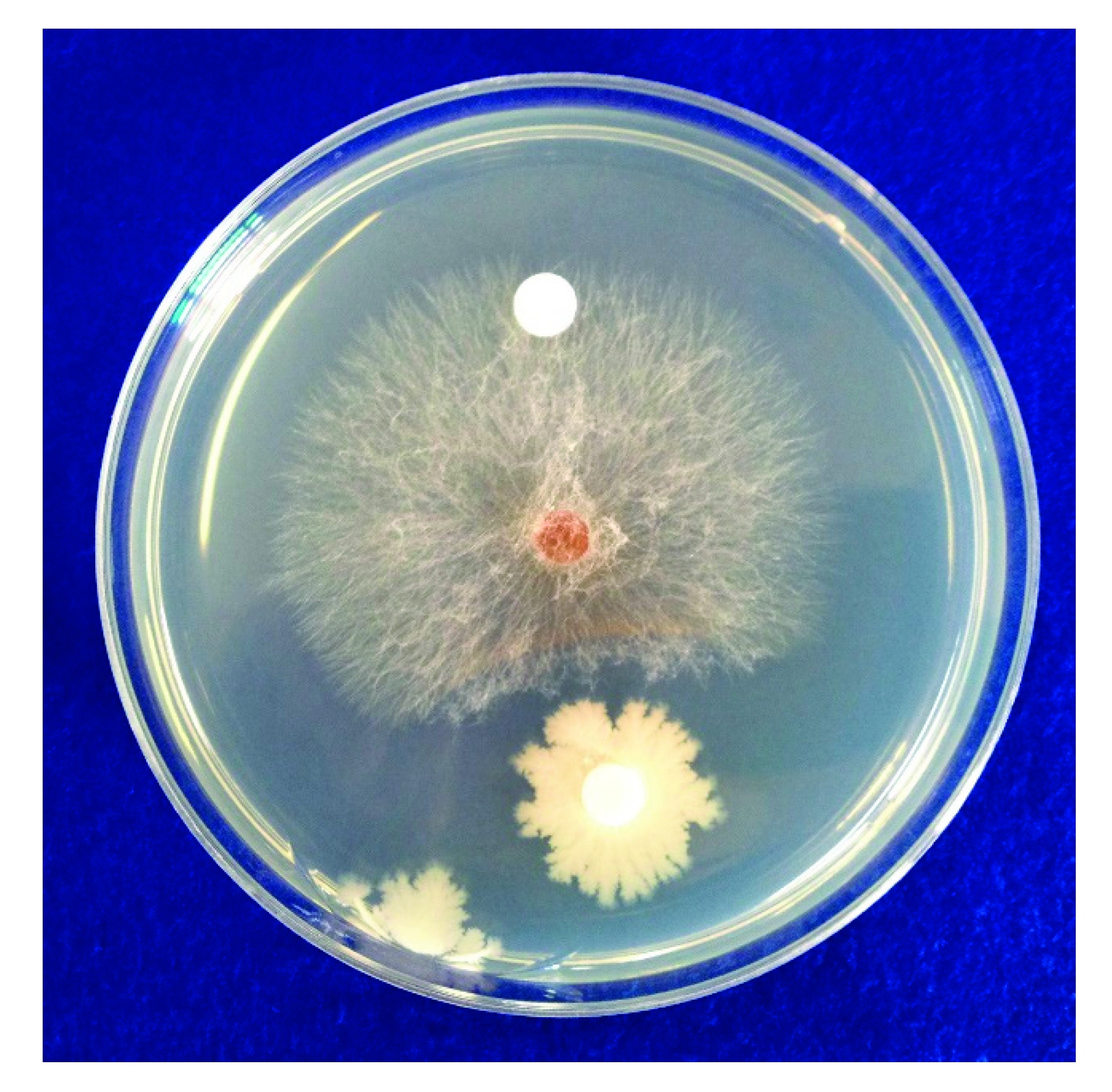
2.1. In Vitro Antifungal Activity of B. subtilis SA-15

The Bacillus subtilis SA-15 strain used in this study was isolated from non-farming soil in Korea and identified in our group by comparing sequences of 16S rRNA and gyrase subunit B gene regions. The strain was then deposited in the Korean Collection for Type Cultures, Korea Research Institute of Bioscience and Biotechnology (Daejeon, Korea) as deposit number 13706 [12].
The antifungal activity of B. subtilis SA-15 was investigated using R. solani AG 2-2 (IV), which was obtained from the Korean Agricultural Culture Collection (KACC). Agar plugs were inoculated at the center of potato dextrose agar (PDA) on a 90 mm-diameter Petri dish, and two paper disks of 8 mm diameter were placed on the opposite side, 30 mm away from the central agar plug. One disk received 100 μL of bacterial suspension and the other disk the same volume of sterilized water. Mycelial growth of R. solani AG 2-2 (IV) was evaluated after incubation at 25 °C for 5 d. Mycelial inhibition (MI) was calculated using the following equation:
MI (%) = (1 − mycelial length of treated sample/mycelial length of control sample) × 100;
where the mycelial length was the distance from the agar plug point.
Cellulase activity was investigated to determine the CWL activity of SA-15 strains with three replications. It was determined on a medium consisting of 15.0 g∙L−1 pancreatic digest of casein, 5.0 g∙L−1 papaic digest of soybean, 5.0 g∙L−1 NaCl, 15.0 g∙L−1 agar, and 10.0 g∙L−1 carboxymethyl cellulose. SA-15 cells were inoculated on this medium and incubated at 30 °C for 48 h, and then, cellulase activity was determined based on the clear zone with 0.1% Congo red solution [7].

2.2. Purification of Antifungal Compound Fractions from B. subtilis SA-15

SA-15 cells were incubated in nutrient broth at 180 rpm for 48 h at 30 °C. A mixture of SA-15 culture (10 L) and Diaion HP-20 resin (2 L) was then mixed with a stirrer for 3 h. SA-15 in liquid medium was absorbed on Diaion HP-20 resin through this process. This resin mixture was then placed in a glass column (100 mm × 300 mm), washed with distilled water, and fractionated with 4 L of methanol. The fractions were then analyzed by high performance liquid chromatography (HPLC; pump 5110, autosampler 5210, column oven 5310; TOSOH, Tokyo, Japan), with an ODS TSK-gel 100V column (4.6 mm × 250 mm, 5.0 μm; TOSOH) using mobile phases A [methanol (MeOH) 100%, at 2–23 min] and B [5% MeOH (0.04% trifluoroacetic acid) in water:MeOH (9:1), at 0–2 and 23–26 min] at a flow rate of 1 mL min−1 and detected using a photo-diode array UV-detector (PDA detector 5430; TOSOH).

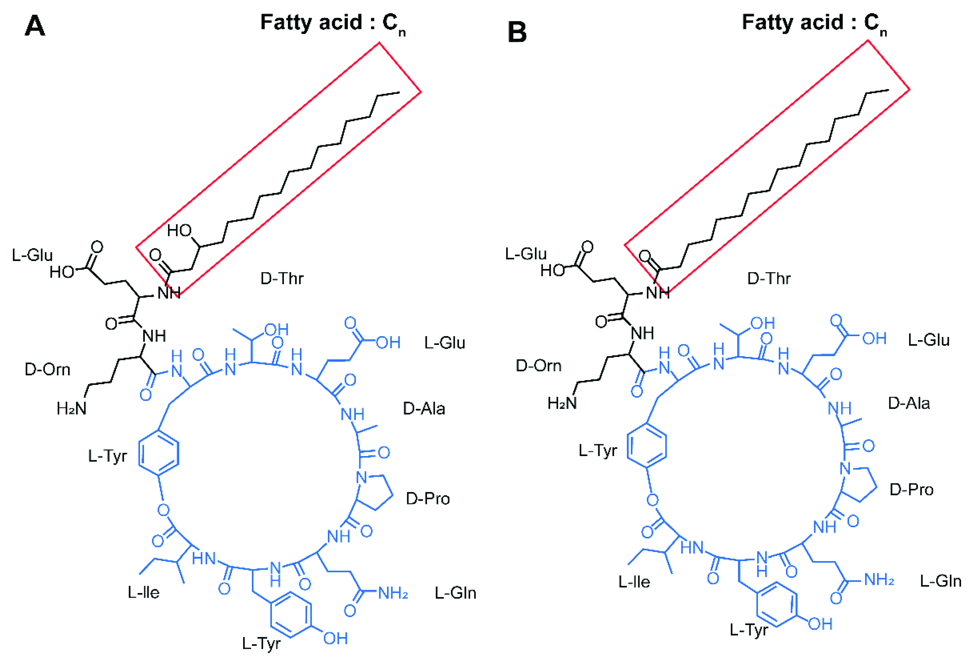
2.3. Identification of Lipopeptide Compounds from B. subtilis SA-15

The solvent of the fraction extracted from the resin mixture was removed using a vacuum rotary evaporator at 40 °C. The pH of the residues was adjusted to 2.0, using 6 N HCl, and placed at 4 °C for 24 h. Precipitated materials and supernatant liquid were collected by centrifugation at 5000× g for 20 min at 4 °C and were stored for HPLC analysis. The precipitated materials dissolved in 50% methanol were used for reversed-phase medium-pressure liquid chromatography (MPLC; System: Combiflash RF+, Teledyne ISCO, Lincoln NE, USA; column C18: ODS resin, 130 g, Teledyne ISCO; flow rate: 10 mL min−1) using 50–100% MeOH with the following gradient program: (1) 50% MeOH for 12 min; (2) 70% MeOH for 11 min and then 3 min after the gradient elution; (3) 90% MeOH for 12 min and then 2 min after the gradient elution; (4) 95% MeOH for 14 min and then 2 min after the gradient elution; and (5) 100% MeOH for 14 min. The solvent of the fractions containing lipopeptides was removed using a vacuum rotary evaporator. The residues were dissolved in methanol and applied to a Sephadex LH-20 column (5 cm × 100 cm; 17-0090-02, GE Healthcare, Chicago, IL, USA) using methanol as the mobile phase.
The lipopeptide fraction, referred to as compound 1 henceforth, was confirmed by inhibiting the fungal plant pathogen Botrytis cinerea. Mycelial disks (5 mm diam.) of B. cinerea were placed on the opposite middle of potato dextrose agar (PDA) medium and an 8 mm-diameter paper disk containing lipopeptide fraction (100 μL) on the other side. After incubation at 25 °C for 48 h, antifungal activity of the fraction was evaluated by measuring mycelial growth length toward the disk on the PDA medium.
The supernatant was removed from the solvent using a vacuum rotary evaporator, and the residue was dissolved in methanol. Purification was carried out by ODS MPLC and antifungal activity was determined by Sephadex LH-20 column chromatography. Hereinafter, this fraction is referred to as compound 2. Compounds 1 and 2 were identified using an NMR spectrometer (JEOL JNM-ECA 600, JEOL Ltd., Tokyo, Japan) and LC-ESI-mass spectrometer (LC-ESI-MS; Agilent 6410, Agilent Technologies, Santa Clara, CA, USA). NMR spectra were obtained on a JEOL JNM-ECA600, 600 MHz FT-NMR spectrometer at 600 MHz for 1H NMR and at 150 MHz for 13C NMR in CD3OD. LC-ESI-MS was conducted using an electrospray ionization (ESI)-QTRAP-3200 mass spectrometer (Applied Biosystems, Waltham, MA, USA). The sample injection volume was set at 30 μL, and chromatography was performed using a gradient of methanol in 0.1% (v/v) formic acid as the mobile phase. Eclipse Plus C18 (4.6 mm × 150 mm, 5 μm column; Agilent) was used as the analytical column. An HPLC ESIMS full scan in the positive modes was performed from 50 to 2000 m/z with a capillary voltage of 4.0 kV and a gas temperature of 300 °C. A gas flow of 121 L/h and a nebulizer of 50 psi were used.

2.4. In Vitro Inhibition Effects of Compounds 1 and 2 on R. solani Growth

Compounds 1 and 2 were purified from SA-15 culture and dissolved in methanol. Their concentration was not determined because they consisted of complex compounds and were only collected with HPLC. An agar plug of R. solani was inoculated at the center of PDA in a 9 cm diameter Petri dish with two 8 mm-diameter paper disks placed on the opposite side, 30 mm away from the central agar plug. Thereafter, 150 μL of compound 1 or 2 was added 30 mm from the R. solani plug. Fungal growth inhibition was evaluated, in triplicate, by investigating MI after incubation at 25 °C for 96 h.

2.5. In Vivo Control of Large Patch Disease with B. subtilis SA-15

SA-15 cells were grown in nutrient broth for 48 h at 28 °C and collected by centrifuging at 3000 rpm. Cells were than mixed in a ratio of 1% cells with 93% bentonite, 5% surfactant (NK-SLS, Coseal Co. Ltd., Seoul, Korea), and 1% white carbon (Bunammine Co., Ltd., Kyeong-buk, Korea) to form a wettable powder denoted as SA-15 WP. Two field experiments were conducted at Daeduck Science Golf Club in Korea from July to October 2014, to determine the efficacy of SA-15 WP to control large patch in zoysiagrass. The fungicide pencycuron (DB Hiteh Co., Ltd., Bucheon, Korea) was also used in the experiments.
R. solani AG 2-2 (IV) on PDA was inoculated in sterilized sand-oatmeal medium prepared with sand (380 g) and oatmeal (20 g) in 70 mL of distilled water and incubated at 25 °C for 20 days. In both experiments, zoysiagrass had been infected with the pathogen at an application rate of 500 g m−2 in the experimental plot on 11 June 2014.
In the first experiment, the following treatments were used to determine the efficacy of SA-15 WP: control (no treatment), pencycuron (1.0 mL∙m−2; 0.2 g∙m−2 active ingredient (a.i.)), WP (1.0 g∙m−2), 2WP (2.0 g∙m−2), and 4WP (4.0 g∙m−2). In the second experiment, we tested various mixtures of SA-15 WP and pencycuron (P) using the following treatments: control (no treatment), P (1.0 mL∙m−2; 0.2 g∙m−2 a.i.), 1/2P + WP (0.1 g∙m−2 P + 1.0 g∙m−2 WP), and 1/2P + 2WP (0.1 g∙m−2 P + 2.0 g∙m−2 WP). There were three replicate plots (0.5 m × 1.0 m) for each treatment, and the experiment was arranged in a randomized complete block design. Like in the first experiment above, applications of antifungal treatments were implemented six times (three times on 11 July, 25 July, and 8 August before and three times on 26 August, 10 September and 25 September after disease symptoms appeared). Treatment of SA-15 WP and pencycuron in a solution was prepared by diluting with water. Symptoms of large patch were first observed on 26 August. The control efficacy of each treatment was calculated on 26 August and 7 October using the following equation [6]:
Control efficiency (%) = (1 − mean disease area of treated sample/mean disease area of control sample) × 100.

2.6. Statistical Analysis

Statistical analyses were conducted using Statistical Products and Service Solutions for Windows (SPSS; version 12.1, IBM, New York, NY, USA). Data were analyzed using analysis of variance (ANOVA), and differences among the means were tested using a Tukey’s test (p < 0.05).

3. Results

3.1. In Vitro Antifungal Activity of B. subtilis SA-15 and Identification of Antifungal Compounds

B. subtilis SA-15 inhibited the mycelial growth of R. solani AG 2-2 (IV), resulting in an MI of 63.3 ± 1.0% (mean ± standard error) after 5 days (Figure 1).
Among the fractions extracted from SA-15, two compounds inhibited R. solani mycelial growth. The HPLC analysis of each compound revealed that they were fengycin-type lipopeptides, not iturin-type lipopeptides (Figure 2).
Compounds 1 and 2 were analyzed by 1NMR spectrometry and LC-ESI-MS. 1NMR spectrometry revealed that compound 1 contained α-methine with a D-alanine functional group (4.6 ppm) and fatty acid with a β-OH functional group (5.5 ppm), indicating that it was fengycin-A. Using LC-ESI-MS, seven peaks were observed for compound 1 at 725/1449, 739/1477, 732/1463, 739/1477, 746/1491, 753/1505, and 760/1519 m/z ([M+2H]2+/[M+H]+) (Table 1). The chemical structure of compound 1 is shown in Figure 3A. Compound 2 was identified as dehydroxyfengycin-A by 1NMR spectrometry because it only had a D-alanine group. Using LC-ESI-MS, eight peaks were observed for compound 2 at 732/1462, 739/1476, 1477/1490, 746/1490, 753/15041, 753/1504, 760/1518, and 767/1532 m/z ([M+2H]2+/[M+H]+) (Table 1). The chemical structure of compound 2 is shown in Figure 3B.
The inhibitory effects of fengycin A complex (compound 1) and dehydroxyfengycin A complex (compound 2) on the mycelial growth of R. solani were 30.4 ± 2.7% and 63.2 ± 2.5%, respectively (Figure 4).

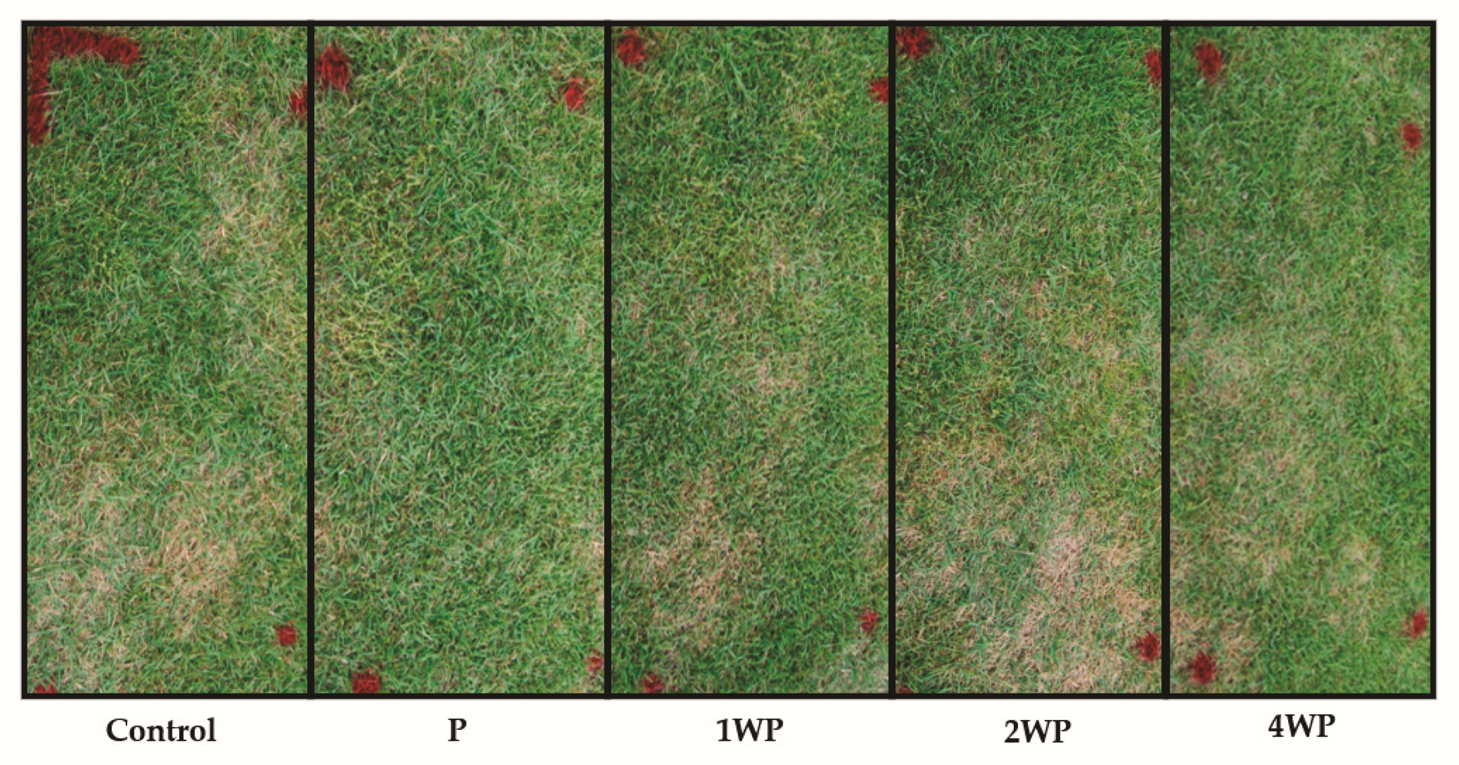
3.2. In Vivo Control of Large Patch Disease after Applying B. subtilis SA-15

In the first field experiment, the disease area and control efficiency on August 26 ranged from 2.5 to 29.2% and from 0.0 to 79.9%, respectively, and on 7 October, they ranged from 2.8 to 35.8% and from 51.2 to 92.0%, respectively. The pencycuron (P) treatment had the highest control efficacy against large patch on August 26, with a control efficacy of 79.9% (Table 2). The control efficacy of the P, 1WP, 2WP, and 4WP treatments on 7 October were 88.5, 92.0, 51.2, and 70.0%, respectively (Table 2 and Figure 5). The control efficacy of the P treatment on 7 October was not significantly different from that of the WP treatments.
In the second field experiment, the disease area and control efficiency on 26 August ranged from 0.5 to 12.3% and from 79.9 to 100.0%, respectively, and on 7 October, they ranged from 0.0 to 35.8% and from 88.5 to 100.0%, respectively (Table 3 and Figure 6). On 7 October, there were no significant differences between the P treatment and the mixture treatments.

4. Discussion

The thatch layer is a mat-like layer that occurs in the root zone due to the accumulation of organic matter [13]. Large patch in zoysiagrass is caused by R. solani AG 2-2 (IV) in the thatch layer in the fairways of golf courses [4]. Various fungicides are used to control large patch; however, microorganisms could be an effective alternative to these fungicides [14,15]. Certain PGPR can inhibit soil-borne diseases, such as large patch, brown patch, and yellow patch diseases [16].
Bacillus spp. are PGPRs that have been isolated from various sources such as organic matter and soil and are known to inhibit the growth of R. solani [17,18]. Specifically, B. subtilis SA-15 has a strong antifungal activity against various phytopathogens that decompose organic matter, including carbohydrates and proteins [12]. As B. subtilis SA-15 has CWL activity and produces antifungal compounds, it may inhibit plant pathogens. In the present study, we found that the SA-15 strain inhibited R. solani AG 2-2 (IV) in vitro and suppressed large patch disease in vivo. These results suggest that B. subtilis SA-15 could be used to control R. solani AG 2-2 (IV).
There were previous reports that the genus Bacillus produces CWLs such as cellulase, chitinase, and β-glucanase [7,19]. Although Bacillus subtilis SA-15 inhibited the mycelial growth of the fungal pathogen by 63.3% (Figure 1), there is no direct evidence inferring the hydrolytic cellulase-derived mycelial inhibition of the non-cellulose type fugus in our study. Secondary metabolites produced by microorganisms can also inhibit the growth of plant pathogens [20]. For example, Bacillus spp. produce lipopeptides such as iturin, fengycin, and surfactin, which are antifungal compounds [11]. Synthesis of lipopeptides by B. subtilis is dependent on the growth phase and culture conditions [21,22].
In particular, fengycins are volatile organic compounds (VOCs) with antifungal activity [23,24]; they can play a short- or long-distance role [25]. Fengycin is a well-known inhibitor of R. solani mycelial growth [26], and its inhibitory effects may vary according to the pathogenic fungus involved [27]. Fengycin produced by B. subtilis forms pores in the cellular membrane by increasing membrane interactions and destabilization [28], as observed by the fluid state of the membrane [29], low anionic phospholipid content [30], and low ergosterol content [31]. Fengycin complexes bind to the membrane through electrostatic and hydrophobic interactions. The insertion of the hydrophobic tails of fengycin into the bio-membrane results in the asymmetrical lateral expansion of the upper leaflet of the lipid bilayer that perturbs hydration by increasing the local membrane curvature, destabilizing the membrane. This causes transient membrane disruption, which leads to permeability change and pore formation in the cellular membrane [32]. The fengycin family members also inhibit spore germination in Fusarium culmorum [33] and reduce toxin production in fungi [34]. The antifungal activity of fengycin differs depending on the treated pathogenic species because of the different characteristics of fungal membranes, such as the composition or content of phospholipids and sterols [28]. Consequently, although the biocontrol mechanism of fengycin is unclear, fengycin readily suppresses spore germination and causes hyphal cell perturbation because of the permeabilization of fungal spores [27].
Fengycin is made up of lipodecapeptides interlinked with a β-hydroxy fatty acid side chain. The branching types are linear, iso, and anteiso, which can be hydroxylated or dehydroxylated [20]. It has been reported that both fengycin A and deghydroxyfengycin A have antifungal activities in many fungi [20], which was confirmed in the present study. Compared to fengycin A, the deghydroxyfengycin A complex had more inhibitory effect. This difference may be due to the molecular structures of fengycin A and dehydroxyfengycin A, such as the chain length of their fatty acids and the composition of the amino acids. It is speculated that the fengycin-type cyclopeptides of both fengycin A and dehydroxyfengycin A complexes collapse resting spores as documented in controlling clubroot disease caused by Plasmodiophora brassiae [20]. From the NMR spectrometry and LC-ESI-MS analysis, fengycin A and dehydroxyfengycin A complexes showed 7 and 8 components, respectively, but more efforts are needed to explain structural and biochemical relations with antifungal activity.
When B. subtilis SA-15 formulated as a wettable powder was applied to zoysiagrass, it yielded 51.2–92.0% control efficacy against large patch in the present study. These results indicate that the application of B. subtilis SA-15 could control large patch of zoysiagrass. Furthermore, microorganisms or their extracts may have synergistic effects if applied together with fungicides [14,35]. Hong et al. [36] reported that the combined application of microbial and chemical fungicides could reduce the amount of chemical fungicides needed. In the present study, the combined application of SA-15 WP and pencycuron appeared to control large patch with a higher efficacy range of 88.6–100% as zoysiagrass enters dormancy gradually in October, while there was no significantly higher disease inhibition when the turfgrass was vigorously growing during the hot August in Korea. Taken together, these results indicate that B. subtilis SA-15 could be used as a biocide for the management of turfgrass diseases and that its application could reduce the amount of fungicides needed when disease severity is excessive during the periods of cool weather around dormancy leaving or entering season in zoysiagrass.

Author Contributions

Conceptualization, Y.-S.K.; methodology, H.-G.K.; software, K.-S.L.; validation, Y.-S.K., H.-G.K. and G.-J.L.; formal analysis, K.-S.L.; investigation, Y.-S.K.; resources, Y.-S.K.; data curation, Y.-S.K.; writing—original draft preparation, Y.-S.K. and K.-S.L.; writing—review and editing, H.-G.K. and G.-J.L.; visualization, Y.-S.K.; supervision, H.-G.K.; project administration, Y.-S.K.; funding acquisition, Y.-S.K. All authors have read and agreed to the published version of the manuscript.

Funding

This research was supported by New Breeding Technologies Development Program (No. PJ016547) funded by the Rural Development Administration and Basic Science Research Program (NRF-2020R1A2C1015119) through the National Research Foundation of Korea (NRF) funded by the Ministry of Science and ICT.

Institutional Review Board Statement

Not applicable.

Informed Consent Statement

Not applicable.

Acknowledgments

We thank Hyosung O&B Co. Ltd. (Asan, Korea) for providing the Bacillus subtilis SA-15 strain used in this study.

Conflicts of Interest

The authors declare no conflict of interest.

References

  1. Choi, D.H.; Park, N.I.; Choi, S.H.; Park, K.W.; Kim, J.W.; Kwak, Y.S.; Lee, J.J. Composition and invading problem of interspecies turfgrass on golf course. Korean J. Weed Sci. 2012, 32, 174–179. [Google Scholar] [CrossRef] [Green Version]
  2. Shim, G.Y.; Kim, H.K. Identification and pathogenicity of Rhizoctonia spp. isolated from turfgrasses in golf course in Korea. Korean Turfgrass Sci. 1995, 9, 235–252. [Google Scholar]
  3. Ahn, Y.T.; Kim, S.T.; Kim, I.S.; Kim, J.W.; Kim, H.J.; Shim, K.Y.; Yang, S.W.; Lee, J.J.; Ham, S.K. Standard and Practice for Management in Golf Course; KTRI: Seongnam, Korea, 1992. [Google Scholar]
  4. Shim, G.Y.; Kim, J.W.; Kim, H.K. Occurrence of Rhizoctonia blight of zoysiagrass in golf course in Korea. Korean J. Plant Pathol. 1994, 10, 54–60. [Google Scholar]
  5. Sidhu, S.S.; Huang, Q.; Carrow, R.N.; Raymer, C.P. Laccase mediated changes in physical and chemical composition properties of thatch layer in creeping bentgrass (Agrostis stolonifera L.). Soil Biol. Biochem. 2013, 64, 48–56. [Google Scholar] [CrossRef]
  6. Lim, H.J.; Kim, Y.S.; Ham, S.K. Screening of cellulose decomposing microorganisms for functional improvement for SCB (slurry composting and biofiltering) liquid fertilizer. Asian J. Turfgrass Sci. 2011, 25, 48–51. [Google Scholar]
  7. Kim, Y.S.; Ma, K.W.; Lee, G.J. Antagonistic mechanisms and culture conditions of isolated microbes applied for controlling large patch disease in zoysiagrass. Hort. Sci. Technol. 2015, 33, 492–500. [Google Scholar] [CrossRef] [Green Version]
  8. Borriss, R. Use of plant-associated Bacillus strains as biofertilizers and biocontrol agents. In Bacteria in Agrobiology: Plant Growth Response; Maheshwari, D.K., Ed.; Springer: Heidelberg, Germany, 2011; pp. 41–76. [Google Scholar]
  9. Borriss, R.; Chen, X.H.; Rückert, C.; Blom, J.; Becker, A.; Baumgarth, B.; Fan, B.; Pukall, R.; Schumann, P.; Sproer, C.; et al. Relationship of Bacillus amyloliquefaciens clades associated with strains DSM 7T and FZB42T: A proposal for Bacillus amyloliquefaciens subsp. amyloliquefaciens subsp. nov. and Bacillus amyloliquefaciens subsp. plantarum subsp. nov. based on complete genome sequence comparisons. Int. J. Syst. Evol. Microbiol. 2011, 61, 1786–1801. [Google Scholar] [CrossRef]
  10. Lee, S.Y.; Kim, B.Y.; Ahn, J.H.; Song, J.Y.; Seol, Y.J.; Kim, W.G.; Weon, H.Y. Draft genome sequence of the biocontrol bacterium Bacillus amyloliquefaciens strain M27. J. Bacteriol. 2012, 194, 6934–6935. [Google Scholar] [CrossRef] [Green Version]
  11. Kim, P.I.; Ryu, J.; Kim, Y.H.; Chi, Y.T. Production of biosurfactant lipopeptides iturin A, fengycin, and surfactin A from Bacillus subtilis CMB32 for control of Colletotrichum gloeosprioides. J. Mirobio. Biotechnol. 2010, 20, 138–145. [Google Scholar] [CrossRef] [Green Version]
  12. Ahn, S.H. Development and Application of New Microbial Agent Bacillus subtilis HSB904. Master’s Thesis, Korean National Open University, Seoul, Korea, 2014. [Google Scholar]
  13. Beard, J.B. Turfgrass: Science and Culture; Prentice Hall Inc.: Englewood Cliffs, NJ, USA, 1973. [Google Scholar]
  14. Shim, G.Y.; Kim, H.K. Control of large patch caused by Rhyzoctonia solani AG 2-2 by combined application of antagonists and chemicals in golf course. Kor. Turfgrass Sci. 1999, 13, 131–138. [Google Scholar]
  15. Kim, Y.S.; Lee, K.S.; Lee, G.J. Strategies of eco-friendly turfgrass management and resource recycling for a sustainable golf course. Weed Turf. Sci. 2019, 8, 83–98. [Google Scholar] [CrossRef]
  16. Ryu, J.H.; Shim, G.Y.; Kim, K.S. Inhibition of in vitro growth of three soil-borne turfgrass diseased by antagonistic bacteria from composted from liquid manure. Hort. Sci. Technol. 2014, 32, 879–886. [Google Scholar] [CrossRef]
  17. Jeong, J.Y.; Kim, Y.S.; Cho, S.H.; Lee, G.J. Isolation and selection of functional microbes for eco-friendly turfgrass management in golf course from livestock manure compost. Weed Turf. Sci. 2017, 6, 157–164. [Google Scholar] [CrossRef]
  18. Ma, K.Y.; Kwark, S.N.; Lee, G.J. Isolation and selection of antagonistic microbes for biological control of zoysiagrass large patch disease. Hort. Sci. Thechnol. 2013, 31, 657–665. [Google Scholar] [CrossRef] [Green Version]
  19. Ryu, J.Y.; Jin, R.D.; Kim, Y.W.; Lee, H.B.; Kim, K.Y. Biocontrol of damping-off (Rhizoctonia solani) in cucumber by Trichoderma asperellum T-5. Korean J. Soil Sci. Fert. 2006, 39, 185–194. [Google Scholar]
  20. Li, X.Y.; Mao, Z.C.; Wang, Y.H.; Wu, Y.X.; He, Y.Q.; Long, C.L. Diversity and active mechanism of fengycin-type cyclopetides from Bacillus subtilis XF-1 against Plasmodiophora brassicae. J. Mirobiol. Biotechnol. 2013, 23, 313–321. [Google Scholar] [CrossRef] [Green Version]
  21. Jacques, P.; Hbid, C.; Destain, J.; Razafindralambo, H.; Paquot, M.; de Pauw, E.; Thonart, P. Optimization of biosurfactant lipopeptide production from Bacillus subtilis S499 by Placket-Burman design. Appl. Biochem. Biotechnol. 1999, 77–79, 223–233. [Google Scholar] [CrossRef]
  22. Tao, Y.; Bie, X.M.; Lv, F.X.; Zhao, H.Z.; Lu, Z.X. Antifungal activity and mechanism of fengycin in the presence and absent of commercial surfactin against Rhizopus stolonifer. J. Microbial. 2011, 49, 146–150. [Google Scholar] [CrossRef]
  23. Thimon, L.; Peypoux, F.; Wallach, J.; Michel, G. Effect of the lipopeptide antibiotic, iturin A, on morphology and membrane ultrastructure of yeast cells. FEMS Microbiol. Lett. 1995, 128, 101–106. [Google Scholar] [CrossRef] [PubMed]
  24. Zeriouh, H.; de Vicente, A.; Pérez-García, A.; Romero, D. Surfactin triggers biofilm formation of Bacillus subtilis in melon phylloplane and contributes to the biocontrol activity. Environ. Microbiol. 2014, 16, 2196–2211. [Google Scholar] [CrossRef] [PubMed]
  25. Yuan, J.; Raza, W.; Shen, Q.; Huang, Q. Antifungal activity of Bacillus amyloliquefaciens NJN-6 volatile compounds against Fusarium oxysporum f. sp. cubense. Appl. Environ. Microbiol. 2012, 78, 5942–5944. [Google Scholar] [CrossRef] [PubMed] [Green Version]
  26. Li, B.; Li, Q.; Xu, Z.; Zhang, N.; Shen, Q.; Zhang, R. Responses of beneficial Bacillus amyloliquefaciens SQR9 to different soil borne fungal pathogens through the alteration of antifungal compounds production. Front. Microbiol. 2014, 5, 636. [Google Scholar] [CrossRef] [PubMed] [Green Version]
  27. Kulimushi, P.Z.; Arias, A.A.; Franzil, L.; Steels, S.; Ongena, M. Stimulation of fengycin-type antifungal lipopeptides in Bacillus amyloliquefaciencs in the presence of the maize fungal pathogen Rhizomucor variabilis. Front. Microbiol. 2017, 8, 850. [Google Scholar] [CrossRef]
  28. Falardeau, J.; Wise, C.; Novitsky, L.; Avis, T.J. Ecological and mechanistic insights into the direct and indirect antimicrobial properties of Bacillus subtilis lipopeptides on plant pathogens. J. Chem. Ecol. 2013, 39, 869–878. [Google Scholar] [CrossRef] [PubMed]
  29. Deleu, M.; Paquot, M.; Mylander, T. Effect of fengycin, a lipopeptide produced by Bacillus subtilis, on model membranes. Biophys. J. 2008, 94, 323–329. [Google Scholar] [CrossRef] [Green Version]
  30. Eeman, M.; Pegado, L.; Dufrêne, Y.F.; Paquot, M.; Deleu, M. Influence of environmental conditions on the interfacial organization of fengycin, a bioactive lipopeptide produced by Bacillus subtilis. J. Colloid Interface Sci. 2009, 329, 253–264. [Google Scholar] [CrossRef] [PubMed]
  31. Avis, T.J. Antifungal compounds that target fungal membranes: Applications in plant disease control. Can. J. Plant Pathol. 2007, 29, 323–329. [Google Scholar] [CrossRef]
  32. Balleza, D.; Alessandrini, A.; Beltran Garcia, M.J. Role of lipid composition, physicochemical interactions, and membrane mechanics in the molecular actions of microbial cyclic lipopeptides. J. Membrane Biol. 2019, 252, 131–157. [Google Scholar] [CrossRef]
  33. Rebib, H.; Hedi, A.; Rousset, M.; Boudabous, A.; Limam, F.; Sadfi-Zouaoui, N. Biological control of Fusarium foot rot of wheat using fengycin-producing Bacillus subtilis isolated from salty soil. Afr. J. Biotechnol. 2012, 11, 8464–8475. [Google Scholar] [CrossRef]
  34. McCormick, S.P. Microbial detoxification of mycotoxins. J. Chem. Ecol. 2013, 39, 907–918. [Google Scholar] [CrossRef]
  35. Kim, Y.S.; Lee, J.P.; Chang, S.W.; Lee, G.J. Synergistic suppression of bio-sulfur of pencycuron on large patch disease caused by Rhizoctonia solani AG 2-2 (IV) in Zoysia japonica. Weed Turf. Sci. 2019, 8, 355–364. [Google Scholar] [CrossRef]
  36. Hong, S.J.; Kim, J.H.; Kim, Y.K.; Jee, H.J.; Shim, C.K.; Kim, M.J.; Park, J.H.; Han, E.J.; Goo, H.J.; Choi, M.J. Control efficacy of mixed application of microbial and chemical fungicides against powdery mildew of red-pepper. J. Korean Pestic. 2014, 18, 409–416. [Google Scholar] [CrossRef] [Green Version]
Figure 1. Antifungal activity of Bacillus subtilis SA-15 against Rhizoctonia solani AG 2-2 (IV) on PDA medium B. subtilis-incoulated paper disk (bottom) compared with control disk (top).
Horticulturae 08 00034 g001
Figure 2. HPLC chromatograms showing inhibiting substances compounds 1 (B) and 2 (C) extracted from the Bacillus subtilis SA-15 strain. The retention time of inturins and fengycins on the lipopeptide standard chromatogram (A) were 37–39 min and 41–49 min, respectively.
Horticulturae 08 00034 g002
Figure 3. Chemical structures of compound 1 (A) and compound 2 (B), which were identified as fengycin A and dehydroxyfengycin A complexes, respectively.
Horticulturae 08 00034 g003
Figure 4. Inhibition of Rhizoctonia solani mycelial growth by fengycin A (compound 1) and dehydroxyfengycin A (compound 2) extracted from Bacillus subtilis SA-15. Values are means and standard errors of three replicates.
Horticulturae 08 00034 g004
Figure 5. Control of large patch by application of Bacillus subtilis SA-15 (WP). Treatments were as follows: control, no treatment; P, 1.0 mL∙m−2 pencycuron; 1WP, 1.0 g∙m−2 WP; 2WP, 2.0 g∙m−2 WP; and 4WP, 4.0 g∙m−2 WP.
Horticulturae 08 00034 g005
Figure 6. Control of large patch by application of Bacillus subtilis SA-15 (WP) with pencycuron (P). Treatments were as follows: control, no treatment; P, 1.0 mL∙m−2 P; 1/2P + 1WP, 0.2 g∙m−2 P + 1.0 g∙m−2 WP; and 1/2P + 2WP, 0.2 g∙m−2 P + 2.0 g∙m−2 WP.
Horticulturae 08 00034 g006
Table 1. Main mass peaks of compounds 1 and 2 from Bacillus subtilis SA-15 characterized by LC-ESI-MS.
PeakRetention Time
(min)
Mass
(m/z)
Assignment
Compound 1
137.53725 [M+2H]2+, 1449 [M+H]+C15fengycinA
238.91739 [M+2H]2+, 1477 [M+H]+C17fengycinA
339.66732 [M+2H]2+, 1463 [M+H]+C16fengycinA
440.04739 [M+2H]2+, 1477 [M+H]+C17fengycinA
540.98746 [M+2H]2+, 1491 [M+H]+C18fengycinA
642.12753 [M+2H]2+, 1505 [M+H]+C19fengycinA
743.67760 [M+2H]2+, 1519 [M+H]+C20fengycinA
Compound 2
138.21732 [M+2H]2+, 1462 [M+H]+C17dehydroxyfengycinA
239.50739 [M+2H]2+, 1476 [M+H]+C18dehydroxyfengycinA
340.651477 [M+H]+, 1490 [M+H]+C18 & C19dehydroxyfengycinA
441.48746 [M+2H]2+, 1490 [M+H]+C19dehydroxyfengycinA
542.35753 [M+2H]2+, 1504 [M+H]+C20dehydroxyfengycinA
642.96753 [M+2H]2+, 1504 [M+H]+C20dehydroxyfengycinA
744.08760 [M+2H]2+, 1518 [M+H]+C21dehydroxyfengycinA
845.30767 [M+2H]2+, 1532 [M+H]+C22dehydroxyfengycinA
Table 2. Diseased area and control efficiency of large patch disease in zoysiagrass after the application of various treatments containing Bacillus subtilis SA-15 (WP) and pencycuron (P).
Treatment 1Diseased Area (%)Control Efficiency (%)
26 August7 October26 August7 October
Control12.3 ± 5.1 a 235.8 ± 1.4 a--
P2.5 ± 1.4 a4.1 ± 2.4 b79.9 a88.5 a
1WP15.3 ±7.3 a2.8 ± 1.6 b0.0 b92.0 a
2WP29.2 ± 11.4 a17.4 ± 2.2 b0.0 b51.2 a
4WP7.1 ± 1.2 a10.7 ± 3.2 b41.9 ab70.0 a
1 Treatments were as follows: control, no treatment; P, 1.0 mL∙m−2 P; 1WP, 1.0 g∙m−2 WP; 2WP, 2.0 g∙m−2 WP; and 4WP, 4.0 g∙m−2 WP. 2 Means with the same letters within a column were not significantly different according to Tukey’s test at p ≤ 0.05. The value of the disease area indicates mean ± standard error. Control efficiency (%) = (1 − disease area of treated sample/mean disease area of control sample) × 100.
Table 3. Diseased area and control efficiency of large patch in zoysiagrass after the application of various combinations of Bacillus subtilis SA-15 (WP) and pencycuron (P).
Treatment 1Diseased Area (%)Control Efficiency (%)
26 August7 October26 August7 October
Control12.3 ± 5.1 a 235.8 ± 1.4 a--
P2.5 ± 1.4 a4.1 ± 2.4 b79.9 a88.5 a
1/2P + WP0.5 ± 0.3 a0.0 ± 0.0 b95.6 a100.0 a
1/2P + 2WP0.5 ± 0.3 a4.1 ± 2.3 b100.0 a88.6 a
1 Treatments were as follows: control, no treatment; P, 1.0 mL∙m−2 P; 1/2P+1WP, 0.2 g∙m−2 P + 1.0 g∙m−2 WP; and 1/2P + 2WP, 0.2 g∙m−2 P + 2.0 g∙m−2 WP. 2 Means with the same letters within a column were not significantly different according to Tukey’s test at p ≤ 0.05. The value of disease area indicates mean ± standard error. Control efficiency (%) = (1 − disease area of treated sample/mean disease area of control sample) × 100.
Publisher’s Note: MDPI stays neutral with regard to jurisdictional claims in published maps and institutional affiliations.

