Article

Establishment of In Vitro Regeneration Protocol for Sabah’s Jewel Orchid, Macodes limii J.J. Wood & A.L. Lamb

by
Devina David
1,
Nor Azizun Rusdi
2,
Ruzaidi Azli Mohd Mokhtar
3,
Ainol Azifa Mohd Faik
4 and
Jualang Azlan Gansau
2,4,*
1
Faculty of Sustainable Agriculture, Universiti Malaysia Sabah, Sandakan 90509, Malaysia
2
Unit for Orchid Studies, Universiti Malaysia Sabah, Kota Kinabalu 88400, Malaysia
3
Biotechnology Research Institute, Universiti Malaysia Sabah, Kota Kinabalu 88400, Malaysia
4
Faculty of Science and Natural Resources, Universiti Malaysia Sabah, Kota Kinabalu 88400, Malaysia
*
Author to whom correspondence should be addressed.
Horticulturae 2022, 8(2), 155; https://doi.org/10.3390/horticulturae8020155
Submission received: 4 January 2022 / Revised: 26 January 2022 / Accepted: 5 February 2022 / Published: 11 February 2022

Abstract

:
Habitat disturbance and excessive collection of wild orchids from their natural habitat have threatened many orchids species at risk of extinction. In this study, the in vitro regeneration protocol for Macodes limii, a jewel orchid endemic to Sabah was established. The effects of explant source and plant growth regulators (PGRs) including naphthaleneacetic acid, picloram, 2,4-dichlorophenoxyacetic acid, 6-benzylaminopurine, kinetin, and thidiazuron on the in vitro regeneration capacity of M. limii plantlets were examined. Both factors showed a significant interaction in promoting axillary shoot formation. Nodal explants from the third and fourth positions cultured with 1.0 mg/L TDZ, induced 95% of shoot regeneration, with an average of three shoots/explant (1.6–1.8 cm of shoot length) after 90 days of culture. The well-developed plantlets went through an acclimatization phase for 60 days with a 60% of survival rate. An inter simple sequence repeat (ISSR) marker analysis confirmed the genetic stability of the in vitro regenerated plants to the mother plant. The successfully acclimatized plantlets were finally transferred to Poring Orchid Conservation Centre for reintroduction. The established protocol provides the means for large-scale production of this endemic jewel orchid, as well as a basis for further research aimed at the conservation and genetic improvement of this plant.

1. Introduction

Orchids are commonly known for their beautiful and distinctive types of flowers, and some of them are appreciated for their beautiful foliage or known as “jewel orchids”. Jewel orchid is a group of orchids that are highly prized for their unique leaf morphology [1] and it comprises orchids from several genera including Anoectochilus, Dossinia, Goodyera, Ludisia, and Macodes. Jewel orchids such as Anoectochilus formosanus, A. roxburghii and A. koshunensis have been used in traditional Chinese medicine and known as the “King of Medicine” [2,3]. Macodes limii J.J. Wood & A.L. Lamb is a jewel orchid that belongs to the genus Macodes Lindl. (Subtribe Goodyerinae, tribe Cranichideae, subfamily Orchidoideae). Macodes limii is endemic to Sabah, Borneo, and the distribution is restricted to the area of hill forest, lower montane ridge forest at elevation ranges between 250 m to 1000 m above sea level and restricted to the ultramafic substrate [4].
Conventional propagation of jewel orchid through stem cutting resulted in a low propagation rate and slow growth [5]. Seed propagation through symbiotic and asymbiotic seed germination that have been introduced in several jewel orchids was less desirable due to the long juvenile period before flowering as reported in Goodyera schlechtendaliana [6], Haemaria discolor [7], A. formosanus [8,9] and M. limii [10], besides leading to the production of heterozygous plants [11]. Alternatively, micropropagation, a rapid clonal propagation has been a recommended approach implemented for years in the orchid floriculture industry as well as for conservation [12,13]. The effect of explant types such as shoot tip and the nodal segment on micropropagation of jewel orchids was reported previously in A. elatus [14], A. roxburghii [15], A. formosanus [16] and Ludisia discolor [17]. Furthermore, the incorporation of plant growth regulators (PGRs) in a culture medium at suitable concentration and combination was reported to be beneficial in the shoot multiplication of A. setaceus [18,19] and A. elatus [20].
A genetic fidelity screening is important to observe the genetic variations among micropropagated plants and mother plant for quality control in plant tissue culture. The type of explant, concentration and types of growth regulators, number and duration of subcultures might influence genetic and epigenetic functions in plant tissue and might cause somaclonal-variation-based changes in propagated plants [21]. Molecular markers such as random amplified polymorphic DNA (RAPD), inter simple sequence repeat (ISSR), start codon targeted (SCoT) and amplified fragment length polymorphism (AFLP) have been used to assess the genetic stability in micropropagated plants of several orchid species [22,23,24,25]. Among these, inter simple sequence repeat (ISSR) markers have successfully revealed a high monomorphism percentage of the in vitro propagated plants to be similar to the mother plants in jewel orchids including A. formosanus [26,27] and A. elatus [28,29]. These findings suggested that genotypically stable true-to-type plants may be utilized in the industrial commercial production as well as germplasm conservation.
Macodes limii has a unique characteristic of ten distinctive sparkling golden-yellow primary nerves on blackish and purple-green background leaves. However, studies on the propagation as well as conservation of this plant were poorly conducted due to its stringent growth requirement, limited availability due to habitat destruction, and indiscriminate collection from its natural habitat for trading purposes. Therefore, the present study aimed to establish a micropropagation protocol by studying the effects of node positions and PGRs treatments on the in vitro regeneration of M. limii plantlets, and subsequently measuring the genetic stability among micropropagated plants and mother plant. The micropropagation protocol established from this study can be used to scale up the production as well as to support the conservation of this native species.

2. Materials and Methods

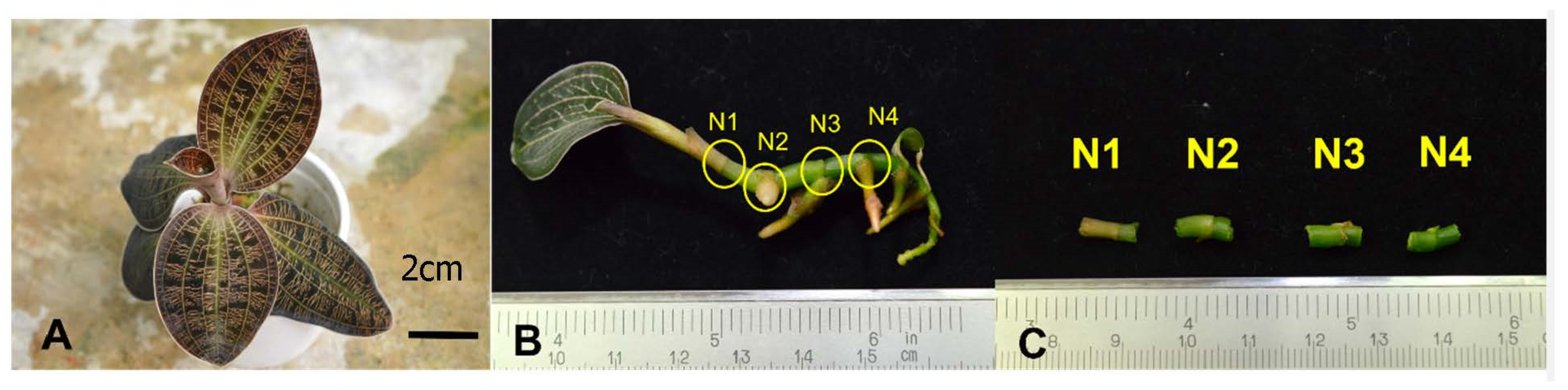
2.1. Establishment of In Vitro Plantlets

Macodes limii (Figure 1A) plants were collected from their natural habitat in the Kota Belud-Ranau area, and the species was identified by Mr. Jamirus Jamian and prior deposited at the Sandakan Herbarium of Forestry Research Centre, Sandakan, Sabah (specimen no: UMSDD-001-005/2018). For surface sterilization, plants were gently brushed and placed under a continuous flow of tap water to remove solid dirt particles. Stem segments were then sterilized in 70% (v/v) ethanol for 20 s, followed by the treatment of 2% (v/v) sodium hypochlorite (Clorox®) for 15 min, and finally rinsed three times with sterilized distilled water. The sterilized nodal explants were cultured on half-strength of Murashige and Skoog (MS) media [30], 3% (w/v) sucrose, and 1% (w/v) activated charcoal.

2.2. Effects of Node Position and PGRs

To study the effect of node positions on in vitro shoot multiplication, explants were excised from the 10-month-old in vitro grown plantlets to four node positions counted from the tip of the plant; N1 referred to shoot tip and N2, N3, and N4 referred to the 2nd, 3rd, and 4th node, respectively (Figure 1B,C). The procedure was conducted in the morning for all treatments to minimize the regulation of endogenous phytohormones during nodal excision [31]. All explants were transferred to a glass jar containing 50 mL of half-strength MS medium with 1% (w/v) activated charcoal, 3% (w/v) sucrose, 0.3% (w/v) of Gelrite, and supplemented with various types of PGRs including auxin such as α-naphthalene acetic acid (NAA), picloram, 2,4-dichlorophenoxyacetic acid (2,4-D), and cytokinin such as 6-benzylaminopurine (BAP), kinetin, and thidiazuron (TDZ) at 1.0 mg/L, respectively. Each treatment consisted of one node with five replicates. Medium devoid of growth regulators served as control. The pH of the medium was adjusted to pH 5.7 with 0.1 N of HCl or NaOH before autoclaving at 121 °C, 101.325 kPa for 20 min. All cultures were incubated for 90 days at 25 ± 2 °C under a 12 h photoperiod with cool white fluorescent lamps (150 µmol m−2 s−1). Explants were subcultured three times onto fresh media at 4-week intervals.

2.3. Rooting and Acclimatization of Plantlets

To promote root initiation, the in vitro raised shoots of M. limii were rooted on half-strength MS medium with 1% (w/v) activated charcoal, 3% (w/v) sucrose, 0.3% (w/v) of Gelrite, and supplemented with indole-3-acetic acid (IAA) at 1.0 mg/L. The cultures were incubated for another 60 days at 25 ± 2 °C under a 12 h photoperiod with cool white fluorescent lamps (150 µmol m−2 s−1). Regenerated plantlets of M. limii with roots were removed from the glass jars and then washed thoroughly with distilled water to remove the residual medium gel. The plantlets were then transferred to a (5 × 7 cm) plastic pot filled with perlite, charcoal, and sphagnum moss (1:1:1). The plantlets were maintained at 30 °C in a greenhouse nursery with a relative humidity of 70–80% for two months. The successfully acclimatized plantlets were transferred back to the Poring Orchid Conservation Centre (POCC), Ranau for reintroduction.

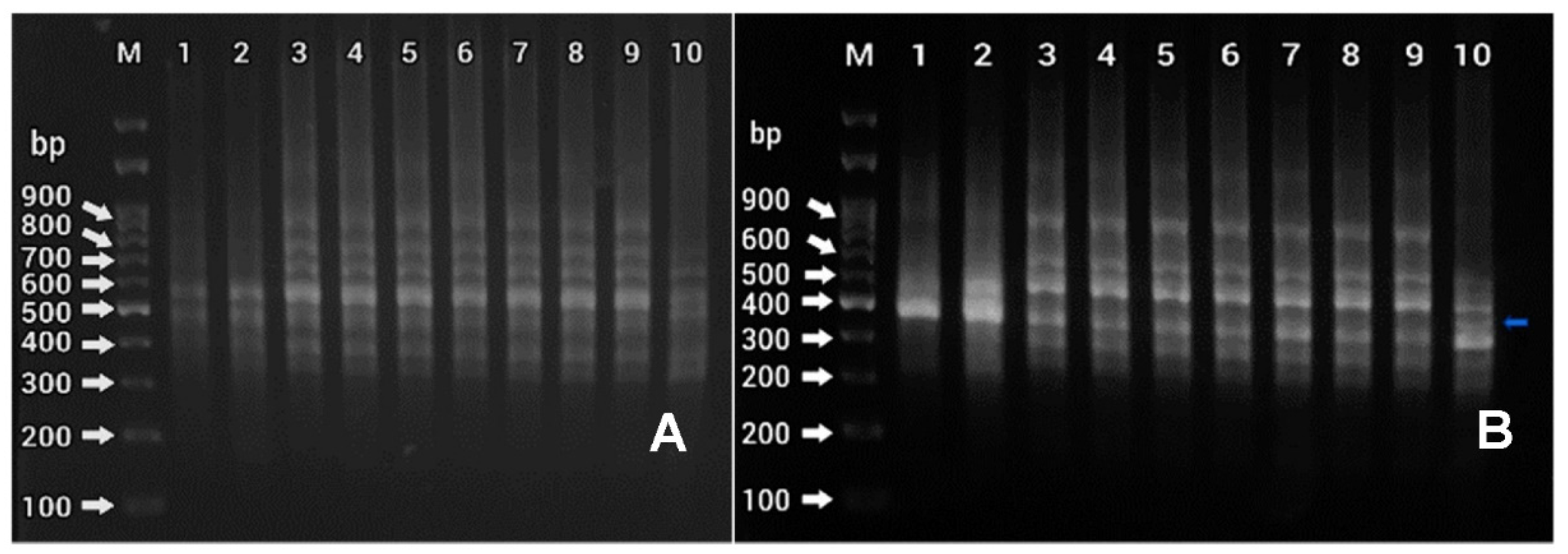
2.4. Analysis of Genetic Fidelity Using ISSR Primers

Genomic DNA was extracted from the young leaves of the mother plant and eight randomly selected in vitro regenerated hardened plants (10 months old) using a salting-out procedure [32]. For genetic stability assessment, the five ISSRs primers (UBC 808, UBC 835, UBC 836, UBC 841, and UBC 842) [26,27] used in this study were purchased from Integrated DNA Technologies, Singapore. PCR was performed in a total of 15 µL reaction containing 100 ng DNA, 0.48 µM of each primer and 1X Power Taq (Bioteke, Wuxi, China). DNA amplification was performed in a thermocycler programmed for 35 cycles at 30 s (90 °C), 30 s (50 °C), and 50 s (72 °C). The PCR products were resolved on a 2% (w/v) agarose gel electrophoresis using 1X Tris–Acetate–Ethylenediamine tetraacetic acid buffer.

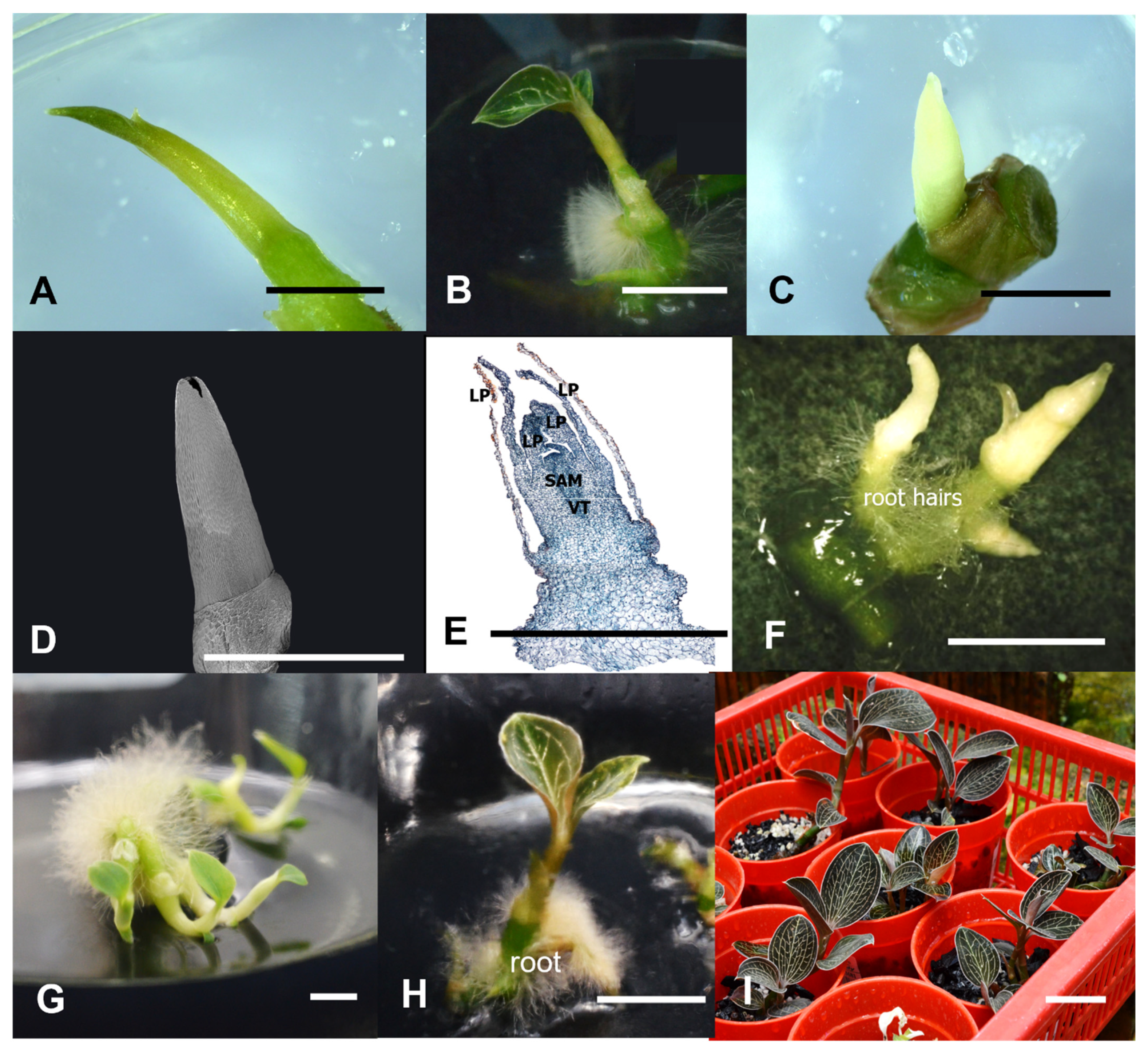
2.5. Histology and Scanning Electron Microscopy (SEM) of Axillary Shoot Induced from Nodal Explant

Tissues for histological observations were fixed in a solution of formaldehyde alcohol acetic acid (95% (v/v) ethyl alcohol + glacial acetic acid + formaldehyde + water, 10:1:2:7), dehydrated in a tertiary butyl alcohol series, embedded in paraffin wax, sectioned at 10 µm thickness and stained with 0.5% (v/v) safranin-O and 0.1% (v/v) Fast Green. Visualization was carried out using a light microscope (Olympus BX-53, Tokyo, Japan). For SEM, a scanning electron microscope (S-3400N; Hitachi, Tokyo, Japan) was used for the examination and photography of the specimens.

2.6. Statistical Analysis

Statistical analysis was performed using IBM SPSS Statistic version 28 (SPSS Inc., Chicago, IL, USA). A factorial two-way analysis of variance (ANOVA) was used to evaluate the effects of two independent factors, namely node position and PGRs treatment, on the in vitro regeneration capacity. Each treatment consisted of five replicates, and the experiment was repeated two times. Data are presented as mean and were compared with Tukey’s test at a 5% probability level.

3. Results

3.1. Effects of Node Position and PGRs on In Vitro Regeneration

The effects of node position and PGRs on the in vitro regeneration of M. limii were investigated. The highest shoot proliferation response (95%) was observed on N3 and N4 explants cultured on half-strength MS basal medium supplemented with 1.0 mg/L of TDZ (Table 1). The two-way ANOVA showed significant effects on the node position (N), PGRs treatment (P), and the interaction between them (N × P) on the average number of shoot production (Table 2). An average of three new shoots were produced from the N3 or N4 explants cultured on half-strength MS medium supplemented with 1.0 mg/L TDZ after 90 days of culture (DAC). In this study, it was obvious that the N1 explant was less responsive in shoot proliferation compared to the N2, N3, and N4 explants. Two types of regeneration pathways were observed between N1 (shoot tip) and N2, N3, and N4 explants (second, third, and fourth nodes). For N1, the shoot elongation was observed from the excised part and eventually developed into leaf after 90 DAC (Figure 2A,B). Meanwhile, for explants N2–N4, a direct organogenesis through a shoot bud formation was observed. It began with a formation of a white bulge (axillary bud) from the swollen node within 14 DAC, followed by a differentiation into multiple shoots (Figure 2C–G). The histological and SEM observations showed the presence of meristematic cells (axillary shoot) developing from the nodal explant and later forming microshoots through direct organogenesis (Figure 2D–G). The formation of root hair on the nodal explants was also observed from the early stage of shoot multiplication (Figure 2F,G).
The highest shoot length was observed on explant N4 cultured on 1.0 mg/L TDZ with an average of 1.8 ± 0.4 cm shoot length after 90 DAC (Table 1). Besides this treatment, the control medium with no addition of any PGRs also gave a promotive effect on shoot length. The current finding indicates that even though node position influenced the shoot proliferation response and the number of shoots produced, there was no significant difference among them in terms of shoot length (Table 2). Meanwhile, the result also revealed that explants cultured on 1.0 mg/L of NAA gave the least response on shoot proliferation as well as shoot length.

3.2. In Vitro Rooting and Acclimatization of M. limii Plantlets

Microshoots regenerated from all explants (1.4–1.8 cm length) (Figure 2G), were excised carefully and cultured on half-strength MS basal medium supplemented with 1.0 mg/L IAA. After eight weeks of culture, the microshoots were well-developed with two leaves (1.0 cm length) and two to three roots (0.5–1.0 cm length) per responded explant, with a 100% rooting response (Figure 2H). The well-developed plantlets of M. limii were selected and acclimatized in a pot containing perlite, charcoal, and sphagnum moss (1:1:1) and maintained under greenhouse conditions for 60 days, achieving a 60% of survivability (Figure 2I). The healthy acclimatized plantlets were selected and transferred back to POCC, Ranau for reintroduction.

3.3. Determination of Genetic Homogeneity Using ISSR

Five ISSR primers were utilized in this analysis, generating a total of 260 amplified DNA fragments ranging from 310 to 1200 bp (Figure 3). In this study, about 255 were monomorphic and 5 were polymorphic (Table 3). The percentage of monomorphism was 98.1% and polymorphism was 1.9%. Both primers of UBC 808 and UBC 836 produced 100% monomorphism, meanwhile the primer of UBC 835 produced one polymorphic band (1.5% polymorphism), and the primers of UBC 841 and UBC 842 produced two polymorphic bands with 3.9% and 3.7% polymorphism, respectively. The finding indicates that the micropropagation of M. limii via nodal explants showed a low risk of genetic instability and therefore it is a reliable method for plantlet regeneration.

4. Discussion

Plant regeneration through shoot tip and nodal cultures has been reported to be the most applicable and reliable method of true-to-type in vitro propagation [12,33]. Different types of explants have different distributions of meristematic cells, which also differ among different species. In the present study, N2, N3, and N4 showed a significant response on shoot proliferation compared to N1 (shoot tip). The formation of axillary bud is controlled by apical dominance and endogenous hormones. Apical dominance is the process whereby the shoot tip inhibits the growth of axillary buds along the stem [34,35,36]. High production of auxin in shoot apex and subsequent basipetal auxin transport inhibits the outgrowth of axillary buds [37]. The removal of the shoot tip (apical meristem) caused lateral buds to break their dormancy and stimulated axillary bud outgrowth [38,39]. The suitability of nodal segment rather than shoot tip explant in the mass propagation of jewel orchids have been previously demonstrated in A. sikkimensis, A. regalis [40], and A. elatus [14]. Previously, Vasudevan and Staden [41] also reported that the absence of axillary shoot proliferation in shoot tip explants of Ansellia africana was due to auxin synthesized at the shoot tip explant, that exerted strong apical dominance. However, when compared to another study, the shoot tip explant was found superior to the nodal explant in axillary shoot regeneration of A. formosanus [16].
Cytokinins play many roles in plant development, often acting in concert with other hormones, most notably auxin, to regulate cell division and differentiation [42], apical dominance, and axillary bud outgrowth [43]. In this study, nodal explants of M. limii cultured on basal medium containing 1.0 mg/L of TDZ for 90 days yielded the highest number of axillary bud formations. TDZ possesses cytokinin- and auxin-like effects [44], which promoted the synthesis and/or accumulation of endogenous cytokinins, which significantly induced the proliferation of axillary shoots and released lateral buds from dormancy [45]. TDZ is widely used for micropropagation of many orchids including jewel orchids because of its incredible ability to induce organogenesis. TDZ has been previously used to induce axillary shoot production in jewel orchids including A. formosanus [46] and A. elatus [28,47]. TDZ, which is a substituted phenyl urea with cytokinin-like activity, has also been reported as an effective growth regulator for in vitro morphogenesis and organogenesis in other orchids including Phalaenopsis cv. Surabaya [48] and Dimorphorcis lowii [49].
The well-rooted plantlets of M. limii were acclimatized for two months, with a survival rate of 60%. The healthy acclimatized plantlets were successfully transferred to the conservation center for reintroduction. Plantlets produced in vitro should be gradually acclimatized to the ex vitro conditions to prevent high mortality after transfer to the environment of the field [50], and also to allow the plantlets to repair abnormalities in morphology, physiology, and anatomy before undergoing normal photosynthesis [51].
Typically, plantlets regenerated via axillary buds or direct somatic embryogenesis are considered to be the most genetically uniform [25,52]. The uniformity of the in vitro cultures against genetic variations despite repeated subcultures can be linked to their respective genotype, which could be genetically resistant to alteration in their genetic materials [53]. However, artificial in vitro conditions, modified nutrients, and exposure to synthetic hormones might generate a genetic instability in cultured plants known as somaclonal variation [21,53,54]. Furthermore, the application of TDZ and prolonged exposure to it increased the chances of heritable somaclonal variation induction within the regenerated plantlets [55,56,57]. Molecular markers suitable for generating DNA profiles have served as an effective tool to assess the varietal identification, phylogenetic analysis, and genetic diversity [58]. In the current study, the genetic homogeneity of the in vitro propagated M. limii assessed via ISSR primers analysis, was 98.1% of monomorphism and 1.9% of polymorphism, indicating that the in vitro regenerated plants were confirmed as clonally uniform and genetically stable. Previously, the ISSR markers have been successfully utilized for the analysis of genetic fidelity within lines of some jewel orchids, including A. elatus [29] and A. formosanus [26].

5. Conclusions

Macodes limii is a Sabah endemic jewel orchid greatly appreciated for its attractive foliar venation but is still underexplored for its ornamental potential. Considering the rare and threatened status of the species, a protocol for in vitro regeneration of M. limii has been successfully established by using nodal explants, cultured on half-strength MS basal media incorporated with 1.0 mg/L TDZ. The in vitro regenerated plantlets showed high genetic similarity to the mother plant via an ISSR analysis, and were finally transferred to POCC for ex situ conservation. The current study has attempted to highlight the importance of the micropropagation protocol in combination with the reintroduction effort as an excellent opportunity for conserving endangered plant biodiversity and might as well be applied for large-scale propagation of this native plant.

Author Contributions

Conceptualization and methodology, D.D. and J.A.G.; formal analysis, D.D. and A.A.M.F.; investigation, D.D.; resources, N.A.R. and J.A.G.; data curation, D.D. and J.A.G.; writing—original draft preparation, D.D.; review and editing J.A.G.; supervision, N.A.R. and R.A.M.M.; project administration, J.A.G. All authors have read and agreed to the published version of the manuscript.

Funding

This study was financially supported by the Ministry of Higher Education (MOHE), Malaysia, Reference Code: FRGS/1/2018/STG05/UMS/02/2, and the Universiti Malaysia Sabah (UMS) Research Grant: UMSGreat GUG0220-2018.

Institutional Review Board Statement

Not applicable.

Informed Consent Statement

Not applicable.

Data Availability Statement

Data are available with a request to the first author.

Acknowledgments

We would like to acknowledge the Unit for Orchid Studies, UMS for the laboratory facilities, and the Poring Orchid Conservation Centre (POCC) under Sabah Parks for supporting the orchid reintroduction program. This study was conducted under permission from the Sabah Biodiversity Centre (license reference no: JKM/MBS.1000-2/2 JLD.10(4)).

Conflicts of Interest

The authors declare no conflict of interest.

References

  1. Bhattacharjee, A.; Chowdhery, H.J. Two frequently confused species of ‘Jewel Orchid’ (Orchidaceae—Goodyerinae) from India. Taiwania 2013, 58, 213–216. [Google Scholar] [CrossRef]
  2. Du, X.M.; Irino, N.; Furusho, N.; Hayashi, J.; Shoyama, Y. Pharmacologically active compounds in the Anoectochilus and Goodyera species. J. Nat. Med. 2008, 62, 132–148. [Google Scholar] [CrossRef]
  3. He, C.N.; Wang, C.L.; Guo, S.X.; Yang, J.S.; Xiao, P.G. A novel flavonoid glucoside from Anoectochilus roxburghii (Wall.) Lindl. J. Integr. Plant Biol. 2006, 48, 359–363. [Google Scholar] [CrossRef]
  4. Wood, J.J.; Lamb, A.; Lun, C.C.; Beaman, J.H.; Beaman, T.E. The Orchids of Mount Kinabalu; Natural History Publication: Kota Kinabalu, Malaysia, 2011; Volume 2, p. 1184. [Google Scholar]
  5. Li, B.; Tang, M.J.; Tang, K.; Zhao, L.F.; Guo, S.X. Screening for differentially expressed genes in Anoectochilus roxburghii (Orchidaceae) during symbiosis with the mycorrhizal fungus Epulorhiza sp. Sci. China Life Sci. 2012, 55, 164–171. [Google Scholar] [CrossRef] [Green Version]
  6. Tomita, M. Symbiotic of seed germination and development of Goodyera schlechtendaliana in vitro. Plant Tissue Cult. Lett. 1995, 12, 151–155. [Google Scholar] [CrossRef]
  7. Shiau, Y.J.; Nalawade, S.M.; Hsai, C.N.; Tsay, H.S. Propagation of Haemaria discolor via in vitro seed germination. Biol. Plant. 2005, 49, 341–346. [Google Scholar] [CrossRef]
  8. Shiau, Y.J.; Sagare, A.P.; Chen, U.C.; Yang, S.R.; Tsay, H.S. Conservation of Anoectochilus formosanus Hayata by artificial cross-pollination and in vitro culture of seeds. Bot. Bull. Acad. Sin. 2002, 43, 123–130. [Google Scholar]
  9. Chou, L.C.; Chang, D.C.N. Asymbiotic and symbiotic seed germination of Anoectochilus formosanus and Haemaria discolor and their F1 hybrids. Bot. Bull. Acad. Sin. 2004, 45, 143–147. [Google Scholar] [CrossRef]
  10. Indan, H.; David, D.; Jawan, R.; Marbawi, H.; Gansau, J.A. Development and characterization of flower and capsule in Borneo jewel orchid Macodes limii J.J. Wood and A.L. Lamb (Orchidaceae: Asparagales). J. Asia-Pacific Biodivers. 2021, 14, 236–241. [Google Scholar] [CrossRef]
  11. Chugh, S.; Guha, S.; Rao, I.U. Micropropagation of orchids: A review on the potential of different explants. Sci. Hortic. 2009, 122, 507–520. [Google Scholar] [CrossRef]
  12. Yam, T.W.; Arditti, J. Micropropagation of Orchids, 3rd ed.; John Wiley & Sons, Inc.: Chischester, UK, 2017; p. 2225. [Google Scholar] [CrossRef]
  13. Pant, B.; Pradhan, S.; Paudel, M.R.; Shah, S.; Pandey, S.; Joshi, P.R. Various culture techniques for the mass propagation of medicinal orchids from Nepal. Acta Hortic. 2019, 1262, 109–124. [Google Scholar] [CrossRef]
  14. Sherif, N.A.; Benjamin, J.H.F.; Muthukrishnan, S.; Kumar, T.S.; Rao, M.V. Regeneration of plantlets from nodal and shoot tip explants of Anoectochilus elatus Lindley, an endangered terrestrial orchid. Afr. J. Biotechnol. 2012, 11, 7549–7553. [Google Scholar] [CrossRef]
  15. Zhang, A.; Wang, H.; Shao, Q.; Xu, M.; Zhang, W.; Li, M. Large scale in vitro propagation of Anoectochilus roxburghii for commercial application: Pharmaceutically important and ornamental plant. Ind. Crops Prod. 2015, 70, 158–162. [Google Scholar] [CrossRef]
  16. Winarto, B.; Samijan. Axillary shoots derived from shoot tips in in vitro mass propagation of Anoectochilus formosanus Hayata. Agraarteadus 2018, 29, 121–130. [Google Scholar] [CrossRef]
  17. Poobathy, R.; Zakaria, R.; Murugaiyah, V.; Subramaniam, S. Surface sterilization and micropropagation of Ludisia discolor. Biocatal. Agric. Biotechnol. 2019, 22, 101380. [Google Scholar] [CrossRef]
  18. Mien, P.; Hang, P.; Ket, N.; Anh, T.; Phe, P.; Thanh, N.T. In vitro Culture of Jewel orchids (Anoectochilus setaceus Blume). Annu. Reports. 1998, 33, 436–444. [Google Scholar]
  19. Thanh, N.T.; Hang, P.L.; Van Ket, N.; Anh, T.T.; Van Phe, P.; Gam, N.T.; Mien, P.T. The role of different medium and plant hormones on multiple shoots of Jewel orchids (Anoectochilus setaceus Blume). VNU J. Sci. Nat. Sci. Technol. 2012, 28, 47–53. [Google Scholar]
  20. Sherif, N.A.; Kumar, T.S.; Rao, M.V. In vitro regeneration by callus culture of Anoectochilus elatus Lindley, an endangered terrestrial jewel orchid. In Vitro Cell. Dev. Biol.-Plant 2016, 52, 72–80. [Google Scholar] [CrossRef]
  21. Krishna, H.; Alizadeh, M.; Singh, D. Somaclonal variations and their applications in horticultural crops improvement. 3 Biotech 2016, 6, 54. [Google Scholar] [CrossRef] [Green Version]
  22. Sherif, N.A.; Kumar, T.S.; Rao, M.V. DNA barcoding and genetic fidelity assessment of micropropagated Aenhenrya rotundifolia (Blatt.) C.S. Kumar and F.N. Rasm.: A critically endangered jewel orchid. Physiol. Mol. Biol. Plants 2020, 26, 2391–2405. [Google Scholar] [CrossRef]
  23. Tikendra, L.; Koijam, A.S.; Nongdam, P. Molecular markers based genetic fidelity assessment of micropropagated Dendrobium chrysotoxum Lindl. Meta Gene 2019, 20, 100562. [Google Scholar] [CrossRef]
  24. Oliya, B.K.; Chand, K.; Thakuri, L.S.; Baniya, M.K.; Sah, A.K.; Pant, B. Assessment of genetic stability of micropropagated plants of Rhynchostylis retusa (L.) using RAPD markers. Sci. Hortic. 2020, 281, 110008. [Google Scholar] [CrossRef]
  25. Bhattacharyya, P.; Ghosh, S.; Mandi, S.S.; Kumaria, S.; Tandon, P. Genetic variability and association of AFLP markers with some important biochemical traits in Dendrobium thyrsiflorum, a threatened medicinal orchid. S. Afr. J. Bot. 2017, 109, 214–222. [Google Scholar] [CrossRef]
  26. Lin, S.F.; Tsay, H.S.; Chou, T.W.; Yang, M.J.; Cheng, K.T. Genetic variation of Anoectochilus formosanus revealed by ISSR and AFLP analysis. J. Food Drug Anal. 2007, 15, 156–162. [Google Scholar] [CrossRef]
  27. Zhang, F.; Lv, Y.; Dong, H.; Guo, S. Analysis of genetic stability through Intersimple sequence repeats molecular markers in micropropagated plantlets of Anoectochilus formosanus H AYATA, a medicinal plant. Biol. Pharm. Bull. 2010, 33, 384–388. [Google Scholar] [CrossRef] [Green Version]
  28. Sherif, N.A.; Kumar, T.S.; Rao, M.V. In vitro propagation and genetic stability assessment of an endangered terrestrial jewel orchid Anoectochilus elatus Lindl. Indian J. Exp. Biol. 2017, 55, 853–863. [Google Scholar]
  29. Sherif, N.A.; Benjamin, J.F.; Kumar, T.S.; Rao, M.V. Somatic embryogenesis, acclimatization and genetic homogeneity assessment of regenerated plantlets of Anoectochilus elatus Lindl., an endangered terrestrial jewel orchid. Plant Cell. Tissue Organ Cult. 2018, 132, 303–316. [Google Scholar] [CrossRef]
  30. Murashige, T.; Skoog, F. A revised medium for rapid growth and bio assays with tobacco tissue cultures. Physiol. Plant. 1962, 15, 473–497. [Google Scholar] [CrossRef]
  31. Otiende, M.A.; Fricke, K.; Nyabundi, J.O.; Ngamau, K.; Hajirezaei, M.R.; Druege, U. Involvement of the auxin–cytokinin homeostasis in adventitious root formation of rose cuttings as affected by their nodal position in the stock plant. Planta 2021, 254, 65. [Google Scholar] [CrossRef]
  32. Miller, S.A.; Dykes, D.D.; Polesky, H.F. A simple salting out procedure for extracting DNA from human nucleated cells. Nucleic Acids Res. 1988, 16, 1215. [Google Scholar] [CrossRef] [Green Version]
  33. Stephin, S.; Gangaprasad, A.; Mathew, S.P.; Muthukrishnan, S. Enhanced in vitro shoot multiplication of Piper sarmentosum by suppression of apical dominance. Proc. Natl. Acad. Sci. India Sect. B-Biol. Sci. 2020, 90, 87–94. [Google Scholar] [CrossRef]
  34. Chabikwa, T.G.; Brewer, P.B.; Beveridge, C.A. Initial bud outgrowth occurs independent of Auxin flow from out of buds. Plant Physiol. 2019, 179, 55–65. [Google Scholar] [CrossRef] [PubMed] [Green Version]
  35. Cline, M.G. Concepts and terminology of apical dominance. Am. J. Bot. 1997, 84, 1064–1069. [Google Scholar] [CrossRef] [PubMed] [Green Version]
  36. Kebrom, T.H. A Growing Stem Inhibits Bud Outgrowth—The Overlooked Theory of Apical Dominance. Front. Plant. Sci. 2017, 8, 1874. [Google Scholar] [CrossRef]
  37. Muller, D.; Waldie, T.; Miyawaki, K.; To, J.P.; Melnyk, C.W.; Kieber, J.J.; Kakimoto, T.; Leyser, O. Cytokinin is required for escape but not release from auxin mediated apical dominance. Plant J. 2015, 82, 874–886. [Google Scholar] [CrossRef] [Green Version]
  38. Barbier, F.F.; Dun, E.A.; Kerr, S.C.; Chabikwa, T.G.; Beveridge, C.A. An Update on the Signals Controlling Shoot Branching. Trends Plant Sci. 2019, 24, 220–236. [Google Scholar] [CrossRef]
  39. Balla, J.; Medveďová, Z.; Kalousek, P.; Matiješčuková, N.; Friml, J. Auxin flow-mediated competition between axillary buds to restore apical dominance. Sci. Rep. 2016, 6, 35955. [Google Scholar] [CrossRef] [Green Version]
  40. Gangaprasad, A.; Latha, P.G.; Seeni, S. Micropropagation of terrestrial orchids, Anoectochilus sikkimensis and Anoectochilus regalis. Indian J. Exp. Biol. 2000, 38, 149–154. [Google Scholar]
  41. Vasudevan, R.; van Staden, J. Cytokinin and explant types influence in vitro plant regeneration of Leopard Orchid (Ansellia africana Lindl.). Plant Cell. Tissue Organ Cult. 2011, 107, 123–129. [Google Scholar] [CrossRef]
  42. Kieber, J.J.; Schaller, G.E. Cytokinin signaling in plant development. Development 2018, 145, dev149344. [Google Scholar] [CrossRef] [Green Version]
  43. Yuan, C.; Xi, L.; Kou, Y.; Zhao, Y.; Zhao, L. Current perspectives on shoot branching regulation. Front. Agric. Sci. Eng. 2015, 2, 38–52. [Google Scholar] [CrossRef] [Green Version]
  44. Deepa, A.V.; Anju, M.; Thomas, D.T. The Applications of TDZ in Medicinal Plant Tissue Culture. In Thidiazuron: From Urea Derivative to Plant Growth Regulator; Ahmad, N., Faisal, M., Eds.; Springer: Singapore, 2018; pp. 297–314. [Google Scholar] [CrossRef]
  45. Li, Y.; Hu, J.; Xiao, J.; Guo, G.; Jeong, B.R. Foliar thidiazuron promotes the growth of axillary buds in Strawberry. Agronomy 2021, 11, 594. [Google Scholar] [CrossRef]
  46. Ket, N.V.; Hahn, E.J.; Park, S.Y.; Chakrabarty, D.; Paek, K.Y. Micropropagation of an endangered orchid Anoectochilus formosanus. Biol. Plant. 2004, 48, 339–344. [Google Scholar] [CrossRef]
  47. Raja, D.H.D. Effect of cytokinins on micropropagation of Anoectochilus elatus Lindl. from shoot tip explants—An endangered medicinal orchid. IOSR J. Biotechnol. Biochem. 2017, 3, 73–76. [Google Scholar] [CrossRef]
  48. Balilashaki, K.; Vahedi, M.; Karimi, R. In vitro direct regeneration from node and leaf explants of Phalaenopsis cv. ‘Surabaya’. Plant Tissue Cult. Biotechnol. 2015, 25, 193–205. [Google Scholar] [CrossRef] [Green Version]
  49. Jainol, J.E.; Gansau, J.A. Embryogenic callus induction from leaf tip explants and protocorm-like body formation and shoot proliferation of Dimorphorchis lowii: Borneon endemic orchid. Agrivita 2017, 39, 1–10. [Google Scholar] [CrossRef] [Green Version]
  50. Hazarika, B.N. Acclimatization of tissue-cultured plants. Curr. Sci. 2003, 85, 1704–1712. [Google Scholar]
  51. Pospóšilová, J.; Tichá, I.; Kadleček, P.; Haisel, D.; Plzáková, Š. Acclimatization of micropropagated plants to ex-vitro conditions. Biol. Plant. 1999, 42, 481–497. [Google Scholar] [CrossRef]
  52. Konar, S.; Karmakar, J.; Ray, A.; Adhikari, S.; Bandyopadhyay, T.K. Regeneration of plantlets through somatic embryogenesis from root derived calli of Hibiscus sabdariffa L. (Roselle) and assessment of genetic stability by flow cytometry and ISSR analysis. PLoS ONE 2018, 13, e0202324. [Google Scholar] [CrossRef]
  53. Chin, C.K.; Lee, Z.H.; Mubbarakh, S.A.; James, A.J.J.; Chew, B.L.; Subramaniam, S. Effects of plant growth regulators and activated charcoal on somaclonal variations of protocorm-like bodies (PLBs) of Dendrobium Sabin Blue orchid. Biocatal. Agric. Biotechnol. 2019, 22, 101426. [Google Scholar] [CrossRef]
  54. Kang, L.; Zheng, K.; Xie, Y.; Deng, Y.; Yu, Y.; Zhu, M.; Xi, R.; Deng, X. Efficient tissue culture protocol for Magnolia lucida (Magnoliaceae) and confirmation of genetic stability of the regenerated plants. Plants 2020, 9, 997. [Google Scholar] [CrossRef] [PubMed]
  55. Dewir, Y.H.; Nurmansyah; Naidoo, Y.; Teixeira da Silva, J.A. Thidiazuron-induced abnormalities in plant tissue cultures. Plant Cell Rep. 2018, 37, 1451–1470. [Google Scholar] [CrossRef] [PubMed]
  56. Samarfard, S.; Kadir, M.A.; Kadzimin, S.B.; Saud, H.M.; Ravanfar, S.A.; Danaee, M. In vitro propagation and detection of somaclonal variation in Phalaenopsis gigantea as affected by chitosan and thidiazuron combinations. HortScience 2014, 49, 82–88. [Google Scholar] [CrossRef]
  57. Roy, A.R.; Sajeev, S.; Pattanayak, A.; Deka, B.C. TDZ induced micropropagation in Cymbidium giganteum Wall. Ex Lindl. and assessment of genetic variation in the regenerated plants. Plant Growth Regul. 2012, 68, 435–445. [Google Scholar] [CrossRef]
  58. Kumari, S.; Yadav, K.; Singh, N. Evaluation of genetic fidelity among micropropagated plants of Salvadora persica L. using DNA-based markers. Meta Gene 2017, 14, 129–133. [Google Scholar] [CrossRef]
Figure 1. Source of explants for in vitro regeneration of M. limii. (A) Wild plant of M. limii; (B) In vitro grown M. limii plantlet (starting material); (C) Defoliated explants with different node positions.
Horticulturae 08 00155 g001
Figure 2. In vitro regeneration of Macodes limii. (A) Shoot elongated from the excised part of N1 (shoot tip) explant; (B) development of true leaves from shoot tip (N1) explant; (C) axillary bud break on N3 explant; (D) SEM of axillary bud on N3 explant; (E) histology of axillary shoot formation from N3 explant (LP—leaf primordia, SAM—shoot apical meristem, VT—vascular tissue); (F) multiple shoots induced from N3 explant; (G) multiple shoots with root hairs from N3 explant; (H) development of true leaves and root formation from the regenerated shoots; (I) fully acclimatized plantlets ready to be transferred to Poring Orchid Conservation Centre. Bars: (AG) 0.5 cm; (H) 1 cm; (I) 3 cm.
Horticulturae 08 00155 g002
Figure 3. Gel electrophoresis of amplified fragments of Macodes limii produced by ISSR Primers (A) UBC 808 (B) UBC 835. Note: M: 100 bp marker (Fermentas); Lanes 1–2: mother plant; lanes 3–10: in vitro regenerated M. limii.
Horticulturae 08 00155 g003
Table 1. Effects of PGRs and explant node position on in vitro regeneration of M. limii after 90 days of culture.
PGRsNode
Position
% Shoot
Formation
No. of ShootsShoot Length (cm)
ControlN1350.7 ± 0.5 defgh1.6 ± 1.1 ab
N2751.4 ± 0.5 bcdefgh1.6 ± 0.5 ab
N3851.9 ± 0.7 abcdefg1.4 ± 0.5 abc
N4602.1 ± 1.5 abcde1.2 ± 0.8 abcde
1.0 mg/L NAAN100.0 h0.00 g
N2350.9 ± 1.3 cdefgh0.2 ± 0.3 fg
N3200.4 ± 0.5 fgh0.3 ± 0.4 efg
N4150.3 ± 0.5 gh0.2 ± 0.3 fg
1.0 mg/L PicloramN1400.8 ± 0.4 defgh1.3 ± 0.8 abcd
N2601.2 ± 0.8 cdefgh0.8 ± 0.4 bcdefg
N3601.2 ± 0.8 cdefgh0.8 ± 0.4 bcdefg
N4501.0 ± 0.9 cdefgh0.7 ± 0.6 bcdefg
1.0 mg/L 2,4-DN1200.4 ± 0.5 fgh0.6 ± 0.8 cdefg
N2601.2 ± 1.0 cdefgh0.9 ± 0.8 abcdefg
N3802.2 ± 1.2 abcde1.2 ± 0.5 abcde
N4301.0 ± 1.6 cdefgh0.4 ± 0.5 defg
1.0 mg/L BAPN1400.8 ± 0.4 defgh0.8 ± 0.7 bcdefg
N2801.8 ± 0.8 abcdefg0.7 ± 0.3 bcdefg
N3802.0 ± 1.1 abcdef0.9 ± 0.7 abcdefg
N4702.2 ± 1.4 abcde0.5 ± 0.3 cdefg
1.0 mg/L KinetinN1300.6 ± 0.5 efgh0.7 ± 0.7 bcdefg
N2801.8 ± 0.8 abcdefg0.7 ± 0.4 bcdefg
N3802.5 ± 1.4 abc0.6 ± 0.4 cdefg
N4802.2 ± 1.2 abcde0.7 ± 0.4 bcdefg
1.0 mg/L TDZN1400.8 ± 0.4 defgh1.0 ± 0.7 abcdef
N2852.3 ± 1.1 abcd1.4 ± 0.2 abc
N3953.1 ± 1.0 a1.6 ± 0.5 ab
N4953.0 ± 1.1 ab1.8 ± 0.4 a
Data are means from two repeated experiments. Each treatment consists of five replicates. Means followed by the same letter within each column are not significantly different according to Tukey’s test at p < 0.05.
Table 2. Two-way ANOVA analysis showing the effects of node position and PGRs on the average number of shoot production and shoot length from the responded explant.
Source of VariationSSdfMean SquareFp Value
No. of shoots
Node position (N)70.414323.47125.539<0.001 **
PGRs (P)88.621614.77016.071<0.001 **
N × P29.636181.6461.7910.027 *
Shoot length
Node position (N)1.27030.4231.3620.255 NS
PGRs (P)49.24068.20726.397<0.001 **
N × P9.757180.5421.7440.033 *
** p value is highly significant at p < 0.001 level, * significant at p < 0.05 level, NS—not significant.
Table 3. List of primers and size of amplified fragments generated by ISSRs markers for DNA fingerprinting of in vitro plantlets of M. limii.
Primer CodePrimer Sequence
(5′-3′)
Nucleotide Fragment Size (bp)Total Bands AmplifiedMonomorphic BandPolymorphic BandMonomorphism (%)Polymorphism
(%)
UBC808AGAGAGAGAGAGAGAGC350–950707001000
UBC835AGAGAGAGAGAGAGAGYC325–12006968198.61.5
UBC836AGAGAGAGAGAGAGAGYC600–1000161601000
UBC841GAGAGAGAGAGAGAGAYC320–9005149296.13.9
UBC842GAGAGAGAGAGAGAGAYG310–8005452296.33.7
Total260255598.11.9
Publisher’s Note: MDPI stays neutral with regard to jurisdictional claims in published maps and institutional affiliations.

