Article

Effects of Azorhizobium caulinodans and Piriformospora indica Co-Inoculation on Growth and Fruit Quality of Tomato (Solanum lycopersicum L.) under Salt Stress

1
College of Horticulture and Gardening, Yangtze University, Jingzhou 434025, China
2
Department of Biology, Faculty of Arts and Sciences, Recep Tayyip Erdogan University, Rize 53100, Turkey
*
Author to whom correspondence should be addressed.
Horticulturae 2022, 8(4), 302; https://doi.org/10.3390/horticulturae8040302
Submission received: 28 January 2022 / Revised: 21 March 2022 / Accepted: 21 March 2022 / Published: 2 April 2022

Abstract

:
Salt stress is a worldwide environmental signal, reducing the growth and yield of crops. To improve crop tolerance to salt, several beneficial microbes are utilized. Here, nitrogen-fixing bacterium Azorhizobium caulinodans and root endophytic fungus Piriformospora indica were used to inoculate tomato (Solanum lycopersicum) under salt stress, and the effects of the co-inoculation were investigated. Results showed that A. caulinodans colonized in the intercellular space in stems and roots of tomato plants, while P. indica colonized in the root cortex. Two weeks following salt treatment, co-inoculated tomato plants grew substantially taller and had larger stem base diameters. Activities of superoxide dismutase (SOD), catalase (CAT), peroxidase (POD), and reduced and oxidized ascorbate and glutathione (i.e., AsA, DHA, GSH, and GSSG, respectively) concentrations along with the ratios of AsA/(AsA + DHA) and GSH/(GSH + GSSG) increased in the leaves of co-inoculated plants under salt stress. The co-inoculation significantly increased soluble proteins and AsA in fruits; however, concentrations of soluble sugars and proanthocyanins did not show significant changes, compared with NaCl only treatment. Data suggest that A. caulinodans and P. indica co-inoculation boosted tomato growth and improved the quality of tomato fruits under salt stress. O-inoculation of A. caulinodans and P. indica might be employed to enhance tomato plant salt tolerance.

1. Introduction

Soil salinity affects more than 6% of the world’s total land area (about 800 million hectares of land globally) [1], and roughly one billion hectares in more than 100 countries are affected (Food and Agriculture Organization, 2015). Poor irrigation techniques, incorrect fertilizer use, and industrial contamination have all contributed to the rise in this instance [2].
Among the various salts in the soil, NaCl is the most prevalent [3]. High salinity is caused by high concentrations of Na+ and Cl in the soil solution, which causes hyperosmotic stress, preventing plant roots from absorbing water and nutrients from the soil [4]. However, most crops such as rice [5,6,7], wheat [8,9], tomato [10,11], and many other plants [12,13,14,15] are glycophytes, and their development and yield are affected by excessive NaCl.
Salt stress causes ionic, osmotic, and secondary stresses in plants, including oxidative damage [16]. Under salt stress, plants need to change their physiological and biochemical mechanisms that regulate ion and osmotic balance in response to salt stress, minimizing salt stress damage and enhancing detoxification [17]. They need to regulate signaling pathways to re-establish cellular ionic, osmotic, and reactive oxygen species (ROS) homeostasis [16,18]. For example, the Salt Overly Sensitive pathway plays a crucial role in maintaining ionic homeostasis via extruding sodium ions into the apoplast [19,20,21]. Mitogen-activated protein kinase cascades also mediate ionic, osmotic, and ROS homeostasis [18,22,23]. SnRK2 (sucrose nonfermenting 1-related protein kinase 2) proteins, on the other hand, are involved in maintaining osmotic homeostasis [18,24].
In natural ecosystems, plants symbiose with microorganisms. These beneficial microorganisms, such as arbuscular mycorrhizal fungi [25,26,27], ectomycorrhizal fungi [28,29,30], ericoid mycorrhizal fungi [31], root endophytic fungi [32,33,34], and plant growth-promoting rhizobacteria [35,36,37], improve plant resistance to salt stress. These microorganisms alter physiological processes in their host plants. For instance, ectomycorrhizal fungi enhanced root NO3 uptake and mediated K+/Na+ homeostasis [38,39], and also improved water transport properties by regulating the role of aquaporins [40], increased biosynthesis of compatible osmolytes [41,42], and finally increased gas exchange and growth of their host plants under salt stress [41,43]. Other types of beneficial microorganisms show similar functions against excessive salt.
Nitrogen-fixing bacteria have a remarkable potential to promote plant development, notably in the Leguminosae family of plants. Since certain nitrogen-fixing bacteria can only colonize the roots of plants belonging to the Leguminosae family, nitrogen-fixing bacteria that can colonize non-legume plants might be important in agricultural practice. Azorhizobium caulinodans, in this sense, is a symbiotic nitrogen-fixing bacterium that colonizes in both roots and stems of the semi-aquatic (water stress-tolerant) tropical legume Sesbania rostrate [44], and it can also colonize other plant species which do not belong to the Leguminosae family, such as tomato [45], rice [46], and wheat [47,48]. Formation of stem nodules is via the crack entry mode at the site of adventitious root primordia located on the stems of S. rostrata. In wheat, the nitrogen-fixing bacterium may colonize a variety of tissues, including leaves, root hairs, lateral root connections, intercellular space in root and stem epidermis, and vascular bundles [48]. These organisms cause abnormal miRNA expression in tissue and time-dependent ways, and responsible miRNAs participate in plant-microbe interactions. On the other hand, a gene encoding c-di-GMP phosphodiesterase (Chp1) may modulate the motility of A. caulinodans in legume host S. rostrata and form the nodulation process by increasing the biosynthesis of extracellular polysaccharides, which could protect rhizobia against H2O2 [49]. Two chemotaxis response regulators, A. caulinodans CheY1 and CheY2, also regulate chemotaxis and competitive colonization of host plants [50]. Furthermore, Si et al. [51] identified and characterized an organic hydroperoxide resistance gene ohr (AZC_2977) and its regulator ohrR (AZC_3555) in A. caulinodans. A. caulinodans mutant Δohr showed less nodulation and reduced the nitrogenase activity; thus, ohr is essential for nodulation and nitrogen fixation. Transmembrane chemoreceptor TlpA1 and CheZ protein in A. caulinodans are also critical for colonization of the host plants [52,53]. Thus, A. caulinodans colonization helps host plants in many aspects, e.g., increased growth [54] and enhanced adaptability to Pb/Zn tailings [55,56]. However, the functions of A. caulinodans remain largely unknown under environmental stresses, particularly salt stress, since it is a halotolerant bacterium. Piriformospora indica, on the other hand, is a root endophytic fungus that can colonize the roots of most desert plants. Plant tolerance to environmental stressors, particularly salt stress, is improved by this fungus [57,58]. Yet, the impact of co-inoculation of P. indica with other beneficial bacteria on plant development and crop quality under salt stress received little attention.
Tomato (Solanum lycopersicum L.) is one of the most popular vegetable species globally because of its importance in agronomy [59]. Despite this, abiotic factors, particularly salt stress, regularly hinder its production and quality. High salt concentrations reduce tomato germination, leaf number, and area, slows down shoot and root growth, increases root/shoot ratio, induces leaf senescence, and ultimately impairs crop production [60]. Improving tomato plant tolerance and quality under salt stress is vital for tomato practice. Therefore, our aim in this work was (1) to investigate the impact of A. caulinodans and P. indica inoculation on tomato plant growth and biomass accumulation under salt stress, and (2) to assess the impact of co-inoculation on the physiological status and tomato fruit quality under salt stress.

2. Materials and Methods

2.1. P. indica and A. caulinodans Cultures

Piriformospora indica DSM 11827 (syn. Serendipita indica, herein abbreviated as Piin) was acquired from Matthias-Schleiden Institute, University of Jena, Germany. The fungus was cultured on a Kafer medium, with the method introduced by Johnson et al. [61]. Azorhizobium caulinodans B81176 (i.e., ORS 571, ATCC 43989, abbreviated as Azca) was bought from Mingzhou Biotechnological Co., Ltd., Ningbo, China (10 June 2020). The bacterial strain was cultured in the modified arabinose gluconate medium/liter: 1.1 g 2-N-morpholinoethane sulfonic acid, 1.0 g yeast extract, 1.0 g arabinose, 1.3 g HEPES, 0.25 g Na2SO4, 1.0 g gluconic acid, 0.22 g KH2PO4, 12.0 mL NH4Cl (16 g/100 mL), 1.0 mL FeCl3 (0.679 g/100 mL), 1.0 mL Na2MoO4·2H2O (1.0 g/100 mL), 0.1 mL NiCl2 (1.2 g/100 mL), 1.0 mL CaCl2 (1.5 g/100 mL), and 1.0 mL MgSO4·7H2O (18 g/100 mL). The pH of the medium was adjusted to 6.6 with KOH and autoclaved at 120 °C for 30 min.

2.1.1. Tomato Growth, Microbial Inoculation, and Salt Treatments

Seeds of tomato (Solanum lycopersicum L. cv. “Sunrise”) were bought from Guanhe Seed Company, Shouguang, China. The seeds were sterilized (2 min) in the ethanol solution (75%) followed by a sodium hypochlorite step (NaClO, 0.75%) for 15 min, and then were washed several times with sterile distilled water. The sterilized seeds were sown in the sterilized culture mix (perlite: peat: coconut shell powder = 1:2:1) bought from Jinrun Biotechnological Co. Ltd., Jinan, China, in plastic pots (16 cm in height, 25 cm in diameter) (1.5 kg of culture mix per pot), and were grown in a greenhouse with 16 h light/8 h dark (PPFD 300–400 μmol∙m−2∙s−1) period, 85%, relative humidity, and 25/20 °C (day/night). After germination, the seedlings with two true leaves and a similar height were selected; two seedlings were left per pot. These seedlings were watered according to the moisture of the growing mix in the pots.
Tomato seedlings were inoculated with microbial solutions two weeks after seed germination: 10 mL of A. caulinodans (5 × 10⁵ bacteria ml−1) or/and a suspension solution of P. indica hyphae (10 g/L) per pot. Two weeks after microbial inoculation, tomato seedlings were treated with 300 mM NaCl, 100 mL per pot. Thus, 7 treatments occurred, i.e., (1) CK treatment (without NaCl, not inoculated with A. caulinodans or P. indica); (2) Azca treatment (not treated with NaCl, inoculated only with A. caulinodans); (3) Piin treatment (not treated with NaCl, inoculated only with P. indica); (4) NaCl treatment (treated with 300 mM NaCl, not inoculated with P. indica or A. caulinodans); (5) NaCl + Azca treatment (treated with 300 mM and inoculated only with A. caulinodans); (6) NaCl + Piin treatment (treated with 300 mM and inoculated only with P. indica); (7) NaCl + Azca + Piin treatment (treated with 300 mM NaCl and inoculated with both A. caulinodans and P. indica), 20 pots for each treatment. The pots which were not inoculated with A. caulinodans or P. indica were treated with 10 mL of sterile water. After salt treatment, seedlings were watered with 100 mL of sterile water or 300 mM NaCl once every two days. Before 300 mM NaCl was added, pots were irrigated with 100 mL of sterile water (100 mL per pot) to avoid salt accumulation.

2.1.2. Micrography

Two weeks after inoculation, tomato roots were taken out from the pots and washed with tap water. To test effective P. indica infection in tomato roots, the roots were dyed with 0.05% trypan blue in lactophenol [61]. As for A. caulinodans, the roots and young stems were dyed with Gram staining solute and 0.05% trypan blue in lactophenol. Then, the photos were taken under a microscope (Nikon Ds-Ri2, Japan).

2.1.3. Determination of Height and Stem Base Diameters of Tomato Plants

The height of tomato plants was measured with a soft tape one week and two weeks after salt treatment, and their diameters were measured with a digital vernier caliper.

2.1.4. Determination of Chlorophyll Fluorescence

The chlorophyll fluorescence was measured using a portable pulse modulation fluorometer (Junior-PAM, Walz, Effeltrich, Germany) three weeks after salt treatment. Three leaves of the same age were chosen to determine chlorophyll fluorescence, as shown in the guidebook of the instrument. Specific chlorophyll fluorescence parameters were recorded after dark adaptation for 30 min, e.g., the quantum yield of the photosystem II (PSII) (Y(II)), electron transfer rate (ETR), the photochemical quenching (qP), and the maximum quantum yield of PSII (Fv/Fm). The biological significances of these parameters were introduced by Maxwell and Johnson [62].

2.1.5. Plant Harvesting and Shoot Biomass Determination

Tomato plants were collected ninety days after transplantation, and ten shoots were picked at random and weighed. The fresh leaves of the same ages and fruits with the same maturity were treated with liquid nitrogen and stored at −80 °C for biochemical analyses.

2.1.6. Determination of Photosynthetic Pigment Concentrations

Photosynthetic pigments (chlorophyll a, b and carotenoids) were extracted by homogenizing 1 g of fresh leaves in 5 mL of 96% ethanol. After centrifugation for 10 min at 3500× g, the concentrations of the pigments were measured spectrophotometrically at 470, 649, and 665 nm, as described by Kumar et al. [63].

2.1.7. Determination of Soluble Sugars and Proteins

Total soluble sugars were determined using anthrone colorimetry at 625 nm [64]. The total soluble proteins were determined as described by Bradford [65].

2.1.8. Assay of Enzymes

About 0.5 g of fresh tomato leaves were mixed with 2 mL of 50 mM HEPS-KOH buffer (pH 7.8, containing 0.1 mM EDTA) and ground into a homogenate on ice. The final volume of the homogenate was 10 mL. The homogenate was then centrifuged at 15,000× g for 15 min under 4 °C. The supernatant was used for SOD, CAT, and POD activity assays. Superoxide dismutase (SOD) (EC 1.15.1.1) activity was measured by the inhibition of the photochemical reduction of NBT, as described by Becana et al. [66]. One unit of SOD was defined as the amount of enzyme activity that produced a 50% inhibition of NBT reduction under the assay conditions. SOD activities were calculated as SOD activity (U/g) = (XCK − XE) × VT/(XCK × W × VS × 0.5). Here, XCK is the absorbance of control under light; XE is the absorbance of sample; VT is the total volume of enzyme extract; W is the weight of the sample; and VS is the volume used for determining the enzyme activity. Catalase (CAT) (EC 1.11.1.6) activity was determined by directly measuring the decomposition of H2O2 at 240 nm, in the 50 mM potassium phosphate buffer, pH 7.0, containing 10 mM H2O2 and enzyme source (ca 35 μg protein) in a final volume of 1 mL at 25 °C, as described by Aebi [67]. The 0.1 unit change in the absorbance during 1 min was defined as 1 unit of CAT activity. CAT activity was calculated as: CAT activity (U/(g × min)) = (ΔA240 × Vt)/(W × VS × 0.1 × t). ΔA240 = AC0 − (AC1 + AC2)/2. VT: the total volume of the enzyme extract (mL); VS: the volume of the solution used for determining CAT activity (mL); t: reaction time (from the time that H2O2 was added into the cuvette to the time that the absorbance was recorded) (min); W: sample weight (g FW); A C0: absorbance of control; and AC1 and AC2: the two absorbance of a sample, obtained, respectively. Peroxidases (POD) (EC 1.11.1.7) were assayed by the method of Chance and Maehly [68], using guaiacol as the reductant. Guaiacol POD activity was measured by following the increase in the absorbance at 470 nm after the formation of tetraguaiacol (26.6 mM−1 cm−1). The reaction mixture contained 50 mM potassium phosphate buffer, pH 7.0, 0.25% (18 mM) guaiacol, 5 mM H2O2 and extract (ca 0.14 μg protein) in a final volume of 1 mL at 25 °C. The change of 0.01 unit in the absorbance during 1 min was defined as 1 unit of POD activity. POD activity was calculated as: POD activity (U/(g × min)) = (ΔA470 × Vt)/(W × VS × 0.01 × t). Here, ΔA470 is the change in the absorbance during the reaction time; Vt is the total volume of enzyme extract; W is the weight of the sample (g FW); V.s is the solution volume used for determining the POD activity (mL); and t is the reaction time (min).

2.2. Assay of Reduced and Oxidized Glutathione

Non-protein thiols were extracted by homogenizing 0.3 g of leaves in 3 mL of 0.1 N HCl (pH 2) and 1 g polyvinylpyrrolidone (PVP). After centrifugation at 10,000× g for 10 min at 4 °C, the supernatants were used for the analysis. Total glutathione, i.e., reduced glutathione and oxidized glutathione (GSH and GSSG), were determined in the homogenates spectrophotometrically at 412 nm, using yeast-glutathione reductase 5,5′-dithiobis-(2-nitrobenzoic acid) (DTNB), and NADPH. GSSG was determined by the same method in 2-vinyl pyridine, and GSH concentration was calculated from the difference between total glutathione and the GSSG [69].

2.3. Determination of Ascorbate Concentrations

Reduced ascorbate (AsA) and dehydroascorbate (DHA) were determined as described by Law et al. [70]. One g of fresh leaves were homogenized in 10% (w/v) TCA, and the supernatants were used for the assay. For the concentration, half of a sample of preparation was assayed for the total AsA concentration, and the other half was assayed for AsA only; DHA concentration was then deduced from the difference. Sodium hydroxide (10 μL, 5 M) was added to 400 μL of extract, were mixed, and the mixture was centrifuged for 2 min at 3500× g. Obtained supernatants (200 μL) were added to 200 μL of 150 mM NaH2PO4 buffer, pH 7.4, and 200 μL of water. 200 μL of buffer and 100 μL of 10 mM dithiothreitol were added to another half (200 μL) of the supernatant, and, after thorough mixing and incubation at room temperature for 15 min, 100 μL of 0.5% (w/v) N-ethylmaleimide were added. Both samples were vortex-mixed and incubated at room temperature for 30 s. 400 μL of 10% (w/v) TCA, 400 μL of 44% (v/v) H3PO4, 400 μL of 4% (w/v) bipyridyl in 70% (v/v) ethanol and 200 μL of 3% (w/v) FeCl3 was then added to each sample. After vortex-mixing, samples were incubated at 37 °C for 60 min, and the absorbance at 525 nm was recorded.

2.4. Determination of Malondialdehyde (MDA) Levels

The MDA concentration was determined according to the method introduced by Fazeli et al. [71].

2.5. Determination of Total Proanthocyanidin Concentrations

Total proanthocyanins’ concentration in tomato fruits was determined using the modified acid-vanillin method [72,73]. First, 1 g of tomato leaves was ground with 10 mL of methanol (70%, v/v), and the homogenate was centrifuged at 15,000× g for 10 min under 4 °C. The supernatants were used for the measurement of the total proanthocyanins. Then, 10 μL of the extract was added to the test tubes and then 990 μL distilled water to dilute the extracts as well as 2 mL vanillin solution (2%, w/v) in H2SO4 (70%, v/v). The solutions were left in the dark for 15 min; subsequently, absorbances were determined at 500 nm, using a standard curve prepared with catechin solution.

2.6. Statistical Analysis

Statistical analyses of all the experimental data were carried out using SPSS 17.0 software (SPSS Inc., Chicago, IL, USA). The mean comparison was performed for different physiological parameters from all the treatments with the LSD test at a significance level p = 0.05.

3. Results

3.1. Colonization of A. caulinodans and P. indica

Stem nodules occurred on tomato seedlings inoculated with A. caulinodans, and the bacterium was observed in the intercellular space in the stem cortex (Figure 1A) and root cortex (Figure 1B). The root endophytic fungus P. indica colonized in the roots of tomato seedlings, and its spores were found in the root cortex cells of tomato seedlings (Figure 1C).

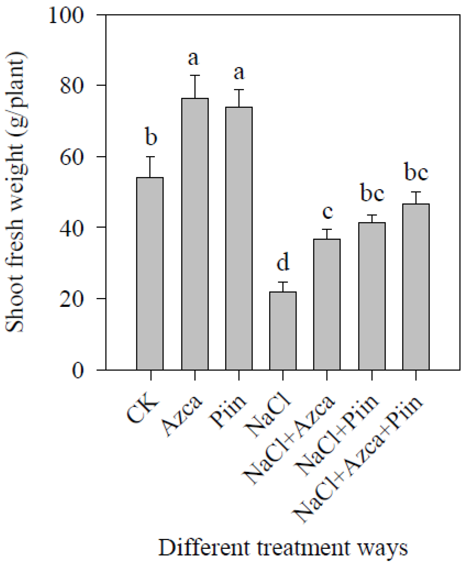
3.2. Effects of Inoculations on Tomato Plant Growth under Different Treatments

The shoot height of tomato seedlings treated with 300 mM NaCl only was considerably lower than that of tomato plants treated with all other treatments one week later (Figure 2A), and there were no significant variations in plant height among all other treatments (Figure 2A). Inoculation of A. caulinodans and P. indica, alone or combined, significantly enhanced stem base diameters under salt stress a week after NaCl treatment, compared to NaCl only treatment (Figure 2A).
Two weeks after salt treatment, NaCl only treatment resulted in a significant reduction in plant height, compared with all the other treatments (Figure 2B). However, co-inoculation of A. caulinodans and P. indica significantly increased the height of tomato plants under salt stress, compared with NaCl only (Figure 2B). Two weeks after salt treatment, inoculation of P. indica only and co-inoculation with A. caulinodans resulted in a significant increase in the stem base diameters under salt stress, compared to NaCl only treatment (Figure 2B). Two weeks after salt stress, the height of tomato plants treated with NaCl + Azca, NaCl + Piin, and NaC + Azca + Piin increased 15.21%, 13.57%, and 23.55%, respectively, compared to NaCl only treatment (Figure 2B).
Tomato plants under different treatments showed phenotypic differences in the growth and maturity of tomato fruits at the end of the tomato growth period (Figure 3). The shoot fresh weight data also showed significant differences (Figure 4). Under no-salt stress conditions, A. caulinodans or P. indica only inoculations significantly increased the shoot fresh weight of tomato plants (Figure 4). In contrast, inoculation of A. caulinodans and P. indica, alone or together, significantly increased the shoot fresh weight of tomato plants under salt stress (Figure 4). On the other hand, the shoot fresh weight of plants treated with NaCl + Azca, NaCl + Piin, and NaC + Azca + Piin increased by 68.04%, 88.69%, and 112.28%, respectively, compared to NaCl only treatment (Figure 4).

3.3. Effects of Inoculations on the Photosynthetic Pigments

The inoculation of A. caulinodans or P. indica only increased the concentration of Chl a and Chl b under no-salt stress conditions, compared to the CK treatment (Table 1). NaCl-only treatment resulted in the lowest concentrations of Chl a and Chl b (Table 1). The inoculation with A. caulinodans and P. indica, alone or together, increased concentrations of Chl a and Chl b under salt stress, compared with NaCl only treatment; however, there were no significant differences (Table 1). The A. caulinodans (i.e., NaCl + Azca) only inoculation and the co-inoculation of the two organisms (i.e., NaCl + Azca + Piin) significantly increased the total chlorophyll concentrations under salt stress, compared to NaCl only treatment (Table 1). Under salt stress, A. caulinodans and P. indica inoculation, alone or together, increased the concentrations of carotenoids, yet, there were no significant differences compared with NaCl only treatment (Table 1).

3.4. Effects of Inoculations on the Chlorophyll Fluorescence Parameters

Chlorophyll fluorescence parameters reflect the photosynthetic process dynamics and are valuable markers for assessing the impact of environmental stressors [74,75,76]. Under non-salt stress, A. caulinodans or P. indica only inoculation significantly increased the Y(II) and ETR, compared to CK treatment (Table 2). Y(II) showed the most significant value in the leaves of tomatoes treated with NaCl + Piin under salt stress among all four treatments. Y(II) was significantly higher than those in the NaCl + Azca + Piin group (Table 2). The two treatments, NaCl + Azca and NaCl + Piin, significantly increased the ETR under salt stress; however, co-inoculation significantly reduced the ETR, compared with NaCl only treatment (Table 2). Under salt stress, inoculation of A. caulinodans and P. indica, alone or together, significantly reduced the chlorophyll fluorescence parameter qP, compared to NaCl only treatment (Table 2). Inoculation of A. caulinodans and P. indica, alone or together, significantly increased the Fv/Fm under salt stress, compared to NaCl only treatment (Table 2).

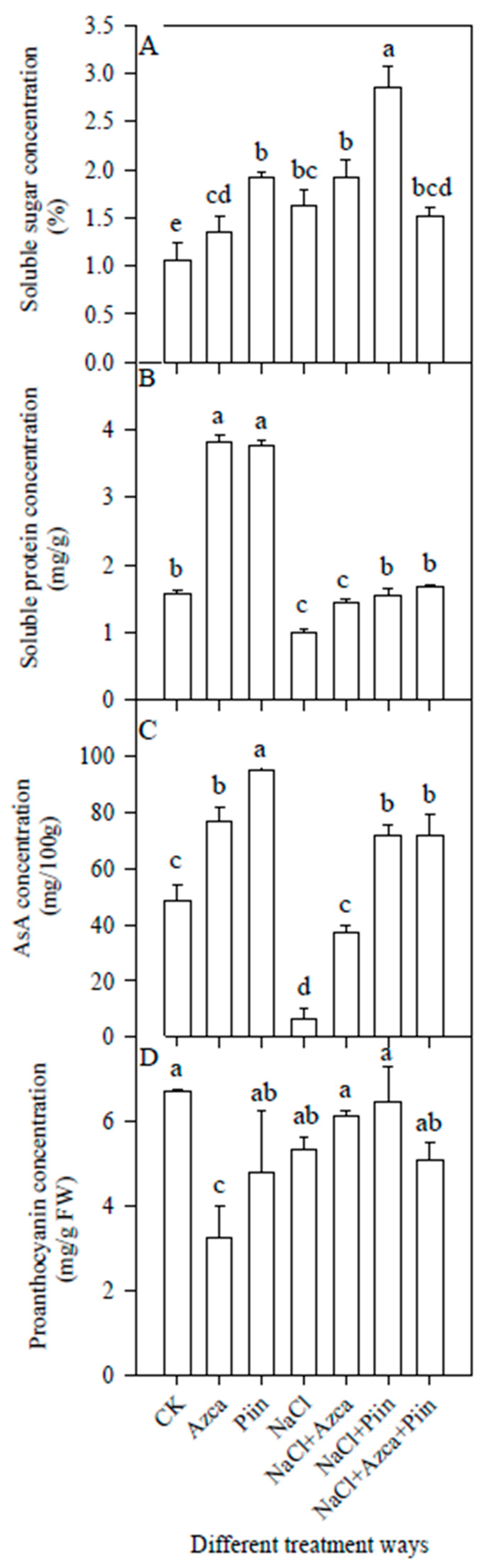
3.5. Effects of Inoculations on the Soluble Sugars and Proteins

The inoculation of A. caulinodans or P. indica significantly increased soluble sugars and proteins concentrations under non-salt stress (Figure 5). Additionally, compared to NaCl only treatment, A. caulinodans, and P. indica, alone and together, significantly increased the concentrations of soluble proteins (Figure 5).

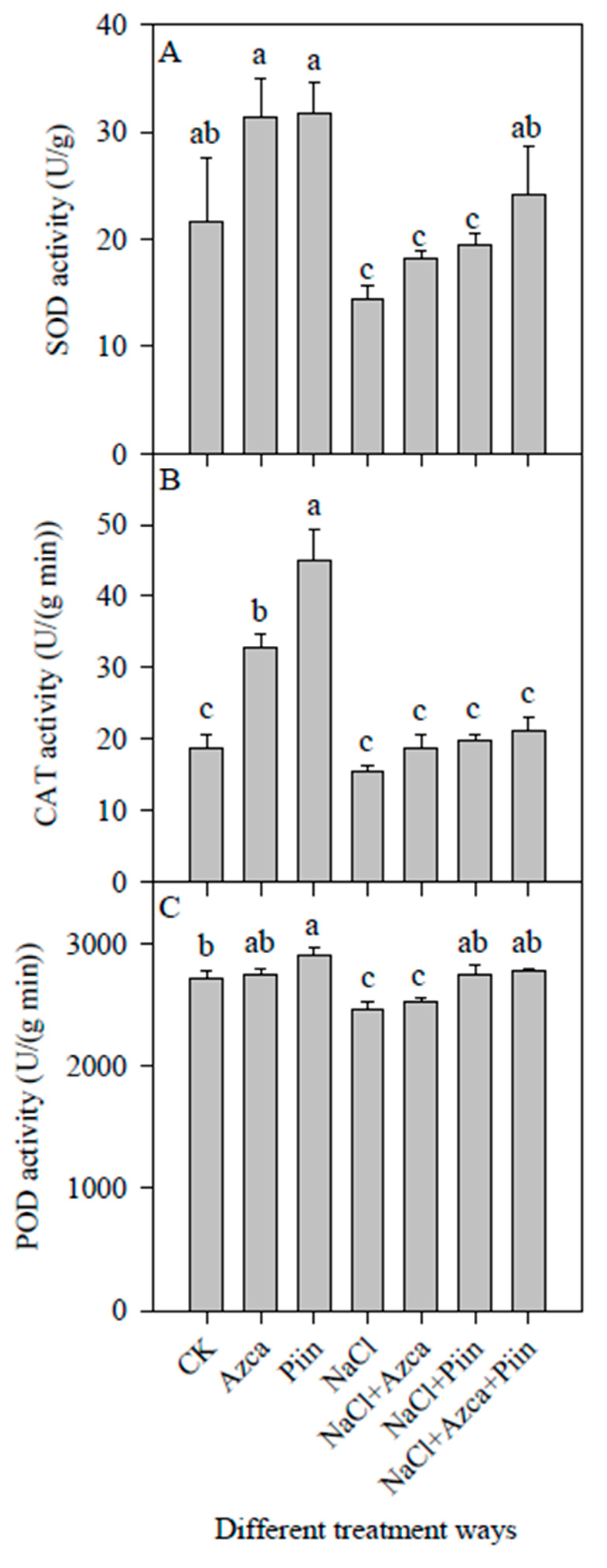
3.6. Effects of Inoculations on the Antioxidant Enzyme Activities

Under non-salt stress, the inoculation of A. caulinodans or P. indica increased the SOD activity; however, there were no significant differences compared to CK treatment (Figure 6A), whereas co-inoculation significantly increased the SOD activities compared to NaCl-only treatment (Figure 6A). A. caulinodans or P. indica significantly increased the CAT activity (Figure 6B) under non-salt stress, yet, inoculation of A. caulinodans and P. indica, alone and together, did not show a significant effect on CAT activities under salt stress (Figure 6B). On the other hand, P. indica inoculation increased the POD activity significantly under non-salt stress (Figure 6C) while A. caulinodans did not, compared with CK treatment (Figure 6C). The two treatments, i.e., NaCl + Piin and NaCl + Azca + Piin, significantly increased the POD activity, compared with NaCl only treatment under salt stress (Figure 6C).

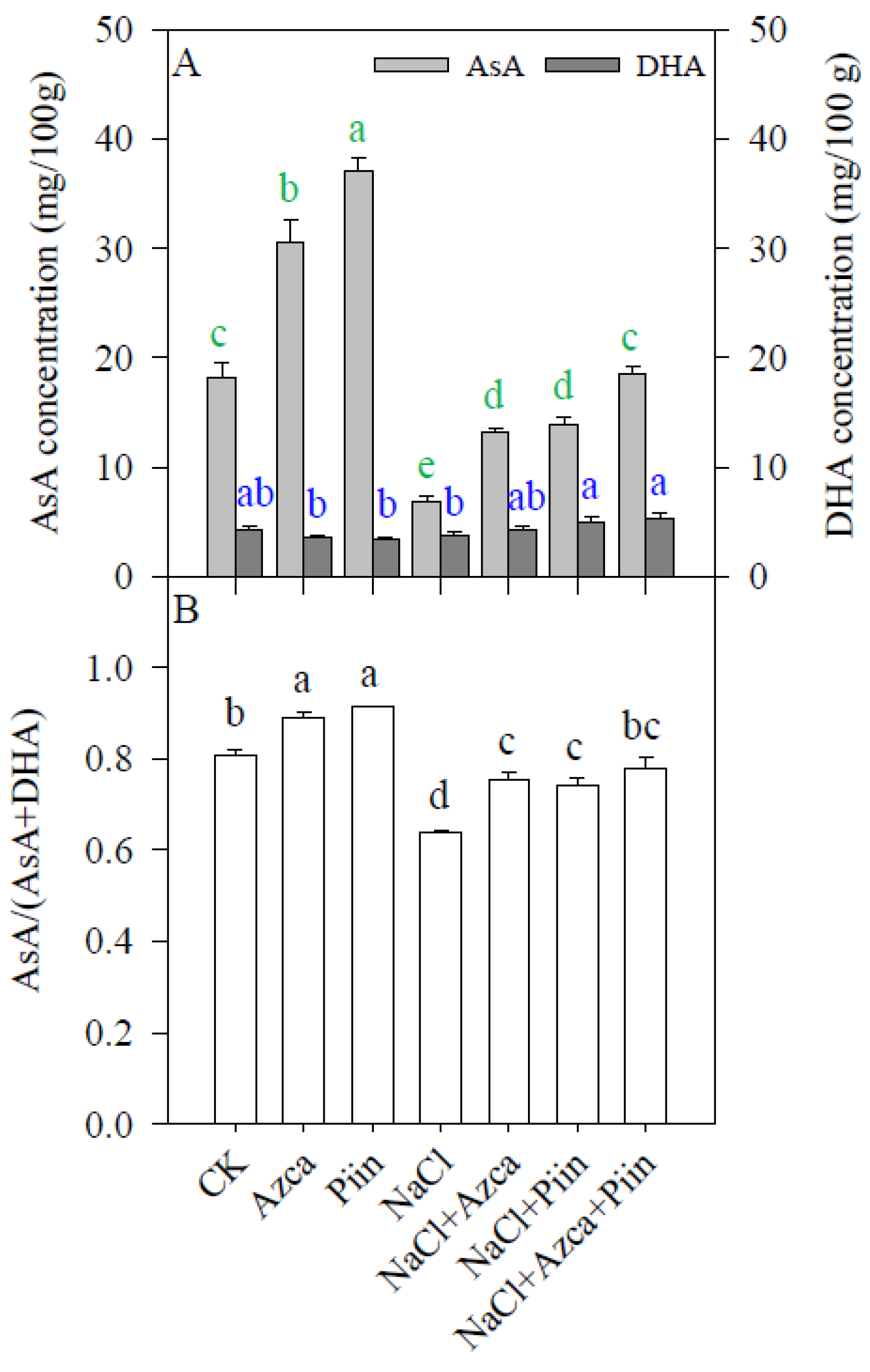
3.7. Effects of Inoculations on the Antioxidants

The inoculation of each microorganism significantly increased concentrations of reduced ascorbate under non-salt stress (AsA, Figure 7A), yet did not affect dehydroascorbate (DHA) concentrations compared with CK (Figure 7A). Similarly, the inoculation of A. caulinodans and P. indica, alone or in combination, increased AsA concentrations compared to NaCl only treatment (Figure 7A). The two treatments, i.e., NaCl + Piin and NaCl + Azca + Piin, significantly increased DHA concentrations under salt stress (Figure 7A).
Similarly, inoculation of A. caulinodans and P. indica affected concentrations of reduced glutathione (GSH) and oxidized glutathione (GSSG). Under both non-salt stress and salt stress, inoculation of A. caulinodans or P. indica significantly increased the GSH concentrations, compared with CK treatment and NaCl only treatment, respectively (Figure 8A). However, A. caulinodans and P. indica inoculation alone and in combination did not affect GSSG concentrations under salt stress (Figure 8A).
Co-inoculation also affected the ratios of AsA/(AsA + DHA) and GSH/(GSH + GSSG) (Figure 7B and Figure 8B). Under both non-salt stress and salt stress, A. caulinodans or P. indica inoculation significantly increased the AsA/(AsA + DHA) values compared to CK and NaCl only, respectively (Figure 7B). The inoculation also considerably increased the GSH/(GSH + GSSG), compared with NaCl-only treatment (Figure 8B).

3.8. Effect of Inoculations on the Malondialdehyde Concentrations

Salt stress-induced decomposition of biomembrane in tomato leaves. Under non-salt stress, A. caulinodans or P. indica inoculation reduced malondialdehyde (MDA) concentrations significantly compared to CK treatment (Figure 9). Under salt stress, A. caulinodans, and P. indica, alone and together, also significantly reduced the MDA concentrations, compared with NaCl only group of plants (Figure 9).

3.9. Effects of Inoculations on the Fruit Quality

The concentrations of soluble sugars and proteins in tomato fruits remarkably increased by A. caulinodans or P. indica under non-salt stress compared with CK treatment (Figure 10A,B). However, among three microbial treatments, only single inoculation of P. indica (i.e., NaCl + Piin treatment) increased the concentrations of soluble sugars under salt stress, compared with NaCl-only treatment (Figure 10A), whereas NaCl + Piin and NaC + Azca + Piin treatment significantly increased concentrations of soluble proteins (Figure 10B).
Under non-salt stress and salt stress conditions, inoculation of A. caulinodans and P. indica, alone and together, significantly increased ascorbate concentrations, compared with CK and NaCl-only treatment, respectively (Figure 10C). A. caulinodans and P. indica showed different effects on proanthocyanin biosynthesis in tomato fruits under non-salt stress. The inoculation of A. caulinodans reduced the pro anthocyanin concentrations in tomato fruits (Figure 10D); however, proanthocyanin concentrations in fruits of tomato plants inoculated with P. indica did not show significant changes (Figure 10D), compared with CK treatment. However, alone and together, both A. caulinodans and P. indica did not affect the proanthocyanin concentrations in tomato fruits under salt stress (Figure 10D).

4. Discussion

4.1. Co-Inoculation of A. caulinodans and P. indica Promotes Growth and Fruit Quality

A. caulinodans and P. indica possess a wide range of plant hosts, including tomato [45,49,50,77,78]. In our study, the two microorganisms were found to colonize in tomato plants (Figure 1). A. caulinodans was detected in the roots and stems of tomato plants (Figure 1A,B), while P. indica spores were found only in roots (Figure 1C). Their inoculation improved tomato growth and development when salt stress was present in the environment. Improved phenotypic characteristics (Figure 2B) and shoot fresh weight (Figure 4) of inoculated plants under salt stress suggest that co-inoculation with A. caulinodans and P. indica possessed the most significant effect on tomato physiology. These data were in accordance with the latest research dealing with the plants co-inoculated with mycorrhizal fungi and PGPRs or root endophytic fungi under salt and drought stress [78,79,80]. Increased biomass accumulation might be attributed to the protected photosynthetic machinery under salt stress since co-inoculation of A. caulinodans, and P. indica increased the intactness of chlorophylls and carotenoid concentrations under salt stress (Table 1).
A. caulinodans functions as a nitrogen-fixing bacterium and can colonize in non-legume plants. No other research has been reported on the functions of A. caulinodans under salt stress to the best of our knowledge. Under non-salt stress and salt stress, inoculation of A. caulinodans only significantly increased the shoot fresh weight of tomato plants (Figure 4), suggesting that the A. caulinodans play a critical role in the biomass development of tomato plants as a nitrogen-fixer. However, the changes in nitrogen metabolism of host plants inoculated with A. caulinodans remain unclear and need to be addressed at the biochemical and molecular levels.
Robust evidence proved that the root endophytic fungus P. indica increased biomass accumulation under salt stress in several previous studies [10,81,82]. The increase in P. indica-induced biomass might be related to its multiple functions under salt stress: (1) increased concentrations of photosynthetic pigments and improved gas exchange, and subsequent photochemical dynamics [10]; (2) maintenance of K+/Na+ homeostasis [58,77]; (3) increased abundance of photosynthetic proteins [83]; and (4) changes in fatty acid composition in phospholipids in the leaves and better maintenance of membrane stability [84]. Our data are in accordance with some of this evidence (Table 1 and Table 2, and Figure 4).
Tomato fruits contain not only proteins, sugars, organic acids, or lycopene, but also several vitamins (e.g., vitamins A, C, E) and other nutrients that are important for a balanced diet for human health and can reduce the risk of cancer and heart disease [85]. The antioxidant AsA is, therefore, might be essential for maintaining a balanced diet and preventing human health risks to a certain extent. Here, in our work, the inoculation of A. caulinodans and P. indica significantly increased AsA concentrations under both non-salt and salt stress conditions (Figure 10C). Anthocyanins, on the other hand, are also essential antioxidants [86,87]. However, inoculation of A. caulinodans and P. indica showed contrary effects on proanthocyanin accumulation in tomato fruits when salt stress is not present in the environment (Figure 10D). Yet, these contrary effects resulted in no meaningful differences in proanthocyanin accumulation in fruits of tomato plants inoculated with A. caulinodans and P. indica, alone and together, compared with NaCl-only treatment under salt stress (Figure 10D). In addition, under non-salt and salt stress conditions, inoculation of A. caulinodans and P. indica, alone and in combination, significantly increased the concentrations of soluble proteins in tomato fruits, compared with CK and NaCl-only treatment, respectively (Figure 10C). Therefore, all the data suggest that co-inoculation with A. caulinodans and P. indica improves the quality of tomato fruits produced under excessive salt.
Interestingly, salt stress promoted the precocity of tomato fruits, presumably due to the hormonal changes, and inoculation of A. caulinodans and P. indica, alone and together, showed a more substantial effect on the plants than NaCl-only treatment (Figure 3). The precocity of tomato fruits is beneficial for earlier harvest and continuous fruit-setting because tomato possesses the characteristic of constant flowering and fruiting.

4.2. Co-Inoculation of A. caulinodans and P. indica Enhances Salt Tolerance

Under abiotic stresses, reactive oxygen species (ROS) might be produced by several different pathways cross-talking each other. Plants have evolved multiple strategies to respond to this oxidative stress, especially various antioxidants and antioxidant enzymes are in charge of scavenging cellular ROS in the plant. Our results showed that the triggered activities of SOD and POD were significant in the leaves of tomato plants treated with NaCl + Azca + Piin (Figure 6A,C). This combination significantly increased the concentrations of reduced ascorbate and glutathione (Figure 7A and Figure 8A). These data suggest that the co-inoculation of A. caulinodans and P. indica improves the ability to scavenge ROS in salt-stressed tomato plants. Increased activities of antioxidant enzymes and concentrations of antioxidants resulted in a significant reduction in the peroxidation of the biomembranes of the leaves of tomato plants under salt stress (Figure 9). These results were found under the latest studies [84,88,89]. For instance, Baltruschat et al. [84] showed that the inoculation of P. indica significantly elevated the amount of ascorbate in quantity and increased the activities of several antioxidant enzymes in the roots of barley (Hordeum vulgare) under salt stress.
High ratios of AsA/(AsA + DHA) and GSH/(GSH + GSSG) are beneficial for ROS scavenging. Consequently, during the ROS scavenging processes, lower concentrations of oxidized ascorbate and glutathione (Figure 7A and Figure 8A) and higher ratios of AsA/(AsA + DHA) and GSH/(GSH + GSSG) (Figure 7B and Figure 8B) measured under salt stress indicate that the antioxidant enzymes, dehydroascorbate reductase (DHAR) and glutathione reductase (GR), played essential roles in the transformation of oxidized ascorbate and glutathione to the reduced form of ascorbate and glutathione, respectively. Accordingly, increased activities of antioxidant enzymes and AsA and GSH concentrations resulted in significantly lower MDA in the leaves of tomato plants inoculated with A. caulinodans and P. indica, alone and together, under salt stress (Figure 9). The findings imply that co-inoculation with A. caulinodans and P. indica improved tomato plant salt tolerance.

5. Conclusions

Co-inoculation of the two microorganisms, A. caulinodans and P. indica, improved tomato plants’ growth and fruit quality under salt stress. The combined inoculation alleviated the deteriorating physiological effects caused by excessive NaCl through improving enzymatic and non-enzymatic antioxidants.

Author Contributions

C.W. planned the whole experiments and revised the manuscript. Z.X. carried out all the experimental work and wrote the rough manuscript. N.P. and A.G. reviewed and revised the rough manuscript. All authors have read and agreed to the published version of the manuscript.

Funding

This work was supported by the National Natural Science Foundation of China (grant number: 31870378).

Institutional Review Board Statement

Not applicable.

Informed Consent Statement

Not applicable.

Data Availability Statement

Not applicable.

Acknowledgments

We thank Ralf Oelmüller for providing the root endophytic fungus Piriformospora indica.

Conflicts of Interest

The authors declare no conflict of interest.

References

  1. Munns, R.; Tester, M. Mechanisms of salinity tolerance. Annu. Rev. Plant Biol. 2008, 59, 651–681. [Google Scholar] [CrossRef] [PubMed] [Green Version]
  2. Ouhibi, C.; Attia, H.; Rebah, F.; Msilini, N.; Chebbi, M.; Aarrouf, J.; Urban, L.; Lachaal, M. Salt stress mitigation by seed priming with UV-C in lettuce plants: Growth, antioxidant activity and phenolic compounds. Plant Physiol. Biochem. 2014, 83, 126–133. [Google Scholar] [CrossRef] [PubMed]
  3. Evelin, H.; Devi, T.S.; Gupta, S.; Kapoor, R. Mitigation of salinity stress in plants by arbuscular mycorrhizal symbiosis: Current understanding and new challenges. Front. Plant Sci. 2019, 10, 470. [Google Scholar] [CrossRef] [PubMed] [Green Version]
  4. Ismail, A.; Takeda, S.; Nick, P. Life and death under salt stress: Same players, different timing? J. Exp. Bot. 2014, 65, 2963–2979. [Google Scholar] [CrossRef] [PubMed]
  5. Bo, C.; Chen, H.; Luo, G.; Li, W.; Zhang, X.; Ma, Q.; Cheng, B.; Cai, R. Maize WRKY114 gene negatively regulates salt-stress tolerance in transgenic rice. Plant Cell Rep. 2020, 39, 135–148. [Google Scholar] [CrossRef]
  6. Jini, D.; Joseph, B. Physiological mechanism of salicylic acid for alleviation of salt stress in rice. Rice Sci. 2017, 24, 97–108. [Google Scholar] [CrossRef]
  7. Karan, R.; DeLeon, T.; Biradar, H.; Subudhi, P.K. Salt stress induced variation in DNA methylation pattern and its influence on gene expression in contrasting rice genotypes. PLoS ONE 2012, 7, e40203. [Google Scholar] [CrossRef]
  8. Niu, C.F.; Wei, W.; Zhou, E.I.; Tian, Q.Y.; Hao, A.G.; Zhang, Y.J.; Zhang, W.K.; Chen, S.Y. Wheat WRKY genes TaWRKY2 and TaWRKY19 regulate abiotic stress tolerance in transgenic Arabidopsis plants. Plant Cell Environ. 2012, 35, 1156–1170. [Google Scholar] [CrossRef]
  9. Oyiga, B.C.; Sharma, R.C.; Shen, J.; Baum, M.; Ogbonnaya, F.C.; Léon, J.; Ballvora, A. Identification and characterization of salt tolerance of wheat germplasm using a multivariable screening approach. J. Agron. Crop Sci. 2016, 202, 472–485. [Google Scholar] [CrossRef]
  10. Ghorbani, A.; Razavi, S.M.; Ghasemi Omran, V.O.; Pirdashti, H. Piriformospora indica inoculation alleviates the adverse effect of NaCl stress on growth, gas exchange and chlorophyll fluorescence in tomato (Solanum lycopersicum L.). Plant Biol. 2018, 20, 729–736. [Google Scholar] [CrossRef]
  11. Keshishian, E.A.; Hallmark, H.T.; Ramaraj, T.; Plačková, L.; Sundararajan, A.; Schilkey, F.; Novák, O.; Rashotteet, A.M. Salt and oxidative stresses uniquely regulate tomato cytokinin levels and transcriptomic response. Plant Direct 2018, 2, e00071. [Google Scholar] [CrossRef] [PubMed] [Green Version]
  12. Li, S.; Wang, N.; Ji, D.; Zhang, W.; Wang, Y.; Yu, Y.; Zhao, S.; Lyu, M.; You, J.; Zhang, Y.; et al. A GmSIN1/GmNCED3s/GmRbohBs feed-forward loop acts as a signal amplifier that regulates root growth in soybean exposed to salt stress. Plant Cell 2019, 31, 2107–2130. [Google Scholar] [CrossRef] [PubMed] [Green Version]
  13. Liu, J.G.; Han, X.; Yang, T.; Cui, W.H.; Wu, A.M.; Fu, C.X.; Wang, B.C.; Liu, L.J. Genome-wide transcriptional adaptation to salt stress in Populus. BMC Plant Biol. 2019, 19, 367. [Google Scholar] [CrossRef] [PubMed]
  14. Çakir Aydemir, B.; Yüksel Özmen, C.; Kibar, U.; Mutaf, F.; Büyük, P.B.; Bakır, M.; Ergül, A. Salt stress induces endoplasmic reticulum stress-responsive genes in a grapevine rootstock. PLoS ONE 2020, 15, e0236424. [Google Scholar] [CrossRef]
  15. Shah, A.N.; Tanveer, M.; Abbas, A.; Fahad, S.; Baloch, M.S.; Ahmad, M.I.; Saud, S.; Song, Y. Targeting salt stress coping mechanisms for stress tolerance in Brassica: A research perspective. Plant Physiol. Biochem. 2021, 158, 53–64. [Google Scholar] [CrossRef]
  16. Yang, Y.; Guo, Y. Elucidating the molecular mechanisms mediating plant salt-stress responses. New Phytol. 2018, 217, 523–539. [Google Scholar] [CrossRef] [Green Version]
  17. Zhu, J.K. Salt and drought stress signal transduction in plants. Annu. Rev. Plant. Biol. 2002, 53, 247–273. [Google Scholar] [CrossRef] [Green Version]
  18. Yang, Y.; Guo, Y. Unraveling salt stress signaling in plants. J. Integr. Plant Biol. 2018, 60, 796–804. [Google Scholar] [CrossRef] [Green Version]
  19. Park, H.J.; Kim, W.Y.; Yun, D.J. A new insight of salt stress signaling in plant. Mol. Cells 2016, 39, 447–459. [Google Scholar] [CrossRef]
  20. Lou, L.; Yu, F.; Tian, M.; Liu, G.; Wu, Y.; Wu, Y.; Xia, R.; Pardo, J.M.; Guo, Y.; Xie, Q. ESCRT-I component VPS23A sustains salt tolerance by strengthening the SOS module in Arabidopsis. Mol. Plant 2020, 13, 1134–1148. [Google Scholar] [CrossRef]
  21. Sun, Y.; Zhao, J.; Li, X.; Li, Y. E2 conjugases UBC1 and UBC2 regulate MYB42-mediated SOS pathway in response to salt stress in Arabidopsis. New Phytol. 2020, 227, 455–472. [Google Scholar] [CrossRef] [PubMed]
  22. Kiegerl, S.; Cardinale, F.; Siligan, C.; Gross, A.; Baudouin, E.; Liwosz, A.; Eklöf, S.; Till, S.; Bögre, L.; Hirt, H.; et al. SIMKK, a mitogen-activated protein kinase (MAPK) kinase, is a specific activator of the salt stress-induced MAPK, SIMK. Plant Cell 2000, 12, 2247–2258. [Google Scholar] [CrossRef] [PubMed]
  23. Tang, Z.; Cao, X.; Zhang, Y.; Jiang, J.; Qiao, D.; Xu, H.; Cao, Y. Two splice variants of the DsMEK1 mitogen-activated protein kinase kinase (MAPKK) are involved in salt stress regulation in Dunaliella salina in different ways. Biotechnol. Biofuels 2020, 13, 147. [Google Scholar] [CrossRef] [PubMed]
  24. Szymańska, K.P.; Polkowska-Kowalczyk, L.; Lichocka, M.; Maszkowska, J.; Dobrowolska, G. SNF1-Related Protein Kinases SnRK2.4 and SnRK2.10 modulate ROS homeostasis in plant response to salt stress. Int. J. Mol. Sci. 2019, 20, 143. [Google Scholar] [CrossRef] [PubMed] [Green Version]
  25. Wang, Y.; Wang, M.; Li, Y.; Wu, A.; Huang, J. Effects of arbuscular mycorrhizal fungi on growth and nitrogen uptake of Chrysanthemum morifolium under salt stress. PLoS ONE 2018, 13, e0196408. [Google Scholar] [CrossRef] [PubMed]
  26. Li, Z.; Wu, N.; Meng, S.; Wu, F.; Liu, T. Arbuscular mycorrhizal fungi (AMF) enhance the tolerance of Euonymus maackii Rupr. at a moderate level of salinity. PLoS ONE 2020, 15, e0231497. [Google Scholar] [CrossRef]
  27. Tisarum, R.; Theerawitaya, C.; Samphumphuang, T.; Polispitak, K.; Thongpoem, P.; Singh, H.P.; Cha-Um, S. Alleviation of salt stress in upland rice (Oryza sativa L. ssp. indica cv. Leum Pua) using arbuscular mycorrhizal fungi inoculation. Front. Plant Sci. 2020, 11, 348. [Google Scholar] [CrossRef] [Green Version]
  28. Thiem, D.; Piernik, A.; Hrynkiewicz, K. Ectomycorrhizal and endophytic fungi associated with Alnus glutinosa growing in a saline area of central Poland. Symbiosis 2018, 75, 17–28. [Google Scholar] [CrossRef] [Green Version]
  29. Guerrero-Galán, C.; Calvo-Polanco, M.; Zimmermann, S.D. Ectomycorrhizal symbiosis helps plants to challenge salt stress conditions. Mycorrhiza 2019, 29, 291–301. [Google Scholar] [CrossRef]
  30. Zwiazek, J.J.; Equiza, M.A.; Karst, J.; Senorans, J.; Wartenbe, M.; Calvo-Polanco, M. Role of urban ectomycorrhizal fungi in improving the tolerance of lodgepole pine (Pinus contorta) seedlings to salt stress. Mycorrhiza 2019, 29, 303–312. [Google Scholar] [CrossRef]
  31. Fadaei, S.; Vaziriyeganeh, M.; Young, M.; Sherr, I.; Zwiazek, J.J. Ericoid mycorrhizal fungi enhance salt tolerance in ericaceous plants. Mycorrhiza 2020, 30, 419–429. [Google Scholar] [CrossRef] [PubMed]
  32. Kord, H.; Fakheri, B.; Ghabooli, M.; Solouki, M.; Emamjomeh, A.; Khatabi, B.; Sepehri, M.; Salekdeh, G.H.; Ghaffari, M.R. Salinity-associated microRNAs and their potential roles in mediating salt tolerance in rice colonized by the endophytic root fungus Piriformospora indica. Funct. Integr. Genom. 2019, 19, 659–672. [Google Scholar] [CrossRef] [PubMed]
  33. Farias, G.C.; Nunes, K.G.; Soares, M.A.; de Siqueira, K.A.; Lima, W.C.; Neves, A.L.R.; de Lacerda, C.F.; Filho, E.G. Dark septate endophytic fungi mitigate the effects of salt stress on cowpea plants. Braz. J. Microbiol. 2020, 51, 243–253. [Google Scholar] [CrossRef] [PubMed]
  34. Bouzouina, M.; Kouadria, R.; Lotmani, B. Fungal endophytes alleviate salt stress in wheat in terms of growth, ion homeostasis and osmoregulation. J. Appl. Microbiol. 2021, 130, 913–925. [Google Scholar] [CrossRef]
  35. Chauhan, P.S.; Lata, C.; Tiwari, S.; Chauhan, A.S.; Mishra, S.K.; Agrawal, L.; Chakrabarty, D.; Nautiyal, C.S. Transcriptional alterations reveal Bacillus amyloliquefaciens-rice cooperation under salt stress. Sci. Rep. 2019, 9, 11912. [Google Scholar] [CrossRef]
  36. Alexander, A.; Singh, V.K.; Mishra, A. Halotolerant PGPR Stenotrophomonas maltophilia BJ01 induces salt tolerance by modulating physiology and biochemical activities of Arachis hypogaea. Front. Microbiol. 2020, 11, 568289. [Google Scholar] [CrossRef]
  37. Sultana, S.; Paul, S.C.; Parveen, S.; Alam, S.; Rahman, N.; Jannat, B.; Hoque, S.; Rahman, M.T.; Karim, M.M. Isolation and identification of salt-tolerant plant-growth-promoting rhizobacteria and their application for rice cultivation under salt stress. Can. J. Microbiol. 2020, 66, 144–160. [Google Scholar] [CrossRef]
  38. Li, J.; Bao, S.; Zhang, Y.; Ma, X.; Mishra-Knyrim, M.; Sun, J.; Sa, G.; Shen, X.; Polle, A.; Chen, S. Paxillus involutus strains MAJ and NAU mediate K+/Na+ homeostasis in ectomycorrhizal Populus × canescens under sodium chloride stress. Plant Physiol. 2012, 159, 1771–1786. [Google Scholar] [CrossRef] [Green Version]
  39. Sa, G.; Yao, J.; Deng, C.; Liu, J.; Zhang, Y.; Zhu, Z.; Zhang, Y.; Ma, X.; Zhao, R.; Lin, S.; et al. Amelioration of nitrate uptake under salt stress by ectomycorrhiza with and without a Hartig net. New Phytol. 2019, 222, 1951–1964. [Google Scholar] [CrossRef]
  40. Lee, S.H.; Calvo-Polanco, M.; Chung, G.C.; Zwiazek, J.J. Role of aquaporins in root water transport of ectomycorrhizal jack pine (Pinus banksiana) seedlings exposed to NaCl and fluoride. Plant Cell Environ. 2010, 33, 69–80. [Google Scholar] [CrossRef]
  41. Bois, G.; Bertrand, A.; Piché, Y.; Fung, M.; Khasa, D.P. Growth, compatible solute and salt accumulation of five mycorrhizal fungal species grown over a range of NaCl concentrations. Mycorrhiza 2006, 16, 99–109. [Google Scholar] [CrossRef] [PubMed]
  42. Bois, G.; Bigras, F.J.; Bertrand, A.; Piché, Y.; Fung, M.Y.; Khasa, D.P. Ectomycorrhizal fungi affect the physiological responses of Picea glauca and Pinus banksiana seedlings exposed to an NaCl gradient. Tree Physiol. 2006, 26, 1185–1196. [Google Scholar] [CrossRef] [PubMed] [Green Version]
  43. Nguyen, H.; Calvo Polanco, M.; Zwiazek, J.J. Gas exchange and growth responses of ectomycorrhizal Picea mariana, Picea glauca, and Pinus banksiana seedlings to NaCl and Na2SO4. Plant Biol. 2006, 8, 646–652. [Google Scholar] [CrossRef] [PubMed]
  44. Dreyfus, B.; Garcia, L.L.; Gillis, M. Characterization of Azorhizobium caulinodans gen. nov., sp. nov., a stem-nodulating nitrogen-fixing bacterium isolated from Sesbania rostrata. Int. J. Syst. Bacteriol. 1988, 38, 89–98. [Google Scholar] [CrossRef] [Green Version]
  45. Cocking, E.C. Xylem colonization of tomato by Azorhizobium caulinodans ORS571. Acta Biol. Hung. 2001, 52, 189–194. [Google Scholar] [CrossRef]
  46. Buvana, R.; Kannaiyan, S. Influence of cell wall degrading enzymes on colonization of N2 fixing bacterium, Azorhizobium caulinodans in rice. Indian J. Exp. Biol. 2002, 40, 369–372. [Google Scholar]
  47. Webster, G.; Jain, V.; Davey, M.R.; Gough, C.; Vasse, J.; Denarie, J.; Cocking, E.C. The flavonoid naringenin stimulates the intercellular colonization of wheat roots by Azorhizobium caulinodans. Plant Cell Environ. 1998, 21, 373–383. [Google Scholar] [CrossRef]
  48. Qiu, L.; Li, Q.; Zhang, J.; Chen, Y.; Lin, X.; Sun, C.; Wang, W.; Liu, H.; Zhang, B. Migration of endophytic diazotroph Azorhizobium caulinodans ORS571 inside wheat (Triticum aestivum L.) and its effect on microRNAs. Funct. Integr. Genom. 2017, 17, 311–319. [Google Scholar] [CrossRef]
  49. Sun, Y.; Liu, Y.; Liu, X.; Dang, X.; Dong, X.; Xie, Z. Azorhizobium caulinodans c-di-GMP phosphodiesterase Chp1 involved in motility, EPS production, and nodulation of the host plant. Appl. Microbiol. Biotechnol. 2020, 104, 2715–2729. [Google Scholar] [CrossRef]
  50. Liu, W.; Bai, X.; Li, Y.; Min, J.; Kong, Y.; Hu, X. CheY1 and CheY2 of Azorhizobium caulinodans ORS571 regulate chemotaxis and competitive colonization with the host plant. Appl. Environ. Microbiol. 2020, 86, e00599-20. [Google Scholar] [CrossRef]
  51. Si, Y.; Guo, D.; Deng, S.; Lu, X.; Zhu, J.; Rao, B.; Cao, Y.; Jiang, G.; Yu, D.; Zhong, Z.; et al. Ohr and OhrR are critical for organic peroxide resistance and symbiosis in Azorhizobium caulinodans ORS571. Genes 2020, 11, 335. [Google Scholar] [CrossRef] [PubMed] [Green Version]
  52. Liu, W.; Yang, J.; Sun, Y.; Liu, X.; Li, Y.; Zhang, Z.; Xie, Z. Azorhizobium caulinodans Transmembrane chemoreceptor TlpA1 involved in host colonization and nodulation on roots and stems. Front. Microbiol. 2017, 8, 1327. [Google Scholar] [CrossRef] [PubMed] [Green Version]
  53. Liu, X.; Liu, W.; Sun, Y.; Xia, C.; Elmerich, C.; Xie, Z. A cheZ-like gene in Azorhizobium caulinodans is a key gene in the control of chemotaxis and colonization of the host plant. Appl. Environ. Microbiol. 2018, 84, e01827-17. [Google Scholar] [CrossRef] [PubMed] [Green Version]
  54. Liu, H.; Wang, X.; Qi, H.; Wang, Q.; Chen, Y.; Li, Q.; Zhang, Y.; Qiu, L.; Fontana, J.E.; Zhang, B.; et al. The infection and impact of Azorhizobium caulinodans ORS571 on wheat (Triticum aestivum, L.). PLoS ONE 2017, 12, e0187947. [Google Scholar] [CrossRef] [Green Version]
  55. Yang, Z.Y.; Yuan, J.G.; Xin, G.R.; Chang, H.T.; Wong, M.H. Germination, growth, and nodulation of Sesbania rostrata grown in Pb/Zn mine tailings. Environ. Manag. 1997, 21, 617–622. [Google Scholar] [CrossRef]
  56. Jian, S.; Shen, W.; Yang, Z. Enhanced adaptability of Sesbania rostrata to Pb/Zn tailings via stem nodulation. J. Environ. Sci. 2009, 21, 1135–1141. [Google Scholar] [CrossRef]
  57. Abdelaziz, M.E.; Kim, D.; Ali, S.; Fedoroff, N.V.; Al-Babili, S. The endophytic fungus Piriformospora indica enhances Arabidopsis thaliana growth and modulates Na+/K+ homeostasis under salt stress conditions. Plant Sci. 2017, 263, 107–115. [Google Scholar] [CrossRef] [Green Version]
  58. Ghorbani, A.; Omran, V.O.G.; Razavi, S.M.; Pirdashti, H.; Ranjbar, M. Piriformospora indica confers salinity tolerance on tomato (Lycopersicon esculentum Mill.) through amelioration of nutrient accumulation, K+/Na+ homeostasis and water status. Plant Cell Rep. 2019, 38, 1151–1163. [Google Scholar] [CrossRef]
  59. FAOSTAT. Available online: http://www.fao.org/country-showcase/selected-product-detail/en/c/1287945 (accessed on 12 June 2020).
  60. Žižková, E.; Dobrev, P.I.; Muhovski, Y.; Hošek, P.; Hoyerová, K.; Haisel, D.; Procházková, D.; Lutts, S.; Motyka, V.; Hichri, I. Tomato (Solanum lycopersicum L.) SlIPT3 and SlIPT4 isopentenyltransferases mediate salt stress response in tomato. BMC Plant Biol. 2015, 15, 85. [Google Scholar] [CrossRef]
  61. Johnson, J.M.; Sherameti, I.; Ludwig, A.; Nongbri, P.L.; Sun, C.; Lou, B.; Varma, A.; Oelmuller, R. Protocols for Arabidopsis thaliana and Piriformospora indica co-cultivation—A model system to study plant beneficial traits. J. Endocytobiol. Cell Res. 2011, 21, 1001–1013. [Google Scholar]
  62. Maxwell, K.; Johnson, G.N. Chlorophyll fluorescence—A practical guide. J. Exp. Bot. 2000, 51, 659–668. [Google Scholar] [CrossRef] [PubMed]
  63. Kumar, M.; Lee, S.-C.; Kim, J.-Y.; Kim, S.-J.; Aye, S.S.; Kim, S.-R. Over-expression of dehydrin gene, OsDhn1, improves drought and salt stress tolerance through scavenging of reactive oxygen species in rice (Oryza sativa L.). J. Plant Biol. 2014, 57, 383–393. [Google Scholar] [CrossRef]
  64. Irigoyen, J.J.; Einerich, D.W.; Sánchez-Díaz, M. Water stress induced changes in concentrations of proline and total soluble sugars in nodulated alfalfa (Medicago sativd) plants. Physiol. Plant. 1992, 84, 55–60. [Google Scholar] [CrossRef]
  65. Bradford, M.M. A rapid and sensitive method for the quantitation of microgram quantities of protein utilizing the principle of protein-dye binding. Anal. Biochem. 1976, 72, 248–254. [Google Scholar] [CrossRef]
  66. Becana, M.; Aparicio-Tejo, P.; Irigoyen, J.J.; Sanchez-Diaz, M. Some enzymes of hydrogen peroxide metabolism in leaves and root nodules of Medicago sativa. Plant Physiol. 1986, 82, 1169–1171. [Google Scholar] [CrossRef] [Green Version]
  67. Aebi, H.E. Catalase. In Methods of Enzymatic Analyses; Berameyer, H.U., Ed.; Verlag Chemie: Weinheim, Germany, 1983; Volume 3, pp. 273–282. [Google Scholar]
  68. Chance, B.; Maehly, A.C. Assay of catalases and peroxidase. In Methods in Enzymology; Colowick, S.P., Kaplan, N.O., Eds.; Academic Press: New York, NY, USA, 1955; Volume 2, pp. 764–775. [Google Scholar]
  69. Anderson, M.E. Determination of glutathione and glutathione disulfide in biological samples. Methods Enzymol. 1985, 113, 548–555. [Google Scholar] [CrossRef]
  70. Law, M.Y.; Charles, S.A.; Halliwell, B. Glutathione and ascorbic acid in spinach (Spinacia oleracea) chloroplast. The effect of hydrogen peroxide and paraquat. Biochem. J. 1983, 210, 899–903. [Google Scholar] [CrossRef] [Green Version]
  71. Fazeli, F.; Ghorbanli, M.; Niknam, V. Effect of drought on biomass, protein content, lipid peroxidation and antioxidant enzymes in two sesame cultivars. Biol. Plant 2007, 51, 98–103. [Google Scholar] [CrossRef]
  72. Queiroz, C.R.A.D.A.; Morais, S.A.L.D.; Nascimento, E.A.D. Characterization of aroeira-preta (Myracrodruon urundeuva) wood tannins. Rev. Árvore 2002, 26, 493–497. [Google Scholar] [CrossRef] [Green Version]
  73. dos Santos, E.L.; da Silva, F.A.; da Silva, F.S.B. Arbuscular mycorrhizal fungi increase the phenolic compounds concentration in the bark of the stem of Libidibia ferrea in field conditions. Open Microbiol. J. 2017, 11, 283–291. [Google Scholar] [CrossRef] [Green Version]
  74. Faraloni, C.; Cutino, I.; Petruccelli, R.; Leva, A.R.; Lazzeri, S.; Torzillo, G. Chlorophyll fluorescence technique as a rapid tool for in vitro screening of olive cultivars (Olea europaea L.) tolerant to drought stress. Environ. Exp. Bot. 2011, 73, 49–56. [Google Scholar] [CrossRef]
  75. Wang, N.Y.; Ho, J.; Yi, L.S.; Gil, C.H.; Hyeon, K.S.; Rae, R.I. Chlorophyll fluorescence as a diagnostic tool for abiotic stress tolerance in wild and cultivated strawberry species. Hortic. Environ. Biotechnol. 2014, 54, 280–286. [Google Scholar] [CrossRef]
  76. Dąbrowski, P.; Baczewska, A.H.; Pawluśkiewicz, B.; Paunov, M.; Alexantrov, V.; Goltsev, V.; Kalaji, M.H. Prompt chlorophyll a fluorescence as a rapid tool for diagnostic changes in PSII structure inhibited by salt stress in perennial ryegrass. J. Photochem. Chem. Photobiol. B-Biol. 2016, 157, 22–31. [Google Scholar] [CrossRef] [PubMed]
  77. Lanza, M.; Haro, R.; Conchillo, L.B.; Benito, B. The endophyte Serendipita indica reduces the sodium content of Arabidopsis plants exposed to salt stress: Fungal ENA ATPases are expressed and regulated at high pH and during plant co-cultivation in salinity. Environ. Microbiol. 2019, 21, 3364–3378. [Google Scholar] [CrossRef] [PubMed]
  78. Heidarianpour, M.B.; Aliasgharzad, N.; Olsson, P.A. Positive effects of co-inoculation with Rhizophagus irregularis and Serendipita indica on tomato growth under saline conditions, and their individual colonization estimated by signature lipids. Mycorrhiza 2020, 30, 455–466. [Google Scholar] [CrossRef]
  79. Heydari, S.; Pirzad, A. Mycorrhizal fungi and Thiobacillus co-inoculation improve the physiological indices of Lallemantia iberica under salinity stress. Curr. Microbiol. 2020, 77, 2523–2534. [Google Scholar] [CrossRef]
  80. Sheteiwy, M.S.; Abd Elgawad, H.; Xiong, Y.C.; Macovei, A.; Brestic, M.; Skalicky, M.; Shaghaleh, H.; Alhaj Hamoud, Y.; El-Sawah, A.M. Inoculation with Bacillus amyloliquefaciens and mycorrhiza confers tolerance to drought stress and improve seed yield and quality of soybean plant. Physiol. Plant 2021, 172, 2153–2169. [Google Scholar] [CrossRef]
  81. Li, L.; Li, L.; Wang, X.; Zhu, P.; Wu, H.; Qi, S. Plant growth-promoting endophyte Piriformospora indica alleviates salinity stress in Medicago truncatula. Plant Physiol. Biochem. 2017, 119, 211–223. [Google Scholar] [CrossRef]
  82. Nivedita; Gazara, R.K.; Khan, S.; Iqrar, S.; Ashrafi, K.; Abdin, M.Z. Comparative transcriptome profiling of rice colonized with beneficial endophyte, Piriformospora indica, under high salinity environment. Mol. Biol. Rep. 2020, 47, 7655–7673. [Google Scholar] [CrossRef]
  83. Sepehri, M.; Ghaffari, M.R.; Khayam Nekoui, M.; Sarhadi, E.; Moghadam, A.; Khatabi, B.; Hosseini Salekdeh, G. Root endophytic fungus Serendipita indica modulates barley leaf blade proteome by increasing the abundance of photosynthetic proteins in response to salinity. J. Appl. Microbiol. 2021, 131, 1870–1889. [Google Scholar] [CrossRef]
  84. Baltruschat, H.; Fodor, J.; Harrach, B.D.; Niemczyk, E.; Barna, B.; Gullner, G.; Janeczko, A.; Kogel, K.H.; Schäfer, P.; Schwarczinger, I.; et al. Salt tolerance of barley induced by the root endophyte Piriformospora indica is associated with a strong increase in antioxidants. New Phytol. 2008, 180, 501–510. [Google Scholar] [CrossRef] [PubMed]
  85. Boffetta, P.; Couto, E.; Wichmann, J.; Ferrari, P.; Trichopoulos, D.; Bueno-de-Mesquita, H.B.; Van Duijnhoven, F.J.; Büchner, F.L.; Key, T.; Boeing, H.; et al. Fruit and vegetable intake and overall cancer risk in the European prospective investigation into cancer and nutrition (EPIC). J. Natl. Cancer Inst. 2010, 102, 529–537. [Google Scholar] [CrossRef] [PubMed] [Green Version]
  86. Pojer, E.; Mattivi, F.; Johnson, D.; Stockley, C.S. The case for anthocyanin consumption to promote human health: A review. Compr. Rev. Food Sci. Food Safe 2013, 12, 483–508. [Google Scholar] [CrossRef] [PubMed]
  87. Lila, M.A.; Burton-Freeman, B.; Grace, M.; Kalt, W. Unraveling anthocyanin bioavailability for human health. Annu. Rev. Food Sci. Technol. 2016, 7, 375–393. [Google Scholar] [CrossRef]
  88. Rodriguez, R.J.; Redman, R.S. Balancing the generation and elimination of reactive oxygen species. Proc. Natl. Acad. Sci. USA 2005, 102, 3175–3176. [Google Scholar] [CrossRef] [Green Version]
  89. Kumar, M.; Yadav, V.; Tuteja, N.; Johri, A.K. Antioxidant enzyme activities in maize plants colonized with Piriformospora indica. Microbiology 2009, 155, 780–790. [Google Scholar] [CrossRef] [Green Version]
Figure 1. Infection of A. caulinodans in tomato stems (A) and roots (B), and infection of P. indica in tomato roots (C). In panel A, the arrows indicate A. caulinodans located in intercellular space in tomato stems, stained with trypan blue; bar = 100 μm. In panel B, the arrows indicate A. caulinodans located in the cortex intercellular space in tomato roots, stained with Gram staining solution; bar = 20 μm. In panel C, the arrows indicate spores of P. indica stained with trypan blue; bar = 10 μm.
Horticulturae 08 00302 g001
Figure 2. Changes in height and stem diameters of tomato plants under different treatments. (A): a week after salt treatment; and (B): two weeks after salt treatment. Results were shown as mean ± SE (n = 3). Green and blue lowercases indicate statistical differences in shoot height and base diameters, respectively. The means with identical lowercase indicate insignificant differences (p > 0.05).
Horticulturae 08 00302 g002
Figure 3. Growth status of tomato plants under different treatments. (A): CK treatment; (B): Azca treatment; (C): Piin treatment; (D): NaCl treatment; (E): NaCl + Azca treatment; (F): NaCl + Piin treatment; and (G): NaCl + Azca + Piin treatment.
Horticulturae 08 00302 g003
Figure 4. Shoot fresh weight of tomato plants under different treatments at the harvest. Shoots included stems, leaves, and fruits. Results were shown as mean ± SE (n = 10). The means with the same lowercases indicate that the differences were insignificant (p > 0.05).
Horticulturae 08 00302 g004
Figure 5. Concentrations of soluble sugars and proteins in leaves of tomato plants under different treatments. Results were shown as mean ± SE (n = 3). The lower-cases of green and blue indicate statistical differences in soluble sugar and soluble protein concentrations, respectively. The means with the same lowercases indicate that the differences were insignificant (p > 0.05).
Horticulturae 08 00302 g005
Figure 6. Changes in antioxidant enzymes in leaves of tomato plants under different treatments. (A): SOD activity; (B): CAT activity; and (C): POD activity. Results were shown as mean ± SE (n = 3). The means with the same lowercases indicate that the differences were insignificant (p > 0.05).
Horticulturae 08 00302 g006
Figure 7. Changes in AsA and DHA concentrations and the ratios of AsA/(AsA + DHA) in leaves of tomato plants under different treatments. (A): AsA and DHA concentrations; and (B): the ratios of AsA/(AsA + DHA). Results were shown as mean ± SE (n = 3). Green and blue lowercases indicate statistical differences of AsA and DHA concentrations, respectively. The means with the same lowercases indicate that the differences were insignificant (p > 0.05).
Horticulturae 08 00302 g007
Figure 8. Changes in GSH and GSSG concentrations and the ratios of GSH/(GSH + GSSG) in leaves of tomato plants under different treatments. (A): GSH and GSSG concentrations; and (B): the ratios of GSH/(GSH + GSSG). Results were shown as mean ± SE (n = 3). Green and blue lowercases indicate statistical differences in GSH and GSSG concentrations, respectively. The means with the same lowercases indicate that the differences were insignificant (p > 0.05).
Horticulturae 08 00302 g008
Figure 9. Changes in MDA concentrations in leaves of tomato plants under different treatments. Results were shown as mean ± SE (n = 3). The means with identical lowercase indicate insignificant differences (p > 0.05).
Horticulturae 08 00302 g009
Figure 10. Changes in nutrition of tomato fruits under different treatments. (A): soluble sugars; (B): soluble proteins; (C): ascorbate; and (D): total proanthocyanins. Results were shown as mean ± SE (n = 3). The means with the same lowercases indicate that the differences were not significant (p > 0.05).
Horticulturae 08 00302 g010
Table 1. Photosynthetic pigment concentrations in leaves of tomato plants under different treatments.
Chl a (mg/g FW)Chl b (mg/g FW)Car (mg/g FW)Total Chlorophyll
CK17.65 ± 0.63 abc6.75 ± 0.34 a3.45 ± 0.30 ab27.84 ± 0.56 abc
Azac18.61 ± 0.73 ab7.95 ± 0.39 a4.16 ± 0.10 a30.72 ± 1.22 a
Piin19.07 ± 1.13 a7.67 ± 1.14 a3.60 ± 0.60 ab30.34 ± 0.79 ab
NaCl15.11 ± 0.41 c6.50 ± 0.25 a2.52 ± 0.20 b24.13 ± 0.80 d
NaCl + Azca16.90 ± 0.96 abc7.29 ± 0.54 a3.25 ± 0.19 ab27.44 ± 0.58 bc
NaCl + Piin15.80 ± 0.90 bc7.21 ± 1.22 a3.20 ± 0.51 ab26.22 ± 1.61 cd
NaCl + Azca + Piin17.56 ± 0.92 abc7.50 ± 0.45 a3.63 ± 0.25 ab28.68 ± 0.78 abc
Note: Data (means ± SD, n = 3) in the same column followed by different letters mean significant differences (p < 0.05; LSD test).
Table 2. Chlorophyll fluorescence parameters of tomato leaves under different treatments.
Y(II)ETRqPFv/Fm
CK0.55 ± 0.00 f29.00 ± 0.08 f0.88 ± 0.00 d0.5591 ± 0.00 e
Azca0.62 ± 0.00 b32.55 ± 0.10 b0.94 ± 0.00 b0.6171 ± 0.00 d
Piin0.60 ± 0.00 d31.36 ± 0.17 d0.90 ± 0.00 d0.6397 ± 0.00 c
NaCl0.56 ± 0.00 e29.55 ± 0.38 e1.00 ± 0.05 a0.4140 ± 0.01 f
NaCl + Azca0.61 ± 0.00 c32.01 ± 0.11 c0.90 ± 0.00 cd0.6768 ± 0.00 b
NaCl + Piin0.64 ± 0.00 a33.82 ± 0.13 a0.93 ± 0.00 bc0.6848 ± 0.00 a
NaCl + Azca + Piin0.55 ± 0.00 f28.61 ± 0.11 f0.85 ± 0.040 e0.6436 ± 0.00 c
Note: Data (means ± SD, n = 30) in the same column with different letters mean significant differences (p < 0.05; LSD test).
Publisher’s Note: MDPI stays neutral with regard to jurisdictional claims in published maps and institutional affiliations.

