Article

Characterization and Expression Analysis of the UDP Glycosyltransferase Family in Pomegranate (Punica granatum L.)

1
College of Horticulture, Nanjing Agricultural University, Nanjing 210095, China
2
Key Laboratory of Horticultural Crop Genetic Improvement and Eco-Physiology of Anhui Province, Institute of Horticultural Research, Anhui Academy of Agricultural Sciences, Hefei 230031, China
3
Key Laboratory of Germplasm Innovation and Utilization of Horticultural Crop (Co-Construction by Ministry and Province), Ministry of Agriculture and Rural Affairs, Institute of Horticultural Research, Anhui Academy of Agricultural Sciences, Hefei 230031, China
*
Authors to whom correspondence should be addressed.
These authors contributed equally to this work.
Horticulturae 2023, 9(1), 119; https://doi.org/10.3390/horticulturae9010119
Submission received: 12 December 2022 / Revised: 6 January 2023 / Accepted: 9 January 2023 / Published: 16 January 2023

Abstract

:
UDP glycosyltransferases (UGTs) play an indispensable role in regulating signaling pathways and intracellular homeostasis in plants by catalyzing the glycosylation of metabolites. To date, the molecular characteristics and potential biological functions of the UGT gene family in pomegranate (Punica granatum L.) remain elusive. In this study, a total of 120 PgUGT genes were identified in the pomegranate genome. Phylogenetic analysis revealed that these PgUGTs were clustered into 15 groups: 13 conserved groups (A–J and L–N) and two newly discovered groups (P and R). Structural analysis showed that most members in the same evolutionary branch shared similar motifs and gene structures. Gene duplication analysis demonstrated that tandem duplication and fragment duplication were the primary driving force for the expansion of the PgUGT family. Expression analysis based on RNA-seq data indicated that PgUGTs exhibited various expression profiles in different pomegranate tissues. We further analyzed the expression patterns of the PgUGTs of groups E and L in the seed coat of the hard-seeded cultivar ‘Dabenzi’ and the soft-seeded cultivar ‘Tunisia’ at different developmental stages. There were eight PgUGTs with high expression levels in the seed coat of both cultivars: PgUGTE10 was highly expressed in inner and outer seed coats; PgUGTE20, PgUGTE21, PgUGTL6, PgUGTL11, and PgUGTL12 were mainly expressed in the inner seed coat; and PgUGTE12 and PgUGTL13 were mainly expressed in the outer seed coat. Interestingly, the relative expression levels of PgUGTE10 and PgUGTL11 in ‘Tunisia’ were higher than in ‘Dabenzi’. In the seedlings, quantitative real-time PCR analysis showed that the expression level of PgUGTE10 was induced by brassinolide treatment, while the expression of PgUGTL11 was up-regulated both by indole-3-acetic acid and the brassinolide treatment. In addition, the expressions of PgUGTE10 and PgUGTL11 were highly correlated with the expression of genes involved in hormone signaling and lignin biosynthesis pathways. These results suggested that PgUGTE10 and PgUGTL11 are potential candidate genes involved in seed hardness development by catalyzing the glycosylation of specific substrates.

1. Introduction

The pomegranate (Punica granatum L.) fruit is rich in secondary metabolites, which have significant medicinal and health care functions, and pomegranate has the reputation as a ‘super fruit’ [1,2]. The edible proportion of the pomegranate fruit depends largely on the size of the seed coat, which is composed of outer and inner seed coats. Due to the differential accumulation of metabolites during seed coat development, the outer seed coat develops into an expanded fleshy tissue, while the inner seed coat develops into a compressed firm tissue. Seed hardness is an important quality characteristic of fresh fruit, which directly affects the fruit’s marketability. Thus, genetic studies of seed hardness development have attracted more attention.
In pomegranates, seed hardness development is a process of lignification of the inner seed coat [3]. Lignin is produced in the cell wall by the oxidative coupling of monolignols that are synthesized in the cytoplasm via the phenylpropanoid pathway [4]. The glycosylation of monolignols is essential for their activity and transport to the cell wall for polymerization or to the vacuole for storage [5,6]. Therefore, monolignol glycosylation plays an important role in the regulation of lignin biosynthesis.
Previous studies showed that seed coat development is regulated by auxin exported from the endosperm [7,8,9]. Auxin regulates most physiological processes through signal transduction, including lignin biosynthesis [10,11,12]. Much work has been conducted to clarify the molecular mechanisms underlying the auxin-mediated signaling pathway that regulates lignin formation. In pomegranates, cell divisions in the early seed coat development stage and lignin accumulation in the later seed coat development stage limit the seed hardness. A lack or excess of auxin is unfavorable to plant growth and development [13]. Brassinolide (BR) has been shown to play a regulatory role in the development of the secondary xylem of Liriodendron tulipifera [14]. The lignin content in peach fruit was increased by BR treatment [15]. In Arabidopsis, BR may participate in signal transduction in cell wall deposition [16]. Both IAA and BR play decisive roles in the pattern of the vascular bundle formation cycle of buds [17], and BR can also induce the formation of lignin in suspension cells [18,19]. Glycosylation is one of the mechanisms maintaining cellular hormone homeostasis through the regulation of the level of biological activity and subcellular distribution of the glycosylated hormone [20]. Therefore, hormone homeostasis mediated by glycosylation may play an important role in regulating seed hardness development.
The Family-1 UDP glycosyltransferases (UGTs) comprise a multigene family in higher plants. The UGTs play an indispensable role in plant growth and development by catalyzing the glycosylation of small molecule compounds with a wide range of substrates, including lipophilic receptors, hormones, and secondary metabolites [21,22]. Some plant UGTs were isolated and functionally characterized. In Arabidopsis thaliana, 112 UGTs were identified and divided into 14 phylogenetic groups (A–N) based on amino acid sequence homology [23]. In addition, several new phylogenetic groups (groups R [24], Q [25], O, and P [26]) were found in other higher plants, indicating that some variation in UGTs occurred in different plants. In previous studies, AtUGT72E1–3 was first proven to be glycosylate lignin precursors [27,28]. The knockout of UGT72E2 in Arabidopsis can significantly reduce the content of coniferyl alcohol and sinapyl alcohol in roots and leaves [29]. Recombinant UGT72B1 protein has a higher catalytic activity for coniferyl alcohol and coniferyl aldehyde in vitro, with the UGT72B1 mutant plant showing intensified ectopic lignification of flower stems [30]. Recent studies showed that PbUGT72AJ2 affects the stone cell development of pear fruit through glycosylated coniferol and sinapinol [31]. The first indole-3-acetic acid (IAA) glycosyltransferase found in corn was IAGLU, which catalyzes IAA to produce indoleacetyl sugar ester [32]. In Arabidopsis, UGT84B1 was shown to glycosylate IAA to form the corresponding sugar ester, and UGT84B1 overexpression resulted in a series of auxin-deficient phenotypes [33,34]. The gene UGT74E2 regulates plant morphogenesis and responses to drought and salt stress through glycosylated indole-3-butyric acid (IBA) [35]. The analysis of biochemical characteristics in vitro showed that UGT74D1 was able to glycosylate IAA, indole-3-propionic acid, and naphthene acid in addition to preferentially glycosylating IBA [36]. UGT73C5 and UGT73C6 play important roles in regulating BR homeostasis in Arabidopsis [37,38]. These results indicate that the glycosylation modification of hormones plays an important role in plant growth and development. At present, only a few pomegranate UGT genes are identified and functionally characterized. Previously, PgUGT84A23 (PgUGTL13 in this study) and PgUGT84A24 (PgUGTL12 in this study) were shown to catalyze gallic acid and UDP-glucose to form β-glucogallin (galloylglucose ester) [39]. PgUGT95B2 (PgUGTR2 in this study) prefers flavones and flavonols as substrates in enzyme activity [40]. However, the other PgUGTs remain to be identified.
In this study, we presented genome-wide identification and classification of UGT family members in pomegranates and described their evolution and expansion. UGT gene expression profiles in pomegranate tissues and seed coats at different development stages were examined to study their potential function in pomegranate, and stimulus-induced expression patterns of selected UGT genes were investigated under IAA and BR treatment. This study could provide useful theoretical support for functionally characterizing and understanding the role of pomegranate UGT genes.

2. Materials and Methods

2.1. Plant Materials and Treatments

Pomegranate cultivar ‘Dabenzi’ were collected from Hefei City, Anhui Province in China. For gene expression analysis under hormones treatments, the seeds of ‘Dabenzi’ were sown in soil and moved to half-strength Hoagland nutrient solution with growth conditions of temperature 25 °C, relative humidity 60%, and photoperiod 16 h/day after the seedlings grew four leaves. When the seedlings reached a height of about 10 cm, they were treated with 0.5 mM IAA for 6 or 12 h [41] or 1 μM BR for 3 and 5 days [42]. At the corresponding time points, pomegranate root tissue was harvested.

2.2. Identification of Pomegranate UGT Gene Family

The whole genome and protein sequences of ‘Dabenzi’ were used in this study [43]. The Arabidopsis UGT protein sequences were downloaded from the CAZy-GT1 website (http://www.cazy.org/GT1.html; accessed on 2 May 2022) as reference sequences, and the conserved PSPG domain was obtained from the Pfam website (http://pfam.xfam.org/; accessd on 3 May 2022). First, the PSPG domain was used as a query sequence to Blastp search the pomegranate protein genome database with Biological Sequence Alignment Editor (BioEdit) software, with E value set at 1 × 10⁻⁵, and obtain the candidate sequences. Multi-sequence alignment was then performed with DNAMAN software, and any sequences without the conserved sequence of the PSPG domain were deleted. Finally, the CDD (https://www.ncbi.nlm.nih.gov/Structure/bwrpsb/bwrpsb.cgi; accessed on 21 May 2022) and SMART (http://smart.embl.de; accessed on 2 June 2022) databases were used to further verify the candidate genes, removing members without conserved domains or with incomplete domains [44,45]. The ProtParam tool (http://web.expas y.org/protparam/; accessed on 15 June 2022) was used for predicting the molecular weight (MW) and isoelectric point (pI) of PgUGTs.

2.3. Phylogenetic Analysis of PgUGTs

The PgUGTs were aligned with 112 AtUGTs to construct a phylogenetic tree by the neighbor-joining method using MEGA10.2.2 software. The bootstrap values were calculated with 1000 replications, and other parameters were defaulted. The PgUGTs were classified and named according to Cui et al. [24] and Li et al. [46].

2.4. Gene Structure and Conservative Motif Analysis of PgUGTs

The conserved motifs of the PgUGT family were identified using the MEME website (http://meme.nbcr.net/meme/cgi-bin/meme.cgi; accessed on 1 June 2022), with the maximum number of motifs as 20 [47]. The exon–intron structures of the PgUGTs were determined through comparison of the coding sequence of each PgUGT with its genomic sequence using TBtools software [48]. The amino acid composition of the PSPG-box of 120 PgUGTs was analyzed using the SeqLogo function in TBtools [49].

2.5. Chromosomal Locations and Gene Duplication of PgUGTs

The specific chromosomal location of each PgUGT was obtained from the pomegranate genome database and visualized using the Gene Location Visualize from GTF/GFF function in TBtools. Two or more adjacent genes within a region of 200 kb located on a chromosome were defined as tandem duplications, while homologous genes located on a single chromosome or different chromosomes were defined as transposition/segmental duplications [50]. The intra-species collinearity analysis was performed using the Synteny Visualization function in TBtools. For each duplication gene pair, the Ka (nonsynonymous substitution rate) and Ks (synonymous substitution rate) were calculated through the TBtools simple Ka/Ks calculator program [51].

2.6. Cis-Regulatory Element Analysis of PgUGTs

The upstream 2000-bp sequences of PgUGTs were extracted as the promoter sequences using TBtools, and cis-regulatory elements (CREs) in the promoter sequences were predicted using the PlantCARE online website (http://bioinformatics.psb.ugent.be/webtools/plantcare/html; accessed on 4 June 2022) [52].

2.7. Expression Pattern Analysis of PgUGTs

The abundance of PgUGT transcripts in different tissues of ‘Dabenzi’ and in inner and outer seed coats of hard-seeded cultivar ‘Dabenzi’ and soft-seeded cultivar ‘Tunisia’ were collected from the NCBI Sequence Read Archive database (accession numbers SRP100581 and SRP212814) [43,53]. The transcriptional abundances of PgUGTs were estimated using the fragments per kilobase per million (FPKM) method and illustrated with a heat map generated by TBtools [48].

2.8. Quantitative Real-Time PCR (qRT-PCR) Analysis

Total RNA was extracted from the samples using the RNAprep Pure Plant Kit (polysaccharide and polyphenolic-rich) (TianGen Bio-chemical Technology Co., Ltd., Beijing, China). The cDNA synthesis was carried out using a Prime ScriptTM RT Reagent Kit (TaKaRa Biomedical Technology Co., Ltd., Tokyo, Japan).
The qRT-PCR was carried out using SYBR Premix Ex Taq II Kit (TaKaRa). A melt curve analysis was used to verify that the primer dimer had not been quantified. PgACTIN7 was used as a reference gene. Relative expressions of genes were compared using the 2−ΔΔCt method. The primers were designed using Primer Premier 5 software and were listed in Table S1. The histogram was constructed using Prism software [54].

2.9. Co-Expression Analysis

The transcriptome data were obtained from the RNA-seq data (accession number SRP100581). Firstly, genes with expression in the seed coat were selected for Kyoto Encyclopedia of Genes and Genomes (KEGG) analysis. Then, genes enriched in phenylpropanoid biosynthesis and hormone signaling pathways were used to construct co-expression analysis with the candidate PgUGTs using Pearson’s correlation coefficient (PCC). The detected associations with PCC > 0.99 were selected. Networks were visualized using Cytoscape v3.7.1 [55].

2.10. Subcellular Localization of PgUGTL11 and PgUGTE10

To investigate the subcellular localization of PgUGTL11 and PgUGTE10, we constructed 35S::PgUGTL11/PgUGTE10–GFP fusion vectors and transformed them into tobacco epidermal cells. The pCAMBIA1305–GFP empty vector was used as the control. After 2–3 days of incubation, the localization of these proteins was observed under a laser confocal microscope (LSM 800, Zeiss, Jena, Germany).

3. Results

3.1. Identification and Phylogenetic Analysis of PgUGTs

A total of 120 PgUGTs were identified in the pomegranate genome. The PgUGTs encoded proteins of 245 (PgUGTA2) to 1364 aa (PgUGTE22), with an MW range of 27.3–151.2 kDa. The predicted pI of PgUGTs was 4.79 (PgUGTL11) to 8.70 (PgUGTE6) (Table S2).
The phylogenetic tree of 120 PgUGTs and 112 AtUGTs showed that PgUGTs were distributed in 15 groups (A–J, L–N, P, and R) (Figure 1), including two newly discovered groups (P and R), which do not exist in Arabidopsis but are present in some plants such as apple and tea (Figure S1) [24,56]. Group E is the largest UGT subfamily (23 genes), while group N is the smallest (only one gene). Compared with Arabidopsis, group G had a significant increase in numbers, while groups K, O, and Q showed a loss of PgUGTs.

3.2. Conserved Motifs and Gene Structure Analysis of PgUGTs

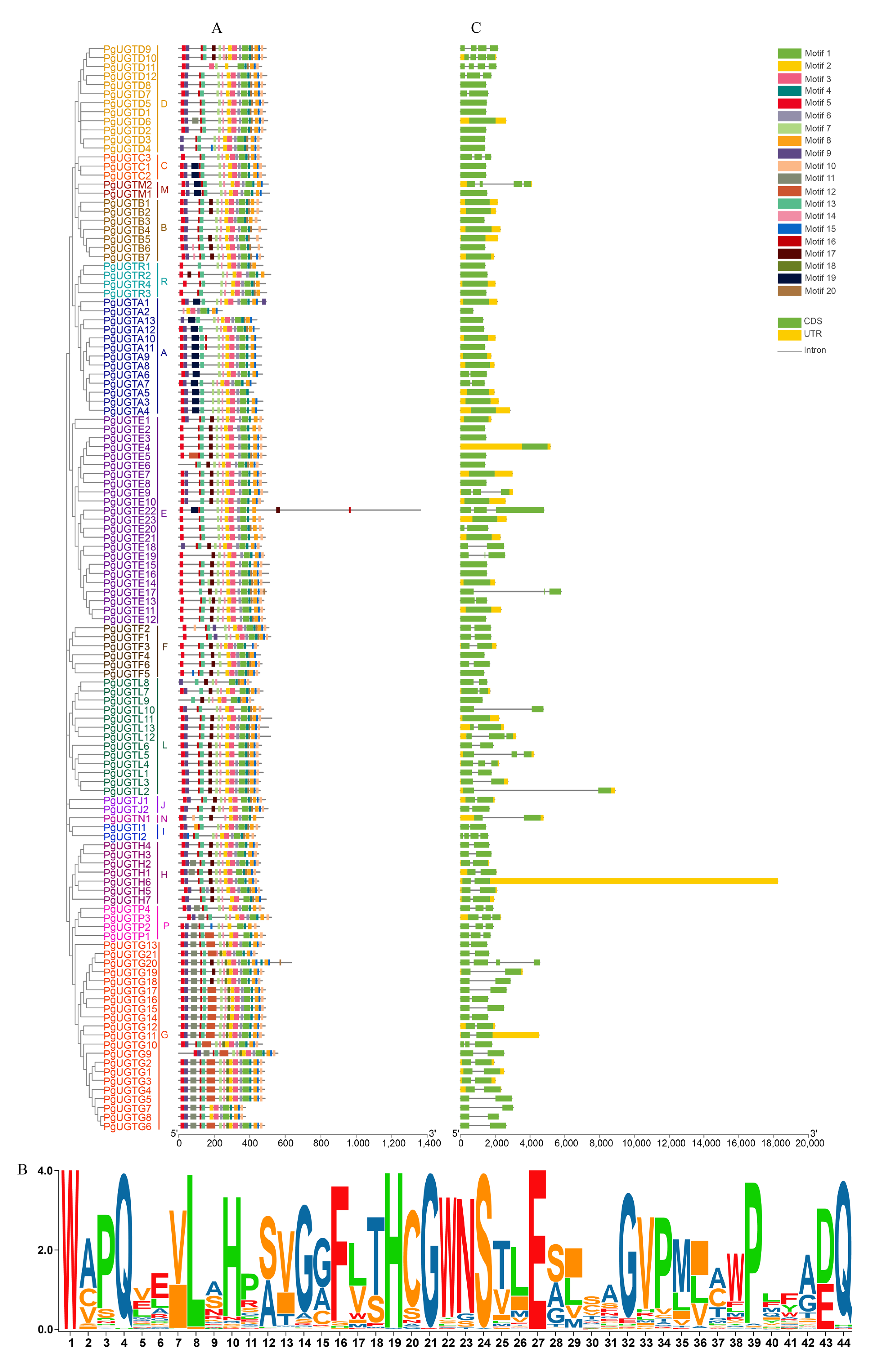
The conserved motifs and gene structure were analyzed to further explore the evolutionary relationship between different PgUGTs. The results showed that the number of motifs of PgUGT family members ranged from 9 to 19, and all members contained motif 1 (PSPG-box) (Figure 2A), indicating that this motif, as a specific aa sequence of the family, is closely related to its biological function and sequence characteristics. The aa composition analysis of the PSPG-box motifs showed that 44 aa made up the PSPG-box motif, some of which were highly conserved and related to the binding of sugar donors, including positions 1 (W), 4 (Q), 8 (L), 10 (H), 19 (H), 21–24 (GWNS), 27 (E), 32–34 (GVP), 39 (P), 43 (D/E), and 44 (Q) (Figure 2B) [57]. In addition, members in different evolutionary groups showed differences in the distribution of conserved motifs. For example, except for groups A, I, M, N, and R, most members of other groups contained motif 10; only members of groups A, C, I, M, and P did not have motif 17; motif 12 was only found in groups E, G, and P; and members of groups F and R did not have motif 9. The differences in the distribution of these motifs may be related to the different biological functions of the members of each group. Members of the same group had similar motif types and arrangement sequences, indicating a close evolutionary relationship between members of the same subgroup. An exon–intron analysis indicated that most members of the same group had similar intron numbers, with 52 out of 120 PgUGTs having no introns, including the members of groups B and R (Figure 2C).

3.3. Gene Duplication and Synteny Analysis of PgUGTs

Genome location analyses showed that 98 PgUGTs were distributed on nine pomegranate chromosomes, and the other PgUGTs were distributed on scaffolds (Figure 3A). Chromosome 3 contained the most PgUGTs (20), followed by chromosome 4 (13) and chromosomes 1 and 2 (12 each). Only one PgUGT was distributed on chromosome 8. For the same group of genes, the distribution of chromosomes was irregular. For example, members of groups E, L, and G were distributed on seven, five, and six chromosomes, respectively. There were also some PgUGTs distributed on chromosomes as gene clusters.
Gene duplication promotes the evolution of gene families. The duplication mode mainly includes tandem duplication, fragment duplication, genome-wide duplication, and rearrangement at the chromosome. Groups E, A, D, L, H, and P possessed the maximum number of tandem duplicated UGTs (five, three, three, three, two, and two, respectively), and B, C, G, N, and R possessed one only (Figure 3A). Groups F, I, J, and M did not have any tandem duplicated UGTs. A total of seven pairs of duplication genes were identified from the pomegranate UGT family, all of which were fragment duplication: Pgr013573.1/Pgr004004.1, Pgr014646.1/Pgr008782.1, Pgr022566.1/Pgr025860.1, Pgr004136.1/Pgr003390.1, Pgr007463.1/Pgr002854.1, Pgr022563.1/Pgr007805.1, and Pgr010381.1/Pgr025860.1 (Figure 3B). These results suggest that tandem duplication and fragment duplication played important roles in the expansion of the PgUGT family. The Ka/Ks values of duplication gene pairs were calculated using TBtools (Table S3). Seven duplication gene pairs had Ka/Ks < 1, indicating that the PgUGT family was mainly affected by purifying selection during the evolution process.

3.4. Expression Profiles of PgUGTs Based on RNA-seq Data

To investigate the potential functions of the PgUGTs, we analyzed their transcript abundances using two sets of published RNA-seq data. First, we detected the expression profiles of 120 PgUGTs in different pomegranate tissues: root, leaf, flower, peel, and outer and inner seed coats (Figure 4A). A total of 46 PgUGTs were expressed in all six tissues, while 20 PgUGTs had no transcript abundance in all these tissues. Some PgUGTs showed tissue-specific expression, for example, PgUGTG1, PgUGTG2, PgUGTG3, PgUGTL2, and PgUGTE18 were specifically expressed in roots; PgUGTE7, PgUGTE16, PgUGTH2, PgUGTL4, and PgUGTL10 were mainly expressed in leaves; PgUGTR4, PgUGTD7, PgUGTN1, PgUGTA13, and PgUGTF3 were specifically expressed in flowers; and PgUGTG5, PgUGTG12, and PgUGTM2 were specifically expressed in peel. In addition, some PgUGTs showed expression in seed coats: PgUGTE12, PgUGTG9, and PgUGTL13 had higher expression levels in the outer than the inner seed coat, while PgUGTE20, PgUGTE21, PgUGTL6, and PgUGTL11 had higher expression levels in the inner than the outer seed coat.
The PgUGTs distributed in groups E and L were considered responsible for the glycosylation modification of monolignols/precursors and hormones [27,28,35,58,59,60]. Therefore, we focused on the members of these two groups for investigating spatiotemporal expression patterns in the seed coats of the hard-seeded cultivar ‘Dabenzi’ and the soft-seeded cultivar ‘Tunisia’ at three developmental stages. Eight PgUGTs (PgUGTE10, PgUGTE12, PgUGTE20, PgUGTE21, PgUGTL6, PgUGTL11, PgUGTL12, and PgUGTL13) were highly expressed in seed coats of both cultivars (Figure 4B): PgUGTE20, PgUGTE21, PgUGTL6, PgUGTL11, and PgUGTL12 were mainly expressed in the inner seed coat, while PgUGTE12 and PgUGTL13 were mainly expressed in the outer seed coat. The relative expression levels of PgUGTE10 and PgUGTL11 in the inner seed coat of ‘Tunisia’ were higher than in ‘Dabenzi’. Accordingly, these eight genes were selected for further study as candidate genes involved in seed hardness development.

3.5. CRE Identification and Relative Expression of PgUGTs Responding to Exogenous Hormones

The CREs in the promoter regions were identified to investigate the transcriptional regulation of the candidate PgUGTs (Figure 5). Several light-responsive elements were present in the PgUGTs. Nine elements related to hormones were identified, including TGA-box, TGA element, and AuxRR-core, which respond to auxin [61]; P-box, TATC-box, and GARE-motif, which respond to gibberellin [62]; TCA-element that responds to salicylic acid [63]; ABRE that responds to abscisic acid [64]; and CGTCA-motif that responds to methyl jasmonate [65]. There were five CREs related to plant growth and development: CAT-box, A-box, GCN4-motif, O2-site, and MBSI [66,67,68]. Interestingly, PgUGTL11 contained MBSI elements, indicating that it may participate in the phenylpropanoid pathway.
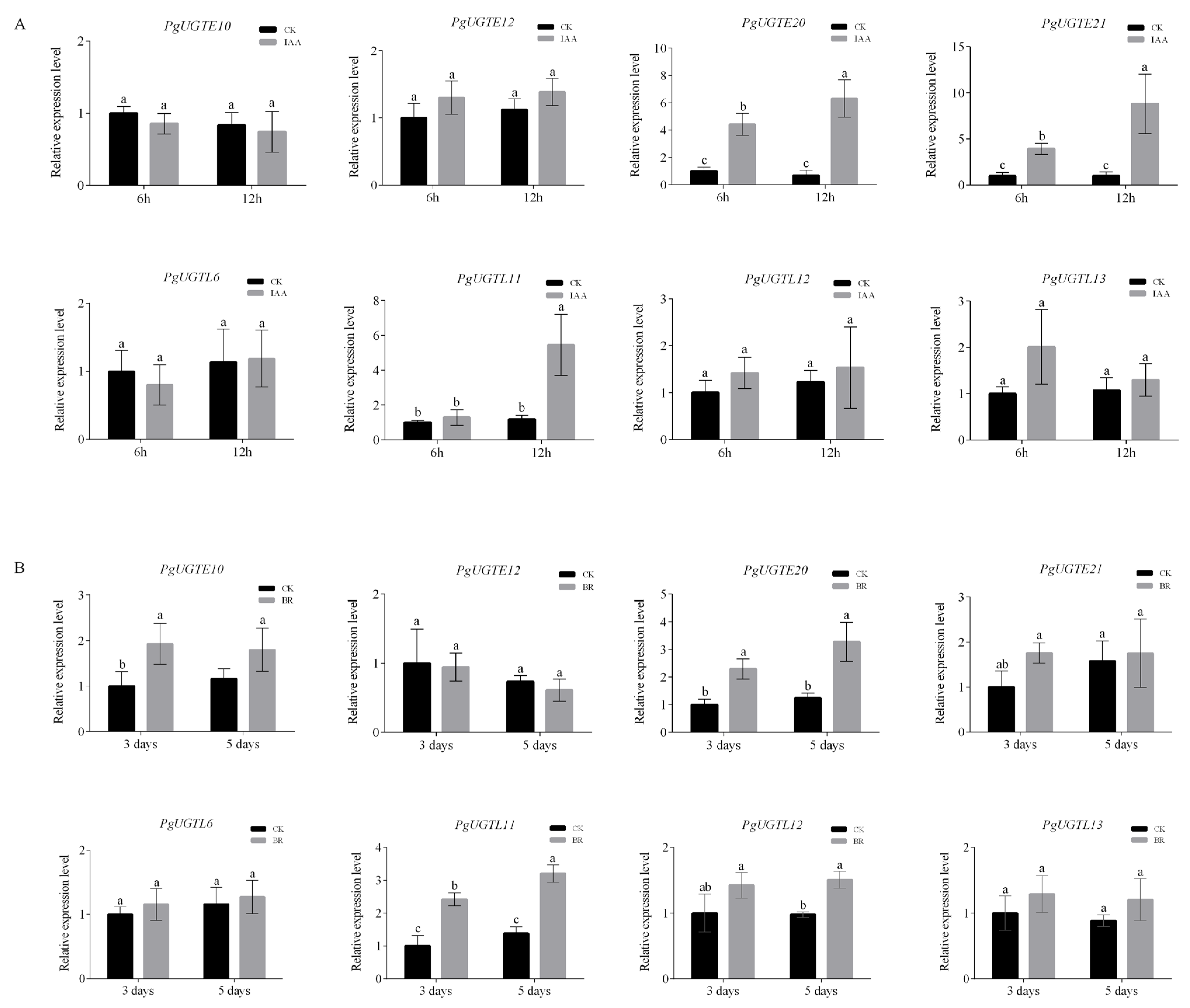
We studied the expression levels of eight candidate PgUGTs under IAA and BR treatments. The expression levels of PgUGTE20, PgUGTE21, and PgUGTL11 were induced by IAA (Figure 6A), while PgUGTE10, PgUGTE20, PgUGTL11, and PgUGTL12 were up-regulated by BR (Figure 6B).

3.6. Co-Expression Analysis of Candidate PgUGTs

According to the expression profiles of PgUGTs, PgUGTE10 and PgUGTL11 were selected to perform co-expression analysis between candidate PgUGTs and genes enriched in phenylpropanoid biosynthesis and hormone signaling pathways. The gene PgUGTL11 was co-expressed with 26 genes involved in the hormone-signaling and lignin biosynthesis pathways (Figure 7), including the Pgr006318.1- and Pgr006319.1-encoding cinnamyl-alcohol dehydrogenase, Pgr016121.1-encoding cinnamoyl-CoA reductase, Pgr011171.1-encoding peroxiredoxin, Pgr000815.1-encoding IAA-responsive protein, and Pgr001703.1-encoding the auxin-responsive protein. The gene PgUGTE10 was co-expressed with six genes, including the Pgr014360.1-encoding shikimate O-hydroxycinnamoyl transferase, Pgr013400.1-encoding cinnamyl-alcohol dehydrogenase, and Pgr011628.1-encoding auxin-response factor (Figure 7). The functional annotation of these co-expression genes is shown in Table S4.

3.7. Subcellular Localization of PgUGTL11 and PgUGTE10

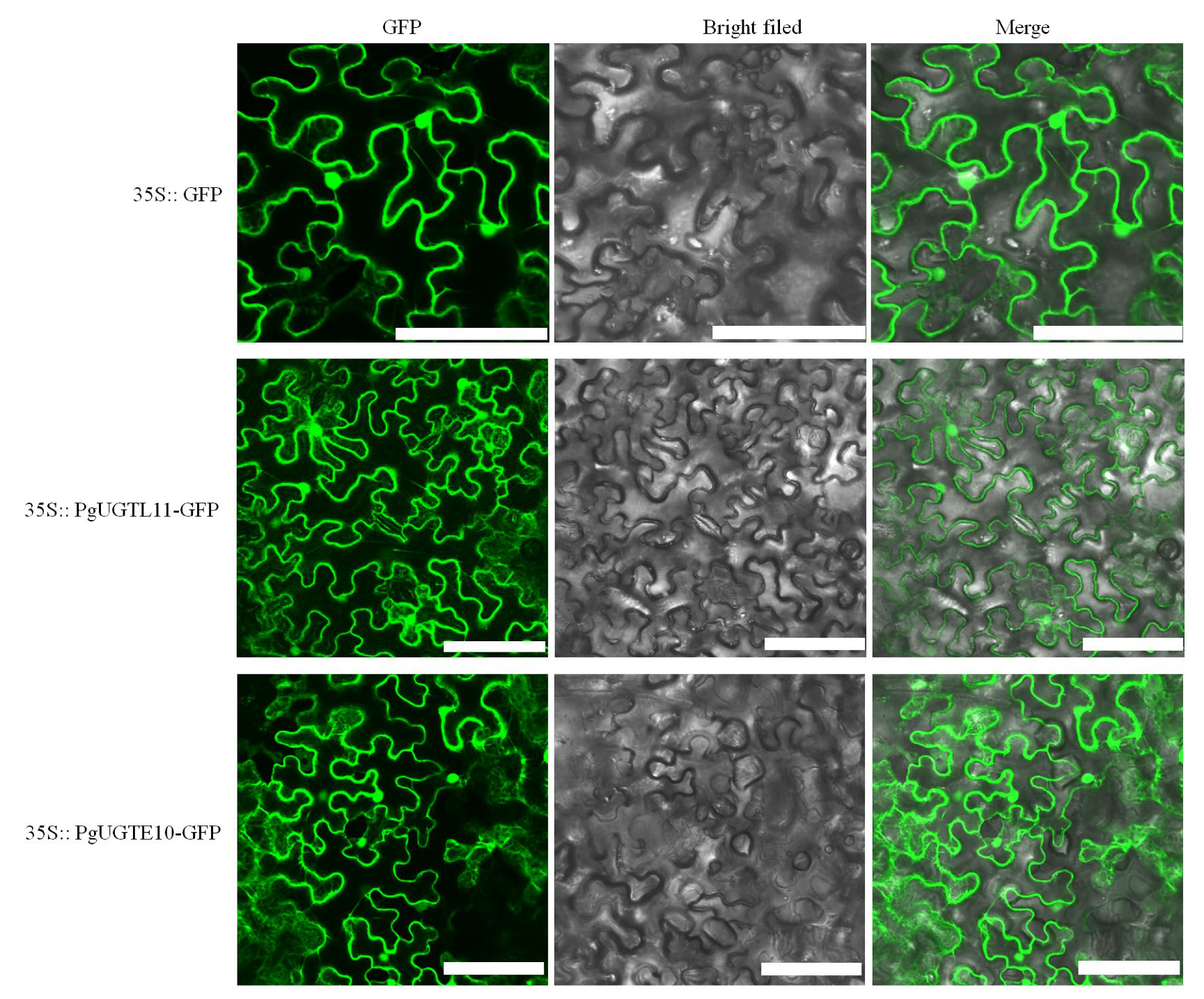
To investigate the subcellular localization of PgUGTL11 and PgUGTE10, we constructed 35S::PgUGTL11/PgUGTE10–GFP fusion vectors and transformed them into tobacco epidermal cells. The results showed that PgUGTL11–GFP and PgUGTE10–GFP presented green fluorescent signals in the nucleus, plasma membrane, and cytoplasm, similar to the control GFP signals (Figure 8).

4. Discussion

Glycosylation modification is ubiquitous in secondary plant metabolites and determines the complex and diverse regulatory network of natural plant products [69]. Glycosylation is involved in regulating the bioactivity of hormones, in the detoxification of xenobiotics, as well as in the biosynthesis, storage, and transport properties of secondary metabolites, suggesting an important role in plant growth and development [70,71,72]. The UGT family is a multigene family of glycosyltransferases with a broad range of substrates [21,22]. In recent years, the UGT family has been identified in plant species, including Arabidopsis [46], rice [26], soybean [73], and fruit species such as grape [21], apple [56], and pear [74]. In pomegranate, three UGTs were identified, but the large-scale analysis was missing.
We identified 120 PgUGTs in the pomegranate genome. Phylogenetic analysis showed the PgUGTs were divided into 15 groups, including 13 conserved groups (A–J and L–N) and two newly discovered groups (P and R). The P group is also found in some other species, including rice [26], grape [75], and apple [56]. The R group is not found in many species but is present in tea [24]. In contrast, groups K and O are present in many species but not in pomegranates. In addition, group G contained 21 PgUGTs but only six AtUGTs, and group H contained only seven PgUGTs but 19 AtUGTs. These findings suggest that specific expansion or gene loss events of the UGT family may have occurred in the evolution of different species. The main driving force of genome and genetic system evolution is gene replication, which promotes plant evolution and leads to the expansion of gene families [76]. The 59 PgUGTs involved in 23 tandem duplication events and the seven pairs of segment duplication genes suggest that tandem duplication and fragment duplication may have been the primary driving force for the expansion of the PgUGTs. The Ka/Ks ≤ 1 of PgUGT repeat gene pairs indicated that the PgUGT family eliminates harmful mutations through purification selection during evolution. The conserved motifs and gene structure analysis showed that all PgUGT protein sequences contained motif 1, a conserved sequence reported as essential for the binding of glycosyl groups. The PgUGT proteins in different phylogenetic groups contained different types and amounts of motifs. In addition, 52 (43%) PgUGTs distributed in different phylogenetic groups did not contain any introns. The diversity of protein and gene structure suggests functional variation in the evolution of this gene family.
The structure and molecular composition of the seed coat profoundly affect seed physiology and have evolved to adapt to diverse environments [77]. The natural diversity of the seed coat structure affects the resistance, dormancy, and germination of seeds [78,79,80]. Angiosperms have evolved two integuments (bitegmic), which develop into the so-called inner and outer seed coats [77]. Interestingly, unlike most angiosperms, the outer seed coat of pomegranate is soft and juicy, and the inner seed coat is highly lignified. The distinguishing structure characteristics of inner and outer seed coats are due to the differential cell expansion and differential accumulation of cellular metabolites, especially of monolignols/monolignol glucosides [53]. However, the molecular regulation mechanism of seed coat development in pomegranate remains unclear. In this study, we attempted to identify candidate PgUGTs involved in seed coat development, especially seed hardness development. We focused on the PgUGTs in phylogenetic groups E and L, which were reported for the glycosylation modification of monolignols/precursors and hormones. We found that eight PgUGTs in these two groups were expressed in seed coats. Among them, PgUGTL11 and PgUGTE10 showed higher expression levels in the seed coat of soft-seeded ‘Tunisia’ compared to the hard-seeded ‘Dabenzi’. The AtUGTs with high homology to PgUGTL11 (i.e., AtUGT84B1 and AtUGT84B2) were reported to glycosylate IAA in vitro and play crucial roles in regulating IAA homeostasis in plants [33,34,81]. The homologous gene cluster AtUGT84A1–4 of PgUGTL11 can glycosylate monolignol precursors [27]. Gene AtUGT72B1, homologous to PgUGTE10, regulates cell wall lignification by glycosylating coniferyl alcohol [30]. In addition, a cluster of homologous AtUGTs, including AtUGT72E1, AtUGT72E2, and AtUGT72E3, was identified as responsible for glucose conjugation of monolignols [29,82]. We found that expression levels of PgUGTE10 were induced by BR treatment, while PgUGTL11 expression was up-regulated both by IAA and BR treatment. In addition, PgUGTL11 and PgUGTE10 were co-expressed with the genes involved in phenylpropanoid biosynthesis and hormone signaling pathways. Auxin has been proven to play a crucial role in seed coat initiation and development by regulating various physiological processes, including cell expansion and lignin biosynthesis [9,83,84]. Several genes involved in lignin synthesis were regulated by BR, thereby promoting cellular lignification and xylem vessel formation [85,86,87]. The UGT proteins are localized in the nucleus, cytoplasm, chloroplast, and other regions, and tend to have substrate diversity, which may be beneficial for metabolic regulation in plants [88,89,90]. Both PgUGTL11 and PgUGTE10 may be localized in the nucleus, plasma membrane, and cytoplasm, but the exact localization requires further verification. Thus, PgUGTL11 and PgUGTE10 are potential candidate genes involved in seed hardness development through glycosylating specific substrates, possibly hormones or monolignols.

5. Conclusions

We identified 120 UGT genes in pomegranate, which were divided into 15 groups according to phylogenetic analysis. Tandem duplication and fragment duplication played important roles in the expansion of the PgUGT gene family. The PgUGTs exhibited various expression profiles in different pomegranate tissues. Comprehensive analysis indicated that PgUGTL11 and PgUGTE10 are potential candidate genes involved in seed hardness development. These results provide new insights into the characteristics of the PgUGT family and its potential functions in pomegranate seed coat development.

Supplementary Materials

The following supporting information can be downloaded at: https://www.mdpi.com/article/10.3390/horticulturae9010119/s1, Figure S1. The phylogenetic tree of UGTs in newly discovered groups P and R in pomegranate, apple, and tea. Table S1. The gene-specific primers used in this study. Table S2. Detailed information of PgUGTs identified in the pomegranate genome. Table S3. Ka/Ks analysis of PgUGT duplicated genes. Table S4. The functional annotation of the genes that co-expressed with the candidate PgUGTs.

Author Contributions

Conceptualization, J.L. and G.Q.; data curation, G.L. and J.L.; methodology, G.L., J.L. and X.L.; resources, Z.C. and B.J.; supervision, C.L.; writing—original draft, G.L. and J.L.; writing—review and editing, G.Q. and H.Z. All authors have read and agreed to the published version of the manuscript.

Funding

This research was supported by the Major Science and Technology Project of Anhui Province (201903b06020017), the Major Science and Technology Project of Huaibei City (HK2021014), and the Natural Science Foundation of Anhui Province (2108085QC129).

Institutional Review Board Statement

Not applicable.

Informed Consent Statement

Not applicable.

Data Availability Statement

All datasets presented in this study are included in the article/Supplementary Material.

Conflicts of Interest

The authors declare no conflict of interest.

References

  1. Niu, J.; Cao, D.; Li, H.; Xue, H.; Chen, L.; Liu, B.; Cao, S. Quantitative proteomics of pomegranate varieties with contrasting seed hardness during seed development stages. Tree Genet. Genomes 2018, 14, 14. [Google Scholar] [CrossRef] [Green Version]
  2. Heber, D. Multitargeted therapy of cancer by ellagitannins. Cancer Lett. 2008, 269, 262–268. [Google Scholar] [CrossRef] [PubMed]
  3. Xie, X.; Huang, Y.; Tian, S.; Li, G.; Cao, S. Relationship of seed hardness development and microstructure of seed coat cell in soft seed pomegranate. Acta Hortic. Sin. 2017, 44, 1174–1180. [Google Scholar] [CrossRef]
  4. Boerjan, W.; Ralph, J.; Baucher, M. Lignin biosynthesis. Annu. Rev. Plant Biol. 2003, 54, 519–546. [Google Scholar] [CrossRef] [PubMed]
  5. Liu, C.J.; Miao, Y.C.; Zhang, K.W. Sequestration and transport of lignin monomeric precursors. Molecules 2011, 16, 710–727. [Google Scholar] [CrossRef] [Green Version]
  6. Dima, O.; Morreel, K.; Vanholme, B.; Kim, H.; Ralph, J.; Boerjan, W. Small glycosylated lignin oligomers are stored in Arabidopsis leaf vacuoles. Plant Cell 2015, 27, 695–710. [Google Scholar] [CrossRef] [Green Version]
  7. Luo, M.; Dennis, E.S.; Berger, F.; Peacock, W.J.; Chaudhury, A. MINISEED3 (MINI3), a WRKY family gene, and HAIKU2 (IKU2), a leucine-rich repeat (LRR) KINASE gene, are regulators of seed size in Arabidopsis. Proc. Natl. Acad. Sci. USA 2005, 102, 17531–17536. [Google Scholar] [CrossRef] [Green Version]
  8. Garcia, D.; Saingery, V.; Chambrier, P.; Mayer, U.; Jurgens, G.; Berger, F. Arabidopsis haiku mutants reveal new controls of seed size by endosperm. Plant Physiol. 2003, 131, 1661–1670. [Google Scholar] [CrossRef] [Green Version]
  9. Figueiredo, D.D.; Batista, R.A.; Roszakt, P.J.; Hennig, L.; Kohler, C. Auxin production in the endosperm drives seed coat development in Arabidopsis. Elife 2016, 5, e20542. [Google Scholar] [CrossRef]
  10. Immanen, J.; Nieminen, K.; Smolander, O.P.; Kojima, M.; Serra, J.A.; Koskinen, P.; Zhang, J.; Elo, A.; Mahonen, A.P.; Street, N.; et al. Cytokinin and auxin display distinct but interconnected distribution and signaling profiles to stimulate cambial activity. Curr. Biol. 2016, 26, 1990–1997. [Google Scholar] [CrossRef]
  11. Xu, C.Z.; Shen, Y.; He, F.; Fu, X.K.; Yu, H.; Lu, W.X.; Li, Y.L.; Li, C.F.; Fan, D.; Wang, H.C.; et al. Auxin-mediated Aux/IAA-ARF-HB signaling cascade regulates secondary xylem development in Populus. New Phytol. 2019, 222, 752–767. [Google Scholar] [CrossRef]
  12. Uggla, C.; Mellerowicz, E.J.; Sundberg, B. Indole-3-acetic acid controls cambial growth in Scots pine by positional signaling. Plant Physiol. 1998, 117, 113–121. [Google Scholar] [CrossRef] [Green Version]
  13. Korasick, D.A.; Enders, T.A.; Strader, L.C. Auxin biosynthesis and storage forms. J. Exp. Bot. 2013, 64, 2541–2555. [Google Scholar] [CrossRef] [Green Version]
  14. Jin, H.; Do, J.; Shin, S.J.; Choi, J.W.; Choi, Y.I.; Kim, W.; Kwon, M. Exogenously applied 24-epi brassinolide reduces lignification and alters cell wall carbohydrate biosynthesis in the secondary xylem of Liriodendron tulipifera. Phytochemistry 2014, 101, 40–51. [Google Scholar] [CrossRef]
  15. Ge, Y.H.; Li, C.Y.; Tang, R.X.; Sun, R.H.; Li, J.R. Effects of postharvest brassinolide dipping on quality parameters and antioxidant activity in peach fruit. In Proceedings of the 3rd International Symposium on Postharvest Pathology—Using Science to Increase Food Availability, Bari, Italy, 7–11 June 2015; pp. 377–384. [Google Scholar] [CrossRef]
  16. Sanchez-Rodriguez, C.; Rubio-Somoza, I.; Sibout, R.; Persson, S. Phytohormones and the cell wall in Arabidopsis during seedling growth. Trends Plant Sci. 2010, 15, 291–301. [Google Scholar] [CrossRef]
  17. Ibanes, M.; Fabregas, N.; Chory, J.; Cano-Delgado, A.I. Brassinosteroid signaling and auxin transport are required to establish the periodic pattern of Arabidopsis shoot vascular bundles. Proc. Natl. Acad. Sci. USA 2009, 106, 13630–13635. [Google Scholar] [CrossRef] [Green Version]
  18. Oda, Y.; Mimura, T.; Hasezawa, S. Regulation of secondary cell wall development by cortical microtubules during tracheary element differentiation in Arabidopsis cell suspensions. Plant Physiol. 2005, 137, 1027–1036. [Google Scholar] [CrossRef] [Green Version]
  19. Shen, H.; Mazarei, M.; Hisano, H.; Escamilla-Trevino, L.; Fu, C.X.; Pu, Y.Q.; Rudis, M.R.; Tang, Y.H.; Xiao, X.R.; Jackson, L.; et al. A genomics approach to deciphering lignin biosynthesis in switchgrass. Plant Cell 2013, 25, 4342–4361. [Google Scholar] [CrossRef] [Green Version]
  20. Ostrowski, M.; Jakubowska, A. UDP-Glycosyltransferases of Plant Hormones. Postep. Biol. Komorki 2013, 40, 141–160. [Google Scholar] [CrossRef]
  21. Yonekura-Sakakibara, K.; Hanada, K. An evolutionary view of functional diversity in family 1 glycosyltransferases. Plant J. 2011, 66, 182–193. [Google Scholar] [CrossRef]
  22. Paquette, S.; Møller, B.L.; Bak, S. On the origin of family 1 plant glycosyltransferases. Phytochemistry 2003, 62, 399–413. [Google Scholar] [CrossRef] [PubMed]
  23. Ross, J.; Li, Y.; Lim, E.-K.; Bowles, D.J. Higher plant glycosyltransferases. Genome Biol. 2001, 2, reviews3004.1. [Google Scholar] [CrossRef] [PubMed]
  24. Cui, L.; Yao, S.; Dai, X.; Yin, Q.; Liu, Y.; Jiang, X.; Wu, Y.; Qian, Y.; Pang, Y.; Gao, L.; et al. Identification of UDP-glycosyltransferases involved in the biosynthesis of astringent taste compounds in tea (Camellia sinensis). J. Exp. Bot. 2016, 67, 2285–2297. [Google Scholar] [CrossRef] [PubMed] [Green Version]
  25. Li, Y.; Li, P.; Wang, Y.; Dong, R.; Yu, H.; Hou, B. Genome-wide identification and phylogenetic analysis of family-1 UDP glycosyltransferases in maize (Zea mays). Planta 2014, 239, 1265–1279. [Google Scholar] [CrossRef]
  26. Caputi, L.; Malnoy, M.; Goremykin, V.; Nikiforova, S.; Martens, S. A genome-wide phylogenetic reconstruction of family 1 UDP-glycosyltransferases revealed the expansion of the family during the adaptation of plants to life on land. Plant J. 2012, 69, 1030–1042. [Google Scholar] [CrossRef]
  27. Lim, E.-K.; Li, Y.; Parr, A.; Jackson, R.; Ashford, D.A.; Bowles, D.J. Identification of glucosyltransferase genes involved in sinapate metabolism and lignin synthesis in Arabidopsis. J. Biol. Chem. 2001, 276, 4344–4349. [Google Scholar] [CrossRef] [Green Version]
  28. Lim, E.-K.; Jackson, R.G.; Bowles, D.J. Identification and characterisation of Arabidopsis glycosyltransferases capable of glucosylating coniferyl aldehyde and sinapyl aldehyde. FEBS Lett. 2005, 579, 2802–2806. [Google Scholar] [CrossRef] [Green Version]
  29. Lanot, A.; Hodge, D.; Jackson, R.G.; George, G.L.; Elias, L.; Lim, E.K.; Vaistij, F.E.; Bowles, D.J. The glucosyltransferase UGT72E2 is responsible for monolignol 4-O-glucoside production in Arabidopsis thaliana. Plant J. 2006, 48, 286–295. [Google Scholar] [CrossRef]
  30. Lin, J.S.; Huang, X.X.; Li, Q.; Cao, Y.; Bao, Y.; Meng, X.F.; Li, Y.J.; Fu, C.; Hou, B.K. UDP-glycosyltransferase 72B1 catalyzes the glucose conjugation of monolignols and is essential for the normal cell wall lignification in Arabidopsis thaliana. Plant J. 2016, 88, 26–42. [Google Scholar] [CrossRef]
  31. Wang, H.; Feng, X.; Zhang, Y.; Wei, D.; Zhang, Y.; Jin, Q.; Cai, Y. PbUGT72AJ2-mediated glycosylation plays an important role in lignin formation and stone cell development in pears (Pyrus bretschneideri). Int. J. Mol. Sci. 2022, 23, 7893. [Google Scholar] [CrossRef]
  32. Szerszen, J.B.; Szczyglowski, K.; Bandurski, R.S. iaglu, a gene from Zea mays involved in conjugation of growth hormone indole-3-acetic acid. Science 1994, 265, 1699–1701. [Google Scholar] [CrossRef]
  33. Jackson, R.G.; Lim, E.-K.; Li, Y.; Kowalczyk, M.; Sandberg, G.; Hoggett, J.; Ashford, D.A.; Bowles, D.J. Identification and biochemical characterization of an Arabidopsis indole-3-acetic acid glucosyltransferase. J. Biol. Chem. 2001, 276, 4350–4356. [Google Scholar] [CrossRef] [Green Version]
  34. Jackson, R.G.; Kowalczyk, M.; Li, Y.; Higgins, G.; Ross, J.; Sandberg, G.; Bowles, D.J. Over-expression of an Arabidopsis gene encoding a glucosyltransferase of indole-3-acetic acid: Phenotypic characterisation of transgenic lines. Plant J. 2002, 32, 573–583. [Google Scholar] [CrossRef]
  35. Tognetti, V.B.; Van Aken, O.; Morreel, K.; Vandenbroucke, K.; van de Cotte, B.; De Clercq, I.; Chiwocha, S.; Fenske, R.; Prinsen, E.; Boerjan, W.; et al. Perturbation of indole-3-butyric acid homeostasis by the UDP-glucosyltransferase UGT74E2 modulates Arabidopsis architecture and water stress tolerance. Plant Cell 2010, 22, 2660–2679. [Google Scholar] [CrossRef] [Green Version]
  36. Jin, S.-H.; Ma, X.-M.; Han, P.; Wang, B.; Sun, Y.-G.; Zhang, G.-Z.; Li, Y.-J.; Hou, B.-K. UGT74D1 is a novel auxin glycosyltransferase from Arabidopsis thaliana. PLoS ONE 2013, 8, e61705. [Google Scholar] [CrossRef]
  37. Poppenberger, B.; Fujioka, S.; Soeno, K.; George, G.L.; Vaistij, F.E.; Hiranuma, S.; Seto, H.; Takatsuto, S.; Adam, G.; Yoshida, S. The UGT73C5 of Arabidopsis thaliana glucosylates brassinosteroids. Proc. Natl. Acad. Sci. USA 2005, 102, 15253–15258. [Google Scholar] [CrossRef] [Green Version]
  38. Husar, S.; Berthiller, F.; Fujioka, S.; Rozhon, W.; Khan, M.; Kalaivanan, F.; Elias, L.; Higgins, G.S.; Li, Y.; Schuhmacher, R. Overexpression of the UGT73C6 alters brassinosteroid glucoside formation in Arabidopsis thaliana. BMC Plant Biol. 2011, 11, 51. [Google Scholar] [CrossRef] [Green Version]
  39. Ono, N.N.; Qin, X.; Wilson, A.E.; Li, G.; Tian, L. Two UGT84 family glycosyltransferases catalyze a critical reaction of hydrolyzable tannin biosynthesis in pomegranate (Punica granatum). PLoS ONE 2016, 11, e0156319. [Google Scholar] [CrossRef] [Green Version]
  40. Wilson, A.E.; Wu, S.; Tian, L. PgUGT95B2 preferentially metabolizes flavones/flavonols and has evolved independently from flavone/flavonol UGTs identified in Arabidopsis thaliana. Phytochemistry 2019, 157, 184–193. [Google Scholar] [CrossRef]
  41. Shi, M.; Liu, X.; Zhang, H.; He, Z.; Yang, H.; Chen, J.; Feng, J.; Yang, W.; Jiang, Y.; Yao, J.-L. The IAA-and ABA-responsive transcription factor CgMYB58 upregulates lignin biosynthesis and triggers juice sac granulation in pummelo. Hortic. Res. 2020, 7, 139. [Google Scholar] [CrossRef]
  42. Soeno, K.; Fujioka, S.; Hiranuma, S.; Seto, H.; Yoshida, S. Metabolic conversion of castasterone and brassinolide into their glucosides in higher plants. J. Plant Growth Regul. 2006, 25, 195–202. [Google Scholar] [CrossRef]
  43. Qin, G.; Xu, C.; Ming, R.; Tang, H.; Guyot, R.; Kramer, E.M.; Hu, Y.; Yi, X.; Qi, Y.; Xu, X. The pomegranate (Punica granatum L.) genome and the genomics of punicalagin biosynthesis. Plant J. 2017, 91, 1108–1128. [Google Scholar] [CrossRef] [PubMed] [Green Version]
  44. Letunic, I.; Doerks, T.; Bork, P. SMART 7: Recent updates to the protein domain annotation resource. Nucleic Acids Res. 2012, 40, D302–D305. [Google Scholar] [CrossRef] [PubMed]
  45. Marchler-Bauer, A.; Lu, S.; Anderson, J.B.; Chitsaz, F.; Derbyshire, M.K.; DeWeese-Scott, C.; Fong, J.H.; Geer, L.Y.; Geer, R.C.; Gonzales, N.R. CDD: A Conserved Domain Database for the functional annotation of proteins. Nucleic Acids Res. 2010, 39, D225–D229. [Google Scholar] [CrossRef] [PubMed] [Green Version]
  46. Li, Y.; Baldauf, S.; Lim, E.K.; Bowles, D.J. Phylogenetic analysis of the UDP-glycosyltransferase multigene family of Arabidopsis thaliana. J. Biol. Chem. 2001, 276, 4338–4343. [Google Scholar] [CrossRef] [Green Version]
  47. Bailey, T.L.; Boden, M.; Buske, F.A.; Frith, M.; Grant, C.E.; Clementi, L.; Ren, J.; Li, W.W.; Noble, W.S. MEME SUITE: Tools for motif discovery and searching. Nucleic Acids Res. 2009, 37, W202–W208. [Google Scholar] [CrossRef]
  48. Chen, C.; Chen, H.; Zhang, Y.; Thomas, H.R.; Frank, M.H.; He, Y.; Xia, R. TBtools: An integrative toolkit developed for interactive analyses of big biological data. Mol. Plant 2020, 13, 1194–1202. [Google Scholar] [CrossRef]
  49. Hoang, M.; Wu, C.-N.; Lin, C.-F.; Nguyen, H.T.T.; Chiou, M.-T.; Lin, C.-N. Genetic characterization of feline panleukopenia virus from dogs in Vietnam reveals a unique Thr101 mutation in VP2. PeerJ 2020, 8, e9752. [Google Scholar] [CrossRef]
  50. Zhang, J. Evolution by gene duplication: An update. Trends Ecol. Evol. 2003, 18, 292–298. [Google Scholar] [CrossRef]
  51. Zhang, Z.; Li, J.; Zhao, X.-Q.; Wang, J.; Wong, G.K.-S.; Yu, J. KaKs_Calculator: Calculating Ka and Ks through model selection and model averaging. Genom. Proteom. Bioinform. 2006, 4, 259–263. [Google Scholar] [CrossRef] [Green Version]
  52. Lescot, M.; Déhais, P.; Thijs, G.; Marchal, K.; Moreau, Y.; Van de Peer, Y.; Rouzé, P.; Rombauts, S. PlantCARE, a database of plant cis-acting regulatory elements and a portal to tools for in silico analysis of promoter sequences. Nucleic Acids Res. 2002, 30, 325–327. [Google Scholar] [CrossRef]
  53. Qin, G.; Liu, C.; Li, J.; Qi, Y.; Gao, Z.; Zhang, X.; Yi, X.; Pan, H.; Ming, R.; Xu, Y. Diversity of metabolite accumulation patterns in inner and outer seed coats of pomegranate: Exploring their relationship with genetic mechanisms of seed coat development. Hortic. Res. 2020, 7, 10. [Google Scholar] [CrossRef] [Green Version]
  54. Kwiatkowska, M.; Norman, G.; Parker, D. PRISM 4.0: Verification of probabilistic real-time systems. In Computer Aided Verification. CAV 2011. Lecture Notes in Computer Science; Gopalakrishnan, G., Qadeer, S., Eds.; Springer: Berlin/Heidelberg, Germany, 2011; pp. 585–591. [Google Scholar]
  55. Smoot, M.E.; Ono, K.; Ruscheinski, J.; Wang, P.L.; Ideker, T. Cytoscape 2.8: New features for data integration and network visualization. Bioinformatics 2011, 27, 431–432. [Google Scholar] [CrossRef] [Green Version]
  56. Zhou, K.; Hu, L.; Li, P.; Gong, X.; Ma, F. Genome-wide identification of glycosyltransferases converting phloretin to phloridzin in Malus species. Plant Sci. 2017, 265, 131–145. [Google Scholar] [CrossRef]
  57. Gachon, C.M.; Langlois-Meurinne, M.; Saindrenan, P. Plant secondary metabolism glycosyltransferases: The emerging functional analysis. Trends Plant Sci. 2005, 10, 542–549. [Google Scholar] [CrossRef]
  58. Lim, E.-K.; Doucet, C.J.; Li, Y.; Elias, L.; Worrall, D.; Spencer, S.P.; Ross, J.; Bowles, D.J. The activity of Arabidopsis glycosyltransferases toward salicylic acid, 4-hydroxybenzoic acid, and other benzoates. J. Biol. Chem. 2002, 277, 586–592. [Google Scholar] [CrossRef] [Green Version]
  59. Tanaka, K.; Hayashi, K.; Natsume, M.; Kamiya, Y.; Sakakibara, H.; Kawaide, H.; Kasahara, H. UGT74D1 catalyzes the glucosylation of 2-Oxindole-3-Acetic Acid in the auxin metabolic pathway in Arabidopsis. Plant Cell Physiol. 2014, 55, 218–228. [Google Scholar] [CrossRef] [Green Version]
  60. Aoi, Y.; Hira, H.; Hayakawa, Y.; Liu, H.Q.; Fukui, K.; Dai, X.H.; Tanaka, K.; Hayashi, K.I.; Zhao, Y.D.; Kasahara, H. UDP-glucosyltransferase UGT84B1 regulates the levels of indole-3-acetic acid and phenylacetic acid in Arabidopsis. Biochem. Biophys. Res. Commun. 2020, 532, 244–250. [Google Scholar] [CrossRef]
  61. Du, Y.; Zhao, Q.; Li, W.; Geng, J.; Li, S.; Yuan, X.; Gu, Y.; Zhong, J.; Zhang, Y.; Du, J. Genome-wide identification of the LBD transcription factor genes in common bean (Phaseolus vulgaris L.) and expression analysis under different abiotic stresses. J. Plant Interact. 2022, 17, 731–743. [Google Scholar] [CrossRef]
  62. Pinweha, N.; Asvarak, T.; Viboonjun, U.; Narangajavana, J. Involvement of miR160/miR393 and their targets in cassava responses to anthracnose disease. J. Plant Physiol. 2015, 174, 26–35. [Google Scholar] [CrossRef]
  63. Zeng, M.-H.; Liu, S.-H.; Yang, M.-X.; Zhang, Y.-J.; Liang, J.-Y.; Wan, X.-R.; Liang, H. Characterization of a gene encoding clathrin heavy chain in maize up-regulated by salicylic acid, abscisic acid and high boron supply. Int. J. Mol. Sci. 2013, 14, 15179–15198. [Google Scholar] [CrossRef] [PubMed] [Green Version]
  64. Tuteja, N. Abscisic acid and abiotic stress signaling. Plant Signal. Behav. 2007, 2, 135–138. [Google Scholar] [CrossRef] [PubMed] [Green Version]
  65. Xu, Z.; Sun, M.; Jiang, X.; Sun, H.; Dang, X.; Cong, H.; Qiao, F. Glycinebetaine biosynthesis in response to osmotic stress depends on jasmonate signaling in watermelon suspension cells. Front. Plant Sci. 2018, 9, 1469. [Google Scholar] [CrossRef] [PubMed] [Green Version]
  66. Kaur, G.; Pati, P.K. Analysis of cis-acting regulatory elements of Respiratory burst oxidase homolog (Rboh) gene families in Arabidopsis and rice provides clues for their diverse functions. Comput. Biol. Chem. 2016, 62, 104–118. [Google Scholar] [CrossRef] [PubMed]
  67. Kaur, A.; Pati, P.K.; Pati, A.M.; Nagpal, A.K. In-silico analysis of cis-acting regulatory elements of pathogenesis-related proteins of Arabidopsis thaliana and Oryza sativa. PLoS ONE 2017, 12, e0184523. [Google Scholar] [CrossRef] [Green Version]
  68. Zhou, J.; Hu, Y.; Li, J.; Yu, Z.; Guo, Q. Genome-wide identification and expression analysis of the plant U-box protein gene family in Phyllostachys edulis. Front. Genet. 2021, 2464. [Google Scholar] [CrossRef]
  69. Song, C.K.; Gu, L.; Liu, J.Y.; Zhao, S.; Hong, X.T.; Schulenburg, K.; Schwab, W. Functional characterization and substrate promiscuity of UGT71 glycosyltransferases from strawberry (Fragaria x ananassa). Plant Cell Physiol. 2015, 56, 2478–2493. [Google Scholar] [CrossRef] [Green Version]
  70. Lim, E.K.; Bowles, D.J. A class of plant glycosyltransferases involved in cellular homeostasis. EMBO J. 2004, 23, 2915–2922. [Google Scholar] [CrossRef] [Green Version]
  71. Bowles, D.; Isayenkova, J.; Lim, E.K.; Poppenberger, B. Glycosyltransferases: Managers of small molecules. Curr. Opin. Plant Biol. 2005, 8, 254–263. [Google Scholar] [CrossRef]
  72. Bowles, D.; Lim, E.K.; Poppenberger, B.; Vaistij, F.E. Glycosyltransferases of lipophilic small molecules. Annu. Rev. Plant Biol. 2006, 57, 567–597. [Google Scholar] [CrossRef]
  73. Rehman, H.M.; Nawaz, M.A.; Bao, L.; Shah, Z.H.; Lee, J.-M.; Ahmad, M.Q.; Chung, G.; Yang, S.H. Genome-wide analysis of family-1 UDP-glycosyltransferases in soybean confirms their abundance and varied expression during seed development. J. Plant Physiol. 2016, 206, 87–97. [Google Scholar] [CrossRef]
  74. Cheng, X.; Muhammad, A.; Li, G.; Zhang, J.; Cheng, J.; Qiu, J.; Jiang, T.; Jin, Q.; Cai, Y.; Lin, Y. Family-1 UDP glycosyltransferases in pear (Pyrus bretschneideri): Molecular identification, phylogenomic characterization and expression profiling during stone cell formation. Mol. Biol. Rep. 2019, 46, 2153–2175. [Google Scholar] [CrossRef]
  75. Wei, Y.; Mu, H.; Xu, G.; Wang, Y.; Li, Y.; Li, S.; Wang, L. Genome-wide analysis and functional characterization of the UDP-glycosyltransferase family in grapes. Horticulturae 2021, 7, 204. [Google Scholar] [CrossRef]
  76. Levasseur, A.; Pontarotti, P. The role of duplications in the evolution of genomes highlights the need for evolutionary-based approaches in comparative genomics. Biol. Direct 2011, 6, 11. [Google Scholar] [CrossRef] [Green Version]
  77. Coen, O.; Magnani, E. Seed coat thickness in the evolution of angiosperms. Cell. Mol. Life Sci. 2018, 75, 2509–2518. [Google Scholar] [CrossRef] [Green Version]
  78. Gardarin, A.; Durr, C.; Mannino, M.R.; Busset, H.; Colbach, N. Seed mortality in the soil is related to seed coat thickness. Seed Sci. Res. 2010, 20, 243–256. [Google Scholar] [CrossRef]
  79. Kelly, K.M.; Vanstaden, J.; Bell, W.E. Seed coat structure and dormancy. Plant Growth Regul. 1992, 11, 201–209. [Google Scholar] [CrossRef]
  80. Nooden, L.D.; Blakley, K.A.; Grzybowski, J.M. Control of seed coat thickness and permeability in soybean: A possible adaptation to stress. Plant Physiol. 1985, 79, 543–545. [Google Scholar] [CrossRef] [Green Version]
  81. Mateo-Bonmati, E.; Casanova-Saez, R.; Simura, J.; Ljung, K. Broadening the roles of UDP-glycosyltransferases in auxin homeostasis and plant development. New Phytol. 2021, 232, 642–654. [Google Scholar] [CrossRef]
  82. Baldacci-Cresp, F.; Le Roy, J.; Huss, B.; Lion, C.; Creach, A.; Spriet, C.; Duponchel, L.; Biot, C.; Baucher, M.; Hawkins, S.; et al. UDP-glycosyltransferase 72E3 plays a role in lignification of secondary cell walls in Arabidopsis. Int. J. Mol. Sci. 2020, 21, 6094. [Google Scholar] [CrossRef]
  83. Aloni, R.; Tollier, M.T.; Monties, B. The role of auxin and gibberellin in controlling lignin formation in primary phloem fibers and in xylem of coleus blumei stems. Plant Physiol. 1990, 94, 1743–1747. [Google Scholar] [CrossRef] [PubMed] [Green Version]
  84. Salazar, R.; Pollmann, S.; Morales-Quintana, L.; Herrera, R.; Caparros-Ruiz, D.; Ramos, P. In seedlings of Pinus radiata, jasmonic acid and auxin are differentially distributed on opposite sides of tilted stems affecting lignin monomer biosynthesis and composition. Plant Physiol. Biochem. 2019, 135, 215–223. [Google Scholar] [CrossRef] [PubMed]
  85. Takeuchi, M.; Kegasa, T.; Watanabe, A.; Tamura, M.; Tsutsumi, Y. Expression analysis of transporter genes for screening candidate monolignol transporters using Arabidopsis thaliana cell suspensions during tracheary element differentiation. J. Plant Res. 2018, 131, 297–305. [Google Scholar] [CrossRef] [PubMed]
  86. Fan, F.H.; Zhou, Z.J.; Qin, H.J.; Tan, J.H.; Ding, G.J. Exogenous brassinosteroid facilitates xylem development in pinus massoniana seedlings. Int. J. Mol. Sci. 2021, 22, 7615. [Google Scholar] [CrossRef]
  87. Oh, M.H.; Honey, S.H.; Tax, F.E. The control of cell expansion, cell division, and vascular development by brassinosteroids: A historical perspective. Int. J. Mol. Sci. 2020, 21, 1743. [Google Scholar] [CrossRef] [Green Version]
  88. Zhang, K.; Sun, Y.; Li, M.N.; Long, R.C. CrUGT87A1, a UDP-sugar glycosyltransferases (UGTs) gene from Carex rigescens, increases salt tolerance by accumulating flavonoids for antioxidation in Arabidopsis thaliana. Plant Physiol. Biochem. 2021, 159, 28–36. [Google Scholar] [CrossRef]
  89. Wu, B.P.; Cao, X.M.; Liu, H.R.; Zhu, C.Q.; Klee, H.; Zhang, B.; Chen, K.S. UDP-glucosyltransferase PpUGT85A2 controls volatile glycosylation in peach. J. Exp. Bot. 2019, 70, 925–936. [Google Scholar] [CrossRef] [Green Version]
  90. Zheng, H.; Zhang, C.L.; Wang, Y.T.; Zhou, W.T.; Chen, J.; Yan, X.Y.; Li, Z.Q.; Huang, S.Y.; Li, M.Y.; Tang, Y.; et al. Overexpression of the glucosyltransferase gene BoaUGT74B1 enhances the accumulation of indole glucosinolates in Chinese kale. Sci. Hortic. 2021, 288, 110302. [Google Scholar] [CrossRef]
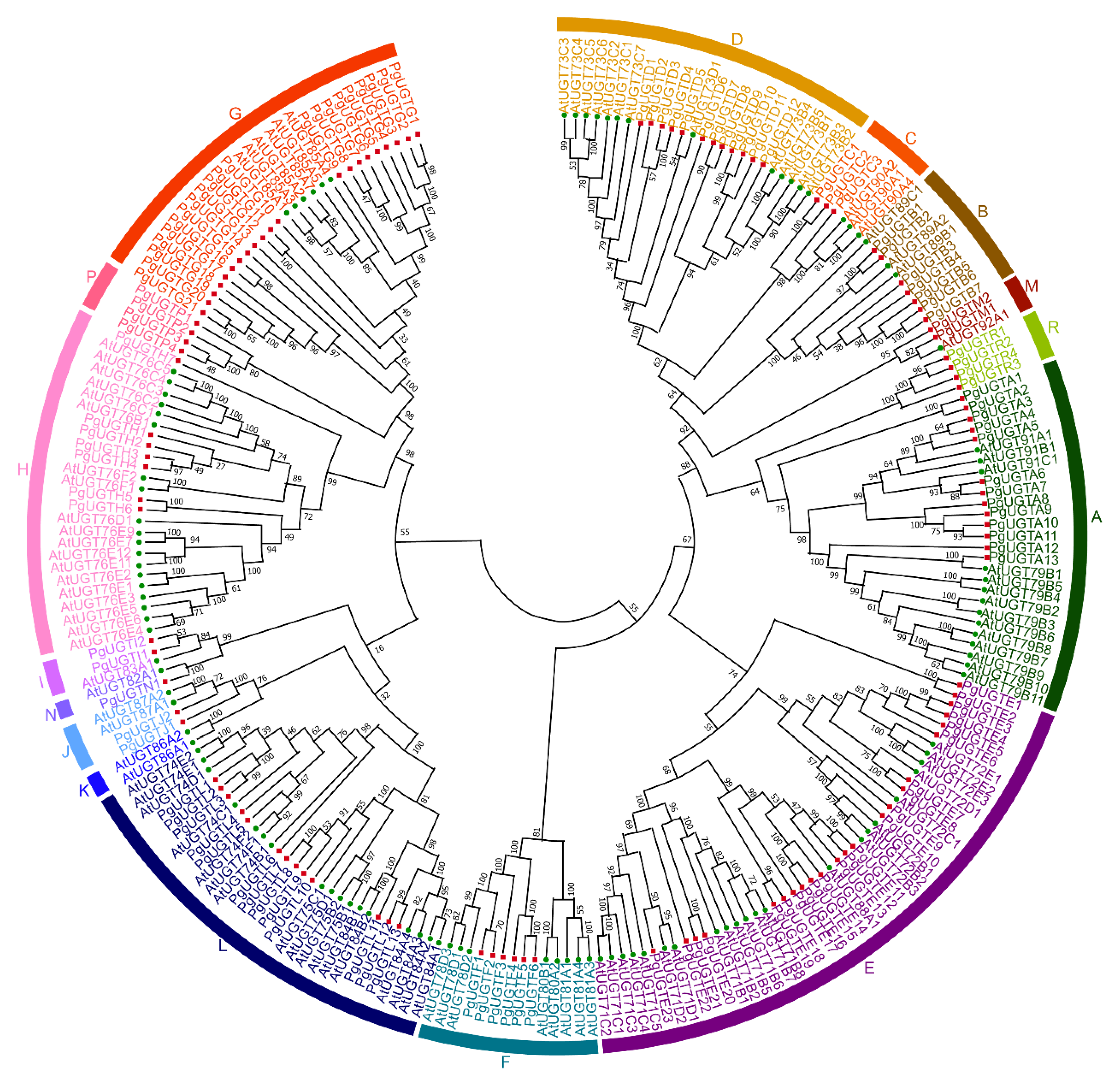
Figure 1. Phylogenetic analysis of UGT proteins in pomegranate and Arabidopsis. The capital letters (A–J, L–N, P, and R) represent different groups. The phylogenetic tree was generated by the neighbor-joining method using MEGA 10 software with 1000 bootstrap replicates.
Horticulturae 09 00119 g001
Figure 2. Conserved motifs and gene structure analysis of PgUGTs. (A) Distribution of the conserved motif for PgUGT proteins. The capital letters(A–J, L–N, P, and R) represent different groups. (B) Amino acid composition analysis of motif 1 (PSPG-box). (C) Gene structure of PgUGTs.
Horticulturae 09 00119 g002
Figure 3. Chromosome distribution and collinearity analysis of PgUGTs. (A) The PgUGTs were mapped to different chromosomes. The capital letters (A–J, L–N, P, and R) represent different groups. (B) Collinearity analysis of PgUGTs in pomegranate. The red lines highlight the syntenic gene pairs.
Horticulturae 09 00119 g003
Figure 4. Expression profiles of PgUGTs. (A) Expression profiles of PgUGTs in different tissues. Transcript data of PgUGTs in root, flower, leaf, peel, and inner and outer seed coat at 50, 95, and 140 days after pollination (DAP) were normalized with log2 transformed FPKM values. ISC, inner seed coat; and OSC, outer seed coat. (B) Expression profiles of the PgUGTs of groups L and E in seed coats of soft-seeded ‘Tunisia’ and hard-seeded ‘Dabenzi’. The transcript data of PgUGTs in the inner and outer seed coats of ‘Dabenzi’ and ‘Tunisia’ at 50, 95, and 140 DAP were normalized with log2 transformed FPKM values. D, ‘Dabenzi’; T, ‘Tunisia’; DAP, days after pollination; I, inner seed coat; and O, outer seed coat.
Horticulturae 09 00119 g004
Figure 5. CREs of the candidate PgUGTs. The CREs are displayed in different colored boxes. The number of each CRE in the gene is displayed in the boxes.
Horticulturae 09 00119 g005
Figure 6. Expression analysis of PgUGTs under hormone treatment by qRT-PCR. (A) Relative expression levels of candidate PgUGTs in response to auxin treatment after 6 and 12 h. (B) Relative expression levels of candidate PgUGTs in response to BR treatment after 3 and 5 days. IAA, auxin; BR, brassinolide. Error bars indicate the standard deviation of three replications. Different letters indicate a significant difference at p < 0.05 using Tukey’s test.
Horticulturae 09 00119 g006
Figure 7. Co-expression network of candidate PgUGTs and genes involved in phenylpropanoid biosynthesis and hormone signaling pathways. The red nodes represent the PgUGTs, orange nodes represent genes involved in the hormone signaling pathway, and blue nodes represent genes involved in the phenylpropanoid biosynthesis pathway. The thickness of lines represents Pearson’s correlation coefficients.
Horticulturae 09 00119 g007
Figure 8. Subcellular localization of PgUGTL11 and PgUGTE10 in tobacco epidermal cells. Scale bar, 100 μm.
Horticulturae 09 00119 g008
Disclaimer/Publisher’s Note: The statements, opinions and data contained in all publications are solely those of the individual author(s) and contributor(s) and not of MDPI and/or the editor(s). MDPI and/or the editor(s) disclaim responsibility for any injury to people or property resulting from any ideas, methods, instructions or products referred to in the content.