Article

Interspecific Hybridization between Ganoderma lingzhi and G. resinaceum by PEG-Induced Double-Inactivated Protoplast Fusion

Institute of Special Animal and Plant Sciences, Chinese Academy of Agricultural Sciences, Changchun 130112, China
*
Authors to whom correspondence should be addressed.
These authors contributed equally to this work.
Horticulturae 2023, 9(10), 1129; https://doi.org/10.3390/horticulturae9101129
Submission received: 29 August 2023 / Revised: 8 October 2023 / Accepted: 10 October 2023 / Published: 12 October 2023

Abstract

:
Ganoderma lingzhi is an important medicinal fungus, and it is particularly important to select strains with high yields and active substance contents. In this study, protoplasts of G. lingzhi were thermally inactivated to destroy intracellular enzyme proteins and preserve DNA. The DNA of G. resinaceum was damaged by ultraviolet (UV) radiation, and other components of the protoplasm except DNA were preserved. Then, the protoplast was induced using polyethylene glycol (PEG) for fusion. The results showed that the optimal thermal inactivation conditions for G. lingzhi were 30 min in a 45 °C water bath, and the optimal UV inactivation conditions for G. resinaceum were 70 s of irradiation using a 20 W UV lamp at a vertical distance of 15 cm. Antagonistic tests, internal transcribed space (ITS) and mitochondrial DNA identification, intersimple sequence repeat (ISSR) molecular markers and morphology were used to distinguish the parents from the fusants. Four true fusants were obtained, and the yield was 2.5%. The fruiting body yield of the fusants was significantly higher than that of G. lingzhi, and the polysaccharide and triterpene contents of the RAD-64 fusant were significantly higher than those of G. lingzhi. The results presented in this paper show that protoplast fusion technology can effectively improve G. lingzhi varieties and support the breeding of new varieties.

1. Introduction

Taxonomically, Ganoderma lingzhi and G. resinaceum belong to two different species of Ganoderma, both of which have high medicinal value [1,2]. G. lingzhi is the main cultivated species in China, and the research on it is more comprehensive. G. lingzhi is rich in polysaccharides, triterpenoids, amino acids, nucleosides, proteins, trace elements and other functional components [3,4,5], which provide immune regulation, antiviral, anti-inflammatory and liver-protective effects [6]. The fruiting body of G. resinaceum has a short stipe or no stipe. The mycelial growth rate of G. resinaceum is faster than that of G. lingzhi, and triterpenoids are abundant and high in content [7,8,9]. G. resinaceum triterpenes have anti-inflammation, antioxidation, antiapoptosis, α-glucosidase inhibition and other biological activities and have potential therapeutic importance for neuro-related diseases, obesity and related metabolic diseases, diabetes, etc. [10,11,12]. Our previous experiments showed that G. lingzhi also had strong insecticidal properties and a high polysaccharide content, and G. resinaceum had a high yield. To incorporate the good characteristics of G. resinaceum into G. lingzhi, which is highly popular in the market, appropriate breeding methods are needed. The mating system of Ganoderma species is tetrapolar heterothallism, and interspecific hyphal crossing of the homokaryons of two species is not possible because of incompatibility [13,14]. However, protoplast fusion breeding with dual heating and ultraviolet (UV) irradiation can overcome this restriction and allow for cell hybridization between G. lingzhi and G. resinaceum [15].
Dual heat inactivation and UV inactivation are common technical means for protoplast fusion breeding [16]. After the heat treatment of protoplasts, the structure of proteins in the cytoplasm is changed due to the breaking of hydrogen bonds, and the proteins are denatured and inactivated. At present, it is believed that the enzyme protein, 16S ribosomal subunit and ribosomal RNA in the cytoplasm are damaged after heat treatment of the protoplasm. UV light can covalently bond adjacent pyrimidines on the DNA chain to form dimers, the most easily formed of which is the thymine dimer, resulting in DNA damage. UV radiation can damage DNA in the protoplasm, resulting in loss of genome function and inactivation [17,18]. The regeneration ability of protoplasts caused by the two methods is lost due to damage to different parts, and complementary regeneration can be achieved after fusion for the purpose of screening fusion strains [19]. The method is simple to perform, reduces the workload of early labeling, retains the genetic traits of the parents, improves the screening efficiency, and avoids regeneration of the parent-type fusion strains. Biparental inactivation technology has been widely used in the breeding of edible mushrooms, such as Pleurotus [20], Lactarius deliciosus [21], Agrocybe aegerita and Copyinds comatus [22].
At present, the selection of G. lingzhi strains is mostly focused on systematic selection and mutagenesis breeding [23], and there have been no reports on the selection of new strains through interspecific protoplast fusion of G. lingzhi and G. resinaceum. To improve the varieties of G. lingzhi, the main genetic material of G. lingzhi was preserved, and a new strain with excellent complementary characteristics of G. lingzhi and G. resinaceum was obtained. In addition, the yield of secondary metabolites was increased. In this study, the method of dual inactivated protoplast fusion induced by polyethylene glycol (PEG) was used. The DNA of G. lingzhi was preserved by heat inactivation of G. lingzhi. The DNA of G. resinaceum was destroyed by UV inactivation. The double-inactivated protoplasts were interspecifically hybridized, and new fusants with stable characteristics were obtained. The production potential and medicinal value of the fusants were determined to enrich the germplasm resources of G. lingzhi in China. The results showed that protoplast fusion technology can be used to effectively breed and improve G. lingzhi varieties.

2. Materials and Methods

2.1. Strains and Culture Media

The two parent strains were collected from cultivated Ganoderma on Changbai Mountain and identified as G. lingzhi and G. resinaceum. The cultivation location of G. lingzhi is South Dingzi village, Huangsongdian town, Jiaohe city, and that of G. resinaceum is Fuxing Tun, Songshan town, Panshi city. The strains are now stored at the Institute of Special Animal and Plant Sciences of the Chinese Academy of Agricultural Sciences under numbers LZ-10 and LZ-13, respectively. The strains were maintained on potato dextrose agar (PDA) with 200 g potato (immersion), 20 g glucose, 1.5 g KH2PO4, 1.5 g K2HPO4, 2 g MgSO4, 20 g agar, and water to 1 L at natural pH. For protoplast preparation, the mycelium was prepared in potato dextrose broth without agar (PDB). Protoplast regeneration medium was prepared by adding an osmotic stabilizer to PDA medium so that the final concentration was 0.6 mol/L, and the pH was natural [24]. The sawdust medium used in this study consisted of 76% sawdust, 20% bran, 2% gypsum, and 2% glucose, with a 40% water content.

2.2. Protoplasm Preparation and Inactivation

In the early stage, enzymatic hydrolysis was used to complete the preparation of G. lingzhi protoplasm and optimization of regeneration conditions [24], and the parental protoplast suspension obtained by enzymatic hydrolysis was adjusted to 105/mL with 0.6 mol/L mannitol. Then, parental protoplast inactivation was used as a marker for screening the fusion strains.
Heat inactivation of G. lingzhi: 0.5 mL of G. lingzhi protoplast suspension was added to a sterile 1.5 mL EP tube, which was placed in a water bath at 40, 45 and 50 °C for constant-temperature treatment for 10, 20, 30, 40, 50, 60, 70 and 80 min, with periodic shock to ensure uniform heating. Protoplasts of LZ-10 without heat inactivation were used as the control group.
UV inactivation of G. resinaceum: 0.5 mL of G. resinaceum protoplast suspension was placed in a 5 cm sterile Petri dish and inactivated 15 cm below a 20 W UV lamp (wavelength: 253.7 nm) on an ultraclean workbench. A gradient of five time points was set: 30, 40, 50, 60 and 70 s. The protoplasts of G. resinaceum that had not been inactivated by UV were used as the control group.
After the inactivation treatment, a single layer of plate coating (100 μL) was applied to the regeneration medium supplemented with 0.5 mol/L mannitol, and the noninactivated protoplasts were used as the control and cultured in a constant-temperature incubator at 25 °C. The mortality rate was calculated after the regenerated colony was established. The mortality rate was calculated as follows:
Mortality = 1 − (number of regenerated colonies in the inactivated group/number of regenerated colonies in the control group) × 100%.

2.3. Protoplast Fusion

As described by Quan et al. [25], PEG-induced chemical fusion was used to fuse the inactivated parental protoplasts, and they were placed under a 400× optical microscope (NLCD500, Jiangnan Yongxin Optical Co., Ltd., Nanjing, China). After the protoplast suspension was diluted in accordance with the gradient, a 0.1 mL plate coating was taken. After a single colony grew in the regeneration medium, it was transferred to PDA medium for culture and identification.

2.4. Preliminary Screening of Clamp Connection and Antagonism Test

The sections of the fusants were placed under a 400× optical microscope to observe the mycelial clamp connection. After 5 rounds of subgeneration, the double-nucleated strains and the two parents were simultaneously inoculated onto the same medium for confrontation culture. The fusants displaying antagonistic phenomena were antagonized according to the experimental method of Chen et al. [26].

2.5. Nuc-ITS and cox2 Gene Amplification

Genomic DNA was extracted using an Ezup Column Fungi Genomic DNA Purification Kit (B518259, Sangon Biotech, Shanghai, China). The nuclear rDNA (nuc-ITS) sequence was amplified with the primers ITS1 (TCCGTAGGTGAACCTGCGG) and ITS4 (TCCTCCGCTTATTGATATGG). The mitochondrial cox2 gene sequence can be used for interspecific identification of Ganoderma [27,28], referring to mitochondrial genome information for G. lingzhi and G. resinaceum [29]. The primers GLcox2-F (TGTAAGTGTATTTTGGGTTATAGGATCA) and GLcox2-R (CAAAAGAACCTTTAGCAATACTCATTTT) were designed for mitochondrial cox2 gene amplification. The reaction system was 25 μL, including 12.5 μL of 2*PCR Mix, 1 μL of upstream and downstream primers, 1 μL of DNA template, and 9.5 μL of ddH2O. The PCR amplification conditions included predenaturation at 94 °C for 3 min; 30 cycles of denaturation at 94 °C for 30 s, annealing at 55 °C for 30 s, and extension at 72 °C for 45 s; elongation at 72 °C for 10 min; and a termination temperature of 4 °C. The primers were synthesized by Sangon Biotech (Shanghai, China).

2.6. ISSR Profiling

With reference to the methods of Xu [30], Xing [31] and Raman [14], mycelium culture, DNA extraction and PCR amplification were performed. The intersimple sequence repeat (ISSR) primer sequences and annealing temperatures used in this study are shown in Table 1.

2.7. Determination of the Mycelial Growth Rate and Fruiting Experiment

Strains inoculated onto PDA medium in a Petri dish (Biosharp, Hefei, China) with a diameter of 9 cm were incubated at 25 °C in darkness. The colony radius was measured on the 6th day after inoculation, and the mycelial growth rate (mm/day) was calculated. The colony pigment was observed after 15 days. The sawdust medium in the test tube (30 mm × 200 mm) was inoculated with mycelial discs with diameters of 0.8 cm and incubated at 25 °C in darkness. The height of the substrate colonized by mycelium was measured on the 12th day after inoculation, and the mycelial growth rate (mm/d) was calculated. The strains were inoculated in bags and cultured in a dark environment at 25 °C. After the mycelium filled the bag, it was placed in the mushroom cultivation room for mushroom management and collected after the fruiting body matured.

2.8. Determination of Polysaccharides and Triterpenes in Fruiting Bodies

After harvesting, the fruiting bodies were dried in an oven (GZX-9140, Boxun, Shanghai, China) at 60 °C and then crushed after drying to a constant weight.
Two grams of dried sample was used for polysaccharide extraction with 60 mL of distilled water at 88.5 °C twice: the first extraction for 4 h and the second extraction for 3 h. The extract was dried at 95 °C, and the residue was dissolved in 5 mL of water. Next, 75 mL of anhydrous ethanol was added, and the mixture was shaken well, placed at 4 °C for 12 h, and centrifuged to obtain the precipitate. The precipitate was dissolved in hot water at 60–70 °C and transferred to a 50 mL volumetric flask. An appropriate amount of solution was centrifuged, and 3 mL of supernatant was accurately measured, placed in a 25 mL volumetric flask, and brought to a constant volume. The standard curve was prepared with anhydrous glucose. The polysaccharide content was determined using anthrone-sulfuric acid spectrophotometry. The detection wavelength of the microplate reader (Epoch, Biotek, VT, USA) was 625 nm [32].
Two grams of dry sample was taken for triterpene extraction, and 50 mL of anhydrous ethanol was added. Then, ultrasonic treatment (power 140 W, frequency 42 kHz) (KQ5200, Shumei, Kunshan, China) was performed for 45 min. The mixture was strained and added to a 100 mL volumetric flask. The triterpene content was determined using the vanillal-glacial acetic acid and perchloric acid colorimetric method. Oleanolic acid was used as the standard substance. The absorbance was determined at a wavelength of 546 nm [32].

2.9. Statistical Analysis

Based on the ISSR amplification electrophoretic map, a 0/1 matrix was established, and the genetic similarity coefficient was calculated using NTSYS-pc 2.10 software based on the Dice coefficient. Then, cluster analysis was performed using the unweighted average pair group method with arithmetic mean (UPGMA) technique. All experiments included three or more biological and technical repetitions. IBM SPSS Statistics 23 software was used for statistical analysis of the experimental data, and GraphPad Prism 8.0 was used for graphing. The lowercase letters in the charts indicate significant differences at the p < 0.05 level.

3. Results

3.1. Protoplast Inactivation Conditions

The DNA of G. lingzhi was retained, and the parent LZ-10 was thermally inactivated. To ensure complete heat inactivation of G. lingzhi during protoplast fusion, the thermal inactivation conditions of LZ-10 were optimized. The test results are shown in Table 2. Thermal inactivation can be achieved in G. lingzhi protoplasts treated at three temperatures at different times, and the inactivation rate of protoplasts increases with increasing inactivation time and temperature. Protoplasts can be completely inactivated by treatment at 40 °C for more than 70 min, 45 °C for more than 30 min and 50 °C for 10 min. After comprehensive consideration, a water bath at 45 °C for 30 min was selected as the optimal thermal inactivation condition of G. lingzhi for the follow-up test.
In this study, LZ-13 was inactivated by UV irradiation of protoplasts, thereby destroying the DNA of LZ-13 and preserving other components of the cytoplasm. The UV inactivation results for LZ-13 are shown in Table 3. After UV inactivation for 70 s, no colonies were found in the regeneration medium, and the mortality rate reached 100%. Therefore, irradiation under a vertically arranged UV lamp for 70 s was chosen as the UV inactivation condition for follow-up tests using LZ-13.

3.2. Protoplast Fusion and Antagonism Test

The process of PEG-induced protoplast fusion is shown in Figure 1A. The parent protoplast gradually approaches the plasma membrane until it fits closely. Then, the cytoplasm of one parent enters the cytoplasm of the other parent; the volume of the other parent gradually shrinks, while the volume of the first parent gradually expands. The volume of the protoplast after fusion was maximized. In this experiment, 160 single colonies were selected from the regeneration medium. The results showed that there were 49 mononuclear strains and 111 binucleated strains among the 160 strains, with binucleated strains accounting for 69.4%. To distinguish the fusants from their parents, antagonism tests were performed after 5 subgenerations. Some of the antagonism test results are shown in Figure 1B. A total of 49 strains showing antagonistic phenomena with their parents were screened out, accounting for 44.1%. A pairwise antagonism experiment was conducted among 49 strains, and 7 strains, including RAD-4, RAD-13, RAD-26, RAD-57, RAD-59, RAD-64 and RAD-66, with large differences were selected.

3.3. Nuc-ITS and Mitochondrial DNA Identification

To determine the source of nuclear DNA of the fusants, nuc-ITS sequencing was performed, and a sequence of approximately 615 bp was obtained after removing the hybrid peaks at both ends and examining the artificial test peak map. The sequence comparison results showed that the similarity between the parents LZ-10 and LZ-13 was 94.79%, and the similarity between the fusants and LZ-10 was 100% (Table 4). The results showed that the nuclear DNA of the fusants was derived from the parent LZ-10.
The results of the cox2 gene sequence comparison between the parent and the fusants are shown in Table 5. The similarity between LZ-10 and LZ-13 was 95.32%, indicating that this sequence can be used for mitochondrial genome identification between the parent and the fusants. The similarity between the fusants and LZ-10 was 100%, and the similarity between the fusants and LZ-13 was 95.32%, indicating that the mitochondrial DNA of the fusants was also from the parent LZ-10.

3.4. ISSR Analysis

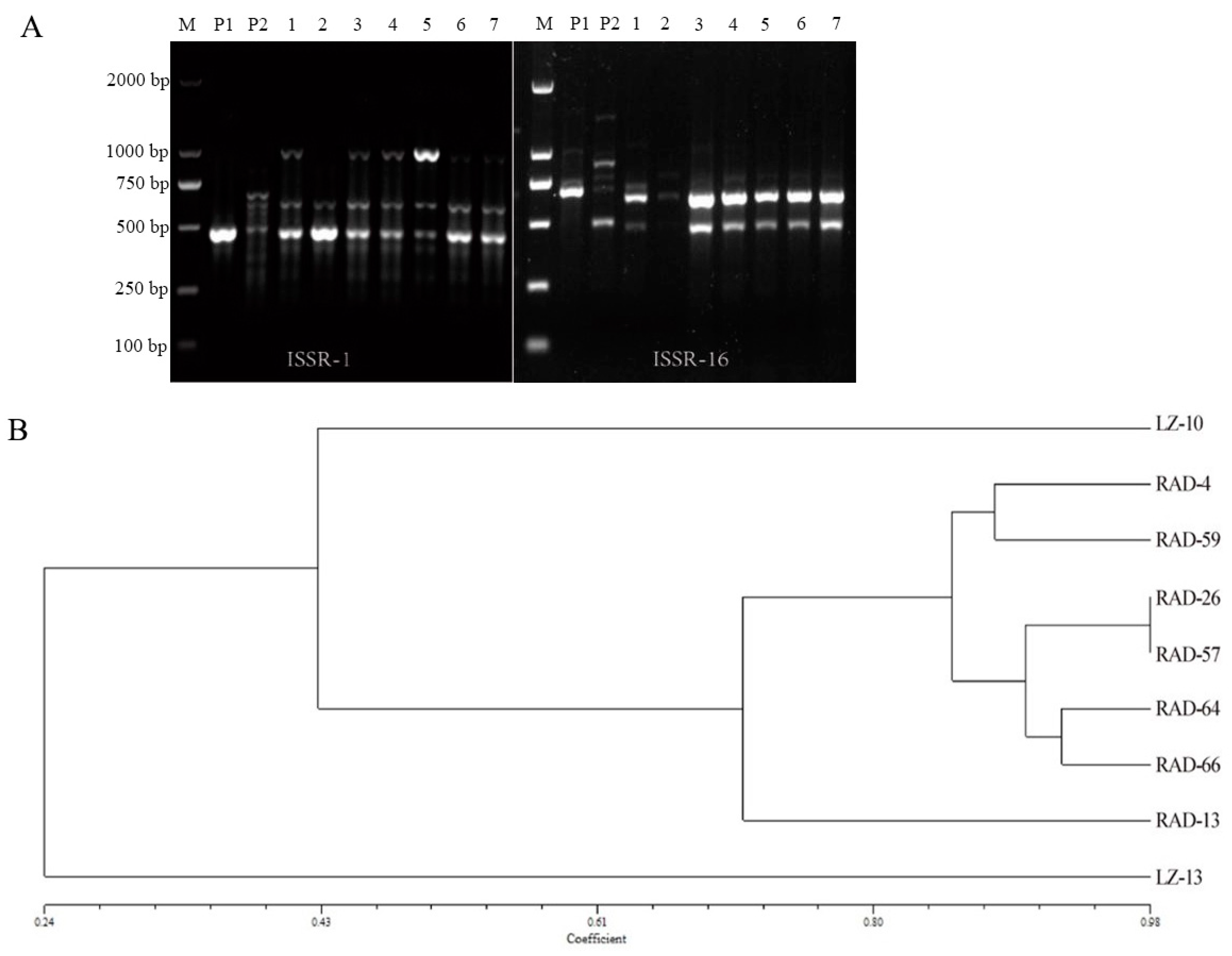
The parents and fusants were identified using ISSR markers. A total of 12 primers were used to amplify the genomic DNA of the 2 parents and 7 fusants by PCR sequencing. The DNA bands amplified by the 12 primers ranged in length from 250 bp to 2000 bp, were easy to identify and showed good polymorphism. A total of 82 DNA bands were obtained by sequence amplification, and the number of DNA bands amplified by each primer was 4–10. The electrophoretic map of the partial primer amplification results is shown in Figure 2A. The results showed that all or part of the parental bands were amplified in the 7 fusants, indicating that the fusants contained the genetic material of the parents. In addition, the fusants also produced specific new bands that were different from those of the parents. For example, in the amplification map of the ISSR-1 primer, 480 bp and 650 bp bands of the fusants and the parent were amplified at the same time. In addition, new specific bands of the fusants were also amplified in addition to that of RAD-13. In the amplification map of ISSR-16 primers, 700 bp bands were amplified for LZ-10, 500 bp bands were amplified for LZ-13, and both types of bands were amplified for the fusants.
To determine the genetic relationship between parents and fusants, a GS value matrix was calculated using ISSR markers, and a cluster map of the genetic relationships between parents and fusants was constructed using the UPGMA technique (Figure 2B). The genetic similarity coefficients of the nine strains ranged from 0.1944 to 0.9825, with an average of 0.6973, and the genetic range varied greatly. The largest genetic similarity coefficient with LZ-10 was observed for RAD-13, which was 0.4918, and the largest genetic similarity coefficient with LZ-13 was observed for RAD-59, which was 0.2813. The genetic similarity coefficient of the fusion genes RAD-26 and RAD-57 was 0.9825, which indicates that the genetic difference between them is the smallest and that they are the most closely related. As shown in Figure 3, when the GS value was 0.43, the strains could be divided into three categories, namely, the parent LZ-10, the parent LZ-13 and the fusants. The genetic distance between LZ-13 and its offspring fusants was relatively long, thus forming an outgroup, while the 7 fusants were more closely related to LZ-10.

3.5. Colony Morphology

According to the ISSR clustering diagram, 4 fusants with large genetic differences were selected for comparisons of colony morphology with that of their parents. There were obvious differences in colony morphology between the fusants and their parents (Figure 3). The parent strains showed a high colony density and regular colony edges. Among the 4 fusants, RAD-13, RAD-26 and RAD-64 showed thicker mycelial fascicles than the two parent strains, and the colony edges were irregular. The colonies grew close to the plate, and the morphology of RAD-59 was basically the same as that of the two parent colonies. The two parent strains and the fusants were cultured for 15 days, and both the parent strain and RAD-64 showed a small amount of pigment production. In contrast, the other three strains showed obvious pigment production.

3.6. Growth Characteristics and Agronomic Traits

The mycelial growth rates of the parents and fusants in PDA plates and cultivation material were measured, and the results are shown in Figure 4A. When mycelia grew on PDA medium, the growth rates of the parents LZ-10 and LZ-13 were significantly higher than those of the fusants; the growth rate of LZ-13 was the fastest (12.23 ± 0.14 mm/d), and that of fusant RAD-64 was the slowest (1.91 ± 0.27 mm/d). Among the cultivated materials, LZ-13 showed the fastest growth (6.00 ± 0.17 mm/d), and LZ-10 showed the slowest growth (4.07 ± 0.61 mm/d). The growth rate of the fusants was between those of the two parents. Among the fusants, RAD-26 had the fastest growth rate of 4.83 ± 0.37 mm/d, and the other fusants showed no significant differences in growth rates. The results of the mushroom emergence experiment showed that the yield of parent LZ-10 (46.83 ± 6.06 g/bag) was significantly lower than that of the other strains, and the yields of LZ-13 and RAD-13 were the highest. However, the yield differences among the other fusants were not significant (Figure 4B). The fruiting bodies of LZ-10, LZ-13, RAD-59 and RAD-64 were fan shaped, while those of RAD-13 and RAD-26 were kidney shaped. The stipes of RAD-13 and RAD-26 were longer than those of the other fusants. The cases of LZ-10 and LZ-13 were dark yellow-red, while the cases of RAD-13 and RAD-26 were lighter in color. The back of the LZ-13 case was pale white, that of the cases of LZ-10 and the other fusants except RAD-26 was pale yellow, and that of the RAD-26 case was yellow. In addition, the surface of the cases of RAD-13, RAD-26 and RAD-64 had obvious ring patterns (Figure 4C). In summary, the fusants showed large differences from their parents in terms of the mycelial growth rate, fruiting body yield and fruiting body morphology.

3.7. Contents of Polysaccharides and Triterpenes in Fruiting Bodies

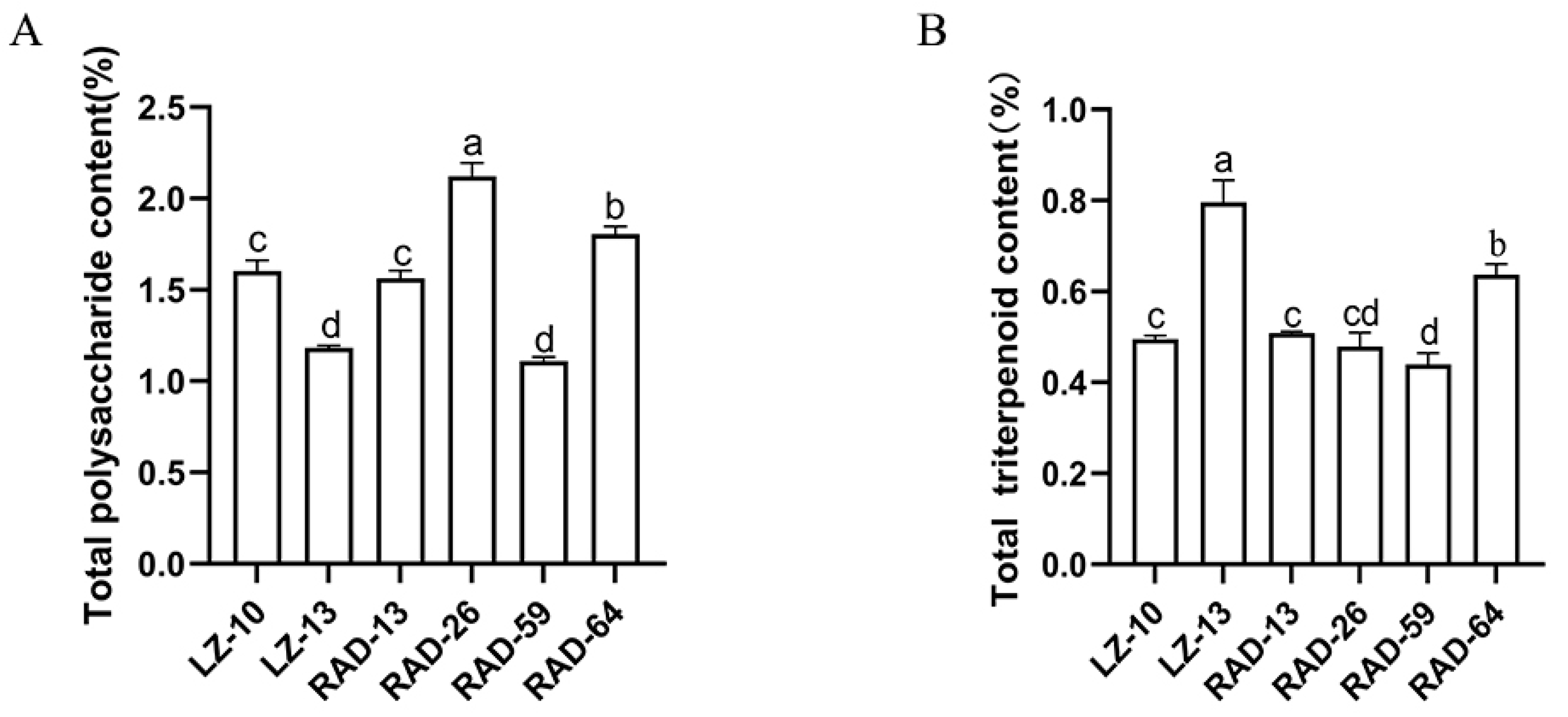
The polysaccharide and triterpene contents in the fruiting bodies of the fusants were determined. As shown in Figure 5A, the polysaccharide content of the parent LZ-10 (1.6 ± 0.06%) was significantly higher than that of the parent LZ-13 (1.18 ± 0.01%). The polysaccharide contents of RAD-26 and RAD-64 were significantly higher than those of their parents. The polysaccharide content of RAD-26 was the highest (2.12 ± 0.07%), while the polysaccharide content of RAD-59 was the lowest (1.11 ± 0.02%), which was not significantly different from that of the parent LZ-13. LZ-13 had the highest triterpene content (0.8 ± 0.05%). Among the fusants, only RAD-64 showed a higher triterpene content (0.64 ± 0.02%) in the fruiting bodies than the parents, which was 28% higher than that of the LZ-10 fruiting body (Figure 5B). The contents of triterpenes and polysaccharides in RAD-64 were significantly higher than those in LZ-10.

4. Discussion

Biparental inactivation is a commonly used method in protoplast fusion breeding, and the technology is well established and has been applied in the breeding of several new strains of edible fungi [33,34,35]. Excessive inactivation will seriously affect the cell structure of protoplasts and affect their regeneration [36]. Therefore, the exploration of inactivation conditions is particularly important. In this paper, the thermal inactivation conditions of the protoplasm of G. lingzhi were investigated. It was found that all the protoplasts (5 × 10⁴) could be inactivated when treated at 40 °C for more than 70 min, 45 °C for more than 30 min and 50 °C for 10 min. Microscopic examination showed that treatment at 45 °C for 30 min resulted in less protoplast damage and required a shorter amount of time, so this condition was selected as the optimal heat inactivation condition for G. lingzhi. The mortality rate of G. resinaceum protoplasts (5 × 10⁴) was 100% after irradiation for 70 s at a distance of 15 cm under a 20 W UV lamp. It has been reported that Cordyceps sinensis protoplasts (1 × 10⁷) require a 60 °C water bath for 10 min to be completely inactivated. All the protoplasts (2 × 10⁶) of Cordyceps militaris could be inactivated by vertical irradiation with a 30 W UV lamp 10 cm away for 13 min [37]. The protoplasts (2 × 10⁷) of Antrodia cinnamomea and C. militaris could be completely thermally inactivated at 55 °C for 10 min. The protoplasts of A. cinnamomea and C. militaris could be completely inactivated by 10 cm irradiation with a 30 W UV lamp for 7 min [34]. Therefore, the sensitivity of protoplasts of different species to inactivation conditions is different, and the number of protoplasts, inactivation temperature and inactivation time should be controlled during thermal inactivation. Attention should be given to the number of protoplasts, irradiation distance, UV intensity, inactivation time and other conditions during UV inactivation.
The identification of fusants is key to successful fusion. Antagonistic experiments, molecular markers, colony morphology and other metrics and methods were used to identify whether fusants were successfully fused, and these methods could be combined and supported each other [38]. The mechanism of protoplast fusion is very complex [39]. The fusants recombine the genetic material of both parents, and antagonistic lines will be generated if the fusants are incompatible with both parents or between the fusion strains. In this paper, a total of 111 strains were selected, and 49 strains (44.1%) showed antagonism with their parents, indicating that there was obvious incompatibility between the fusants and their parents. The pair-to-pair antagonism experiment was carried out among 49 strains. Because the protoplasm could retain the genetic material of the parents well and the differences between the inactivated protoplasts were not significant, the differences between the fusants were not significant, and only 7 fusants with large differences were preliminarily screened out. The same phenomenon was also found in protoplast fusion experiments with Agaricus bisporus and Agaricus blazi. Specifically, 85% of the 60 strains with fusion formed lines obviously antagonistic with their parents. There was also antagonism between the fusants, but it was not obvious [40]. The results of nuc-ITS and mitochondrial gene cox2 sequencing showed that UV inactivation was complete, and the DNA in the fusants was from G. lingzhi. ISSR markers are universal, rapid, easy to apply, highly repeatable and polymorphic and are often used for molecular identification of protoplast fusions in edible fungi [38,41]. In this study, it was found that the fusants had unique DNA bands from both of the parents, indicating that the fusants had genetic material from both parents. According to the genetic relationship cluster diagram, when the GS value was 0.43, the strains could be divided into three categories, namely, the LZ-10 parent, the LZ-13 parent and the fusants. Because UV inactivation greatly damaged the DNA of LZ-13, the fusants retained very little DNA information from LZ-13, resulting in long genetic distances between LZ-13 and its offspring fusants, thus forming an outgroup. The phenomenon by which one parent has a long genetic distance has also been confirmed in breeding with inactivated protoplast fusion between the two parents of Morchella importuna and Morchella sextelata [33]. The maximum genetic similarity coefficient with G. lingzhi was observed for RAD-13 (0.4918), and that with G. resinaceum was observed for RAD-59 (0.2813), indicating genetic diversity in the fusants. The life processes of eukaryotes, such as cell differentiation, morphogenesis, senescence and disease, result from the interaction between the nucleus and cytoplasm [42]. After protoplast fusion, the morphology of the fusants significantly differed from that of the parents, and the fusants and parents could be distinguished [14]. Among the four fusants, RAD-13, RAD-26 and RAD-64 showed mycelial fascicles that were thicker than those of the two parent strains, and the colony edges were irregular, showing obvious morphological differences from those of the parents, indicating that the alloplasmic introduction changed the original nucleoplasmic coordination and formed a new phenotype. We obtained a new protoplast fusion strain through antagonistic experiments, molecular markers and colony morphological comparison.
The growth state, fruiting body yield, active substance content and stability of the new strain are particularly notable. In this paper, the fusants were studied after five subgenerations. The growth rate of the fusants in wood chips was significantly higher than that of the G. lingzhi parent (4.07 ± 0.61 mm/d), and the fusant RAD-26 had the fastest growth rate of 4.83 ± 0.37 mm/d. The fruiting experiments showed that the yield of the selected G. lignzhi parent was lowest (46.83 ± 6.06 g/bag), but the yield of the fruiting body of the fusants was significantly higher than that of G. lignzhi. In particular, the yield of RAD-13 was as high as 76.70 ± 6.20 g/bag. The pharmacological properties of Ganoderma are primarily attributed to its polysaccharides and triterpenoids [43], which directly reflect the quality of Ganoderma. The fruiting body of G. lingzhi was rich in polysaccharides but relatively low in triterpenes, and that of G. resinaceum was high in triterpenes and low in polysaccharides. RAD-64 strains obtained by fusion had superior parental traits, and the contents of polysaccharides (1.81 ± 0.04%) and triterpenes (0.64 ± 0.02%) were significantly higher than those of G. lingzhi. The above trait tests confirmed that we obtained fusants whose traits were superior to those of the parents. In the future, we will conduct tissue separation of the fruiting body to determine the progeny stability of the fusants. Biological traits are mainly controlled by nuclear DNA, followed by cytoplasm, which interact with each other [42]. The fusants are the result of recombination of the nucleus of G. lignzhi and the cytoplasm of G. resinaceum. Recent studies have shown that the cytoplasm can affect colony morphology, the mycelial growth rate, enzyme activity, the agronomic traits of fruiting bodies and polysaccharide and triterpene content [28,42,44,45], consistent with our results. The contents of adenosine, biomass, cordycepic acid, cordycepin, total polysaccharides and total triterpenes of the fusants obtained using protoplast fusion of A. cinnamomea and C. militaris were significantly increased [34]. These results showed that the use of protoplast fusion technology to recombine the nucleus and cytoplasm of parents can significantly increase the content of active substances [46,47].

5. Conclusions

This experiment showed that double-inactivated protoplast fusion technology can achieve somatic hybridization of G. lingzhi and G. resinaceum and increase the genetic potential and genetic diversity of Ganoderma strains. The new strain obtained by protoplast fusion resulted in variety improvement of G. lingzhi, concentrated the excellent traits of both parents, and significantly increased the yield of fruiting bodies and the content of polysaccharides and triterpenes, which are very important for the development of the G. lingzhi industry. Finally, the results of this study will support breeding research on G. lingzhi.

Author Contributions

M.Y. and C.S. designed the experiments; S.W. and N.Z. prepared the materials; J.L. and L.X. carried out the experiments; J.L. and L.L. analyzed the data and wrote the manuscript. M.Y. revised the manuscript. All authors have read and agreed to the published version of the manuscript.

Funding

This study was funded by the Agricultural Science and Technology Innovation Program (CAAS-ASTIP-2021-ISAPS).

Data Availability Statement

The data presented in this study are available in the article.

Conflicts of Interest

The authors declare no conflict of interest.

References

  1. Cao, Y.; Wu, S.H.; Dai, Y.C. Species clarification of the prize medicinal Ganoderma mushroom “Lingzhi”. Fungal Divers. 2012, 56, 49–62. [Google Scholar] [CrossRef]
  2. Cui, B.K.; Pan, X.H.; Pan, F.; Sun, Y.F.; Xing, J.H.; Dai, Y.C. Species diversity and resources of Ganoderma in China. Mycosystema 2023, 40, 49–62. [Google Scholar] [CrossRef]
  3. Nakagawa, T.; Zhu, Q.C.; Tamrakar, S.; Amen, Y.; Mori, Y.; Suhara, H.; Kaneko, S.; Kawashima, H.; Okuzono, K.; Inoue, Y.; et al. Changes in content of triterpenoids and polysaccharides in Ganoderma lingzhi at different growth stages. J. Nat. Med. 2018, 72, 734–744. [Google Scholar] [CrossRef] [PubMed]
  4. Ma, G.H.; Qin, X.; Zhang, J.S.; Yan, M.Q.; Zhou, S.; Feng, J.; Zhou, J.; Liu, Y.F. β-glucooligosaccharide separated and purified from Ganoderma lingzhi and its in vitro anti-inflammation activities. Mycosystema 2023, 42, 1360–1369. [Google Scholar] [CrossRef]
  5. Feng, N.; Yue, Y.W.; Cheng, C.L.; Yang, M.; Wang, C.; Zhang, J.S. Research progress of triterpenes from mycelia of Ganoderma lingzhi and its pharmacological effects. Mycosystema 2022, 41, 1341–1353. [Google Scholar] [CrossRef]
  6. Zhu, J.; Shi, L.; Ren, A.; Liu, R.; Zhao, M.W. Research progress on the biosynthesis of ganoderic acid. J. Nanjing Agric. Univ. 2022, 45, 981–989. [Google Scholar] [CrossRef]
  7. Chen, B.Z.; Ke, B.R.; Ye, L.Y.; Jin, S.S.; Jie, F.; Zhao, L.L.; Wu, X.P. Isolation and varietal characterization of Ganoderma resinaceum from areas of Ganoderma lucidum production in China. Sci. Hortic. 2017, 224, 109–114. [Google Scholar] [CrossRef]
  8. Huang, Y.J.; Li, X.; Peng, X.R.; Adegoke, A.T.; Chen, J.C.; Su, H.G.; Hu, G.L.; Wei, G.; Qiu, M.H. NMR-based structural classification, Identification, and Quantification of triterpenoids from edible mushroom Ganoderma resinaceum. J. Agric. Food Chem. 2020, 68, 2816–2825. [Google Scholar] [CrossRef]
  9. Shi, Q.Q.; Huang, Y.J.; Su, H.G.; Gao, Y.; Lu, S.Y.; Peng, X.R.; Li, X.N.; Zhou, L.; Qiu, M.H. Structurally diverse lanostane triterpenoids from medicinal and edible mushroom Ganoderma resinaceum Boud. Bioorganic Chem. 2020, 100, 103871. [Google Scholar] [CrossRef]
  10. Kou, R.W.; Xia, B.; Wang, Z.J.; Li, J.N.; Yang, J.R.; Gao, Y.Q.; Yin, X.; Gao, J.M. Triterpenoids and meroterpenoids from the edible Ganoderma resinaceum and their potential anti-inflammatory, antioxidant and anti-apoptosis activities. Bioorganic Chem. 2022, 121, 105689. [Google Scholar] [CrossRef]
  11. Chen, X.Q.; Zhao, J.; Chen, L.X.; Wang, S.F.; Wang, Y.; Li, S.P. Lanostane triterpenes from the mushroom Ganoderma resinaceum and their inhibitory activities against alpha-glucosidase. Phytochemistry 2018, 149, 103–115. [Google Scholar] [CrossRef]
  12. Huang, Y.J.; Wei, G.; Peng, X.R.; Hu, G.L.; Su, H.G.; Liu, J.L.; Chen, X.; Qiu, M.H. Triterpenoids from functional mushroom Ganoderma resinaceum and the novel role of Resinacein S in enhancing the activity of brown/beige adipocytes. Food Res. Int. 2020, 136, 109303. [Google Scholar] [CrossRef] [PubMed]
  13. Adaskaveg, J.E.; Gilbertson, R.L. Cultural Studies and genetics of sexuality of Ganoderma lucidum and G. tsugae in relation to the taxonomy of the G. lucidum complex. Mycologia 2018, 78, 694–705. [Google Scholar] [CrossRef]
  14. Raman, J.; Jang, K.Y.; Oh, Y.L.; Oh, M.; Im, J.H.; Lakshmanan, H.; Kong, W.S. Interspecific hybridization between Ganoderma lingzhi and G. applanatum through protoplast fusion. World J. Microbiol. Biotechnol. 2021, 37, 114. [Google Scholar] [CrossRef]
  15. Dong, Y.T.; Miao, R.Y.; Feng, R.C.; Wang, T.; Yan, J.J.; Zhao, X.; Han, X.; Gan, Y.; Lin, J.B.; Li, Y.J.; et al. Edible and medicinal fungi breeding techniques, a review: Current status and future prospects. Curr. Res. Food Sci. 2022, 5, 2070–2080. [Google Scholar] [CrossRef] [PubMed]
  16. Ren, N.; Yao, L.; Chen, S.J. Study on inactivation conditions of protoplasts of vincamine indole alkaloids producing endophytic fungus CH1. Guangdong Chem. Ind. 2021, 48, 11–12+19. [Google Scholar]
  17. Pan, M. Studies on Protoplast Fusion and Synthetic Complete Medium of Mortierella alpina. Master’s Thesis, University of Science and Technology of China, Hefei, China, 2017. [Google Scholar]
  18. Wang, K. Study on Morphology of Sclerotial Development, IMV Detection and Protoplast Fusion of Morchella Mushrooms. Master’s Thesis, Zhengzhou University of Light Industry, Zhengzhou, China, 2019. [Google Scholar]
  19. Zhao, K.; Ping, W.X.; Zhang, L.N.; Liu, J.; Lin, Y.; Jin, T.; Zhou, D.P. Screening and breeding of high taxol producing fungi by genome shuffling. Sci. China C Life Sci. 2008, 51, 222–231. [Google Scholar] [CrossRef]
  20. He, J.C.; He, Y.X. Research on inactivated protoplast fusion breeding of Pleurotus mushroom. Edible Fungi 1999, 6, 10–11. [Google Scholar]
  21. Sun, J.J. Preliminary Study on Protoplast Fusion of Lactarius delicious and Pleurotus ostreatus. Master’s Thesis, Jiangxi Agricultural University, Nanchang, China, 2012. [Google Scholar]
  22. Jiang, L.; Huang, J.W.; Ci, L.K.; Lu, Y.F. Protoplast fusion and regeneration of Agrocybe aegerita and Copyinds comatus. Food Sci. 2011, 32, 141–144. [Google Scholar]
  23. Xu, L.; Zhang, X.C.; Zhou, Y.J.; Yan, M.X. Research progress of Ganoderma lucidum breeding. Spec. Wild Econ. Anim. Plant Res. 2022, 1–7. [Google Scholar] [CrossRef]
  24. Xu, L.; Liu, Y.; Zhang, X.C.; Hu, Q.X.; Yan, M.X. Optimization of preparation and regeneration conditions of two kinds of Ganoderma lucidum protoplasts. J. Jilin Agric. Univ. 2022, 1–10. [Google Scholar] [CrossRef]
  25. Quan, W.F.; He, J.X.; Wang, J.; Liu, G.J.; Ji, H.G.; Xue, J. Study on selection and breeding of selenium-rich Ganoderma lucidum by protoplast fusion. Edible Fungi 2012, 34, 19–21+31. [Google Scholar]
  26. Chen, Q.; Zhang, J.X. An improved method to characterize fungal vegetative compatible group. J. Microbiol. 2010, 30, 45–47. [Google Scholar]
  27. Meng, G.L.; Zeng, P.M.; Wang, Z.Y.; Cheng, B.; Ye, L.Y.; Wu, X.P. Application of the Ganoderma multipileum mitogenome to identification of Ganoderma species. Mycosystema 2020, 39, 23–33. [Google Scholar] [CrossRef]
  28. Meng, G.L.; Cheng, B.; Hao, J.B.; Li, S.Y.; Ye, L.Y.; Wu, X.P. Analyses of mitochondrial genome and its influence on mycelial growth and active component of Ganoderma lingzhi. Mycosystema 2020, 39, 42–56. [Google Scholar] [CrossRef]
  29. Ye, L.Y.; Meng, G.L.; He, X.F.; Ma, S.L.; Wu, X.P. Comparative analysis of mitochondrial genomes among Ganoderma Species. GAB 2022, 41, 1723–1731. [Google Scholar] [CrossRef]
  30. Xu, K. Based on Molecular Markers Assisted Breeding for Ganoderma lucidum. Master’s Thesis, Nanjing Agriculture University, Nanjing, China, 2014. [Google Scholar]
  31. Xing, Z.W. Germplasm Identification of Different Varieties and Screening of Excellent Strains of Ganoderma lucidum. Master’s Thesis, Hebei University of Engineering, Handan, China, 2016. [Google Scholar]
  32. National Pharmacopoeia Committee. Pharmacopoeia of the People’s Republic of China (Part 1), 2020th ed.; China Medical Science and Technology Press: Beijing, China, 2020; pp. 195–196. [Google Scholar]
  33. He, P.X.; Yu, M.; Wang, K.; Cai, Y.L.; Li, B.; Liu, W. Interspecific hybridization between cultivated morels Morchella importuna and Morchella sextelata by PEG-induced double inactivated protoplast fusion. World J. Microbiol. Biotechnol. 2020, 36, 58. [Google Scholar] [CrossRef]
  34. Lai, W.K.; Wu, Y.C.; Yeh, T.S.; Hsieh, C.R.; Tsai, Y.H.; Wei, C.K.; Li, C.Y.; Lu, Y.C.; Chang, F.R. The protoplast two-way fusions and fusant characteristics of Antrodia cinnamomea and Cordyceps militaris. Food Sci. Hum. Wellness 2022, 11, 1240–1251. [Google Scholar] [CrossRef]
  35. Huo, G.H.; Sun, J.J.; Luo, G.X.; Han, Q.C. Protoplast Hybridization between Lactarius deliciosus and Pleurotus ostreatus and Characteristic of Fusant Strains. J. Chin. Inst. Food Sci. Technol. 2014, 14, 39–46. [Google Scholar] [CrossRef]
  36. Sun, X.R. Technical Research of Protoplast Fusion between Pleurotus geesterani and pleurotus citrinopileatus Sing. Master’s Thesis, Shanxi Agricultural University, Taigu, Cina, 2018. [Google Scholar]
  37. Guo, C.J.; Zhao, R.; Zhu, W.B. Protoplast Fusion between Cordyceps sinensis and Cordyceps militaris. Food Sci. 2010, 31, 165–171. [Google Scholar]
  38. Li, Y.J.; Sun, G.Q.; Guo, J.F.; Wang, H.Y.; Yu, C.Z.; Pang, J. Research progress on the protoplast fusion technique in edible mushroom. J. North Agric. 2021, 49, 121–127. [Google Scholar] [CrossRef]
  39. Wang, R.; Li, X.Z.; Zhu, S.F.; Zhang, D.M.; Han, S.J.; Li, Z.G.; Lu, J.H.; Chu, H.W.; Xiao, J.M.; Li, S.L. Integrated flow cytometric and proteomics analyses reveal the regulatory network underlying sugarcane protoplast responses to fusion. Plant Physiol. Biochem. 2023, 202, 107918. [Google Scholar] [CrossRef] [PubMed]
  40. He, J.C.; He, C.Y.; Lu, M.H.; Ma, Y.H.; Wang, M.L. Study on the breeding of Agaricus bisporus and Agaricus blazei by inactivated protoplast fusion. Edible Fungi China 2014, 33, 9–10. [Google Scholar] [CrossRef]
  41. Bornet, B.; Branchard, M. Nonanchored inter simple sequence repeat (ISSR) markers: Reproducible and specific tools for genome fingerprinting. Plant Mol. Biol. Rep. 2012, 19, 209–215. [Google Scholar] [CrossRef]
  42. Song, X.X.; Zhang, L.J.; Song, C.Y.; Zhao, Y.; Fei, R.G.; Tan, Q.; Chen, M.J.; Wang, Q.; Zha, L. Effect of alloplasmic cytoplasm on agronomic traits of Lentinula edodes L808. Acta Edulis Fungi 2020, 27, 1–7. [Google Scholar] [CrossRef]
  43. Zheng, C.W.; Rangsinth, P.; Shiu, P.H.T.; Wang, W.; Li, R.K.; Li, J.J.; Kwan, Y.W.; Leung, G.P.H. A review on the sources, structures, and pharmacological activities of lucidenic Acids. Molecules 2023, 28, 1756. [Google Scholar] [CrossRef]
  44. Lu, X. Pleurotus pulmonarius Crossbreeding and Their Alloplasm-Homokaryon Identification. Master’s Thesis, Fujian Agriculture and Forestry University, Fuzhou, China, 2017. [Google Scholar]
  45. Ye, L.Y.; He, X.F.; Su, C.B.; Feng, H.Y.; Meng, G.L.; Chen, B.Z.; Wu, X.P. The Effect of mitochondria on Ganoderma lucidum growth and bioactive components based on transcriptomics. J. Fungi 2022, 8, 1182. [Google Scholar] [CrossRef]
  46. Li, N.; Lu, J.; Wang, Z.R.; Du, P.; Li, P.W.; Su, J.; Xiao, J.; Wang, M.; Wang, J.Q.; Wang, R.M. Improving the regeneration rate of deep lethal mutant protoplasts by fusion to promote efficient L-lysine fermentation. BMC Biotechnol. 2023, 23, 22. [Google Scholar] [CrossRef]
  47. Asitok, A.; Ekpenyong, M.; Akwagiobe, E.; Asuquo, M.; Rao, A.; Ubi, D.; Lheanacho, J.; Ikharia, E.; Antai, A.; Essien, J. Interspecific protoplast fusion of atmospheric and room-temperature plasma mutants of Aspergillus generates an L-asparaginase hyper-producing hybrid with techno-economic benefits. Prep. Biochem. Biotechnol. 2023, 53, 827–840. [Google Scholar] [CrossRef]
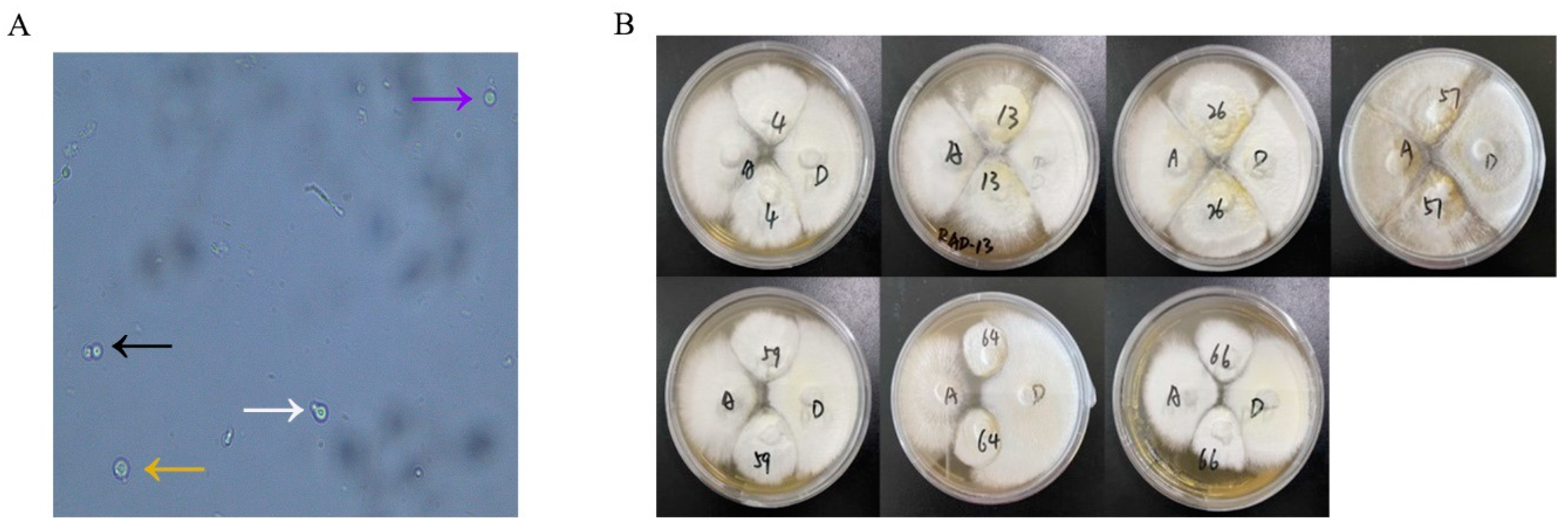
Figure 1. Protoplast fusion process (A) and antagonism test between the fusants and parents (B). The purple arrow in (A) indicates that the protoplast is not fused, the black arrow indicates that the protoplast is in close contact before fusion, the white arrow indicates the protoplast fusion process, and the orange arrow indicates that the fusion is complete. (B), A represents G. lingzhi, and D represents G. resinaceum.
Horticulturae 09 01129 g001
Figure 2. ISSR fingerprints (A) and cluster analysis (B) of parent strains and fusants. (A), M: DL2000; P1: LZ-10; P2: LZ-13; and 1, 2, 3, 4, 5, 6, and 7 are RAD-4, RAD-13, RAD-26, RAD-57, RAD-59, RAD-64, and RAD-66, respectively.
Horticulturae 09 01129 g002
Figure 3. Colony morphology and pigment production.
Horticulturae 09 01129 g003
Figure 4. Growth characteristics and agronomic traits of the parents and fusants. (A) Growth rate of strains in PDA and cultivation materials; (B) fruiting body yield of strains; (C) strain fruiting body morphology. The values are the means ± SDs of three independent experiments. Different letters indicate significant differences in the comparison of samples (p < 0.05 according to Duncan’s test).
Horticulturae 09 01129 g004
Figure 5. Polysaccharide content (A) and triterpene content (B) in the fruiting bodies of the parents and fusants. The values are the means ± SDs of three independent experiments. Different letters indicate significant differences in the comparison of samples (p < 0.05 according to Duncan’s test).
Horticulturae 09 01129 g005
Table 1. ISSR primer information.
Primer CodeSequence (5′–3′)Annealing Temperature (°C)
ISSR-1CACCACACACACACACA46
ISSR-3GAGAGAGAGAGAGAGACC46
ISSR-4AGCAGCAGCAGCAGCAGCG49
ISSR-5TGCACACACACACAC46
ISSR-6GAGAGAGAGAGAGAGAT43
ISSR-7AGAGAGAGAGAGAGAGC46
ISSR-8CACACACACACACACAT46
ISSR-9GAGAGAGAGAGAGAGACT52
ISSR-10TTCCCTTCCCTTCCC50
ISSR-11GTGACACACACACAC45
ISSR-12AGTGTGTGTGTGTGT45
ISSR-16GGATGCAACACACACACAC56
Table 2. Thermal inactivation rate of G. lingzhi protoplasts under different conditions.
StrainsTemperature (°C)Time (min)
1020304050607080
G. lingzhi4040.445.286.197.898.799.6100100
4592.299.1100100100100100100
50100100100100100100100100
Table 3. UV inactivation rate of G. resinaceum protoplasts under different conditions.
StrainsTime (s)
1020304050607080
G. resinaceum31.541.958.966.178.289.5100100
Table 4. Results of nuc-ITS sequence alignment.
StrainsLZ-10LZ-13RAD-4RAD-13RAD-26RAD-57RAD-59RAD-64RAD-66
LZ-10100%94.79%100%100%100%100%100%100%100%
LZ-1394.79%100%94.79%94.79%94.79%94.79%94.79%94.79%94.79%
Table 5. Results of mitochondrial cox2 gene sequence alignment.
StrainsLZ-10LZ-13RAD-4RAD-13RAD-26RAD-57RAD-59RAD-64RAD-66
LZ-10100%95.32%100%100%100%100%100%100%100%
LZ-1395.32%100%95.32%95.32%95.32%95.32%95.32%95.32%95.32%
Disclaimer/Publisher’s Note: The statements, opinions and data contained in all publications are solely those of the individual author(s) and contributor(s) and not of MDPI and/or the editor(s). MDPI and/or the editor(s) disclaim responsibility for any injury to people or property resulting from any ideas, methods, instructions or products referred to in the content.