Article

Integrated Transcriptome and Metabolome Analysis Revealed the Molecular Mechanism of Anthocyanin Synthesis in Purple Leaf Pepper (Capsicum annuum L.) under Different Light Intensities

1
Engineering Research Center of Education Ministry for Germplasm Innovation and Breeding New Varieties of Horticultural Crops, Key Laboratory of Vegetable Biology of Hunan Province, College of Horticulture, Hunan Agricultural University, Changsha 410128, China
2
Vegetable Institution of Hunan Academy of Agricultural Science, Changsha 410125, China
*
Authors to whom correspondence should be addressed.
These authors contributed equally to this work.
Horticulturae 2023, 9(7), 814; https://doi.org/10.3390/horticulturae9070814
Submission received: 17 June 2023 / Revised: 10 July 2023 / Accepted: 12 July 2023 / Published: 14 July 2023

Abstract

:
Light is a crucial environmental component for plant growth, and light intensity plays a crucial function in controlling pigment anabolism in plants. We performed physiological characterisation, transcriptome, and metabolome investigations on purple leaf peppers treated with different light intensities to evaluate the effect on plant leaf colour. The results showed that the leaves of the peppers became significantly purplish under high light, with significantly higher anthocyanin, chlorophyll a, and carotenoid contents. A total of 44,263 genes were quantified using RNA-Seq, with the photoprotein-related genes LRP and LIP maintaining high expression levels under high and medium light. The anthocyanin synthesis pathway was variously enriched among the comparison groups, according to KEGG. The expression of the genes involved in the anthocyanin synthesis pathway, such as CHI, F3H, DFR, and BZ1, was significantly higher under high light. In addition, MYB and bHLH gene families were the most abundant, and MYB1R1, MYB113-like, and bHLH90-like were significantly expressed under high light and highly positively correlated with the above anthocyanin synthesis genes. According to our metabolomic analysis, delphinidin-3-O-rutinoside and delphinidin-3-O-glucoside accumulated in significant concentrations in purple leaves under high light. This study is useful for understanding the process of anthocyanin synthesis and metabolism in pepper leaves that is generated and regulated by varied light intensity.

1. Introduction

Pepper (Capsicum annuum L.) is one of the largest vegetable crops grown in the world; is widely used as a raw material for food seasoning, medicine, and cosmetics; and is an essential vegetable of the Solanaceae family [1]. Pepper leaves come in a wide variety of hues, and the variation in leaf colour in plants is often thought to be caused by an imbalance in the ratio of pigments, so the purple colouring of pepper leaves due to anthocyanin accumulation has an important biological function [2]. Anthocyanins, as a major natural pigment, are flavonoids with a special molecular structure and strong water solubility, mainly in the form of anthocyanin glycosides [3,4]. According to their molecular structure, anthocyanins are divided into six categories: malvidin, petunidin, pelargonidin, paeonidin, delphinidin, and cyanidin. In plants, anthocyanins maintain a wide range of colours, are the main colouring substances, and play an essential role in shielding plants from a range of stresses, such as drought, low temperatures, UV radiation, and pathogenic infestation [4,5,6,7,8,9,10].
In plants like Arabidopsis thaliana, the biosynthesis process for anthocyanins has been extensively characterised. As with many metabolites, the anthocyanin biosynthetic pathway is an essential component of the phenylpropanoid biosynthesis process, and studies on maize and Arabidopsis have demonstrated that anthocyanin biosynthesis is accomplished via the phenylpropanoid pathway, where PAL, CHS, CHI, F3H, DFR, ANS, and others directly encode the synthesis of related enzymes [11,12,13]. Additionally, a variety of transcription factors control the intricate route that produces anthocyanins. The anthocyanin biosynthetic pathway is most significantly known to be regulated by the MYB class of transcription factors, which has been revealed to play a key regulatory function in plant anthocyanin metabolism. The expression of structural genes for anthocyanin synthesis is controlled by MYB; however, various MYBs control distinct structural genes.
Light, as a significant environmental element, has been shown in several studies to affect anthocyanin synthesis in plants to varying degrees. Light not only provides energy for plant growth but also acts as a signal for growth and development, influencing multiple processes and the entire growth cycle [14]. Studies have revealed that the biosynthesis of anthocyanin glycosides is influenced by light intensity; high light can motivate lots of plants to produce and accumulate anthocyanins [15]. Niu et al. [16] found that shading inhibited the expression of all anthocyanin biosynthesis genes as well as MrMYB1 through the bagging of prunes (Myrica rubra). High light has also been demonstrated to encourage the structural genes in plants that are involved in the biosynthesis of anthocyanins, like vaccinium, raspberries, tomato, and eggplant [17,18,19,20,21]. Meanwhile, in green plant tissues or cells, light regulates the synthesis and accumulation of anthocyanins through photoreceptors and photosynthetic electron transfer, thus regulating the synthesis of plant colour and shielding plant tissues from stressors like reactive oxygen species (ROS) [22].
Previous studies have focused on seedling development, photosynthetic physiology, and stress resistance. However, little has been reported on the alterations in the dynamics, transcriptional control, and metabolic pathways of pepper anthocyanin glycosides in leaves. In this study, RNA samples that had been exposed to different light intensities were sequenced using Illumina deep-sequencing technology to elucidate the influence of light intensity on anthocyanin synthesis in pepper leaves. The purpose of this study is to establish the groundwork for future molecular studies by elucidating the molecular mechanism of how light intensity affects anthocyanin production using high-throughput sequencing and expression data.

2. Materials and Methods

2.1. Plant Materials and Treatment

The pepper material used in the experiment was dark purple ‘A178’ provided by the pepper team of the College of Horticulture, Hunan Agricultural University. After germination, the seeds were sown in cavity trays and incubated in a greenhouse (temperature 28 ± 2 °C/20 ± 2 °C, humidity 65% ± 3%, light and dark treatment 16 h/8 h). After the seedlings reached the four-leaf stage, they were transplanted into pots, and after 3 d of incubation under the same conditions, 45 seedlings of uniform growth were selected and placed in three groups under high light (H, 500 ± 5 μmol·m−2·s−1), medium light (M, 200 ± 5 μmol·m−2·s−1), and low light (L, 50 ± 5 200 ± 5 μmol·m−2·s−1). After 15 d of each light treatment, the colour index, photosynthetic rate, and chlorophyll fluorescence parameters of the three groups of seedlings were measured during the daytime, seedlings of uniform growth were selected from each group, and 5 leaves were taken from the 3rd to 8th true leaves from the top to the bottom of the seedlings, mixed, and stored at −80 °C.

2.2. Determination of Physiological Index Parameters

The colour luminance value (ΔL), red value (|Δa|), and yellow value (Δb) of pepper leaves under different light intensities were determined using a spectrophotometer (Ts7600, Shenzhen 3nh Technology Co., Ltd., Shenzhen, China), and total chromaticity value (ΔE) was calculated. Photosynthetic parameters of the apical 3rd–4th leaves of functional leaves of pepper seedlings were measured using an LI-6400XT photosynthesizer (LI-COR, USA). The light intensity and CO2 concentration parameters of the photosynthesizer were set with reference to the method of Liu et al. [23]. After photosynthetic parameters were measured, dark treatment was performed for 30 min, and chlorophyll fluorescence parameters were measured using a Fluorpen (FP110/D) portable chlorophyll fluorometer. The maximum photochemical efficiency for photosystem II (Fv/Fm), photochemical burst coefficient (qP), and non-photochemical burst coefficient (NPQ) were calculated with reference to the method of Maxwell and Johnson [24]. For each parameter determination, the operation was repeated three times for each treatment group.

2.3. RNA Extraction, Library Preparation, and Sequencing

In accordance with the TRI reagent (Sigma Life Sciences, USA) instructions, we extracted the total tissue RNA. RNA was detected with non-ribonuclease agarose gel electrophoresis to avoid possible degradation and contamination and then validated using an Agilent 2100 Bioanalyzer (Agilent Technologies, USA). Poly (A) mRNA was subsequently isolated using Oligo-dT beads (Qiagen, Germany), and then mRNA was randomly interrupted into short fragments by adding Fragmentation Buffer. The first cDNA strand was synthesised with random primers using mRNA as a template, and then the second cDNA strand was synthesised by adding buffer, dNTPs, RNaseH, and DNA polymerase I. The purified double-stranded cDNA was end-repaired, A-tail was added, sequencing junction was attached, and then fragment size selection was performed using AMPure XP beads. Finally, cDNA libraries were obtained with PCR enrichment. Then, sequencing was performed with Illumina sequencing platform (Illumina-nova6000, USA) using double-end sequencing technology.

2.4. Transcriptome Analysis

The raw data were converted to sequences, and low-quality sequences were removed with trim-galore. After that, the processed sequences were aligned to the pepper reference genome (C. annuum L_Zunla-1 database, https://www.ncbi.nlm.nih.gov/genome/10896, accessed on 16 July 2022) using Top Hat2. All successfully localised transcripts were identified and analysed using R package edgeR, and the expression of each gene was calculated and normalised to FPKM. Genes with fold change ≥ 2 and false discovery rate (FDR) < 0.01 were identified as differentially expressed genes (DEGs) using DESeq2 (http://www.bioconductor.org/packages/release/bioc/html/DESeq.html). Finally, DEGs were functionally annotated, GO terms were corrected for p values < 0.05, and KEGG pathways with p values < 0.05 were considered significantly enriched for differentially expressed genes. GO analysis was performed using the R package clusterProfiler, KEGG pathway analysis was performed with BLAST software, and KEGG pathway enrichment analysis was performed with KOBAS 2.0 software. The GO (http://www.geneontology.org/) functional database and KEGG (https://www.genome.jp/kegg/) pathway database analyses were performed on the differential gene set.

2.5. Metabolite Extraction

Pepper leaves were freeze-dried and ground to powder using MM400 grinder (Retsch, Germany), and 50 mg of tissue samples was weighed and extracted using 500 μL solution (50% aqueous methanol solution containing 0.1% hydrochloric acid). Referring to Naranjo et al. [25], the above extracts were vortexed, sonicated, and centrifuged, and the supernatant was aspirated. After repeating the operation once, the two supernatants were combined and filtered through a microporous membrane (0.22 μm pore size) and collected in a bottle for analysis in the LC–MS/MS system [26]. The anthocyanin composition was determined with ultra-performance liquid chromatography (UPLC) using the method provided by Qi et al. [27].

2.6. Quantitative Real-Time PCR

The RT-qPCR method was carried out using Taylor et al. [28] as a guide. The cDNA was obtained with reverse transcription of total RNA using the Vazyme kit (Jiangsu, China) according to the manufacturer’s instructions, which were used as a template for validation. A total of 12 related genes were selected to design gene-specific primers for qPCR on the basis of the sequences selected in RNA-seq (Table S1). Relative gene expression was normalised with the 2−ΔΔCt method [29].

3. Results

3.1. Phenotype Identification and Pigment Content Analysis

Pepper leaves showed significant differences under different light intensities, with the purple colour of the leaves deepening as the light intensity increased (Figure 1A). By gauging the amount of pigment content of the leaves under different light intensities, we discovered that the anthocyanin, chlorophyll a, and carotenoid contents of the leaves were significantly lower in the low light treatment group treatment than in the high light group, while the chlorophyll b content was not appreciably altered (Figure 1B). Simultaneously, the values of ΔL, |Δa|, Δb, and ΔE were significantly higher under the low light treatment, as well as the Pn, Ci, Tr, and Gs (Figure 1C,D). In addition, Fv/F0, qP, and NPQ were likewise discovered to be substantially lower in the low light treatment group than in the high light group (Figure 1E).

3.2. Transcriptome Analysis of Different Light Intensities

An aggregate of 61.23 Gb Clean Data was found from nine pepper samples using Illumina sequencing, with 5.96 Gb Clean Data for each pepper sample and Q30 base percentages at 93.82% and above, indicating high throughput and quality of the RNA-Seq data. Additionally, 204,689,817 clean reads were recovered in total after junctions and low-quality sequences were removed, and these were compared to the C. annuum L_Zunla-1 database (https://www.ncbi.nlm.nih.gov/genome/10896, accessed on 16 July 2022), with the comparison rate ranging from 88.91% to 93.78%. The samples contained 44,263 genes in total, including 8927 novel genes (Figure 2A, Table S2). Of the known genes quantified, 24,013 genes were co-quantified in the high, medium, and low light treatment groups, with 417, 315, and 689 genes specific to each group, respectively (Figure 2B). Furthermore, 7528 new genes were also co-quantified, with 203, 128, and 205 new genes specific to each group, respectively (Figure 2C).

3.3. Analysis of Differentially Expressed Genes (DEGs)

The screening criteria for differential gene screening were fold change ≥ 2 and FDR < 0.01. In total, 6981 DEGs were acquired in each comparison group, and just 1902, 117, and 570 genes were found in the H/L, H/M, and M/L groups, respectively (Figure 2D). There were 6183 (3526 up-regulated and 2557 down-regulated), 991 (724 up-regulated and 267 down-regulated), and 4526 (2040 up-regulated and 2486 down-regulated) DEGs in each of the different comparison groups for H/L, H/M, and M/L, respectively (Figure 2E, Table S3). Further investigation found that varying light intensity treatments resulted in differential expression of genes related to carotenoid anabolism. Among these, PDS (Capana03g000054) maintained a high expression level under high and medium light, with a significant decrease in expression under low light. In addition, the expression trends of photoprotein-related genes under different light intensities were similar to those of PDS, with light-regulated protein (LRP, Capana03g004339) and light-induced protein (LIP, Capana02g002261) being expressed 4.0-fold and 4.2-fold more under high light than under low light, respectively. The differences in leaf colour under various light intensities may be strongly related to the differential expression of these genes associated with the carotenoid biosynthesis pathway as well as photoprotein-related genes.

3.4. GO Analyses of DEGs

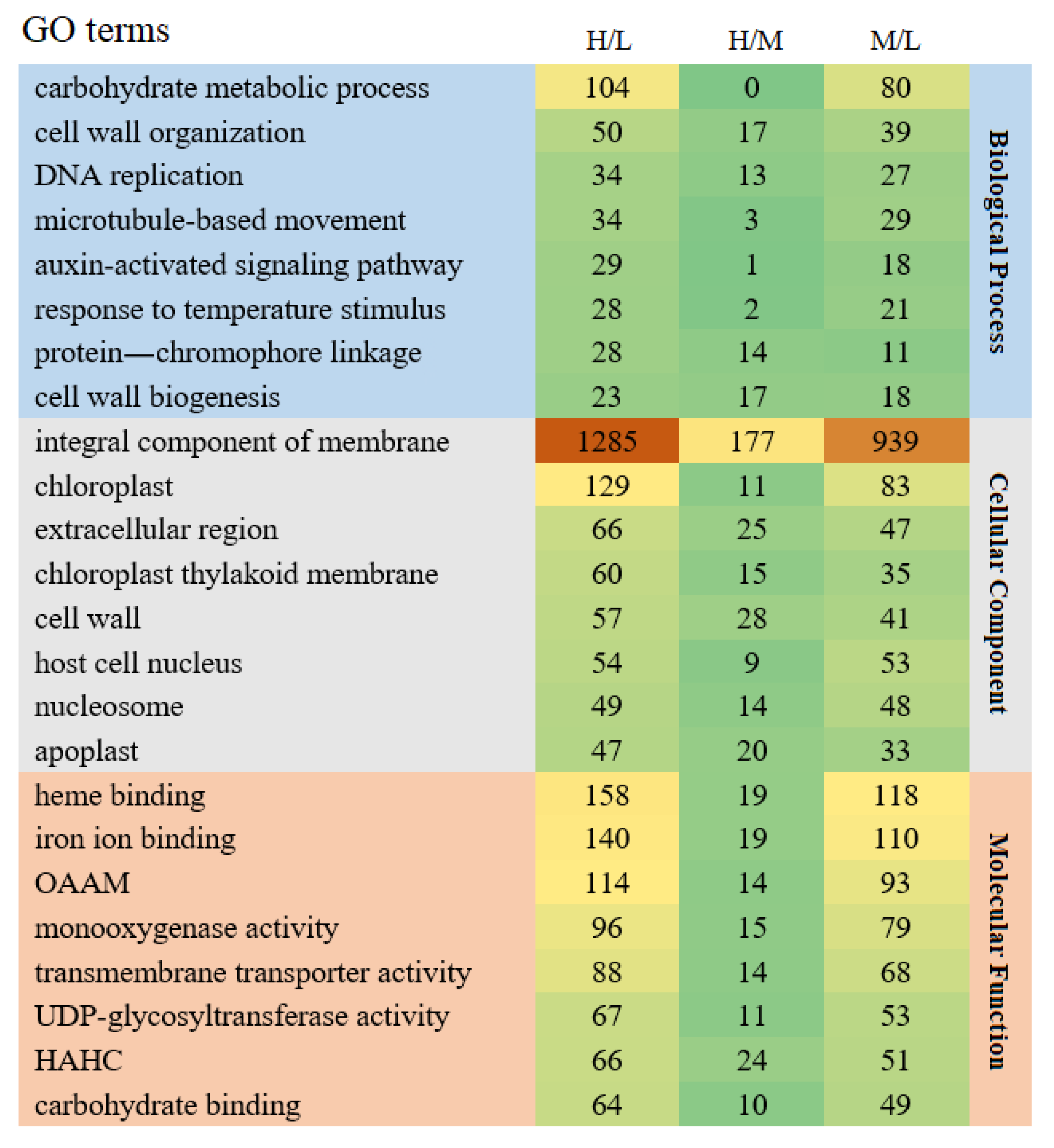
GO enrichment analysis of the DEGs revealed that the DEGs were widely distributed in three functional groups, and the top eight functions containing the greatest substantial enrichment were chosen from each of the functional groups using the H/L group as a screening condition for analysis (Figure 3). There were noticeably fewer DEGs enriched in each GO function in the H/M group than in the M/L and H/L groups in the group comparing light intensities. Regarding biological processes (BP), all three groups were heavily enriched in cell wall organization, and in addition, the DEGs in the H/L and M/L groups were the most enriched in the carbohydrate metabolic process, DNA replication, and microtubule-based movement. In terms of cellular components (CCs), the largest taxon in the three comparison groups was the integral component of membrane, with the DEGs in the H/L and M/L groups mainly enriched in the chloroplast and plasma membranes. In the cell wall, extracellular region, and apoplast, the DEGs of all three groups were enriched to some extent. With regard to molecular functions (MF), all three groups showed some enrichment of the DEGs in hydrolase activity, hydrolysing O-glycosyl compounds, and protein heterodimerisation activity. These results suggest that different light intensity treatments mainly induced differential expression of genes in the functional classes of carbohydrate metabolic process, integral component of membrane, heme binding, and iron ion binding in three functional groups of pepper leaves; the above functions were more enriched in the H/L and M/L groups; and the degree of enrichment was significantly higher than that in the H/M group. The significant variations in leaf colour may be caused by alterations in how these functional groups are expressed.

3.5. KEGG Pathway Analyses of DEGs

To gain more insight into the metabolic pathways that are crucial for pepper leaves, we further analysed the 20 KEGG pathways that were the most enriched in DEGs from each comparison group (Figure 4). The results showed that the DEGs were basically enriched in the plant–pathogen interaction, MAPK signalling pathway–plant, and plant hormone signal transduction. Further analysis revealed that the number of DEGs enriched in each pathway was generally lower in the H/M group compared to the H/L and M/L groups. In the H/L and M/L groups, DEGs were abundant in the ribosome, carbon metabolism, glycolysis/gluconeogenesis, and flavonoid biosynthesis pathways. In the H/M group, the number of DEGs enriched in these four pathways was significantly lower, and there was some enrichment of DEGs in fatty acid elongation. In addition, DEGs were enriched to some extent in the anthocyanin biosynthesis pathway in both the H/L and M/L groups but were weakly enriched in the H/M group and were not found in the first 20 KEGG pathways. According to the results, all three groups of DEGs were highly enriched in MAPK signalling pathway–plant and plant hormone signal transduction, which may affect the main KEGG pathway for leaf colour change in pepper. Moreover, according to the changes in leaf colour, M/L showed a more significant purple colour shift than H/M, indicating that the anthocyanin production pathway may be largely responsible for these alterations.

3.6. Metabolome Analysis of Anthocyanins

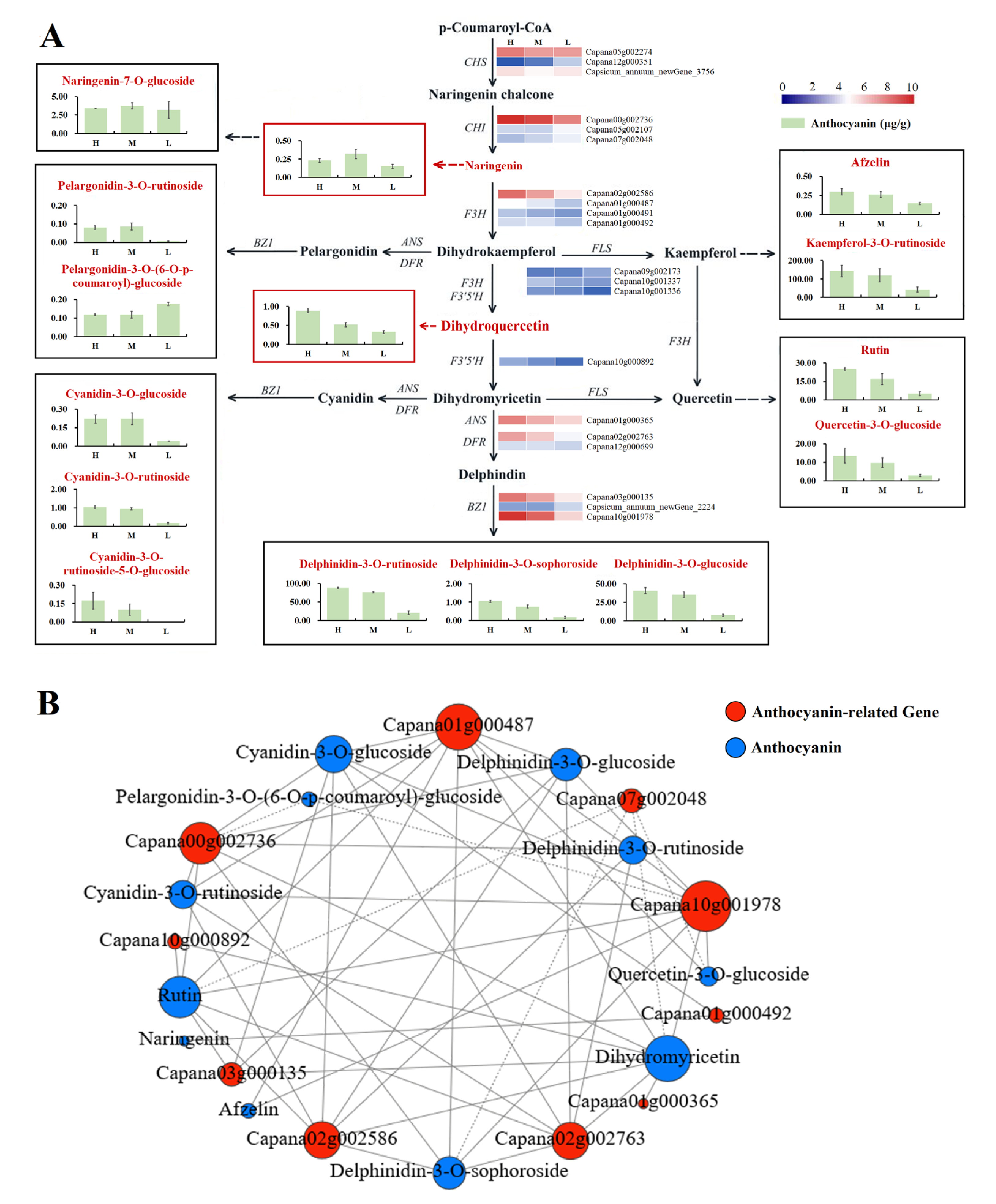
An aggregate of 29 anthocyanin species were found under three light intensities (Table S4). Of these 29 anthocyanins, 25 (22 down-regulated, 3 up-regulated), 2 (2 down-regulated), and 23 (19 down-regulated, 4 up-regulated) metabolites were significantly different in content in H/L, H/M, and M/L, respectively. Further analysis revealed that the six anthocyanins with higher content were kaempferol-3-O-rutinoside, quercetin-3-O-glucoside, rutin, delphinidin-3-O-sophoroside, delphinidin-3-O-rutinoside, and delphinidin-3-O-glucoside, all of which were significantly elevated under high light and had lower levels under medium and low light conditions. We also found that delphinidin-3-O-rutinoside and delphinidin-3-O-glucoside were not only higher in content but also varied to a significantly lesser extent in the H/M group than in the M/L group. The contents of delphinidin-3-O-rutinoside and delphinidin-3-O-glucoside were only 15.36% and 15.95% higher, respectively, under high light compared to the medium light treatment, while their content under medium light was significantly increased by 2.67-fold and 3.62-fold, respectively, compared to that under low light. The purple colour of the pepper leaves significantly deepened from the low to medium light conditions but not from the medium to high light conditions, which is consistent with the accumulation of delphinidin-3-O-rutinoside and delphinidin-3-O-glucoside in the pepper leaves. These findings imply that the primary anthocyanin component influencing how purple the leaves are may be delphinidin derivatives.

3.7. Analysis of Genes Involved in Anthocyanin Biosynthesis

The expression of the genes involved in the anthocyanin metabolic pathway was examined in order to further investigate the effect of light intensity on anthocyanin biosynthesis. The results revealed that there were notable differences in the expression of the relevant genes under various light intensities (Figure 5A). The expression of CHI (Capana00g002736) and BZ1 (Capana10g001978) was at a higher level under different light intensities and was significantly higher than the expression of other genes. Additional investigation revealed that the expression of CHS (Capana05g002274), CHI (Capana00g002736), F3H (Capana02g002586, Capana01g000487), FLS (Capana10g001337, Capana10g001336), DFR (Capana02g002763), ANS (Capana01g000365), and BZ1 (Capana03g000135, Capana10g001978) was maintained at high expression levels under high light, which was significantly different from that under low light treatment. To further understand how anthocyanin metabolites are impacted by structural genes, we also analysed anthocyanin-pathway-related structural genes using co-expression with anthocyanin metabolites (Figure 5B, Table S5). The results revealed that the structural genes CHI (Capana00g002736), F3H (Capana01g000487), and BZ1 (Capana10g001978) were associated with the anthocyanin synthesis pathway metabolites cyanidin-3-O-glucoside, rutin, cyanidin-3-O-rutinoside, dihydromyricetin, delphinidin-3-O-rutinoside, and delphinidin-3-O-glucoside, and all showed a high positive correlation. The anthocyanin biosynthetic pathway, which influences the colour of pepper leaves, may be regulated by the structural genes mentioned above, according to our speculation based on the results.

3.8. Analysis of Transcription Factors Involved in Anthocyanin Biosynthesis

Transcription factors (TFs) play an essential role in anthocyanin synthesis. Under different light intensity treatments, we detected a total of 15 transcription factor families, of which the MYB gene family and the bHLH gene family were the most numerous with 146 and 100 genes, respectively, followed by the NAC, B3, WD40, WRKY, G2-like, and Dof families (Figure 6A, Table S6). The MYB and the bHLH gene families were further analysed with the screening condition that FPKM > 10 was identified in at least one sample, and a total of 35 MYB TFs and 32 bHLH TFs were quantified (Figure 6B,C, Table S5). Among them, MYB1R1 (Capana03g001041) was significantly more expressed than the other transcription factor genes under high light. MYB1R1 (Capana03g001041, Capsicum_annuum_newGene_10195), MYB48 (Capana11g000757), bHLH90-like (Capana11g001290), bHLH35-like (Capana01g004240), and bHLH041 (Capana09g000184) were expressed at high levels under high light and showed decreasing expression levels under medium and low light treatment. Furthermore, in contrast to the above gene trends, MYB82 (Capana00g002415), MYB82-like (Capana00g003240), bHLH149, (Capana08g001640) and bHLH76 (Capana04g000713) were expressed at a lower level under high light treatment, while expression gradually increased under medium and low light conditions. The expression of the two species was gradually increased under medium and low light conditions.

3.9. Real-Time PCR Validation

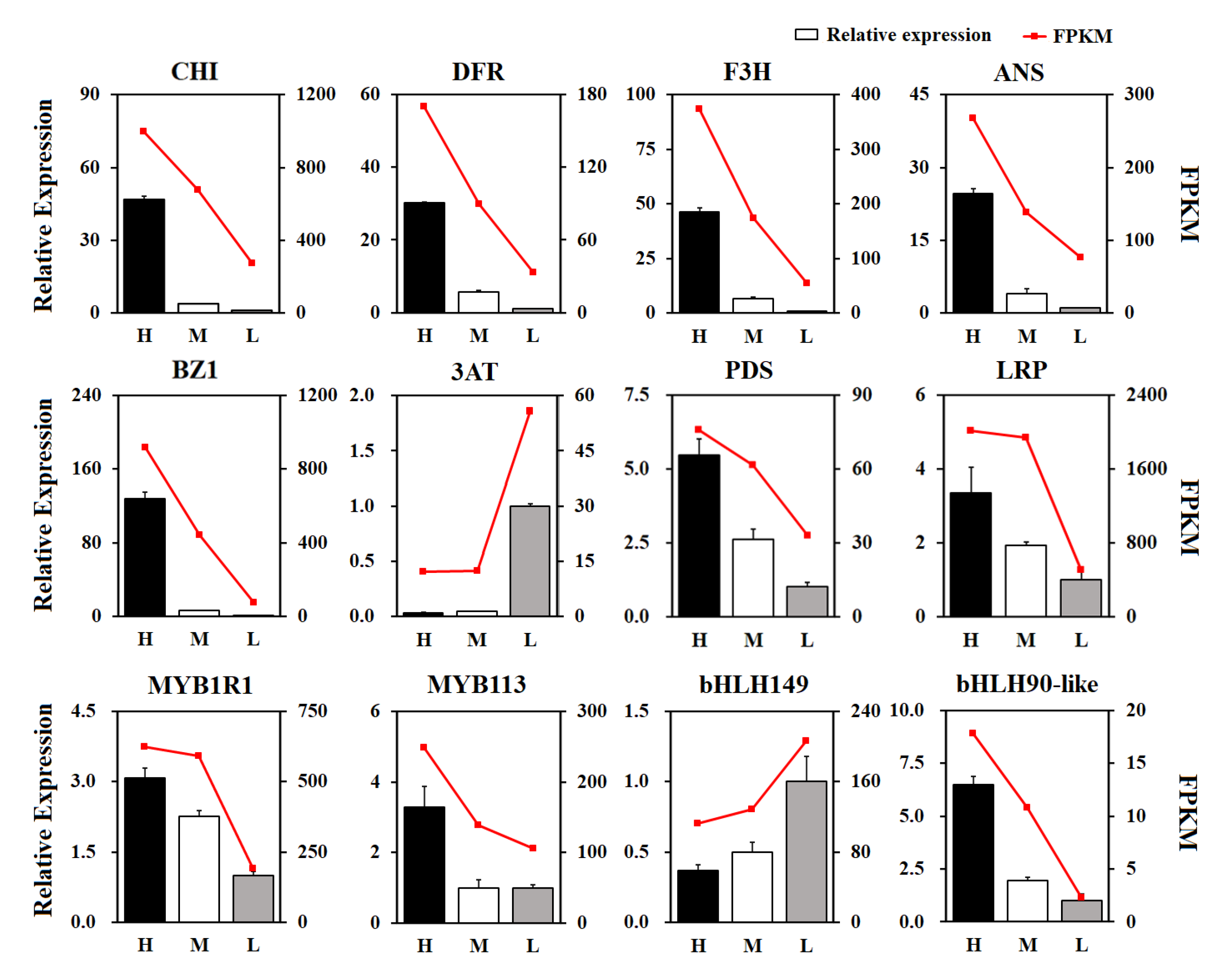
We selected important genes related to anthocyanin synthesis for RT-qPCR validation. The results showed (Figure 7) that the relative expression and transcriptome sequencing results of the 12 selected DEGs showed similar expression trends under different light intensities, indicating that the transcriptome sequencing data in this study were reliable.

4. Discussion

Light is necessary for the growth and development of plants, and high levels of light can alter the colour of some plant reproductive and nutritive organs. It is believed that light-induced colour changes in plants are mainly due to light-induced anthocyanin accumulation [21]. In this study, the purple leaves deepened as the light intensity increased. Analysis of the relevant physiological indicators showed that the content of anthocyanin, chlorophyll a, and carotenoid all increased to a certain extent, and it is assumed that the increase in the chlorophyll a and carotenoid content under different light intensities did not affect the colour of the pepper leaves to a significant extent, with anthocyanin being the main cause of the change in leaf colour.
In general, high levels of light can enhance the expression of both structural and regulatory genes, leading to a rise in the accumulation of anthocyanin glycosides, whereas low levels of light or darkness can inhibit or down-regulate gene expression, squelching the synthesis of anthocyanin glycosides. CHS and CHI serve as vital enzymes in the anthocyanin synthesis pathway, and numerous studies have discovered a favourable link between CHS, CHI, and anthocyanin concentration. For example, the GCHS1 gene in Gerbera hybrida is responsible for the biosynthesis of all flavonoid groups in the petals of Gerbera hybrida and the GCHS4 gene catalyses the biosynthesis of anthocyanins in nutritional tissues [30]. In arabidopsis tt4 and tt5 mutants, the CHS and CHI genes of maize (Zea mays) were introduced, respectively, and both tt4 and tt5 were found to have significantly increased anthocyanin content [31]. In addition, Lc petunia plants accumulated a large number of anthocyanins under high light, with plants showing a dark purple colour and up-regulated expression of CHS, CHI, DFR, and ANS [32], and Ahn et al. [33] found that the production of red leaves in red varieties of Zoysiagrass (Zoysia japonica Steud.) was caused by the elevated expression of DFR and ANS. In the purple-red leaves of chokecherry (Padus virginiana), the expression of BZ1-related genes was significantly higher than in the green leaves of this species [34]. In this study, the expression of CHI (Capana00g002736), ANS (Capana01g000365), DFR (Capana02g002763), and BZ1 (Capana10g001978 and Capana03g000135) was at a high level under high light and was significantly reduced under low light treatment, which is consistent with previous findings. Furthermore, the analysis revealed that the anthocyanin content associated with kaempferol and quercetin was significantly higher than that associated with pelargonidin and cyanidin, suggesting that there may be a strong competition between DFR and FLS acting on dihydrokaempferol and dihydromyricetin, a result that was corroborated in the study of grape hyacinth [35]. Considering these findings, we further hypothesise that light influences the expression of genes involved in anthocyanin metabolism in pepper leaves, which in turn regulates the differential accumulation of anthocyanins in leaves under various lighting conditions, changing the colour of the leaves.
On top of structural genes, the anthocyanin synthesis pathway is also influenced by regulatory genes like MYB TFs and bHLH TFs [36]. Related transcription factors such as the MYB–bHLH–WD40 protein complex, bZIP, NAC, and WRKY have also been implicated in flavonoid biosynthesis in higher plants [37,38,39,40,41]. Studies have shown that the apple MdMYB1 TF is a light-induced R2R3-MYB TF, and the light induces the expression of MdMYB1, which in turn induces anthocyanin synthesis [42]. In Arabidopsis, AtMYB11, AtMYB12, and AtMYB11 regulated the expression of structural genes CHS, CHI, and F3H in the anthocyanin synthesis pathway in various tissues, while the expression of structural genes such as DFR and ANS was positively correlated with the expression of PAP1, PAP2, AtMYB113, and AtMYB114 [43,44]. In addition, recent studies have also identified carrot DcMYB7 as a key gene determining the synthesis of anthocyanins in carrot flesh and petioles [45]. Furthermore, related studies have revealed the key regulatory role of a second transcription factor, bHLH, and a high correlation with the transcription factor MYB. In mangosteen (Garcinia mangostana L.) fruit, transcription factor GmMYB10 changes the most during the ripening process from green to purplish red, and co-transformation of it with Arabidopsis AtbHLH2 into tobacco effectively activates the GmDFR and AtDFR promoters [46]. Xiang et al. [47] found in chrysanthemum that CmMYB6 and CmbHLH2 form a binary complex through physical interaction to regulate the expression of CmDFR during chrysanthemum anthocyanin biosynthesis. In the tobacco MrMYB1-MrbHLH1 overexpression line, the colour of the tobacco leaves changed significantly and the anthocyanin-related structural genes NtCHS, NtDFR, and NtANS were significantly up-regulated [48]. In this study, MYB1R1 (Capana03g001041) and MYB113-like (Capana10g001433) expression was significantly elevated in response to high light treatment. According to weighted gene co-expression network analysis (WGCNA), MYB48 (Capana11g000757), MYB113-like (Capana10g001433), and MYB1R1 (Capana03g001041) were found to have a high positive correlation with structural genes DFR and BZ1. Among the bHLH gene family, bHLH90-like (Capana11g001290) was also highly correlated with the structural genes CHI, ANS, F3H, and DFR. Moreover, the negative regulatory transcription factor bHLH149 (Capana08g001640) was found to have a high negative correlation with CHI, DFR, and BZ1. We hypothesise that the aforementioned transcription factors might affect anthocyanin production by controlling the expression of structurally related genes.
Studies have found that the most common and abundant anthocyanins in fruits and vegetables of the Solanaceae are based on delphinidin derivatives, which are predominantly blue/purple in hue [49,50]. Aromatic acylation is responsible for the blue colour of anthocyanins, while the non-aromatic acylated anthocyanins still show the red colour of anthocyanins [51,52]. The combination of blue and red pigments produces a purple colour, while the more acylated the attached anthocyanin aromatic groups are, the more purple and darker the leaves become. Jin et al. [53] found that the ratio of non-aromatised to aromatised anthocyanins and the ratio of anthocyanin content to chlorophyll content in the composition of purple ornamental cabbage are responsible for the different leaf colours. Furthermore, Veberic et al. [54] found that delphinidin occupied the highest share of 60.7% of the anthocyanins in blackcurrant. Ponder et al. [55] detected delphinidins (delphinidin-3-O-glucoside and delphinidin-3-O-rutinoside) as the dominant anthocyanins. In the purple fruits of aubergine and violet pepper, the major anthocyanins were found to be delphinidin-3-O-rutinoside and delphinidin-3-trans-coumaroylrutinoside-5-glucoside, respectively, according to HPLC chromatograms [56]. In purple broccoli, high levels of delphinidin-3-O-glucoside, delphinidin-3-O-galactoside, cyanidin-3-O-glucoside, and cyanidin-3-O-galactoside were identified [57]. In this study, the levels of kaempferol-3-O-rutinoside, rutin, quercetin-3-O-glucoside, delphinidin-3-O-rutinoside, and delphinidin-3-O-glucoside were found to be significantly higher in purple leaves under high light treatment compared to leaves under low light, with the levels of the anthocyanin metabolites delphinidin-3-O-rutinoside and delphinidin-3-O-glucoside being higher under high light. We, therefore, speculate that delphinidin-3-O-rutinoside and delphinidin-3-O-glucoside may be the dominant substances responsible for the purple coloration of pepper leaves.

5. Conclusions

According to this study, we discovered that pepper leaves’ anthocyanin, chlorophyll a, and carotenoid contents significantly increased after being exposed to high light. An aggregate of 44,263 genes were quantified using transcriptome analysis, with 6183, 991, and 4526 differential genes obtained in the three comparison groups H/L, H/M, and M/L, respectively. At the metabolic level, delphinidin-3-O-rutinoside and delphinidin-3-O-glucoside accumulated in large amounts under high light, leading to a deepening of the purple colour in the pepper leaves. At the gene level, high light treatment increased the expression of CHI, F3H, and BZ1, which are structural genes related to the anthocyanin metabolic pathway; affected the expression of genes related to the MYB transcription factor family and the bHLH transcription factor family; and increased the content of delphinidin derivatives. Among these, co-expression network analysis revealed that MYB1R1, MYB113-like, and bHLH90-like had a high positive correlation with structural genes, and bHLH149 had a high negative correlation with structural genes. The biological basis for the selection and breeding of peppers with purple leaves is put forth by this work, which offers fresh insights into our understanding of anthocyanin synthesis and accumulation in peppers.

Supplementary Materials

The following supporting information can be downloaded at: https://www.mdpi.com/article/10.3390/horticulturae9070814/s1, Table S1: Primers used for RT-qPCR in this study; Table S2: All genes identified in the samples; Table S3: Differentially expressed genes in each of the different comparison groups; Table S4: All anthocyanins detected in the samples; Table S5: Transcription factors identified in this study; Table S6: Transcription factors identified in this study.

Author Contributions

Conceptualisation, Z.L. and C.L.; methodology, Y.S. (Yiyu Shen); software, L.M.; validation, Y.S. (Yiyu Shen), L.M. and Y.Z.; formal analysis, Y.S. (Yiyu Shen); investigation, L.M. and Y.Z.; resources, Z.L.; data curation, Y.S. (Yiyu Shen); writing—original draft preparation, Y.S. (Yiyu Shen) and L.M.; writing—review and editing, Z.L. and C.L.; visualization, Y.S. (Ying Sun); supervision, C.L.; project administration, Z.L.; funding acquisition, Z.L. All authors have read and agreed to the published version of the manuscript.

Funding

Special Project of Biological Seed Industry and Fine and Deep Processing of Agricultural Products (202202AE090031) and the Natural Science Foundation of Hunan Province (2021JJ40240).

Data Availability Statement

The data presented in this study are available on request from the corresponding author. The RNA-Seq data generated in this study are available from the SRA-Archive (http://www.ncbi.nlm.nih.gov/sra, accessed on 13 June 2023) with accession number PRJNA976502.

Acknowledgments

We thank BioMarker for providing RNA-Seq services and Wuhan Metware for providing metabolome sequencing services.

Conflicts of Interest

The authors declare no conflict of interest.

References

  1. Zou, Z.; Zou, X. Geographical and Ecological Differences in Pepper Cultivation and Consumption in China. Front. Nutr. 2021, 8, 718517. [Google Scholar] [CrossRef]
  2. Zhao, M.-H.; Li, X.; Zhang, X.-X.; Zhang, H.; Zhao, X.-Y. Mutation Mechanism of Leaf Color in Plants: A Review. Forests 2020, 11, 851. [Google Scholar] [CrossRef]
  3. Holton, T.A.; Cornish, E.C. Genetics and Biochemistry of Anthocyanin Biosynthesis. Plant Cell 1995, 7, 1071–1083. [Google Scholar] [CrossRef]
  4. Tanaka, Y.; Sasaki, N.; Ohmiya, A. Biosynthesis of Plant Pigments: Anthocyanins, Betalains and Carotenoids. Plant J. 2008, 54, 733–749. [Google Scholar] [CrossRef] [PubMed]
  5. Castellarin, S.D.; Pfeiffer, A.; Sivilotti, P.; Degan, M.; Peterlunger, E.; DI Gaspero, G. Transcriptional Regulation of Anthocyanin Biosynthesis in Ripening Fruits of Grapevine under Seasonal Water Deficit. Plant Cell Env. 2007, 30, 1381–1399. [Google Scholar] [CrossRef] [PubMed] [Green Version]
  6. He, J.; Giusti, M.M. Anthocyanins: Natural Colorants with Health-Promoting Properties. Annu. Rev. Food Sci. Technol. 2010, 1, 163–187. [Google Scholar] [CrossRef] [PubMed]
  7. Li, P.; Li, Y.-J.; Zhang, F.-J.; Zhang, G.-Z.; Jiang, X.-Y.; Yu, H.-M.; Hou, B.-K. The Arabidopsis UDP-Glycosyltransferases UGT79B2 and UGT79B3, Contribute to Cold, Salt and Drought Stress Tolerance via Modulating Anthocyanin Accumulation. Plant J. 2017, 89, 85–103. [Google Scholar] [CrossRef] [PubMed] [Green Version]
  8. Sarma, A.D.; Sharma, R. Anthocyanin-DNA Copigmentation Complex: Mutual Protection against Oxidative Damage. Phytochemistry 1999, 52, 1313–1318. [Google Scholar] [CrossRef]
  9. Sivankalyani, V.; Feygenberg, O.; Diskin, S.; Wright, B.; Alkan, N. Increased Anthocyanin and Flavonoids in Mango Fruit Peel Are Associated with Cold and Pathogen Resistance. Postharvest Biol. Technol. 2016, 111, 132–139. [Google Scholar] [CrossRef]
  10. Wu, X.; Prior, R.L. Systematic Identification and Characterization of Anthocyanins by HPLC-ESI-MS/MS in Common Foods in the United States: Fruits and Berries. J. Agric. Food Chem. 2005, 53, 2589–2599. [Google Scholar] [CrossRef]
  11. Koes, R.; Verweij, W.; Quattrocchio, F. Flavonoids: A Colorful Model for the Regulation and Evolution of Biochemical Pathways. Trends Plant Sci. 2005, 10, 236–242. [Google Scholar] [CrossRef]
  12. Naing, A.H.; Kim, C.K. Roles of R2R3-MYB Transcription Factors in Transcriptional Regulation of Anthocyanin Biosynthesis in Horticultural Plants. Plant Mol. Biol. 2018, 98, 1–18. [Google Scholar] [CrossRef]
  13. Saito, K.; Yonekura-Sakakibara, K.; Nakabayashi, R.; Higashi, Y.; Yamazaki, M.; Tohge, T.; Fernie, A.R. The Flavonoid Biosynthetic Pathway in Arabidopsis: Structural and Genetic Diversity. Plant Physiol. Biochem. 2013, 72, 21–34. [Google Scholar] [CrossRef] [Green Version]
  14. Shi, Y.; Ke, X.; Yang, X.; Liu, Y.; Hou, X. Plants Response to Light Stress. J. Genet. Genom. 2022, 49, 735–747. [Google Scholar] [CrossRef]
  15. Maier, A.; Hoecker, U. COP1/SPA Ubiquitin Ligase Complexes Repress Anthocyanin Accumulation under Low Light and High Light Conditions. Plant Signal Behav. 2015, 10, e970440. [Google Scholar] [CrossRef] [Green Version]
  16. Niu, S.-S.; Xu, C.-J.; Zhang, W.-S.; Zhang, B.; Li, X.; Lin-Wang, K.; Ferguson, I.B.; Allan, A.C.; Chen, K.-S. Coordinated Regulation of Anthocyanin Biosynthesis in Chinese Bayberry (Myrica rubra) Fruit by a R2R3 MYB Transcription Factor. Planta 2010, 231, 887–899. [Google Scholar] [CrossRef]
  17. Zhou, Y.; Singh, B.R. Effect of Light on Anthocyanin Levels in Submerged, Harvested Cranberry Fruit. J. Biomed. Biotechnol. 2004, 2004, 259–263. [Google Scholar] [CrossRef] [Green Version]
  18. Uleberg, E.; Rohloff, J.; Jaakola, L.; Trost, K.; Junttila, O.; Häggman, H.; Martinussen, I. Effects of Temperature and Photoperiod on Yield and Chemical Composition of Northern and Southern Clones of Bilberry (Vaccinium myrtillus L.). J. Agric. Food Chem. 2012, 60, 42. [Google Scholar] [CrossRef] [Green Version]
  19. Wang, S.Y.; Chen, C.-T.; Wang, C.Y. The Influence of Light and Maturity on Fruit Quality and Flavonoid Content of Red Raspberries. Food Chem. 2009, 112, 676–684. [Google Scholar] [CrossRef]
  20. Løvdal, T.; Olsen, K.; Slimestad, R.; Verheul, M.; Lillo, C. Synergetic Effects of Nitrogen Depletion, Temperature, and Light on the Content of Phenolic Compounds and Gene Expression in Leaves of Tomato. Phytochemistry 2010, 71, 605–613. [Google Scholar] [CrossRef]
  21. Li, J.; He, Y.-J.; Zhou, L.; Liu, Y.; Jiang, M.; Ren, L.; Chen, H. Transcriptome Profiling of Genes Related to Light-Induced Anthocyanin Biosynthesis in Eggplant (Solanum melongena L.) before Purple Color Becomes Evident. BMC Genom. 2018, 19, 201. [Google Scholar] [CrossRef] [PubMed] [Green Version]
  22. Isaak, C.K.; Petkau, J.C.; Blewett, H.; Karmin; Siow, Y. L. Lingonberry Anthocyanins Protect Cardiac Cells from Oxidative-Stress-Induced Apoptosis. Can. J. Physiol. Pharmacol. 2017, 95, 904–910. [Google Scholar] [CrossRef] [PubMed] [Green Version]
  23. Liu, Z.; Dai, Y.; Mao, L.; Yang, S.; Yang, B.; Sun, Y.; Zhou, Y.; Wang, S.; Zou, X.; Ou, L. Integrated Metabolome and Transcriptome Analysis Revealed Carotenoid Metabolism Difference in Pepper (Capsicum annuum L.) Yellowing Mutants under Different Light Quality. Veg. Res. 2022, 2, 21. [Google Scholar] [CrossRef]
  24. Maxwell, K.; Johnson, G.N. Chlorophyll Fluorescence—A Practical Guide. J. Exp. Bot. 2000, 51, 659–668. [Google Scholar] [CrossRef] [PubMed]
  25. Di Paola-Naranjo, R.D.; Sánchez-Sánchez, J.; González-Paramás, A.M.; Rivas-Gonzalo, J.C. Liquid Chromatographic-Mass Spectrometric Analysis of Anthocyanin Composition of Dark Blue Bee Pollen from Echium Plantagineum. J. Chromatogr. A 2004, 1054, 205–210. [Google Scholar] [CrossRef]
  26. Want, E.J.; Masson, P.; Michopoulos, F.; Wilson, I.D.; Theodoridis, G.; Plumb, R.S.; Shockcor, J.; Loftus, N.; Holmes, E.; Nicholson, J.K. Global Metabolic Profiling of Animal and Human Tissues via UPLC-MS. Nat. Protoc. 2013, 8, 17–32. [Google Scholar] [CrossRef]
  27. Qi, Y.; Lou, Q.; Li, H.; Yue, J.; Liu, Y.; Wang, Y. Anatomical and Biochemical Studies of Bicolored Flower Development in Muscari Latifolium. Protoplasma 2013, 250, 1273–1281. [Google Scholar] [CrossRef]
  28. Taylor, S.C.; Nadeau, K.; Abbasi, M.; Lachance, C.; Nguyen, M.; Fenrich, J. The Ultimate QPCR Experiment: Producing Publication Quality, Reproducible Data the First Time. Trends Biotechnol. 2019, 37, 761–774. [Google Scholar] [CrossRef] [Green Version]
  29. Mestdagh, P.; Van Vlierberghe, P.; De Weer, A.; Muth, D.; Westermann, F.; Speleman, F.; Vandesompele, J. A Novel and Universal Method for MicroRNA RT-QPCR Data Normalization. Genome Biol. 2009, 10, R64. [Google Scholar] [CrossRef] [Green Version]
  30. Deng, X.; Bashandy, H.; Ainasoja, M.; Kontturi, J.; Pietiäinen, M.; Laitinen, R.A.E.; Albert, V.A.; Valkonen, J.P.T.; Elomaa, P.; Teeri, T.H. Functional Diversification of Duplicated Chalcone Synthase Genes in Anthocyanin Biosynthesis of Gerbera Hybrida. New Phytol. 2014, 201, 1469–1483. [Google Scholar] [CrossRef]
  31. Dong, X.; Braun, E.L.; Grotewold, E. Functional Conservation of Plant Secondary Metabolic Enzymes Revealed by Complementation of Arabidopsis Flavonoid Mutants with Maize Genes. Plant Physiol. 2001, 127, 46–57. [Google Scholar] [CrossRef] [Green Version]
  32. Albert, N.W.; Lewis, D.H.; Zhang, H.; Irving, L.J.; Jameson, P.E.; Davies, K.M. Light-Induced Vegetative Anthocyanin Pigmentation in Petunia. J. Exp. Bot. 2009, 60, 2191–2202. [Google Scholar] [CrossRef] [Green Version]
  33. Ahn, J.H.; Kim, J.-S.; Kim, S.; Soh, H.Y.; Shin, H.; Jang, H.; Ryu, J.H.; Kim, A.; Yun, K.-Y.; Kim, S.; et al. De Novo Transcriptome Analysis to Identify Anthocyanin Biosynthesis Genes Responsible for Tissue-Specific Pigmentation in Zoysiagrass (Zoysia japonica Steud.). PLoS ONE 2015, 10, e0124497. [Google Scholar] [CrossRef] [Green Version]
  34. Li, X.; Li, Y.; Zhao, M.; Hu, Y.; Meng, F.; Song, X.; Tigabu, M.; Chiang, V.L.; Sederoff, R.; Ma, W.; et al. Molecular and Metabolic Insights into Anthocyanin Biosynthesis for Leaf Color Change in Chokecherry (Padus virginiana). Int. J. Mol. Sci. 2021, 22, 10697. [Google Scholar] [CrossRef]
  35. Lou, Q.; Liu, Y.; Qi, Y.; Jiao, S.; Tian, F.; Jiang, L.; Wang, Y. Transcriptome Sequencing and Metabolite Analysis Reveals the Role of Delphinidin Metabolism in Flower Colour in Grape Hyacinth. J. Exp. Bot. 2014, 65, 3157–3164. [Google Scholar] [CrossRef] [Green Version]
  36. Baudry, A.; Heim, M.A.; Dubreucq, B.; Caboche, M.; Weisshaar, B.; Lepiniec, L. TT2, TT8, and TTG1 Synergistically Specify the Expression of BANYULS and Proanthocyanidin Biosynthesis in Arabidopsis Thaliana. Plant J. 2004, 39, 366–380. [Google Scholar] [CrossRef]
  37. Terrier, N.; Torregrosa, L.; Ageorges, A.; Vialet, S.; Verriès, C.; Cheynier, V.; Romieu, C. Ectopic Expression of VvMybPA2 Promotes Proanthocyanidin Biosynthesis in Grapevine and Suggests Additional Targets in the Pathway. Plant Physiol. 2009, 149, 1028–1041. [Google Scholar] [CrossRef] [Green Version]
  38. Wang, N.; Liu, W.; Zhang, T.; Jiang, S.; Xu, H.; Wang, Y.; Zhang, Z.; Wang, C.; Chen, X. Transcriptomic Analysis of Red-Fleshed Apples Reveals the Novel Role of MdWRKY11 in Flavonoid and Anthocyanin Biosynthesis. J. Agric. Food Chem. 2018, 66, 7076–7086. [Google Scholar] [CrossRef]
  39. Xu, W.; Dubos, C.; Lepiniec, L. Transcriptional Control of Flavonoid Biosynthesis by MYB-BHLH-WDR Complexes. Trends Plant Sci. 2015, 20, 176–185. [Google Scholar] [CrossRef]
  40. Zhang, F.; Huang, J.; Guo, H.; Yang, C.; Li, Y.; Shen, S.; Zhan, C.; Qu, L.; Liu, X.; Wang, S.; et al. OsRLCK160 Contributes to Flavonoid Accumulation and UV-B Tolerance by Regulating OsbZIP48 in Rice. Sci. China Life Sci. 2022, 65, 1380–1394. [Google Scholar] [CrossRef]
  41. Zhao, C.; Liu, X.; Gong, Q.; Cao, J.; Shen, W.; Yin, X.; Grierson, D.; Zhang, B.; Xu, C.; Li, X.; et al. Three AP2/ERF Family Members Modulate Flavonoid Synthesis by Regulating Type IV Chalcone Isomerase in Citrus. Plant Biotechnol. J. 2021, 19, 671–688. [Google Scholar] [CrossRef]
  42. Takos, A.M.; Jaffé, F.W.; Jacob, S.R.; Bogs, J.; Robinson, S.P.; Walker, A.R. Light-Induced Expression of a MYB Gene Regulates Anthocyanin Biosynthesis in Red Apples. Plant Physiol. 2006, 142, 1216–1232. [Google Scholar] [CrossRef] [PubMed] [Green Version]
  43. Chiu, L.-W.; Zhou, X.; Burke, S.; Wu, X.; Prior, R.L.; Li, L. The Purple Cauliflower Arises from Activation of a MYB Transcription Factor. Plant Physiol. 2010, 154, 1470–1480. [Google Scholar] [CrossRef] [PubMed] [Green Version]
  44. Yuan, Y.; Chiu, L.-W.; Li, L. Transcriptional Regulation of Anthocyanin Biosynthesis in Red Cabbage. Planta 2009, 230, 1141–1153. [Google Scholar] [CrossRef]
  45. Xu, Z.-S.; Yang, Q.-Q.; Feng, K.; Xiong, A.-S. Changing Carrot Color: Insertions in DcMYB7 Alter the Regulation of Anthocyanin Biosynthesis and Modification. Plant Physiol. 2019, 181, 195–207. [Google Scholar] [CrossRef] [PubMed]
  46. Palapol, Y.; Ketsa, S.; Lin-Wang, K.; Ferguson, I.B.; Allan, A.C. A MYB Transcription Factor Regulates Anthocyanin Biosynthesis in Mangosteen (Garcinia mangostana L.) Fruit during Ripening. Planta 2009, 229, 1323–1334. [Google Scholar] [CrossRef]
  47. Xiang, L.; Liu, X.; Li, X.; Yin, X.; Grierson, D.; Li, F.; Chen, K. A Novel BHLH Transcription Factor Involved in Regulating Anthocyanin Biosynthesis in Chrysanthemums (Chrysanthemum morifolium Ramat.). PLoS ONE 2015, 10, e0143892. [Google Scholar] [CrossRef]
  48. Liu, X.-F.; Yin, X.-R.; Allan, A.C.; Lin-Wang, K.; Shi, Y.-N.; Huang, Y.-J.; Ferguson, I.B.; Xu, C.-J.; Chen, K.-S. The Role of MrbHLH1 and MrMYB1 in Regulating Anthocyanin Biosynthetic Genes in Tobacco and Chinese Bayberry (Myrica rubra) during Anthocyanin Biosynthesis. Plant Cell Tiss. Organ. Cult. 2013, 115, 285–298. [Google Scholar] [CrossRef]
  49. Niño-Medina, G.; Urías-Orona, V.; Muy-Rangel, M.D.; Heredia, J.B. Structure and Content of Phenolics in Eggplant (Solanum melongena)—A Review. S. Afr. J. Bot. 2017, 111, 161–169. [Google Scholar] [CrossRef]
  50. Tanaka, Y.; Ohmiya, A. Seeing Is Believing: Engineering Anthocyanin and Carotenoid Biosynthetic Pathways. Curr. Opin. Biotechnol. 2008, 19, 190–197. [Google Scholar] [CrossRef]
  51. Park, C.H.; Bong, S.J.; Lim, C.J.; Kim, J.K.; Park, S.U. Transcriptome Analysis and Metabolic Profiling of Green and Red Mizuna (Brassica rapa L. Var. Japonica). Foods 2020, 9, 1079. [Google Scholar] [CrossRef]
  52. Abdel-Aal, E.-S.M.; Young, J.C.; Rabalski, I. Anthocyanin Composition in Black, Blue, Pink, Purple, and Red Cereal Grains. J. Agric. Food Chem. 2006, 54, 4696–4704. [Google Scholar] [CrossRef]
  53. Jin, S.-W.; Rahim, M.A.; Jung, H.-J.; Afrin, K.S.; Kim, H.-T.; Park, J.-I.; Kang, J.-G.; Nou, I.-S. Abscisic Acid and Ethylene Biosynthesis-Related Genes Are Associated with Anthocyanin Accumulation in Purple Ornamental Cabbage (Brassica oleracea Var. Acephala). Genome 2019, 62, 513–526. [Google Scholar] [CrossRef] [Green Version]
  54. Anthocyanin Composition of Different Wild and Cultivated Berry Species. Available online: https://www.cabdirect.org/cabdirect/abstract/20143407037 (accessed on 10 June 2023).
  55. Ponder, A.; Hallmann, E.; Kwolek, M.; Średnicka-Tober, D.; Kazimierczak, R. Genetic Differentiation in Anthocyanin Content among Berry Fruits. Curr. Issues Mol. Biol. 2021, 43, 36–51. [Google Scholar] [CrossRef]
  56. Sadilova, E.; Stintzing, F.C.; Carle, R. Anthocyanins, Colour and Antioxidant Properties of Eggplant (Solanum melongena L.) and Violet Pepper (Capsicum annuum L.) Peel Extracts. Z. Für Naturforschung C 2006, 61, 527–535. [Google Scholar] [CrossRef]
  57. Liu, C.; Yao, X.; Li, G.; Huang, L.; Xie, Z. Transcriptomic Profiling of Purple Broccoli Reveals Light-Induced Anthocyanin Biosynthetic Signaling and Structural Genes. PeerJ 2020, 8, e8870. [Google Scholar] [CrossRef]
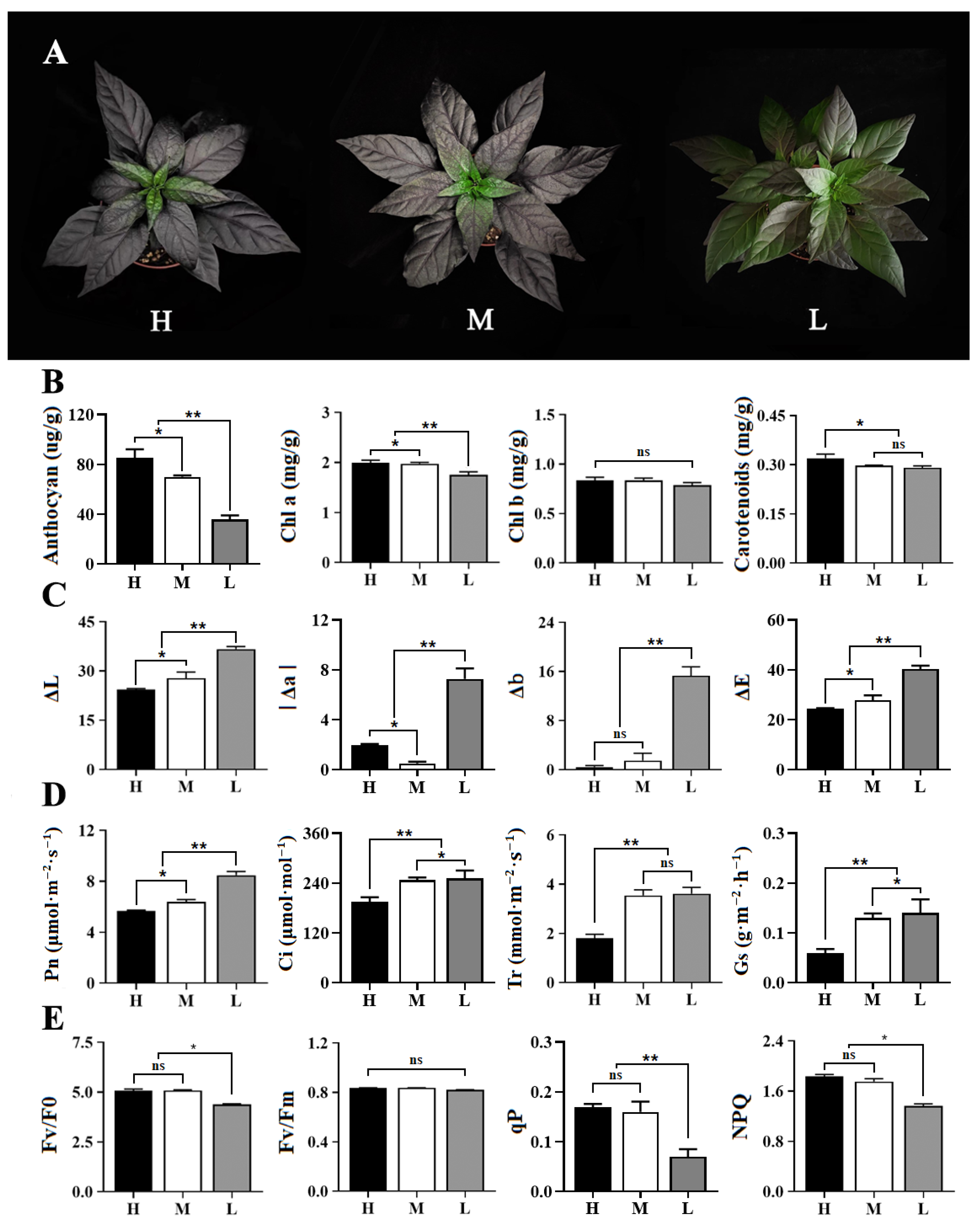
Figure 1. Phenotypic and physiological characterisation of pepper under different light intensities. (A) Phenotypic identification of pepper leaves. H, high light; M, medium light; L, low light. (B) Photosynthetic pigment content; *, p value < 0.05; **, p value < 0.01; ns, no significance. (C) Chromaticity values. (D) Photosynthetic indicators. (E) Chlorophyll fluorescence properties.
Horticulturae 09 00814 g001
Figure 2. Genes discovered with transcriptome analysis subjected to statistical analysis. (A) Venn diagram of total genes quantified under different light intensities. H, high light; M, medium light; L, low light. (B) Venn diagram of known genes quantified under different light intensities. (C) Venn diagram of new genes quantified under different light intensities. (D) Venn diagram of differentially expressed genes between light intensities. (E) The numbers of differentially expressed genes under different light intensities.
Horticulturae 09 00814 g002
Figure 3. GO analysis of differentially expressed genes. The top 8 GO terms with the highest DEG enrichment in the biological processes, cellular components, and molecular functions were screened in comparison with H/L group. Each row represents a GO term, and the number of genes per module is shown above. OAAM: oxidoreductase activity, acting on paired donors, with incorporation or reduction of molecular oxygen; HAHC: hydrolase activity, hydrolysing O-glycosyl compounds.
Horticulturae 09 00814 g003
Figure 4. KEGG enrichment of differentially expressed genes. The top 20 KEGG pathways that were most enriched in DEGs from 3 comparison groups of H/L, H/M and M/L. ASNM, amino sugar and nucleotide sugar metabolism; GBIS, glycosphingolipid biosynthesis-globo and isoglobo series; GBNS, glycosphingolipid biosynthesis-lacto and neolacto series.
Horticulturae 09 00814 g004
Figure 5. Analysis of anthocyanin biosynthesis pathway genes expression and metabolites. (A) Integrated analysis of gene expression and metabolites. H, high light; M, medium light; L, low light. Solid line, generation process; red dashed line, the content of anthocyanins; dashed line, generation of subsequent reactions. CHS, chalcone synthase; CHI, chalconeisomerase; F3H, flavonoid 3-hydroxylase; F3′5′H, flavonoid 3′5′-hydroxylase; DFR, dihydroflavonol 4-reductase; ANS, anthocyanidin synthase; FLS, flavonolsynthase; BZ1, anthocyanidin 3-O-glucosyltransferase. (B) Co-expression network analysis of structural genes and metabolites. Only Pearson correlation coefficient (PCC) ≥ 0.90 or ≤−0.90 are displayed. Solid line, positive correlation between genes; dashed line, negative correlation between genes.
Horticulturae 09 00814 g005
Figure 6. Analysis of transcription factors associated with the anthocyanin biosynthesis pathway. (A) Families of related transcription factors. (B) Heat map of MYB transcription factors. H, high light; M, medium light; L, low light. (C) Heat map of bHLH transcription factors. (D) Analysis of co-expression networks of structural genes and MYB TFs. Only Pearson correlation coefficient (PCC) ≥ 0.90 or ≤−0.90 are displayed. Solid line, positive correlation between genes; dashed line, negative correlation between genes. (E) Analysis of co-expression networks of structural genes and bHLH TFs.
Horticulturae 09 00814 g006
Figure 7. Validation and expression analysis of selected genes using RT-qPCR. The error bars indicate the SDs of three biological replicates. H, high light; M, medium light; L, low light. CHI, Capana00g002736; DFR, Capana02g002763; F3H, Capana02g002586; ANS, Capana01g000365; BZ1, Capana10g001978; 3AT, Capana10g000432; PDS, Capana03g000054; LRP, Capana03g004339; MYB1R1, Capana03g001041; MYB113, Capana10g001433; bHLH149, Capana08g001640; bHLH90-like, Capana11g001290.
Horticulturae 09 00814 g007
Disclaimer/Publisher’s Note: The statements, opinions and data contained in all publications are solely those of the individual author(s) and contributor(s) and not of MDPI and/or the editor(s). MDPI and/or the editor(s) disclaim responsibility for any injury to people or property resulting from any ideas, methods, instructions or products referred to in the content.