Article

Effectiveness of a Novel Endophyte Bacillus velezensis Strain B1 in the Biocontrol of Pear Postharvest Ring Rot

1
Institute of Radiation Technology, Beijing Academy of Science and Technology, Beijing 100875, China
2
National Natural History Museum of China, Beijing 100010, China
3
School of Electronic Engineering, Beijing University of Posts and Telecommunications, Beijing 100876, China
*
Author to whom correspondence should be addressed.
These authors contributed equally to this work.
Horticulturae 2023, 9(9), 1049; https://doi.org/10.3390/horticulturae9091049
Submission received: 23 August 2023 / Revised: 10 September 2023 / Accepted: 12 September 2023 / Published: 19 September 2023
(This article belongs to the Special Issue Biological Control of Pre and Postharvest Diseases II)

Abstract

:
Pear ring rot caused by Botryosphaeria dothidea severely affects the quality and storage life of pear fruit. Plant endophytes are potential new sources of biocontrol agents due to their abilities to produce novel bioactive products. In this work, we focused on an endophytic strain B1, which was isolated from roots of Dendrobium huoshanense. Notably, strain B1 exhibited strongly inhibitory effects against the growth of Botryosphaeria dothidea with an inhibition rate of 73.2% after 1 week of co-cultivation. Furthermore, it displayed a broad-spectrum inhibitory activity. Strain B1 also effectively delayed the onset of ring rot caused by B. dothidea in pear fruit and reduced the lesion diameter by 73.4%. Strain B1 was identified as Bacillus velezensis based on core-genome phylogeny. Genome mining with antiSMASH revealed 13 potential gene clusters involved in antimicrobial metabolites. Three main groups of lipopeptides (surfactin, iturin and fengycin) were identified with MALDI-TOF-MS, and the lipopeptides in the inhibition zone were greatly upregulated by B. dothidea; especially, fengycin isoforms were detected with higher abundance. These results proved that lipopeptides produced by strain B1 had significant antagonistic effect on B. dothidea. In conclusion, our results suggest that strain B1 has potential applications in the biocontrol of B. dothidea in postharvest fruit.

1. Introduction

Postharvest diseases caused by plant pathogens have a detrimental impact on fruit yield, resulting in significant economic losses [1]. Pear is one of widely cultivated fruits in the world, and China is the largest producer and consumer of pears [2]. Throughout the cultivation process, pear is susceptible to various pests and diseases. Botryosphaeria dothidea is one of the most destructive pathogens in pear production. This pathogen can infect not only the fruits but also leaves and trunks of pear trees, causing pear ring rot and canker and dieback diseases on the tree [3]. Notably, B. dothidea exhibits no host preference and has been reported to affect a wide range of plants important in agriculture, forestry and horticulture, making it a global concern [3]. Furthermore, B. dothidea have occasionally infected humans. For instance, Noguchi et al. [4] showed that the causal agent of ungual phaeohyphomycosis was B. dothidea. A recent study also demonstrated that B. dothidea tended to accumulate compounds toxic to mammalian cells [5].
Currently, chemical fungicides are widely used to control B. dothidea, causing environmental and public health issues. Hence, there is a requirement to find an alternative to chemical pesticides that are both safe for food production and environmentally friendly. The utilization of microbial agents for the biological control of pathogens presents a promising strategy. In recent years, microbial agents have been reported to produce bioactive compounds with biocontrol properties [6,7,8,9,10]. While most studies have focused on bacteria and fungi from plant rhizospheres, plant endophytes have gained much more attention from researchers worldwide in recent years due to their abilities to produce novel bioactive compounds [11,12,13,14,15]. Plant endophytes encompass a diverse array of microorganisms residing inside plants without causing any sign of infection [15]. They occupy a similar ecological niche to phytopathogens, making them viable candidates for biocontrol agents [16]. Endophytes have been detected in every plant sampled; although there are nearly 300,000 plant species on Earth, only a fraction of them have been thoroughly investigated relative to endophytes. Therefore, great efforts to study endophytes from diverse plant species in various ecosystem could bring us the opportunity to obtain new and valuable endophytes with potential applications in medicine and agriculture.
Dendrobium huoshanense, a unique medicinal plant, has significant medicinal value and health benefits. Its endophytic bacteria and fungi exhibit rich and diverse [17,18]. The dominant bacterial endophytes are Sphingomonas, Acinetobacter, Enterococcus, Bacillus and Methylobacterium [17]. Streptomyces is the largest genus of Actinobacteria and is well known for producing various secondary metabolites. An endophytic Streptomyces sp. HS-3-L-1 from D. huoshanense is capable of biosynthesis of three new polyketide dimmers known ashuoshanmycins A, B and C, and two of these (A and B) show moderate cytotoxicity against MV4–11 human leukemia cells [19]. Stagonosporopsis oculihominis, Alternaria eichhorniae, Phyllosticta aristolochiicola, Aspergillus flavus and Fusarium lactis are the dominant fungi endophytes of D. huoshanense which exhibit anti-inflammatory and antibacterial effects similar to those of the host plant [18]. In the present study, we aimed to isolate and screen endophytic bacteria with antagonistic activity from D. huoshanense, assessing the protective effect of the isolated strains against B. dothidea on postharvest pears. Additionally, we conducted genomics analysis to mine biosynthetic gene clusters encoding antimicrobial metabolites with antiSMASH. Furthermore, the production of these active metabolites was confirmed with MALDI-TOF-MS. These results could unravel the genetic and metabolic bases of the biocontrol traits exhibited by the antagonistic endophyte. This knowledge will enable us to develop rational strategies for its application in agriculture.

2. Materials and Methods

2.1. Isolation of Antifungal Bacterial Strains

Dendrobium huoshanense sampled in Anhui Province, China, were used to isolate root endophytic bacteria using the methods described by Wu et al. [20]. Briefly, roots were washed thoroughly with tap water and surface-sterilized in 75% ethanol for 30 s and in 3% NaClO for 3 min, then rinsed in sterile water 5 times. Sterilized roots were cut into 1 cm pieces, placed in sterile Petri dishes containing beef extract peptone medium (BPN medium) and incubated at 28 °C. Both endophytic bacteria and fungi emerged from the roots in a Petri dish. Bacteria with obvious antagonistic activities towards fungi were isolated and purified, then stored at −80 °C in 20% glycerol.

2.2. Screening of Antagonistic Bacteria against Botryosphaeria dothidea

Pear ring rot pathogen Botryosphaeria dothidea was isolated from diseased pear fruit and preserved in our laboratory. To assess the antagonistic abilities of isolated endophytes against B. dothidea, a dual-culture method was used [21]. Each treatment consisted of three replicate plates and incubated at 28 °C for 7 d. The antagonistic effect was evaluated by measuring the inhibition zones. Among 9 isolated strains, B1 showed the strongest antagonistic activity and was selected for further characterization. The antagonistic effect of B1 on mycelial growth of B. dothidea was observed under a light microscope (Olympus CX-40), and the inhibition rate (IR) of the pathogen was calculated using the formula as follows: IR = [(C2 − C1)/C2] × 100%, where C2 is the colony radius of the control and C1 is the average colony radius of the treatment.

2.3. Evaluating the Inhibitory Effect of Culture Filtrate of Strain B1

To evaluate the production of antimicrobial compounds by strain B1 in liquid culture against B. dothidea, the well-diffusion method on PDA was used. Strain B1 was grown in liquid BPN. Overnight bacterial cultures (500 μL) were inoculated into 250 mL flasks containing 50 mL of liquid BPN. The flasks were incubated on a rotary shaker at 30 °C and 150 rpm. After incubation for 72 h, the culture was centrifuged at 13,800× g for 15 min at 4 °C. The obtained cells were washed with sterilized water three times then resuspended in sterilized water to obtain cell suspensions (CSs), while the resulting supernatant was filtered through 0.22 μm filters to obtain cell-free supernatants (CFSs).
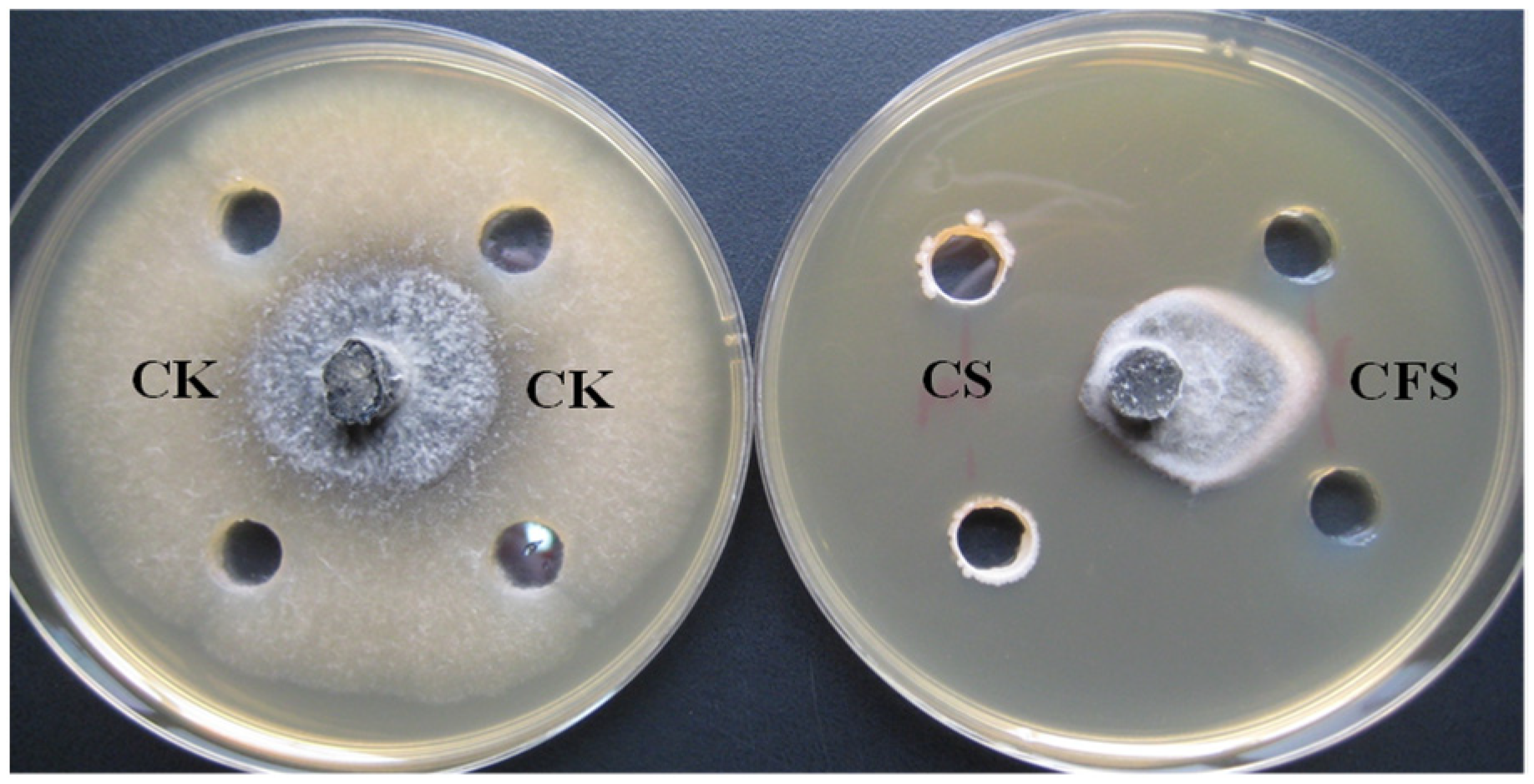
The inhibition of fungal growth was assessed on PDA plates, around the periphery of which wells (0.6 cm) were made equidistantly, and 100 μL CS, CFS, or sterilized BPN broth (control) was added to the wells. Then, mycelia plugs (0.6 cm diameter) taken from the edge of the 7-day-old colony of B. dothidea were inoculated in the center of the same plates. The plates were incubated at 28 °C for seven days, then inhibition zones were measured. The experiment was conducted two times in a completely randomized design with three replicates.

2.4. Inhibition Spectrum of Strain B1

Several important plant pathogens purchased from the Agricultural Culture Collection of China were kept in our laboratory. Antagonistic ability of strain B1 against these pathogens was tested as described in Section 2.2.

2.5. Biocontrol of Pear Fruit Ring Rot by Strain B1

Pear fruit (Pyrus pyrifolia Nakai cv. Shuijing) with uniformity of size and no mechanical injury and infection were selected, sterilized with 75% alcohol for 1 min and air dried. The fruit were wounded according to the method of Wu et al. [21]. To evaluate the biocontrol efficacy of strain B1, each wound of fruit was inoculated with 30 μL of B1 (5 × 10⁷ cfu/mL), and sterilized distilled water served as a control. After 12 h, mycelia plugs (6 mm in diameter) of B. dothidea taken from the margins of the 7-day-old colony were also applied to the wounds. The treated pears were incubated in climatic chambers at 25 °C and 85% relative humidity for 7 d, with three replicates and 8 pears per replicate. Disease incidence and lesion diameter of pear fruit were determined, and the inhibition rate was calculated as follows:
Inhibition rate (%) = (Rot diameter of control − Rot diameter of treatment)/Rot diameter of control × 100%

2.6. Genome Sequencing, Annotation and Phylogenetic Analysis

The genomic DNA of strain B1 was extracted with the SDS method. Genome sequencing was performed on the Illumina HiSeq 2500-PE125 platform with massively parallel sequencing (MPS) Illumina technology. The reads were quality trimmed to the Q30 confidence level, and the de novo genome assembly was performed using SOAPdenovo (http://soap.genomics.org.cn/soapdenovo.html) URL (accessed on 26 March 2018). The genome was annotated using the NCBI Prokaryotic Genomes Automatic Annotation Pipeline (PGAAP). The full genome sequence of strain B1 was deposited in NCBI under the GenBank accession number CP097895.
Various Bacillus strains were downloaded from the NCBI database. Genomic alignment between the B1 genome and reference genomes were performed using the MUMmer [22] and LASTZ [23] tools. Core and specific genes were analyzed by the CD-HITsoftware V4.6.8 [24], employing a threshold of 50% pairwise identity and a 0.7 length difference cutoff in amino acids. A pan- and core-genome plot was generated, while MUSCLE was used for aligning the core genes [25]. The phylogenomic tree based on the core genome was constructed using TreeBeST [26] with the neighbor-joining method.

2.7. Genome Mining of Secondary Metabolites Biosynthetic Gene Clusters

AntiSMASH is a comprehensive bioinformatic tool for the automatic genome mining of biosynthetic gene clusters (BGCs) involved in antimicrobial compounds, such as NRPs, PKs, NRPs-PKs hybrids, bacteriocins and terpenes. The B1 genome was analyzed using the antiSMASH web server (https://antismash.secondarymetabolites.org) to identify BGCs responsible for antimicrobial secondary metabolites [27].

2.8. Mass Spectrometry Analysis of Secondary Metabolites Produced by Strain B1

The interaction between strain B1 and B. dothidea on PDA produced an inhibition zone. In co-culture and control plates, the secondary metabolites with antifungal properties present in both cells and the inhibition zone were analyzed with MALDI-TOF-MS after 2 and 4 days of culturing. To prepare cell extracts, two loops of bacterial cells from co-culture and control plates were suspended in an Eppendorf tube containing 500 μL extraction solution (acetonitrile (70%) with trifluoroacetic acid (0.1%)). Simultaneously, agar samples of about 6 mm were obtained from the inhibition zone and deposited into an Eppendorf tube containing 500 μL extraction solution. In the single bacterial culture, control samples were taken from similar areas. Each tube was vigorously vortexed for 1 min and centrifuged at 10,000× g for 10 min. The collected supernatant was kept at −20 °C until MALDI-TOF-MS analysis. The MALDI-TOF-MS analysis was performed using a Bruker Daltonics MALDI-TOF instrument containing a 337 nm nitrogen laser for desorption and ionization at Institute of Chemistry, Chinese Academy of Sciences, Beijing, China.

2.9. Statistical Analysis

Statistical analyses were performed using SPSS (SPSS Statistics 20.0). The values are expressed as means of three replicates ± the standard deviation (SD). Differences between mean values were compared using Student’s t test, with p < 0.05 considered as a significant level.

3. Results

3.1. Isolation and Screening of Antagonistic Endophytes

The growth of B. dothidea was markedly inhibited by strain B1 (Figure 1B), with an inhibition rate of 73.2% after 1 week incubation compared with the control (Figure 1A). Light microscopic observations revealed that the mycelial morphology of B. dothidea was severely distorted by strain B1. The hyphae structure exhibited irregular thickness, with some hyphae forming swollen bubbles that eventually ruptured, resulting in leakage of the intracellular material (Figure 2A). In contrast, the control mycelium was uniform in thickness and smooth in surface, characterized by vigorous growth (Figure 2B). These results indicated that strain B1 severely damaged the mycelium morphology and inhibited the growth of B. dothidea.

3.2. Inhibitory Effect of Cell Suspension and Cell-Free Supernatants on Growth of B. dothidea

Strain B1 was examined for the production of antimicrobial compounds against B. dothidea in liquid culture, and the well-diffusion method was used to compare the antifungal properties of cell suspensions (CSs) and cell-free supernatants (CFSs). After 1 week incubation, the hyphal extension of B. dothidea was significantly inhibited on PDA medium (Figure 3). Notably, CS was more effective compared to CFS, producing a wider zone of inhibition (Table 1). The higher level of antagonistic activity of CS could be linked to additional biosynthesis of antifungal metabolites by the actively growing B1 cells on PDA medium compared to the CFS.

3.3. Antifungal Spectrum of Strain B1

Other important fungal pathogens were applied to test the antifungal activity of B1 using the same methods as above, and remarkable antagonistic activity was observed (Table 2). Strain B1 showed a broad inhibitory spectrum.

3.4. Biocontrol Effect of Strain B1 on B. dothidea In Vivo

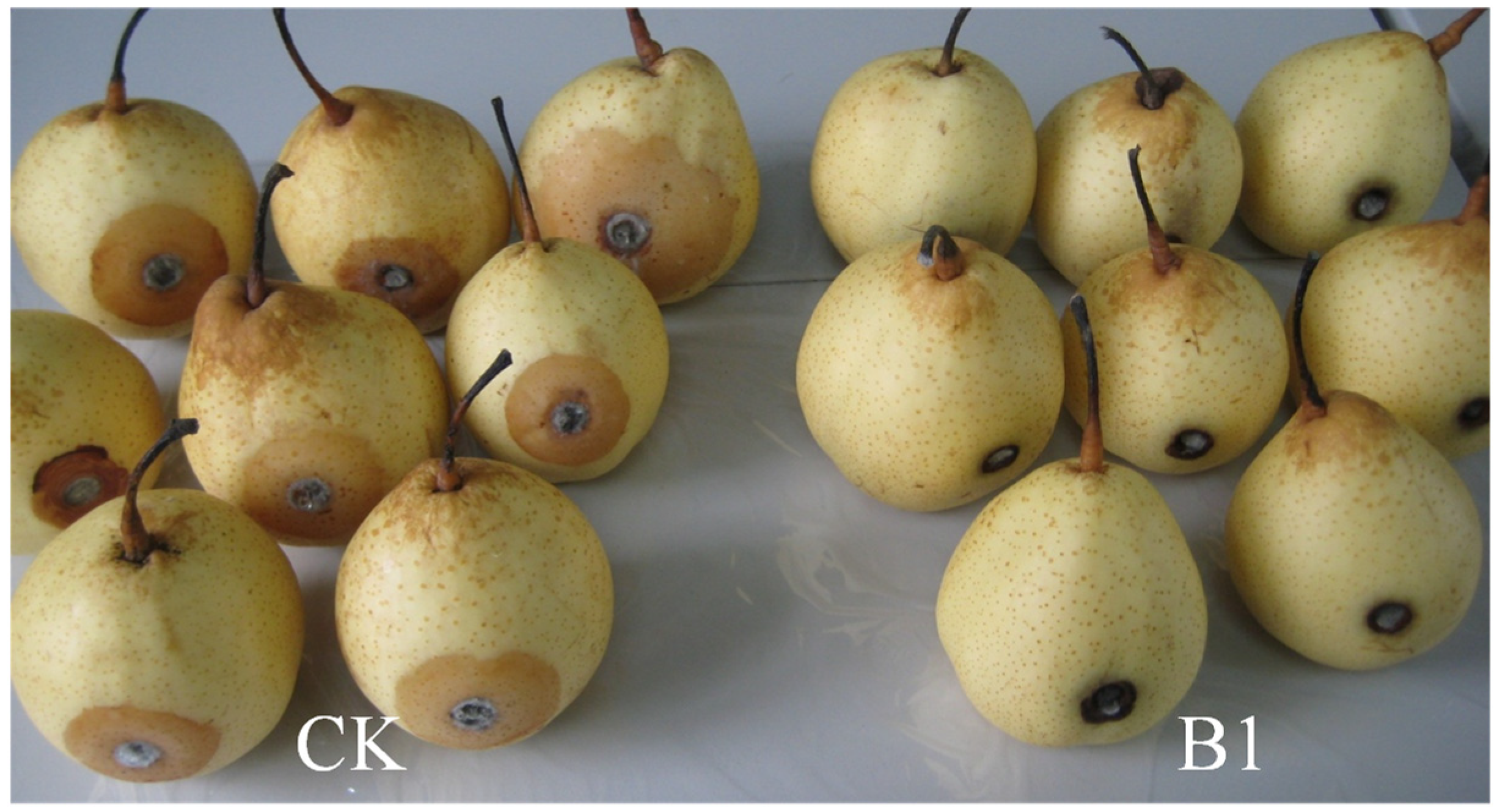
The incidence of pear fruit ring rot caused by B. dothidea was significantly reduced in the B1 treatment group compared to the control group (Figure 4, Table 3). After 3 days of inoculation, ring rot started to appear in the control pear group, whereas the B1 treatment could completely suppress the disease incidence in pear fruit until 5 days of inoculation. Subsequently, ring rot symptoms initiated in the B1 treatment group. After 7 days of inoculation, the control group displayed over 50% of the fruit area affected by rot, with the average lesion diameter of 4.31 cm. In contrast, the lesion diameter of pear fruit in the B1 treatment group was 1.3 cm on average, 73.4% lower than the control (Table 3). Strain B1 effectively slowed down the occurrence and development of ring rot of pear fruit, achieving a good control effect.

3.5. Genome General Characterization and Phylogenetic Analysis of Strain B1

The strain B1 genome had a sequencing coverage of 25×. The generated raw data were assembled and approximately 4.0 Mbp in length, consisting of 1 contig with a GC value of 46.54%. This genome harbored 3725 predicted protein-coding genes, 27 rRNA operons and 86 tRNAs (Table S1).
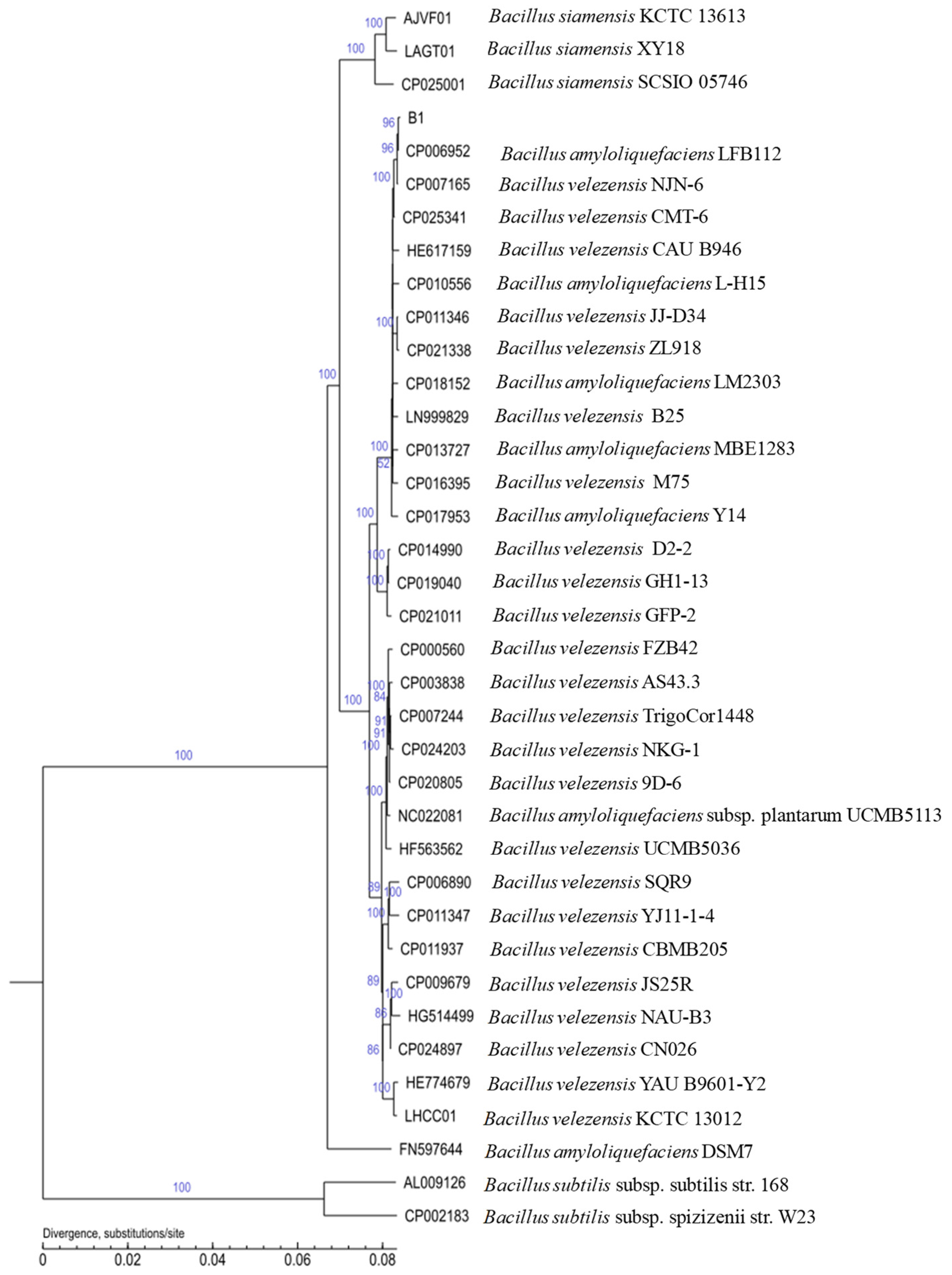
To infer the phylogenomic relationship between strain B1 and its closely related species, an unrooted phylogenomic tree was constructed based on the core genome (Figure 5). Strain B1 was most closely related to Bacillus amyloliquefaciens LFB112 and formed a distinct branch with B. velezensis and B. amyloliquefaciens strains including B. amyloliquefaciens FZB42 (Figure 5). B. amyloliquefaciens species were previously separated into two taxa “B. amyloliquefaciens subspecies amyloliquefaciens” and “B. amyloliquefaciens subspecies plantarum” [28], and the well-known B. amyloliquefaciens FZB42 was described as the type strain of this “plantarum” subspecies. B. amyloliquefaciens FZB42 and LFB112 have been re-categorized as B. velezensis [29]. Due to their high phenotypic and 16S rRNA similarities, it was difficult to differentiate B. velezensis, B. amyloliquefaciens and B. siamensis [30]. Dunlap et al. [31] demonstrated that B. velezensis NRRLB-41580T, B. methylotrophicus KACC 13105T,” B. oryzicola” KACC 18228 and B. amyloliquefaciens subsp. plantarum FZB42T were conspecific through comparative genomics, DNA–DNA relatedness calculations and phylogenomic analysis based on the core genome. Consequently, B. velezensis was considered a heterotypic synonym of B. amyloliquefaciens subsp. plantarum and was distinguished from its nearest neighbors B. amyloliquefaciens and B. siamensis [6,31,32]. Consistent with these reports, our phylogenomic analysis placed strain B1 within the B. velezensis branch (Figure 5), confirming its identification as B. velezensis.

3.6. Genome Mining of Secondary Metabolites Biosynthetic Gene Clusters (BGCs)

The strong antagonistic activity of well-known biocontrol bacterial strains was mostly due to the production of bioactive secondary metabolites. To better understand the biocontrol properties of strain B1, we aimed to determine genome mining BGCs potentially related to diverse bioactive compounds using antiSMASH. Strain B1 harbored BGCs encoding for 13 secondary metabolites, including NRPSs, polyketide synthases (PKSs), hybrid lipopeptides (NRPS-PKS), terpenes and lanthipeptide (Table S2). Clusters 6, 7, 8, 11, 12 and 13 showed 100% similarity to macrolactin H, bacillaene, fengycin, difficidin, bacillibactin and bacilysin, respectively; cluster 2 showed 86% similarity to surfactin. However, cluster 1 showed 21% similarity to locillomycin/locillomycin B/locillomycin C, and gene cluster 3 showed 7% similarity to butirosin A/butirosin B. Notably, clusters 4, 5, 9 and 10 did not match any known secondary metabolites. BGCs that shared less than 70% amino acid identity against known clusters were regarded as novel, suggesting that clusters 1, 3, 4, 5, 9 and 10 might produce novel compounds.
It is noteworthy that the iturin biosynthetic gene clusters were not found in the strain B1 genome. This is particularly intriguing as gene clusters responsible for the production of iturin were typically present in all the genomes of B. amyloliquefaciens, B. siamensis and B. velezensis [33,34,35]. Through manual verification of the results from antiSMASH, we found that the fengycin biosynthetic gene cluster actually contains two adjacent gene clusters, related to fengycin (fengycin or fengycin-like compounds plipastatin) and iturin (bacillomycin D or mycosubtilin, iturin-like compounds) biosynthesis respectively. Remarkably, the fengycin and iturin gene clusters exhibited 100% similarity on an amino acid level to the respective B. velezensis FZB42 gene clusters (Figure 6).
AntiSMASH was unable to distinguish between these clusters, possibly due to the close proximity of the fengycin and iturin gene clusters in the genome. Similar observations had been reported previously in B. velezensis FZB42; a large antibiotic DNA island containing the bmy gene clusters involved in the biosynthesis of bacillomycin D was found inserted close to the fen operon [36]. In the genome of B. subtilis ATCC 6633, the BGC responsible for mycosubtilin biosynthesis instead of the bmy gene clusters was found in the same location [37]. These results indicated that the B1 genome contained three lipopeptide BGCs responsible for the synthesis of surfactin, fengycin and bacillomycin D or mycosubtilin and three polyketide gene clusters involved in synthesis of macrolactin, bacillaene and diffcidin. These findings suggested that strain B1 had the potential to be used as biocontrol agent.

3.7. MALDI-TOF-MS Analysisof Secondary Metabolites

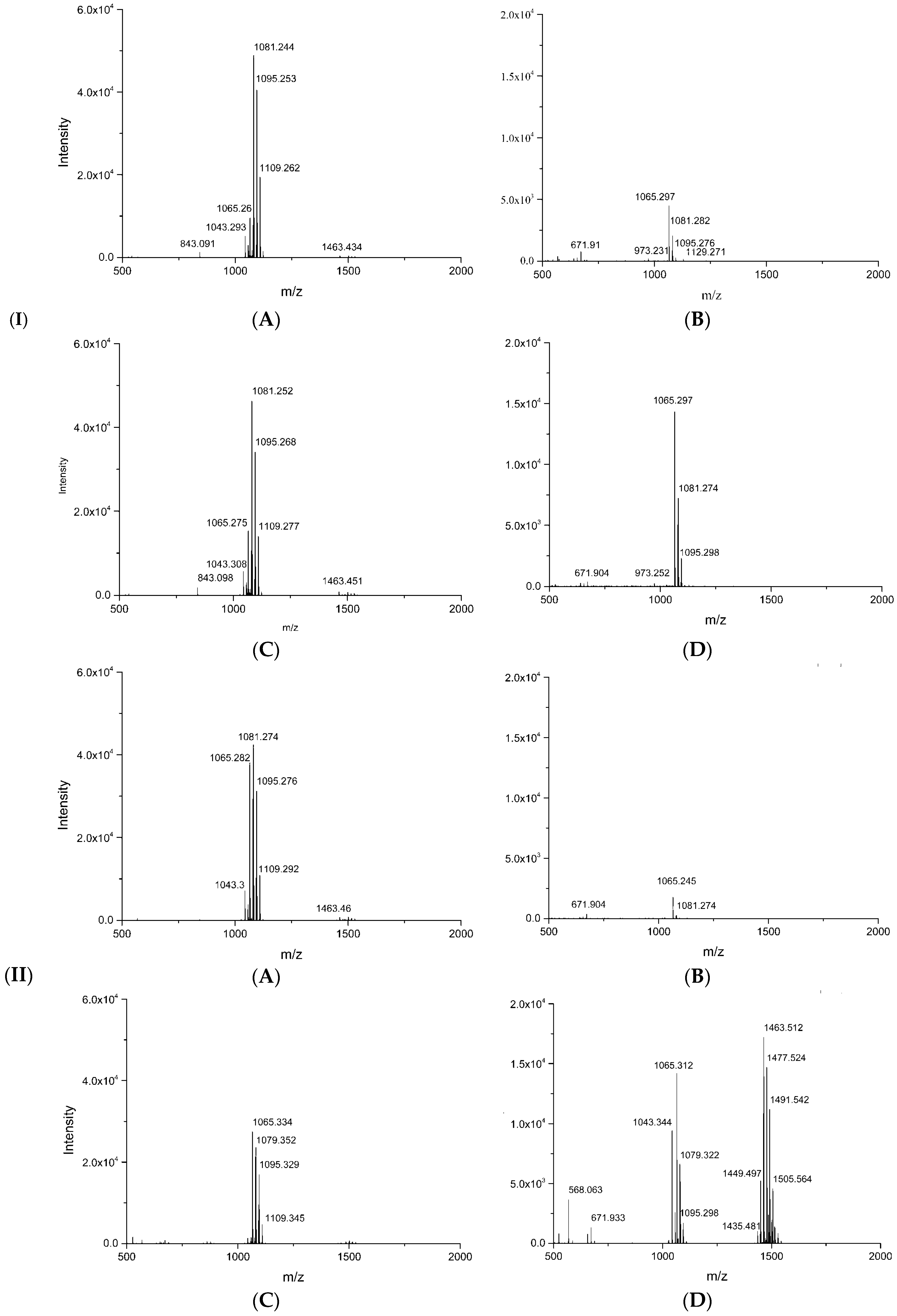
MALDI-TOF-MS analysis of secondary compounds synthesized by strain B1 in cells and inhibition zones could identify those active compounds with antifungal properties. Two main groups of mass peaks were detected in the MALDI-TOF-MS spectra (Figure 7). The literature search indicated that compounds with the most clusters of peaks could be attributed to lipopeptides, including surfactin (C14 to C15), iturin (C14 to C17) and fengycin (C14 to C17) [36,38] (Table 4).
In the co-culture of strain B1 and B. dothidea, the antifungal activity was initially observed after 2 d, but the apparent inhibition zone appeared at 4 d. MALDI-TOF-MS spectra of the cell extracts of strain B1 revealed that lipopeptide production remained relatively stable during the growth periods of strain B1, except that relative intensities of iturin isoforms changed (Figure 7(IA,B,IIA,B)). However, the production and diffusion of lipopeptides were significantly altered in response to B. dothidea, especially in the late stage of coculture (Figure 7(IC,D,IIC,D)). Analyzing the lipopeptide profiles of the inhibition zone, it was noticed that iturins appeared at all times tested (2 and 4 d), while fengycins and surfactins were only detectable at 4 days of co-culture. In addition, the lipopeptides in the inhibition zone were greatly induced by the pathogen; especially, fengycin isoforms were detected with higher abundance at 4 d of co-culture (Table 5), indicating that the strain B1 was able to perceive signals from the fungus and, in response, promoted the production and diffusion of active antimicrobials from the bacteria colony to the inhibition zone. From these results, we found that iturins contributed to the suppression of B. dothidea at the early stage of incubation, and the stronger antifungal activity coincided with the time at which three lipopeptides were produced, demonstrating that lipopeptides produced by strain B1 had a significant antagonistic effect on B. dothidea.

4. Discussion

The use of biological control agents (BCAs) has emerged as an eco-friendly alternative to the chemical compounds for managing fruit diseases. Plant endophytes have received much attention due to their ability to produce bioactive substances with structural diversity, making them potential antagonists [39,40,41]. For example, Wicaksono et al. [42] found that endophytes from medicinal plant Leptospermum scoparium could be artificially inoculated into kiwifruit plants and control kiwifruit canker disease caused by Pseudomonas syringae pv. actinidae, indicating that endophytes could be transferred to non-host plants and maintain their biocontrol features. Similarly, the endophytic strain B. subtilis R31 isolated from the leaves of Dendrobium orchid had a significant biocontrol effect on fusarium wilt of susceptible banana varieties, providing another typical example of an exogenous endophyte suppressing non-host banana fusarium wilt [14]. Therefore, endophytes from plants have the potential to be developed as promising biocontrol agents.
In our study, the endophytic bacterium strain B1 exhibited remarkable antagonistic activity against Botryosphaeria dothidea and effectively controlled postharvest ring rot in pears. It was identified as Bacillus velezensis based on the phylogenomic analysis of core genes (Figure 3). B. velezensis is closely related to B. amyloliquefaciens and B. siamensis, collectively referred to as the “operational group B. amyloliquefaciens” [34]. While biosynthetic genes capable of producing surfactin, bacillibactin, amylocyclicin and iturin may be a common genomic feature of the B. amyloliquefaciens group [33,34,35], genes responsible for synthesis of macrolactin, bacilysin, difficidin, LCI and fengycin were only identified from the core genome of B. velezensis [33]. Our results were consistent with these reports. B velezensis strain B1 harbored gene clusters responsible for synthesis of lipopeptides (iturin, fengycin, surfactin and bacillibactin), polyketides (macrolactin, bacillaene and difficidin) and bacilysin. These studies indicated that B. velezensis strains have the ability to produce a variety of active metabolites with stronger and broader-spectrum antimicrobial properties compared to B. amyloliquefaciens and B. siamensis strains. Consequently, they have attracted attention as potential BCAs. In accordance with our results, recent studies have verified that endophytic B. velezensis was an important source of antagonist. B. velezensis strain P2-1, isolated from apple branches, significantly inhibited the growth of B. dothidea and reduced the apple ring rot caused by B. dothidea [43]. B. velezensis OEE1, isolated from olive tree roots, showed pronounced inhibition on mycelia growth of Verticillium dahliae and effectively controlled fusarium wilt of olive trees under greenhouse and field conditions [44]. B. velezensis BLE7, isolated from sisal (Agave sisalana), showed a strong biocontrol effect on anthracnose caused by Colletotrichum musae in bananas, reducing disease progression up to 97% compared to the positive control [45].
Among the biocontrol mechanisms of this Bacillus group, cyclic lipopeptides from Bacillus have already been reported as major inhibitors against various phytopathogens [46]. Bacillus species were considered to be the most efficient producers of lipopeptides, including the three main families: surfactin, iturin and fengycin, each consisting of closely related isoforms with differences in peptide amino acid sequences and fatty acid chain lengths [47]. Strain B1 exhibited strong antifungal activity against various plant pathogens and caused a significant inhibition zone for pathogens after 7 days of co-culture, indicating that antifungal compounds could be produced and diffused from the bacteria colony into the inhibition zone. MALDI-TOF-MS analysis demonstrated that lipopeptides were the only compounds detected. Strains incapable of producing lipopeptides, such as B. subtilis 168, were unable to inhibit the pathogen growth [36]; Bacillus mutants lacking the ability to produce surfactin, bacillomycin and fengycin failed to suppress various plant diseases [48]. These findings led us to speculate that one of the antimicrobial mechanisms of B1 might be attributed to the lipopeptide production.
Although lipopeptides were not directly related to the growth of the producing bacteria, the synthesis of lipopeptide species could change in different growth periods [36]. Surfactins and bacillomycins were the major lipopeptides in strain FZB42, peaking at different incubation times [36]. Contrary to these observations, our study revealed that lipopeptide production remained relatively stable during the growth periods of strain B1, except for changes in the relative intensities of iturin isoforms. However, the production and diffusion of lipopeptides were significantly induced in response to B. dothidea, especially in the late stage of coculture, with all three lipopeptides—surfactins, fengycins and iturins—experiencing significant stimulation. The simultaneous production of three lipopeptides is not entirely usual, as such co-production of multiple yet different lipopeptides is also found in other Bacillus species [36,49]. The simultaneous production of three kinds of lipopeptides might synergistically improve their antimicrobial activities. Thimon et al. [50] reported that co-production of surfactin along with iturin and fengycin was an advantageous characteristic because surfactin could increase the effect of the other lipopeptides. However, Koumoutsi et al. [36] revealed that although B. velezensis FZB42 could co-produce three kinds of lipopeptides, bacillomycin D was the main compound responsible for antifungal activity against F. oxysporum. Similarly, iturin was a primary contributor to inhibited F. oxysporum in B. velezensis Y6 [51]. Another study reported that in B. subtilis UMAF6639 and EA-CB001, fengycin was the major antimicrobial compound against different phytopathogens [52,53]. Therefore, these results revealed that the relative roles of different LPs in antimicrobial activity might be a species-specific reaction between the beneficial microbe and pathogen. To clarify the role of each individual lipopeptide produced by strain B1 in antimicrobial function, further work, such as experiments with the wild type and mutants that are unable to produce lipopeptides, are necessary.

5. Conclusions

The present study demonstrated that the endophytic isolate Bacillus velezensis B1 exhibited a strong inhibitory effect against the pear ring rot pathogen Botryosphaeria dothidea in vitro and in vivo. Strain B1 harbored gene clusters responsible for the production of iturin, fengycin and surfactin serving as potent biocontrol agents against B. dothidea. Notably, the production and diffusion of these lipopeptides in the inhibition zone was significantly enhanced in the presence of B. dothidea. Further studies involving experiments with wild-type strains and lipopeptide-deficient mutants are needed to verify the exact role of each individual lipopeptide in antimicrobial function, and the active antimicrobial lipopeptides could be overproduced through engineering approaches such as promotor engineering, condensation domain engineering and adenylationdomain engineering. This work may provide a safe biological approach to managing pear ring rot.

Supplementary Materials

The following supporting information can be downloaded at: https://www.mdpi.com/article/10.3390/horticulturae9091049/s1. Table S1: General features of the strain B1 genome; Table S2: Secondary metabolite biosynthetic gene clusters identified in the genome of strain B1 with antiSMASH.

Author Contributions

Conceptualization, S.Y., M.S. and L.W.; formal analysis, M.S. and L.Q.; funding acquisition, L.Q. and L.W.; methodology, G.L. and S.Z.; project administration, L.W.; resources, L.W.; validation, Y.Q., P.W. and H.G.; writing—original draft, L.W.; writing—review and editing, S.Y., M.S. and L.W. All authors have read and agreed to the published version of the manuscript.

Funding

This research was funded by Beijing Municipal Natural Science Foundation of China (No. 6162007) and National Natural Science Foundation of China (No. 62175016).

Data Availability Statement

The complete genome sequences of strain B1 have been deposited in the GenBank database under the accession number CP097895.

Conflicts of Interest

The authors declare no conflict of interest.

References

  1. Xu, B.; Zhang, H.; Chen, K.; Xu, Q.; Yao, Y.; Gao, H. Biocontrol of postharvest Rhizopus decay of peaches with Pichia caribbica. Curr. Microbiol. 2013, 67, 255–261. [Google Scholar] [CrossRef]
  2. Qu, H.; Zhao, L.Y.; Zhao, F.C.; Liu, Y.F.; Yang, Z.Y. Biocontrol of gray mold decay in pear by Bacillus amyloliquefaciens strain BA3 and its effect on postharvest quality parameters. Pol. J. Microbiol. 2016, 65, 171–176. [Google Scholar] [CrossRef]
  3. Marsberg, A.; Kemler, M.; Jami, F.; Nagel, J.H.; Postma-Smidt, A.; Naidoo, S.; Wingfield, M.J.; Crous, P.W.; Spatafora, J.W.; Hesse, C.N.; et al. Botryosphaeria dothidea: A latent pathogen of global importance to woody plant health. Mol. Plant Pathol. 2017, 18, 477–488. [Google Scholar] [CrossRef]
  4. Noguchi, H.; Hiruma, M.; Matsumoto, T.; Kano, R.; Tanaka, M.; Yaguchi, T.; Sonoda, K.; Ihn, H. Fungal melanonychia: Ungual phaeohyphomycosis caused by Botryosphaeria dothidea. Acta Derm. Venereol. 2017, 97, 765–766. [Google Scholar] [CrossRef]
  5. Pour, F.N.; Ferreira, V.; Félix, C.; Serôdio, J.; Alves, A.; Duarte, A.S.; Esteves, A.C. Effect of temperature on the phytotoxicity and cytotoxicity of Botryosphaeriaceae fungi. Fungal Biol. 2020, 124, 571–578. [Google Scholar] [CrossRef]
  6. Andrić, S.; Meyer, T.; Ongena, M. Bacillus responses to plant-associated fungal and bacterial communities. Front. Microbiol. 2020, 11, 1350. [Google Scholar] [CrossRef]
  7. Arnaouteli, S.; Bamford, N.C.; Stanley-Wall, N.R.; Kovács, Á.T. Bacillus subtilis biofilm formation and social interactions. Nat. Rev. Microbiol. 2021, 19, 600–614. [Google Scholar] [CrossRef]
  8. Erdogan, O.; Benlioglu, K. Biological control of Verticillium wilt on cotton by the use of fluorescent Pseudomonas spp. under field conditions. Biol. Control 2010, 53, 39–45. [Google Scholar] [CrossRef]
  9. Luo, J.; Ran, W.; Hu, J.; Yang, X.; Xu, Y.; Shen, Q. Application of bio-organic fertilizer significantly affected fungal diversity of soils. Soil Sci. Soc. Am. J. 2010, 74, 2039–2048. [Google Scholar] [CrossRef]
  10. Mizumoto, S.; Hirai, M.; Shoda, M. Enhanced iturin A production by Bacillus subtilis and its effect on suppression of the plant pathogen Rhizoctonia solani. Appl. Microbiol. Biotechnol. 2007, 75, 1267–1274. [Google Scholar] [CrossRef]
  11. Bhore, S.J.; Preveena, J.; Kandasamy, K.I. Isolation and identification of bacterial endophytes from pharmaceutical agarwood-producing Aquilaria species. Pharmacogn. Res. 2013, 5, 134–137. [Google Scholar] [CrossRef]
  12. Brader, G.; Stephane, C.; Birgit, M.; Friederike, T.; Angela, S. Metabolic potential of endophytic bacteria. Curr. Opin. Biotechnol. 2014, 27, 30–37. [Google Scholar] [CrossRef]
  13. Christina, A.; Christapher, V.; Bhore, S.J. Endophytic bacteria as a source of novel antibiotics: An overview. Pharmacogn. Rev. 2013, 7, 11–16. [Google Scholar]
  14. Li, C.; Cheng, P.; Zheng, L.; Li, Y.; Chen, Y.; Wen, S.; Yu, G. Comparative genomics analysis of two banana fusarium wilt biocontrol endophytes Bacillus subtilis R31 and TR21 provides insights into their differences on phytobeneficial trait. Genomics 2021, 1139, 900–909. [Google Scholar] [CrossRef]
  15. Ryan, R.P.; Germaine, K.; Franks, A.; Ryan, D.J.; Dowling, D.N. Bacterial endophytes: Recent developments and applications. FEMS Microbiol. Lett. 2008, 278, 1–9. [Google Scholar] [CrossRef]
  16. Liu, Y.; Wang, R.H.; Cao, Y.H.; Chen, C.Y.; Bai, F.R.; Xu, T.J.; Zhao, R.; Zhang, X.; Zhao, J.R.; Cheng, C. Identification and antagonistic activity of endophytic bacterial strain Paenibacillus sp. 5 L8 isolated from the seeds of maize (Zea mays L., Jingke 968). Ann. Microbiol. 2016, 66, 653–660. [Google Scholar] [CrossRef]
  17. Chen, S.T.; Dai, J.; Song, X.W.; Jiang, X.P.; Zhao, Q.; Sun, C.B.; Chen, C.W.; Chen, N.F.; Han, B.X. Endophytic microbiota comparison of Dendrobium huoshanense root and stem in different growth years. Planta Med. 2020, 86, 967–975. [Google Scholar] [CrossRef]
  18. Yue, J.Y.; Yu, Y. Isolation and identification of endophytic fungi from Dendrobium huoshanense with their antibacterial and anti-inflammatory activities. Pak. J. Pharm. Sci. 2022, 35, 1143–1151. [Google Scholar]
  19. Zhu, Y.J.; Kong, Y.C.; Hong, Y.; Zhang, L.; Li, S.M.; Hou, S.R.; Chen, X.B.; Xie, T.; Hu, Y.; Wang, X.C. Huoshanmycins A–C, new polyketide dimers produced by endophytic Streptomyces sp. HS-3-L-1 from Dendrobium huoshanense. Front. Chem. 2022, 9, 807508. [Google Scholar] [CrossRef]
  20. Wu, L.Q.; Shang, H.Z.; Wang, Q.; Gu, H.K.; Liu, G.J.; Yang, S.L. Isolation and characterization of antagonistic endophytes from Dendrobium candidum Wall ex Lindl., and the biofertilizing potential of a novel Pseudomonas saponiphila strain. Appl. Soil Ecol. 2016, 105, 101–108. [Google Scholar] [CrossRef]
  21. Wu, L.Q.; Shang, H.Z.; Gu, H.K.; Zheng, J. Bacterial iturins mediate biocontrol activity of Bacillus sp. against postharvest pear fruitrotting fungi. J. Phytopathol. 2019, 167, 501–509. [Google Scholar] [CrossRef]
  22. Kurtz, S.; Phillippy, A.; Delcher, A.L.; Smoot, M.; Shumway, M.; Antonescu, C.; Salzberg, S.L. Versatile and open software for comparing large genomes. Genome Biol. 2004, 5, R12. [Google Scholar] [CrossRef] [PubMed]
  23. Harris, R.S. Improved Pairwise Alignment of Genomic DNA. Ph.D. Thesis, The Pennsylvania State University, University Park, PA, USA, 2007. [Google Scholar]
  24. Li, W.; Godzik, A. Cd-hit: A fast program for clustering and comparing large sets of protein or nucleotide sequences. Bioinformatics 2006, 22, 1658–1659. [Google Scholar] [CrossRef] [PubMed]
  25. Edgar, R.C. MUSCLE: Multiple sequence alignment with high accuracy and high throughput. Nucleic Acids Res. 2004, 32, 1792–1797. [Google Scholar] [CrossRef] [PubMed]
  26. Vilella, A.J.; Severin, J.; Ureta-Vidal, A.; Heng, L.; Durbin, R.; Birney, E. EnsemblCompara Gene Trees: Complete, duplication-aware phylogenetic trees in vertebrates. Genome Res. 2009, 19, 327–335. [Google Scholar] [CrossRef]
  27. Blin, K.; Shaw, S.; Kloosterman, A.M.; Charlop-Powers, Z.; van Wezel, G.P.; Medema, M.H.; Weber, T. antiSMASH 6.0: Improving cluster detection and comparison capabilities. Nucleic Acids Res. 2021, 49, W29–W35. [Google Scholar] [CrossRef]
  28. Borriss, R.; Chen, X.H.; Rueckert, C.; Blom, J.; Becker, A.; Baumgarth, B.; Fan, B.; Pukall, R.; Schumann, P.; Spröer, C.; et al. Relationship of Bacillus amyloliquefaciens clades associated with strains DSM7T and FZB42T: A proposal for Bacillus amyloliquefaciens subsp. amyloliquefaciens subsp. nov. and Bacillus amyloliquefaciens subsp. plantarum sub sp. nov. based on complete genome sequence comparisons. Int. J. Syst. Evol. Microbiol. 2011, 61, 1786–1801. [Google Scholar]
  29. Belbahri, L.; ChenariBouket, A.; Rekik, I.; Alenezi, F.N.; Vallat, A.; Luptakova, L.; Petrovova, E.; Oszako, T.; Cherrad, S.; Vacher, S.; et al. Comparative Genomics of Bacillus amyloliquefaciens strains reveals a core genome with traits for habitat adaptation and a secondary metabolites rich accessory genome. Front. Microbiol. 2017, 8, 1438. [Google Scholar] [CrossRef]
  30. Rooney, A.P.; Price, N.P.; Ehrhardt, C.; Swezey, J.L.; Bannan, J.D. Phylogeny and molecular taxonomy of the Bacillus subtilis species complex and description of Bacillus subtilis subsp. Inaquosorum subsp. nov. Int. J. Syst. Evol. Microbiol. 2009, 59, 2429–2436. [Google Scholar] [CrossRef]
  31. Dunlap, C.A.; Kim, S.J.; Kwon, S.W.; Rooney, A.P. Bacillus velezensis is not a later heterotypic synonym of Bacillus amyloliquefaciens; Bacillus methylotrophicus, Bacillus amyloliquefaciens subsp. plantarum and ‘Bacillus oryzicola’ are later heterotypic synonyms of Bacillus velezensis based on phylogenomics. Int. J. Syst. Evol. Microbiol. 2016, 66, 1212–1217. [Google Scholar]
  32. Rabbee, M.F.; Ali, M.S.; Choi, J.; Hwang, B.S.; Jeong, S.C.; Baek, K.H. Bacillus velezensis: A valuable member of bioactive molecules within plant microbiomes. Molecules 2019, 224, 1046. [Google Scholar] [CrossRef] [PubMed]
  33. Chun, B.H.; Kim, K.H.; Jeong, S.E.; Jeon, C.O. Genomic and metabolic features of the Bacillus amyloliquefaciens group–B. amyloliquefaciens, B. velezensis, and B. siamensis–revealed by pan-genome analysis. Food Microbiol. 2019, 77, 146–157. [Google Scholar] [PubMed]
  34. Fan, B.; Blom, J.; Klenk, H.P.; Borriss, R. Bacillus amyloliquefaciens, Bacillus velezensis, and Bacillus siamensis form an “operational group B. Amyloliquefaciens” within the B. Subtilis species complex. Front. Microbiol. 2017, 8, 3843. [Google Scholar] [CrossRef] [PubMed]
  35. Palazzini, J.M.; Dunlap, C.A.; Bowman, M.J.; Chulze, S.N. Bacillus velezensis RC 218 as a biocontrol agent to reduce Fusarium head blight and deoxynivalenol accumulation: Genome sequencing and secondary metabolite cluster profiles. Microbiol. Res. 2016, 192, 30–36. [Google Scholar] [CrossRef]
  36. Koumoutsi, A.; Chen, X.H.; Henne, A.; Liesegang, H.; Hitzeroth, G.; Franke, P.L.; Vater, J.; Borriss, R. Structural and functional characterization of gene clusters directing nonribosomal synthesis of bioactive cyclic lipopeptides in Bacillus amyloliquefaciens Strain FZB42. J. Bacteriol. 2004, 186, 1084–1096. [Google Scholar] [CrossRef] [PubMed]
  37. Duitman, E.H.; Hamoen, L.W.; Rembold, M.; Venema, G.; Seitz, H.; Saenger, W.; Bernhard, F.; Reinhardt, R.; Schmidt, M.; Ulrich, C.; et al. The mycosubtilin synthetase of Bacillus subtilis ATCC 6633: A multifunctional hybrid between a peptide synthetase, an amino transferase, and a fatty acid synthase. Proc. Natl. Acad. Sci. USA 1999, 96, 13294–13299. [Google Scholar] [CrossRef] [PubMed]
  38. Vater, J.; Kablitz, B.; Wilde, C.; Franke, P.; Mehta, N.; Cameotra, S.S. Matrix-assisted laser desorption ionization–time of flight mass spectrometry of lipopeptide biosurfactants in whole cells and culture filtrates of Bacillus subtilis C-1 isolated from petroleum sludge. Appl. Environ. Microbiol. 2002, 68, 6210–6219. [Google Scholar]
  39. Shao, Y.; Yan, H.; Yin, T.; Sun, Z.; Xie, H.; Song, L.; Sun, K.; Li, W. New azaphilones from Penicillium variabile, a fungal endophyte from roots of Aconitum vilmorinianum. J. Antibiot. 2020, 73, 77–81. [Google Scholar] [CrossRef]
  40. Shiono, Y.; Yokoi, M.; Koseki, T.; Murayama, T.; Aburai, N.; Kimura, K. Allantopyrone A, a new α-pyrone metabolite with potent cytotoxicity from an endophytic fungus, Allantophomopsis lycopodina KS-97. J. Antibiot. 2010, 63, 251–253. [Google Scholar] [CrossRef]
  41. Yang, Z.; Wu, K.; Xu, Y.; Xia, X.; Wang, X.; Ge, M.; Shao, L. Three novel chromanones with biological activities from the endophytic fungus Phomopsis CGMCC No. 5416. J. Antibiot. 2020, 73, 194–199. [Google Scholar]
  42. Wicaksono, W.A.; Jones, E.E.; Casonato, S.; Monk, J.; Ridgway, H.J. Biological control of Pseudomonas syringae pv. Actinidiae (Psa), the causal agent of bacterial canker of kiwifruit, using endophytic bacteria recovered from a medicinal plant. Biol. Control 2018, 116, 103–112. [Google Scholar] [CrossRef]
  43. Yuan, H.; Shi, B.; Wang, L.; Huang, T.; Zhou, Z.; Hou, H.; Tu, H. Isolation and characterization of Bacillus velezensis strain P2-1 for biocontrol of apple postharvest decay caused by Botryosphaeria dothidea. Front. Microbiol. 2022, 12, 808938. [Google Scholar]
  44. Azabou, M.C.; Gharbi, Y.; Medhioub, I.; Ennouri, K.; Barham, H.; Tounsi, S.; Triki, M.A. The endophytic strain Bacillus velezensis OEE1: An efficient biocontrol agent against Verticillium wilt of olive and a potential plant growth promoting bacteria. Biol. Control 2020, 142, 104168. [Google Scholar]
  45. Damasceno, C.L.; Duarte, E.A.A.; Santos, L.B.P.R.D.; Oliveira, T.A.S.D.; Jesus, F.N.D.; Oliveira, L.M.D.; Góes-Neto, A.; Soares, A.C.F. Postharvest biocontrol of anthracnose in bananas by endophytic and soil rhizosphere bacteria associated with sisal (agave sisalana) in brazil. Biol. Control 2019, 137, 104016. [Google Scholar]
  46. Mnif, I.; Ghribi, D. Review lipopeptides biosurfactants: Mean classes and new insights for industrial, biomedical, and environmental applications. Biopolymers 2015, 104, 129–147. [Google Scholar] [PubMed]
  47. Rautela, R.; Singh, A.K.; Shukla, A.; Cameotra, S.S. Lipopeptides from Bacillus strain AR2 inhibits biofilm formation by Candida albicans. Antonie Leeuwenhoek. 2014, 105, 809–821. [Google Scholar] [CrossRef]
  48. Li, B.; Li, Q.; Xu, Z.; Zhang, N.; Shen, Q.; Zhang, R. Responses of beneficial Bacillus amyloliquefaciens SQR9 to different soil borne fungal pathogens through the alteration of antifungal compounds production. Front. Microbiol. 2014, 5, 636. [Google Scholar] [CrossRef]
  49. Stein, T. Bacillus subtilis antibiotics: Structures, syntheses and specific functions. Mol. Microbiol. 2005, 56, 845–857. [Google Scholar]
  50. Thimon, L.; Peypoux, F.; Dana Maget, R.; Roux, B.; Michel, G. Interactions of bioactive lipopeptides, iturin A and surfactin from Bacillus subtilis. Biotechnol. Appl. Biochem. 1992, 16, 144–151. [Google Scholar]
  51. Cao, Y.; Pi, H.L.; Chandrangsu, P.; Li, Y.T.; Wang, Y.Q.; Zhou, H.; Xiong, H.Q.; Helmann, J.D.; Cai, Y.F. Antagonism of two plant-growth promoting Bacillus velezensis isolates against Ralstonia solanacearum and Fusarium oxysporum. Sci. Rep. 2018, 8, 4360. [Google Scholar]
  52. Gonzalez-Jaramillo, L.M.; Aranda, F.J.; Teruel, J.A.; Villegas-Escobar, V.; Ortiz, A. Antimycotic activity of fengycin C biosurfactant and its interaction with phosphatidylcholine model membranes. Colloids Surf. B Biointerfaces 2017, 156, 114–122. [Google Scholar] [PubMed]
  53. Romero, D.; De, V.A.; Rakotoaly, R.H.; Dufour, S.E.; Veening, J.W.; Arrebola, E.; Cazorla, F.M.; Kuipers, O.P.; Paquot, M.; Perez-Garcia, A. The iturin and fengycin families of lipopeptides are key factors in antagonism of Bacillus subtilis toward Podosphaerafusca. Mol. Plant Microbe Interact. 2007, 20, 430–440. [Google Scholar] [CrossRef] [PubMed]
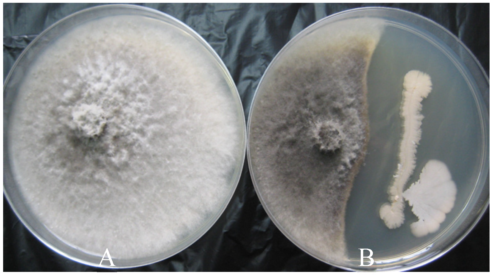
Figure 1. In vitro inhibition of Botryosphaeria dothidea by strain B1 after seven days of co-incubation on PDA. (A): control, (B): B. dothidea co-incubation with strain B1.
Horticulturae 09 01049 g001
Figure 2. Light microscopy observations of the changes in hyphal morphology of B. dothidea mycelium after seven days of co-incubation with B1 on PDA. (A): Treated mycelium of B. dothidea with strain B1, (B): control mycelium.
Horticulturae 09 01049 g002
Figure 3. Effect of antifungal compounds from strain B1 on B. dothidea growth. CK: BPN broth; CS: cell suspension; CFS: cell-free supernatant. Photographs were taken following seven days incubation on PDA medium at 28 °C.
Horticulturae 09 01049 g003
Figure 4. Biocontrol activity of strain B1 against B. dothidea on wounded pears. Wounded pear fruit treated with strain B1 (B1) and water (CK). Photographs were taken after 7 days of incubation at 25 °C.
Horticulturae 09 01049 g004
Figure 5. Phylogenomic tree of the strain B1 based on the core genomes.
Horticulturae 09 01049 g005
Figure 6. BGC organization of the bacillomycin D or mycosubtilin and fengycin or plipastatinoperon in strain B1 genome. Gene clusters within the solid rectangle, exhibited 100% similarity to gene cluster synthesizing bacillomycin D or mycosubtilin; Gene clusters within dashed rectangle, exhibited 100% similarity to gene cluster synthesizing fengycin or plipastatin.
Horticulturae 09 01049 g006
Figure 7. MALDI-TOF-MS analysis of lipopeptides from strain B1 after 2 (I) and 4 (II) days of culturing. Lipopeptides from bacterial colony in presence (C) or absence (A) of B. dothidea and from inhibition zone in presence (D) or absence (B) of B. dothidea.
Horticulturae 09 01049 g007
Table 1. In vitro inhibition of B. dothidea by CS and CFS of strain B1.
TreatmentInhibition Zones (cm) *
CS1.30 ±0.07 a
CFS0.52 ± 0.06 b
Different letters in the same column indicate significantly different results (p < 0.05). * The value represented the mean ± standard deviation of three replicates.
Table 2. Assessment of antifungal spectrum of strain B1.
PathogensInhibition Zone/cm
Alternaria alstroemeriae1.5 ± 0.14
Verticillium dahliae2.4 ± 0.28
Rhizoctonia solani1.2 ± 0.13
Alternaria brassicae1.3 ± 0.12
Botrytis cinerea1.2 ± 0.21
Fusarium graminaerum1.3 ± 0.22
Fusarium oxysporum0.4 ± 0.02
Pyricularia oryzae2.0 ± 0.23
Table 3. Incidence and severity of ring rot in wounded pear fruit treated with strain B1 and water (CK). The trials were performed at 25 °C, and disease was assessed 3, 5 and 7 days after B. dothidea inoculation. Different letters in the same column indicate significant difference (p < 0.05) between treatments according to Student’s t test. All data were recorded as means ± standard deviation of three replicates.
Treatment3d5d7d
Rot Diameter (cm)Inhibition Rate (%)Rot Diameter (cm)Inhibition Rate (%)Rot Diameter (cm)Inhibition Rate (%)
CK1.3 ± 0.3 a 2.81 ± 0.7 a 4.31 ± 1.1 a
B10 b1000 b1001.3 ± 0.6 b73.4 ± 18.6
Table 4. Assignments of lipopeptide mass peaks of strain B1 detected with MALDI-TOF-MS [36,38].
Mass Peak (m/z)FamilyAssignment
1044.3, 1060.3SurfactinC14Surfactin [M + Na, K]+
1058.3, 1074.3C15Surfactin [M + Na, K]+
1043.3, 1065.3IturinC14Iturin A/C14Mycosubtilin [M + H, Na]+
1057.3, 1079.3C15Iturin A/C15Mycosubtilin [M + H, Na]+
1081.3C14Iturin A/C14Mycosubtilin [M + K]+/C16Bacillomycin D [M + Na]+
1095.3C15Iturin A/C15Mycosubtilin [M + K]+/C17Bacillomycin D [M + Na]+
1109.3C16Iturin A/C16Mycosubtilin [M + K]+
1435.5FengycinAla-6-C14 Fengycin [M + H]+
1449.5Ala-6-C15 Fengycin [M + H]+
1463.5, 1485.5, 1501.5Ala-6-C16 Fengycin [M + H, Na, K]+
1477.5, 1515.5Ala-6-C17 Fengycin [M +H, K]+
1491.5, 1529.5Val-6-C16 Fengycin [M + H, K]+
1505.5Val-6-C17 Fengycin [M + H]+
Table 5. Lipopeptide production and diffusion from strain B1in the inhibition zone after 4 days of culturing.
TreatmentMass Peak (m/z)AssignmentIntensity
Control1065.3C14Iturin A/C14Mycosubtilin [M + Na]+1745
1079.3C15 Iturin A/C15Mycosubtilin [M + Na]+237
1081.3C14Iturin A/C14Mycosubtilin [M + K]+/C16Bacillomycin D [M + Na]+285
Dual culture1044.3C14Surfactin [M + Na]+3976
1058.3C15Surfactin [M + Na]+893
1043.3C14Iturin A/C14Mycosubtilin [M + H]+9431
1057.3C15 Iturin A/C15Mycosubtilin [M + H]+2561
1065.3C14IturinA/C14Mycosubtilin [M + Na]+14,196
1079.3C15 Iturin A/C15Mycosubtilin [M + Na]+6615
1081.3C14Iturin A/C14Mycosubtilin [M + K]+/C16Bacillomycin D [M + Na]+5150
1095.3C15 Iturin A/C15Mycosubtilin [M + K]+/C17Bacillomycin D [M + Na]+1714
1435.5Ala-6-C14 Fengycin [M + H]+1020
1449.5Ala-6-C15 Fengycin [M + H]+5251
1463.5Ala-6-C16 Fengycin [M + H]+17,214
1477.5Ala-6-C17 Fengycin [M + H]+14,696
1485.5Ala-6-C16 Fengycin [M + Na]+2368
1491.5Val-6-C16 Fengycin [M + H]+11,158
1501.5Ala-6-C16 Fengycin [M + K]+1943
1505.5Val -6-C17 Fengycin [M + H]+4584
1515.5Ala-6-C17 Fengycin [M + K]+1288
1529.5Val-6-C16 Fengycin [M + K]+825
Disclaimer/Publisher’s Note: The statements, opinions and data contained in all publications are solely those of the individual author(s) and contributor(s) and not of MDPI and/or the editor(s). MDPI and/or the editor(s) disclaim responsibility for any injury to people or property resulting from any ideas, methods, instructions or products referred to in the content.