Article

Preparation, Characterization, and Application of Citrate-Functionalized Cobalt-Doped Iron Oxide Nanoparticles for Rhodamine Dye and Lead Ion Sequestration

by
Sangeetha Jayakumar
1,
Barid Baran Lahiri
1,2,* and
Arup Dasgupta
1,2
1
Smart Materials Section, Physical Metallurgy Division, Materials Characterization Group, Metallurgy and Materials Group, Indira Gandhi Centre for Atomic Research, Kalpakkam 603102, India
2
Homi Bhabha National Institute, Training School Complex, Anushaktinagar, Mumbai 400094, India
*
Author to whom correspondence should be addressed.
Magnetochemistry 2025, 11(4), 24; https://doi.org/10.3390/magnetochemistry11040024
Submission received: 26 February 2025 / Revised: 23 March 2025 / Accepted: 26 March 2025 / Published: 29 March 2025
(This article belongs to the Special Issue Applications of Magnetic Materials in Water Treatment)

Abstract

:
The toxicity of hazardous dyes like rhodamine B and heavy metal ions like lead warrants the need for wastewater remediation. We describe here the functionalization of cobalt-doped iron oxide (Co0.1Fe2.9O4) magnetic nanoparticles (MNPs) with citrate moieties for the effective sequestration of rhodamine B dye and lead ions from contaminated water. Citrate-functionalized MNPs are prepared using a co-precipitation technique. For the uncoated MNPs, the hydrodynamic diameter and zeta potential are found to be 21 nm and ~45 ± 3.1 mV, respectively. The hydrodynamic diameters are found to increase to ~51, ~59, and ~68 nm for the MNPs functionalized with ~20, ~40, and ~60 mg/mL of citrate, respectively, whereas the corresponding zeta potentials are found to be ~−27.95 ± 3.5 mV, ~−32.5 ± 3.6 mV, and ~−33.9 ± 3.5 mV, respectively. The chemisorption of the citrate moieties over the MNPs cause the zeta potential to be negative, a phenomenon which is further verified from the citrate-specific absorption bands in the Fourier transform infrared (FTIR) spectra of the surface-functionalized MNPs. UV-visible spectrophotometry is employed to probe the MNP-aided elimination of rhodamine B dye and lead ions from aqueous media, where the absorption bands at ~554 nm and ~375 nm (for lead (II)-5-dimercapto-1,3,4-thiadiazole chelate) are utilized for quantitative analyses. These citrate-functionalized nanoparticles are found to successfully remove the toxic rhodamine B dye and lead ions from water, with removal efficiencies of ~93.7 ± 2.6% and ~90 ± 2.4%, respectively. The unbound -COO functional groups of the citrate-functionalized MNPs electrostatically interact with the cationic rhodamine B dye or lead (II) ions, thereby leading to the adsorption onto the surface-functionalized MNPs and the subsequent magnetic-field-assisted removal. The experimental findings show the efficacy of the citrate-functionalized cobalt-doped iron oxide MNPs for the sequestration of dye pollutants and lead ions from contaminated water.

1. Introduction

Magnetically polarizable nanometric platforms have found significant applications in various industrial fields, such as toxic chemical sensors [1], optical elements [2], defect sensors [3], water purification [4], catalysis [5], and in numerous biomedical fields, including magnetic fluid hyperthermia [4,6], drug delivery [7], magnetic resonance imaging [8], etc. In recent years, wastewater treatment via magnetic nanoparticles (MNPs) for the sequestration of dye pollutants has gained momentum [9,10,11].
The most common techniques for the removal of heavy metals and hazardous dyes include ion exchange [12], photocatalytic degradation [13], hydrodynamic cavitation [14], membrane separation [15], and adsorption [16,17]. Amongst these, the adsorption-based technique has attracted much interest because of its efficiency, environmental friendliness, and ease of operation [18]. Currently, magnetic nanoabsorbents, such as magnetic nanoparticles (MNPs) [19,20,21,22,23,24] and magnetic metal–oxide frameworks [25,26], are the most promising candidates for wastewater remediation due to various advantages, such as the ease of removal of dye-laden nanostructures using an external magnetic fields, the availability of a large fraction of adsorption sites, and the potential for regeneration and reuse [27,28].
Amongst various types of magnetic nanoabsorbents for wastewater treatment, the spinel ferrite MNPs are advantageous for external magnetic field-guided control and reduced secondary pollution [10,29,30]. Spinel ferrites are metal–oxide nanoparticles with a general formula AB2O4, where the metal cations occupy the tetrahedral (A) and octahedral (B) sites at varied ratios. In an inverse spinel structure (like magnetite, cobalt ferrite, etc.), the trivalent metal ion shares the A and B sites, whereas the divalent metal ion occupies the B sites only [31]. Though iron oxide magnetic nanoparticles (MNPs) are the most widely exploited spinel ferrites for wastewater remediation [32,33], several other types of spinel ferrites are also used as nanoadsorbents, such as nickel ferrite MNPs [34], zinc ferrite MNPs [35], copper ferrite MNPs [36], cobalt ferrite MNPs [37], and manganese ferrite MNPs [38]. The strong magneto-crystalline anisotropy energy density, moderately high saturation magnetization, and high coercivity of CoFe2O4 MNPs cause them to rapidly respond to an external magnetic field, a phenomenon that is suitable for external magnetic-field-assisted quick separation during wastewater treatment [39]. Al-Wasidi and Abdelrahman have reported the use of CoFe2O4 MNPs for indigo carmine dye removal with an efficiency of ~83% [23]. Iqbal et al. reported good removal efficiencies for CoFe2O4 nanocomposites when treated with wastewater containing heavy metals and various dyes such as crystal violet, brilliant green, methyl orange, and Congo red [37]. Further, cobalt ferrite MNPs-aided enhanced removal efficiencies were reported for Remazol red and methylene blue dyes by Ibrahim et al. [20], and for crystal violet and red 88 dyes by Al-Wasidi et al. [36]. The adsorption and removal of rhodamine B dye (efficiency ~89%) and lead (II) ions (efficiency ~42–82%) using biotin and lawsone-functionalized cobalt-doped iron oxide MNPs was reported by Sangeetha et al. [19]. Ain et al. [21] reported an anionic azo dye removal efficiency of ~45–82% using amine-functionalized cobalt–iron oxide nanoparticles. Yahya et al. [40] reported the sequestration of Cr and lead (II) ions from tannery waste using CoFe2O4-loaded activated carbon, with corresponding removal efficiencies of ~98%, and ~96%, respectively. Vargas et al. [41] studied the adsorption and removal potential (efficiency ~92–95%) of arsenic [As(III)] ions using cobalt-substituted ferrite nanoparticles. Therefore, cobalt-based magnetic nanoparticles are a versatile system for toxic dye and metal ion removal.
Rhodamine B is a fluorescent xanthene dye which has widespread applications in the leather, paint, ink, and textile industries [42,43], as well as in the biomedical field as a fluorescent marker [44]. In aqueous media, weakly basic and nitrogenous rhodamine B dissociates to form a stable and non-biodegradable colorful cation [25]. According to previous studies, rhodamine B is carcinogenic and mutagenic, and reports indicate exposure-induced infections of the skin, eyes, respiratory organs, and gastrointestinal tracts in human and animals [45]. Rhodamine B has been shown to be hazardous even when present in wastewater at a low concentration of ~1.0 mg/L [46]. In addition to the rhodamine B dye, lead ions are yet another highly toxic wastewater pollutant. Lead, at all concentrations, is considered unsafe [47], and lead toxicity has been reported to result in oxidative stress causing free radical formation and cascading cell damage [48]. Clinical manifestations, such as anemia, have been reported for lead concentrations in the blood >50 μg/dL, whereas higher (>100 μg/dL) lead concentrations may result in encephalopathy, coma, and seizures [48]. Further, lead has been reported to accumulate in the human body through absorption, and can interfere with the neurological, cardiovascular, renal, hematopoietic, and reproductive systems. The high toxicity of rhodamine B dye and lead ions warrants the need for wastewater remediation, a factor that motivated the present investigation.
Though numerous types of surface-functionalizing moieties have been reported for nanoadsorbents, citrate moieties are particularly useful because of the three -COO groups, which aid in the electrostatic interactions with the dye and heavy metal pollutants. Earlier studies on citrate-functionalized MNPs have revealed superior removal efficiencies for heavy metals like Cd(II) and Cr(VI) [49]. However, an in-depth understanding on the stable functionalization of magnetic nanoparticles for multiple sequestration still exists as a research gap. In the present study, the stable functionalization of cobalt-doped iron oxide nanoparticles with citrate moieties at room temperature is demonstrated. The citrate-functionalized magnetic nanoparticles are used for the sequestration of rhodamine B dye and lead ions from water. Further, the superior magneto-structural properties of the cobalt-doped iron oxide nanoparticles make post-treatment retrieval of the MNPs easier.

2. Materials and Methods

2.1. Materials

The divalent cobalt salt (CoCl2·6H2O), the trivalent (FeCl3·6H2O) and divalent (FeSO4·7H2O) iron salts, acetone, HNO3, HCl, and ammonium hydroxide (30%) were obtained from E-Merck. Trisodium citrate, rhodamine B, lead (II) nitrate salt, and 2,5-dimercapto-1,3,4-thiadiazole (DMTD) were procured from Sigma Aldrich. All chemicals were of GR grade and were used without additional purification. Milli-Q water (resistivity ~18 MΩcm−1) was utilized for sample preparation.

2.2. Synthesis, Surface Functionalization, and Characterization of the Co-Doped Iron Oxide MNPs

Cobalt-doped iron oxide nanoparticles (Co0.1Fe2.9O4) were synthesized following a chemical co-precipitation technique described previously [19,50]. Briefly, 1M solution of the trivalent iron salt and 0.5M solution of the divalent iron and cobalt salts were freshly prepared and mixed in an optimized proportion to maintain the stoichiometric ratio of Co:Fe = 0.1:2.9 [50]. NH4OH solution (~30%) was added to the mixture, which was then maintained at ~85 °C for ~30 min under constant stirring at ~800 rpm. The synthesized magnetic nanoparticles (MNPs) were collected through magnetic decantation, rinsed multiple times with Milli-Q water, and subsequently dried at 150 °C for 30 min in a vacuum oven.
The dried MNPs were treated with a few drops of 2M HNO3 and left for ~180 min [51], after which the MNPs were rinsed with Milli-Q water and redispersed in an aqueous medium (pH: 6–6.4), and horn-sonicated for 30 min. The nitric acid treatment imparted a positive charge to the MNPs, leading to an electrostatic stabilization in the aqueous medium, a step that is essential for citrate functionalization [19]. A fixed quantity of the HNO3-treated MNPs was mixed with ~20, 40, and 60 mg/mL of aqueous solution of sodium citrate, and the mixtures were incubated for 48 h under constant stirring at 400 rpm. Afterward, the citrate-functionalized MNPs were gathered through magnetic decantation and rinsed twice with Milli-Q water to eliminate excess surfactant. The citrate-functionalized MNPs were ultimately redispersed in water and used for additional studies.
The citrate-functionalized MNPs were characterized using dynamic light scattering (DLS), thermogravimetric analysis (TGA), Fourier transform infrared (FTIR) spectroscopy, powder X-ray diffraction (XRD), vibrating sample magnetometry (VSM), transmission electron microscopy (TEM), and phase contrast optical microscopy. The relevant description of the characterization techniques is provided in Section S1 of the Supplementary Information.

2.3. Rhodamine B and Lead (II) Ions Removal Using the Citrate-Functionalized MNPs

To prepare a stock solution of ~1 mg/mL concentration, ~10 mg of rhodamine B (RhB) was dissolved in ~10 mL of Milli-Q water. RhB has a strong absorbance at ~554 nm and, therefore, the dye concentration was determined spectrophotometrically by making use of a LabIndia make UV-visible (~200–700 nm) spectrophotometer (model no. 3200+). To obtain a standard plot, the absorbances of RhB were measured at varied concentrations, viz. ~0.1, ~0.2, ~0.4, ~0.6, ~0.8, ~1, ~2, ~4, ~6, ~8, and ~10 µg/mL. Aqueous solutions of RhB (at a concentration of ~10 µg/mL) were treated with ~3 mg of uncoated and citrate-functionalized MNPs (with a citrate concentration of ~20 mg/mL). This was followed by separation of the supernatant by magnetic decantation and ~20 min of centrifugation at ~6000 rpm, utilizing a Remi C-24 centrifuge. The concentration of RhB in the supernatant was estimated from the standard plot. The removal efficiency (Re, in %) of RhB was estimated using the following equation, where ci and cf indicate the initial (before the treatment with the MNPs) and final (after the treatment with the MNPs) dye concentrations, respectively [19].
R e   ( in   % ) = ( c i c f ) c i × 100
An aqueous solution of lead (II) nitrate salt was used as the source of lead ions, and a stock solution with a concentration of ~1 mg/mL was prepared and supplemented with ~1 mL of diluted HNO3 to delay hydrolysis. To obtain varied lead concentration references, the stock solution was subsequently diluted to ~0.1, ~0.2, ~0.4, ~0.6, ~0.8, ~1, ~2, ~4, ~6, ~8, and ~10 µg/mL concentrations. The absorbance values of lead (II) nitrate at varied concentrations were measured using a previously reported protocol [19,52], where chelation with 2,5-dimercapto-1,3,4-thiadiazole (DMTD) was allowed and the resultant exhibited a distinct absorption at ~375 nm. A stock solution with a concentration of ~1 mg/mL was prepared by dissolving ~200 mg of DMTD in Milli-Q water. Subsequently, ~1 mL of lead reference concentration was mixed with ~1 mL of the stock DMTD solution and ~1 mL of ~5 mM HCl was added to the mixture. Finally, the mixture was diluted to ~10 mL by adding the required amount of Milli-Q water. A standard plot was obtained by varying the lead concentration from ~0.1 µg/mL to ~10 µg/mL. For probing the lead ion removal efficiency, the slightly acidic, DMTD-mixed lead solution (with a concentration of ~10 µg/mL) was treated with ~3 mg of uncoated and citrate-functionalized MNPs (with a citrate concentration of ~20 mg/mL). Subsequently, the supernatant was separated using magnetic decantation and centrifugation, followed by spectrophotometric determination of the lead concentration using the standard plot and the removal efficiency using Equation (1).

3. Results and Discussions

3.1. Physicochemical Properties of the Citrate-Functionalized MNPs

For the acid-treated uncoated MNPs, Figure 1a illustrates the pH-dependent variations of the zeta potential. It was observed that the zeta potential was positive for acidic pH and rapidly decreased to a negative value for the alkaline conditions, a phenomenon that is in agreement with the previous findings of Carlson and Kawatra [53]. The HNO3 treatment imparted a positive surface charge on the MNPs, causing the zeta potential to become positive [19,51]. The estimated isoelectric point (IEP) of the uncoated MNPs was at a pH of ~8.5 (Figure 1a), which is consistent with the previously reported pH range of ~7.0–8.4, especially for air-oxidized MNPs [54]. Earlier studies have reported citrate-stabilization of iron oxide MNPs at an acidic pH of ~2.5 (adjusted using perchloric acid and NaOH) [51] or at a pH of ~5.5 (adjusted using 0.1 N HCl) [49]. On the other hand, citrate functionalization of the MNPs was performed in the current study at a slightly acidic condition (pH ~6–6.4 < IEP) to ensure proper electrostatic interaction-mediated attachment between the negatively charged citrate moieties and the positively charged MNPs. Figure 1b compares the zeta potentials for the uncoated and ~20, ~40, and ~60 mg/mL citrate-functionalized MNPs. The zeta potential of the uncoated MNPs was ~45 ± 3.1 mV, whereas the zeta potentials of the citrate-functionalized MNPs were ~−27.95 ± 3.5, ~−32.5 ± 3.6, and ~−33.9 ± 3.5 mV for the ~20, ~40, and ~60 mg/mL, respectively. In agreement with previous results, the reversal of the zeta potential demonstrated the attachment between the citrate moieties and the MNPs [55,56]. It has been reported that, for pH values > 3.5, the unbound carboxylate groups of citrate moieties are deprotonated, thereby imparting a negative zeta potential to the citrate-functionalized MNPs [51,56]. Reports indicate that, for a zeta potential >±20 mV, the aqueous dispersions exhibited colloidal stability, whereas the colloidal stability is superior for zeta potentials ≥ 30 mV [57]. Hence, the citrate-functionalized MNPs exhibit good colloidal stability in the aqueous medium. The increase in zeta potential with citrate concentrations (Figure 1b) was due to the higher surface density of the adsorbed moieties. Figure 1c shows the distributions of the hydrodynamic diameters (dh) for the uncoated MNPs and for the ~20, ~40, and ~60 mg/mL citrate-functionalized MNPs. For the uncoated MNPs, the dh was ~21 ± 5 nm. On the other hand, the dh values were ~51 ± 6, ~59 ± 4, and ~68 ± 4 nm for the MNPs functionalized with ~20, 40, and 60 mg/mL of citrate moieties, respectively. The increase in dh for the citrate-functionalized MNPs can be attributed to the existence of hydrophilic citrate moieties and the possibility of slight agglomeration, probably mediated through hydrogen bonding [19,58]. Moreover, dh increased with citrate concentration, a phenomenon that is in agreement with a previous report showing a similar concentration-dependent increase in dh for lawsone-functionalized MNPs [19].
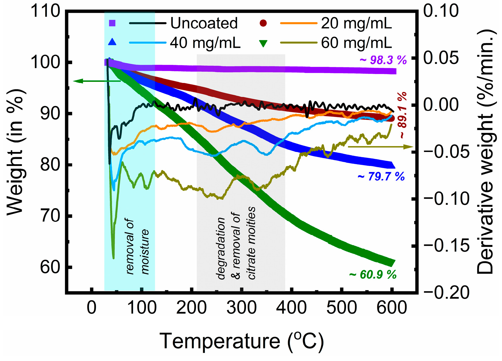
Figure 2 displays the temperature-dependent weight changes (in %) for the uncoated MNPs and MNPs functionalized with citrate concentrations of ~20, ~40, and ~60 mg/mL. The derivative weight data are also shown in the figure. Up to ~100 °C, the uncoated MNPs exhibited a minor weight loss of ~1%, which was due to the removal of water. Up to ~600 °C, the uncoated MNPs showed a minor weight loss of ~1.7%, without any discernible weight loss beyond ~120 °C. The elimination of absorbed moisture was responsible for the observed initial weight losses of ~2.8%, ~3.6%, and ~5.6% for the ~20, ~40, and ~60 mg/mL citrate-functionalized MNPs, respectively. The removal of physically and chemically bound water molecules primarily occurred in the temperature range of ~30–120 °C (light-blue-shaded in Figure 2), where a single broad peak or multiple isolated peaks were observed in the derivative weight data for the uncoated or citrate-functionalized MNPs. With broad peaks centered at ~242–261 °C and ~332–351 °C in the derivative weight data, the most significant weight loss for the citrate-functionalized MNPs was observed in the temperature range of ~200–400 °C (the grey-shaded region in Figure 2). This was attributed to the degradation and subsequent removal of the heat-labile citrate moieties bound to the MNPs and is in agreement with the previously reported primary degradation temperature ranges of ~147–315 °C [59] and ~200–400 °C [60] for citrate-functionalized MNPs. Trisodium citrate has been reported to exhibit a three-stage weight loss in the temperature range of ~30–600 °C [59], whereas in the present study, the weight loss for the citrate-functionalized MNPs was not abrupt, but occurred over an extended temperature range. Such gradual weight losses have been previously reported for citrate-functionalized MNPs [55,60], and probably indicated strong interactions between the citrate moieties and the MNPs. At 600 °C, the weight losses amounted to ~10.9%, ~20.3%, and ~39.1% for the ~20, ~40, and ~60 mg/mL citrate-functionalized MNPs, respectively. From the weight losses of the uncoated MNPs (~1.7%) and ~20 mg/mL citrate-functionalized MNPs (~10.9%), the amount of citrate present was estimated at ~9.2%, which is remarkably close to the previously reported value of ~9.2% of citrate content for ~10.9 ± 2.6 nm sized citrate-functionalized iron oxide MNPs [61]. The citrate contents were estimated at ~18.6% and ~37.4% for the ~40 and ~60 mg/mL citrate-functionalized MNPs, respectively. The weight loss increased with the citrate concentration due to the availability of excess surface stabilizing agent, a phenomenon that is in agreement with the observations made from the dynamic light scattering studies, where the zeta potential (Figure 1b) and hydrodynamic diameter (Figure 1c) increased with the citrate concentration. The moderately high zeta potential and unimodal hydrodynamic diameter distribution for the ~20 mg/mL citrate-functionalized MNPs ensured good colloidal stability without a significant loss of magnetic properties. Hence, the rhodamine B dye and lead ion removal studies were performed using the ~20 mg/mL citrate-functionalized MNPs.
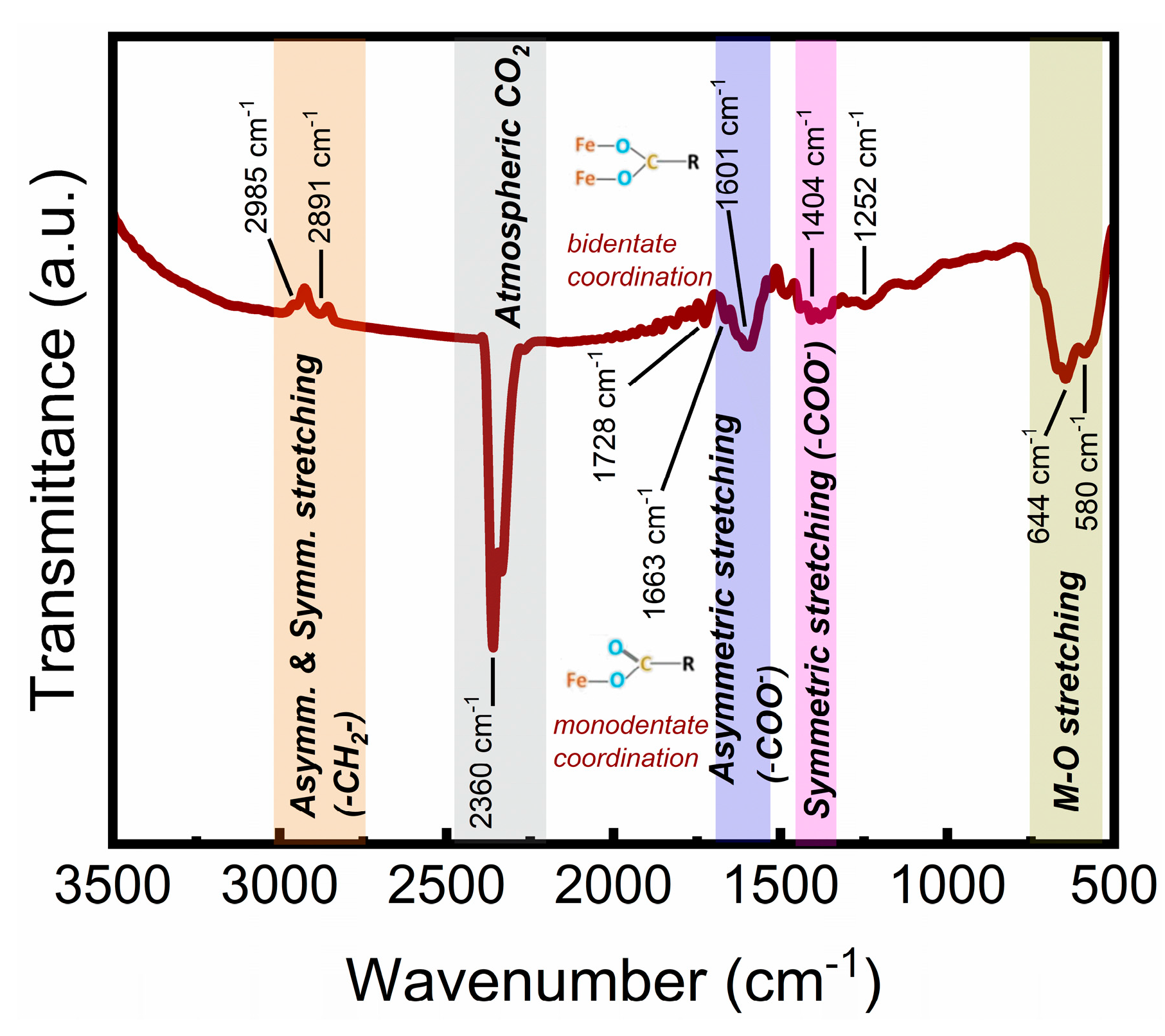
Dynamic light scattering studies revealed an inversion of the zeta potential and an increased hydrodynamic diameter of the citrate-functionalized MNPs, indicating the adherence of the citrate moieties to the MNPs, a phenomenon that was further confirmed by the FTIR spectra of the citrate-functionalized MNPs, as shown in Figure 3. The ambient CO2 caused the absorption band at ~2360 cm−1, whereas the metal–oxygen stretching within the MNPs resulted in absorption bands at ~580 and ~644 cm−1 [62]. The absorption bands at ~2985 and ~2891 cm−1 were due to the asymmetric and symmetric stretching of the -CH2- groups, respectively [63]. The absorption band at ~1404 cm−1 corresponded to the symmetric stretching of the -COO groups [49,63]. On the other hand, the asymmetric stretching of the -COO groups resulted in two absorption bands at ~1601 and ~1663 cm−1 [49,55]. The corresponding wavenumber differences ( Δ ν = ν a s y m m ν s y m m ) were ~197 and ~259 cm−1, thereby indicating bridging bidentate- and monodentate-type coordination between the metal atom and the citrate moieties, respectively [64]. Most of the earlier studies have predominantly reported monodentate-type coordination between citrate and MNP surface with Δ ν ~236–246 cm−1 [49,55,63]. However, a few previous studies have identified the formation of bidentate-type coordination in addition to the monodentate coordination for citrate-transition metal–ion complexes [65,66], where the monodentate coordination has been reported to be a precursor for the bidentate-type coordination [65]. For trisodium citrate, the asymmetric and symmetric stretching resulted in absorptions at ~1591 and ~1399 cm−1, respectively, indicating ~10–72 and 5 cm−1 shifts in the corresponding absorption bands for the citrate-functionalized MNPs, a phenomenon that was due to the chemisorption of the carboxyl groups [55]. The absorption bands corresponding to ~1728 and ~1252 cm−1 were attributed to the C=O [65] and C-OH [65] stretching of the -COOH radical, respectively. Citric acid has three carboxylic acid groups with distinct pKa values: pKa1~3.13, pKa2~4.76, and pKa3~6.4 [65]. In the current study, citrate functionalization was performed at pH values of ~6–6.4, and, most likely, one of the carboxylic acid groups remained protonated (-COOH), resulting in the abovementioned absorptions. The FTIR spectra provided compelling evidence supporting the citrate functionalization of the MNPs.
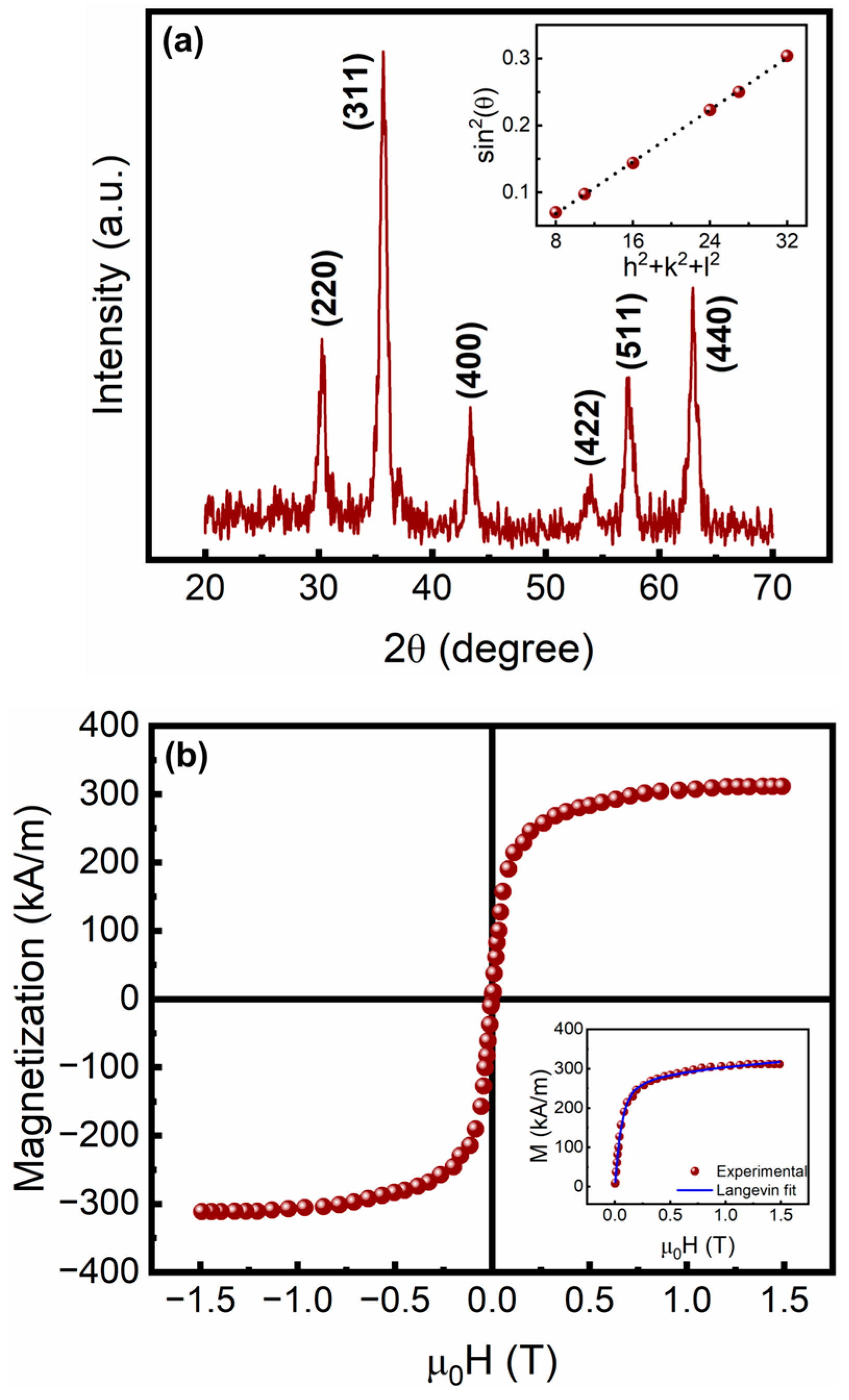
The Bragg reflections from the (220), (311), (400), (422), (511), and (440) crystal planes were clearly visible in the XRD pattern of the citrate-functionalized MNPs (Figure 4a), indicating a spinel ferrite structure (JCPDS Card No. 88-0315, for Fe3O4 [50]). The slightly broadened diffraction patterns were attributed to the nanometric dimension of the MNPs. By using Scherrer’s equation for the most intense (311) peak, the average crystallite size was estimated at ~12 ± 1 nm. From the slope of the linear regression analysis (adj. R2 ~0.99) between sin2(θ) and (h2 + k2 + l2), as shown in the inset in Figure 4a, the lattice parameter (a) was estimated at ~8.341 ± 0.006 Å, which is in agreement with the previously reported values ranging from ~8.33 to 8.36 Å [50]. Figure 4b depicts the magnetization data for the citrate-functionalized MNPs at ~300 K, where the saturation magnetization (Ms) was ~311 kA/m (~60 emu/g). Due to the non-magnetic citrate layer, the Ms was slightly lower than the earlier reported Ms of ~69 emu/g for the uncoated cobalt-doped iron oxide MNPs [50]. The smaller size of the MNPs increased the surface-to-volume ratio, resulting in a higher surface spin disorder [67] which caused the Ms to become lower compared to the bulk Ms of Fe3O4 (~90–92 emu/g [68]) or CoFe2O4 (~80 emu/g [69]). Moreover, Figure 4b shows no hysteresis opening, indicating that the synthesized MNPs were superparamagnetic, a phenomenon that was due to the smaller crystallite size (~12 ± 1 nm) compared to the superparamagnetic size limit (~20 nm [70]) for Fe3O4. A Langevin-type expression (Equation (2)) was used to fit the ~300 K M-H data [4].
M ( H ) = c 1 M s [ coth ( c 2 H ) 1 c 2 H ] + c 3 χ p H
Here, χ p ~ 1.1 × 10 4   emu / g - Oe and Ms, H, and c1c3 indicate the saturation magnetization, magnetic field, and the fitting parameters, respectively [4]. The experimental data and the fitted curve are shown in the inset in Figure 4b. The magnetic domain size was calculated as ~9.9 ± 1.6 nm, which is similar to the crystallite size, demonstrating that the MNPs were most likely single-domain in nature.
A typical TEM image depicting the roughly spherical morphology of the citrate-functionalized MNPs is shown in Figure 5a, and Figure 5b shows the corresponding size distribution, along with the log-normal fit: Y = Y 0 + A 2 π σ x exp [ ( ln ( x ) ln ( x c ) ) 2 2 σ 2 ] , where Y0, A, σ, and xc are the fit parameters and the regression analysis (adj. R2~0.8) yielded an xc of ~10.92 ± 0.29 nm and a σ of ~0.149 ± 0.034. The following expressions were used to determine the average particle size ( d p ) and the standard deviation ( σ d p ) [71].
d p = x c exp [ σ 2 2 ]
σ d p = d p [ e x p ( σ 2 ) 1 ]
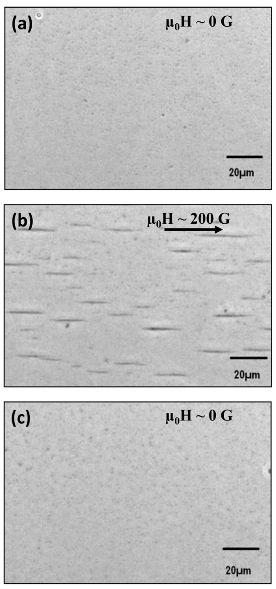
The estimated average particle size was ~11.0 ± 1.7 nm, which is in agreement with the typical crystallite size obtained from the XRD analysis. Figure 6a–c show the PCOM images of the citrate-functionalized MNPs at µ0H ~0 G, at µ0H ~200 G (indicated by an arrow in Figure 6b), and after field removal (µ0H ~0 G), respectively. The MNPs formed chain-like structures parallel to the external magnetic field (Figure 6b) which were not discernible for µ0H ~0 G (Figure 6a). Over time, the chain grew longitudinally and laterally due to zippering, leading to thicker micron-sized chains that were observable through optical microscopy, as seen in Figure 6b. It was further observed from Figure 6c that the chain-like structures disappeared after the removal of the magnetic field, indicating the reversibility of the chain formation. Field-induced reversible chain formation in magnetic nanofluids has been previously reported using optical light scattering [2,72], phase contrast microscopy [2,19], and atomic force microscopy [73] techniques. The citrate-functionalized MNPs were colloidally stable and retained significant magnetization to respond to an external magnetic field, as demonstrated by the reversible chain formation induced by an external magnetic field (Figure 6a–c) which was utilized for rhodamine B and lead ion removal from an aqueous medium.

3.2. Citrate-Functionalized MNPs for the Sequestration of Rhodamine B Dye and Lead (II) Ions

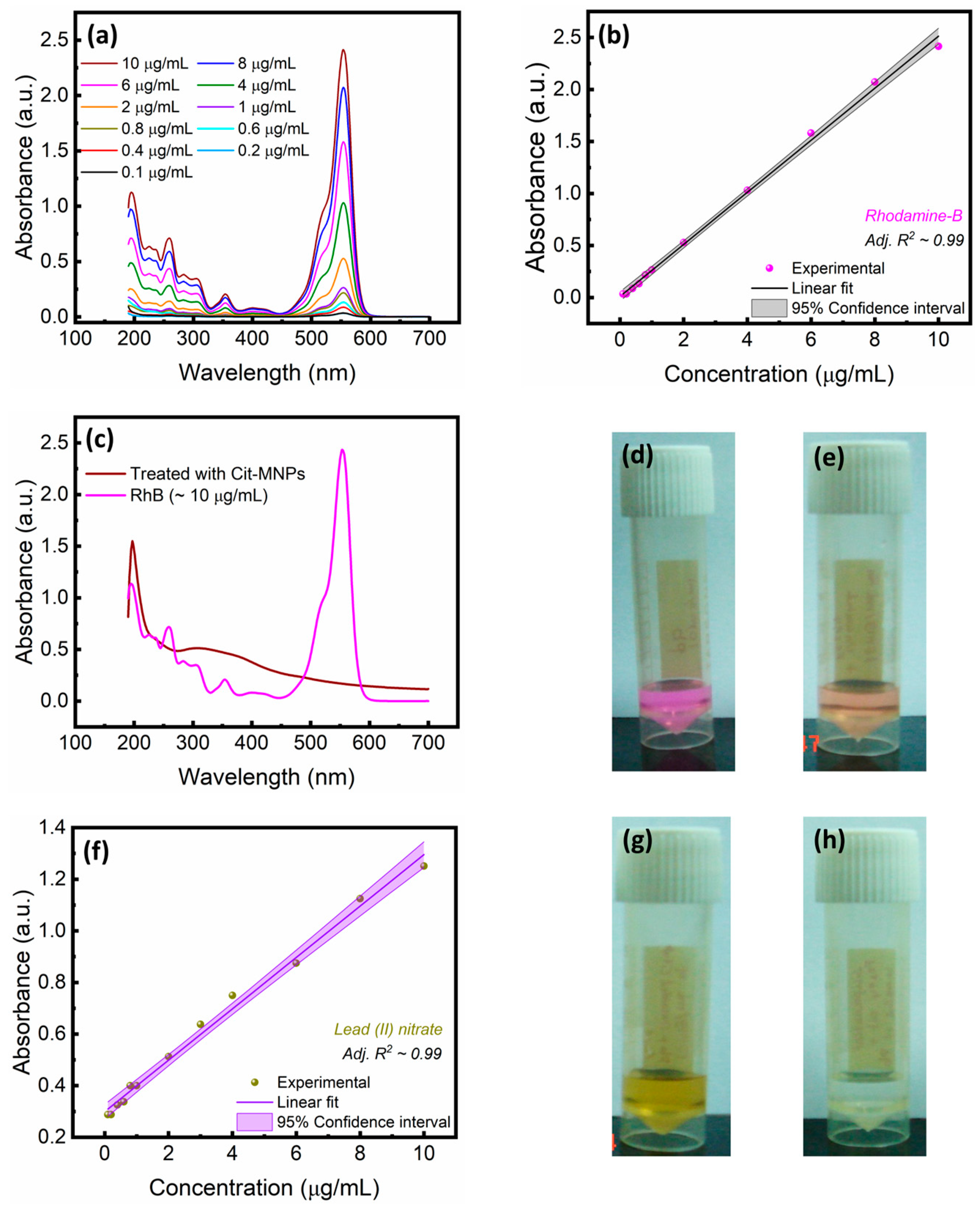
Rhodamine B (molecular formula: C28H31N2O3Cl) is a basic dye that exists in the di-protonated form at acidic pH values, whereas the carboxylic acid group is deprotonated at higher pH values to give rise to the zwitterionic form [74]. Rhodamine B (RhB) exhibits five absorption bands in the UV-visible spectra, viz. ~210 nm, ~245 nm, ~350 nm, ~450 nm, and ~554 nm, with the last being the primary absorption band, thereby aiding in the spectrophotometric determination of the RhB concentration [19,75,76]. The UV-visible spectra of the aqueous solutions containing different concentrations of RhB are displayed in Figure 7a. The UV-visible spectra showed five absorption bands corresponding to ~195 nm, ~259 nm, ~353 nm, and ~554 nm, along with a shoulder at ~52 nm. The strongest absorption was at ~554 nm, and, therefore, the absorbance at ~554 nm was utilized for the spectrophotometric determination of the RhB concentration. Figure 7a illustrates the highest absorbance for the ~10 µg/mL RhB concentration, whereas the absorbance progressively decreased for lower dye concentrations. Figure 7b shows the standard plot for absorbance as a function of RhB concentration, along with the corresponding calibration equation (adj. R2~0.99). Figure 7c shows the UV-visible absorption spectra for an aqueous solution of RhB (with a dye concentration of ~10 µg/mL) and the supernatant of an aqueous dye solution treated with citrate-functionalized MNPs (with a citrate concentration of ~20 mg/mL), where it was observed that the absorbance was considerably lower for the supernatant of the aqueous dye solution treated with citrate-functionalized MNPs. Utilizing the calibration equation, the RhB concentration in the supernatant was determined as ~0.631 µg/mL, indicating a ~93.7 ± 2.6% removal efficiency, which is marginally better than the previously obtained values of ~89.0 ± 0.1% and ~89.0 ± 0.8% for cobalt-doped iron oxide MNPs surface-functionalized with biotin and lawsone, respectively [19]. This, as discussed subsequently, was ascribed to the three -COOH groups present in the citrate moieties that augmented the adsorption of the dye molecules. Figure 7d,e display the photographs of the untreated dye solution and of the supernatant of the dye solution treated with the citrate-functionalized MNPs, respectively. The color intensity decreased for the supernatant of the MNP-treated aqueous dye solution, showing that the citrate-functionalized MNPs interacted with RhB and that the adsorbed dye molecules were subsequently removed along with the MNPs in the presence of an external magnetic field. The findings were well corroborated by the spectrophotometry data (Figure 7c) and showed the suitability of the citrate-functionalized MNPs for the efficient removal of RhB.
The tetrahydroxy-p-benzoquinone, dithizone, and tetrahydroxy quinone methods are utilized for the colorimetric determination of trace amounts of lead [77,78,79]. On the other hand, the 5-dimercapto-1,3,4-thiadiazole (DMTD) technique is suitable for the determination of lead ions in industrial and environmental samples [52,80,81]. In the present study, the DMTD method was utilized, and the chelation reaction was carried out following a protocol reported by Ahmed and Mamun [52], where DMTD acts as a potentiometric reagent for the quantitative analysis of lead ions. When DMTD and lead (II) ions react under slightly acidic conditions, a greenish-yellow chelation product is formed, suggesting the binding of the mercapto group with lead (II) ions to form the [Pb(DMTD)2] complex with an absorbance maximum at ~375 nm [52,80]. The UV-visible spectra of aqueous solutions containing varied concentrations of lead (II) nitrate salts were measured, from which a standard plot was obtained, as shown in Figure 7f along with the calibration equation (adj. R2 ~0.99). Utilizing the calibration equation, the lead ion concentration in the supernatant was determined as ~1.0 µg/mL, indicating a removal efficiency of ~90 ± 2.4%, which is significantly higher than the ~42 ± 1% removal efficiency obtained for biotin-functionalized cobalt-doped iron oxide MNPs and is comparable to the removal efficiency of ~82 ± 9.8% reported for lawsone-functionalized cobalt-doped iron oxide MNPs [19]. Figure 7g,h show the typical photographs of the untreated lead (II)-DMTD solution and the supernatant of the MNP-treated lead (II)-DMTD solution, respectively. The greenish-yellow color is clearly visible in Figure 7g, indicating the presence of lead ions, whereas the color intensity significantly decreased in the supernatant of the MNP-treated solution (Figure 7h), indicating the suitability of the citrate-functionalized MNPs for the removal of lead (II) ions. However, further studies are in progress to ascertain the adsorption kinetics, adsorption isotherms, and regeneration properties of the citrate-functionalized cobalt-doped iron oxide MNPs, such studies being essential to gain an in-depth understanding of the adsorption efficacy [82,83].
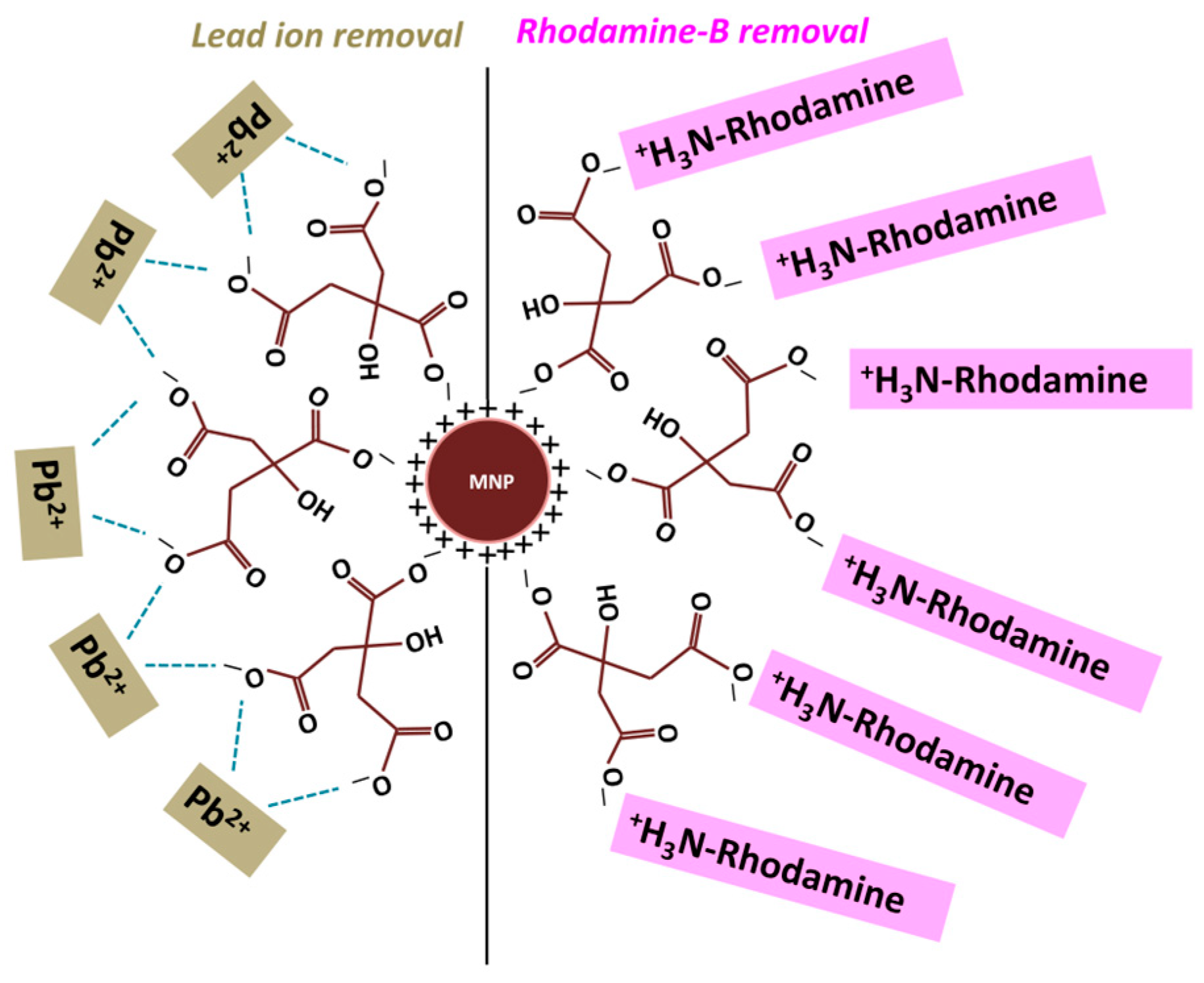
The probable mode of interaction among the citrate-functionalized MNPs, RhB dye molecules, and lead (II) ions is schematically depicted in Figure 8. As observed from the FTIR spectra (Figure 3), the citrate moieties were chemisorbed onto the surface of the MNPs via the carboxylate ions. Depending on pka2 < pH < pka3 or pH > pka3, either one or two -COO groups of the bound citrate moieties were then free to electrostatically interact with the cationic RhB dye molecules or lead (II) ions, as shown in Figure 8. Therefore, when an external magnetic field is applied, the RhB dye or the lead (II) ions, adsorbed onto the MNP surface, were readily separated from the aqueous medium.
Additional studies to probe the steric hindrance effect of the larger RhB dye molecules, the probability of dynamic switching of the double bonds of the carboxylate groups during interactions with the lead (II) ions, the roles of salt concentrations, and pH variations in relation to electrostatic interactions are required to gain an in-depth understanding of the adsorption mechanism [84]. Nevertheless, our experimental results clearly show the suitability of the citrate-functionalized cobalt-doped iron oxide MNPs for the sequestration of RhB dye and lead (II) ions from aqueous media.

4. Conclusions

Using a wet chemical approach, citrate-functionalized cobalt-doped iron oxide MNPs (with a typical crystallite size of ~12 ± 1 nm) were prepared. Room temperature magnetization studies indicated a saturation magnetization of ~311 kA/m without significant hysteresis openings. The as-synthesized MNPs exhibited a zeta potential of ~45 ± 3.1 mV, whereas the zeta potentials were found to be ~−27.95 ± 3.5 mV, ~−32.5 ± 3.6 mV, and ~−33.9 ± 3.5 mV for the MNPs surface-functionalized with ~20, 40, and 60 mg/mL of citrate moieties. This was also accompanied by an increase in the hydrodynamic diameters to ~51, ~59, and ~68 nm, respectively, from ~21 nm (for the as-synthesized MNPs). The attachment of the citrate moieties to the surface of the MNPs was evident from the reversal of the zeta potential, as for pH values > 3.5, the unbound carboxylate groups of the citrate moieties were deprotonated, thereby imparting a negative zeta potential to the citrate-functionalized MNPs. Further, the FTIR spectra, showing the absorption bands corresponding to the symmetric (at ~1404 cm−1) and asymmetric (at ~1601–1663 cm−1) stretching vibrations of the carboxylate group, clearly indicated that the citrate moieties were chemisorbed onto the surface of the MNPs. Thermogravimetric analysis showed ~1.7% and ~10.9% weight losses for the uncoated and ~20 mg/mL citrate-functionalized MNPs, indicating that the non-magnetic citrate coating increased the weight by ~9.2%. Phase contrast microscopy studies revealed a reversible alignment of the MNPs with an external magnetic field, such a phenomenon being due to the electrostatic-interaction-mediated good colloidal stability in the aqueous medium. Spectrophotometric studies indicated ~93.7 ± 2.6% and ~90 ± 2.4% removal efficiencies for the rhodamine B dye and lead ions, respectively, from the aqueous medium, utilizing the citrate-functionalized MNPs, where the unbound -COO groups electrostatically interacted with the dye molecules and the lead (II) ions, leading to an efficient adsorption and subsequent magnetic-field-assisted removal. The findings clearly demonstrate that the citrate-functionalized cobalt-doped iron oxide MNPs are suitable for the sequestration of dye pollutants like rhodamine B and toxic metal ions from wastewater.

Supplementary Materials

The following supporting information can be downloaded at: https://www.mdpi.com/article/10.3390/magnetochemistry11040024/s1, Section S1: Characterization of the citrate-functionalized MNPs.

Author Contributions

Conceptualization, S.J. and B.B.L.; methodology, S.J.; formal analysis, B.B.L.; investigation, S.J.; writing—original draft preparation, S.J. and B.B.L.; writing—review and editing, S.J., B.B.L. and A.D.; visualization, B.B.L.; supervision, A.D. All authors have read and agreed to the published version of the manuscript.

Funding

This research received no external funding.

Institutional Review Board Statement

Not applicable.

Informed Consent Statement

Not applicable.

Data Availability Statement

The raw data supporting the conclusions of this article will be made available by the corresponding author upon reasonable request.

Acknowledgments

The authors thank Anish Kumar, R. Divakar, and Shri C. G. Karhadkar for their encouragement and support.

Conflicts of Interest

The authors declare no conflicts of interest.

References

  1. Soliz, J.R.; Ranjit, S.; Phillips, J.J.; Rosenberg, R.A.; Hauser, A.J. Magnetic and impedance analysis of Fe2O3 nanoparticles for chemical warfare agent sensing applications. Magnetochemistry 2023, 9, 206. [Google Scholar] [CrossRef]
  2. Brojabasi, S.; Mahendran, V.; Lahiri, B.B.; Philip, J. Near infrared light absorption in magnetic nanoemulsion under external magnetic field. Opt. Commun. 2014, 323, 54–60. [Google Scholar] [CrossRef]
  3. Nandy, M.; Lahiri, B.B.; Philip, J. Visual detection of defects in carbon steel using magnetic nanoemulsions: Effect of stabilizing moieties on the defect detection sensitivity. Sens. Actuators A 2020, 314, 112220. [Google Scholar] [CrossRef]
  4. Mahendravada, S.; Lahiri, B.B.; Jayakumar, S.; Sathyanarayana, A.T.; Vetrivendan, E.; Khan, F.; Vidya, R.; Philip, J.; Dasgupta, A. Hyaluronic acid-coated iron oxide nanoparticles for magnetic fluid hyperthermia and methylene blue dye removal: Preparation, physicochemical characterization, and enhancing the heating efficiency. J. Mol. Liq. 2024, 415, 126314. [Google Scholar] [CrossRef]
  5. Raimundo, R.A.; Silva, V.D.; Ferreira, L.S.; Loureiro, F.J.A.; Fagg, D.P.; Macedo, D.A.; Gomes, U.U.; Soares, M.M.; Gomes, R.M.; Morales, M.A. NiFe alloy nanoparticles tuning the structure, magnetism, and application for oxygen evolution reaction catalysis. Magnetochemistry 2023, 9, 201. [Google Scholar] [CrossRef]
  6. Dutz, S.; Buske, N.; Landers, J.; Grafe, C.; Wende, H.; Clement, J.H. Biocompatible magnetic fluids of Co-doped iron oxide nanoparticles with tunable magnetic properties. Nanomaterials 2020, 10, 1019. [Google Scholar] [CrossRef]
  7. Soetaert, F.; Korangath, P.; Serantes, D.; Fiering, S.; Ivkov, R. Cancer therapy with iron oxide nanoparticles: Agents of thermal and immune therapies. Adv. Drug Deliv. Rev. 2020, 163–164, 65–83. [Google Scholar] [CrossRef]
  8. Korolev, D.V.; Shulmeyster, G.A.; Istomina, M.S.; Nikiforov, A.I.; Aleksandrov, I.V.; Semenov, V.G.; Galagudza, M.M. Indocyanine green-containing magnetic liposomes for constant magnetic field-guided targeted delivery and theranostics. Magnetochemistry 2022, 8, 127. [Google Scholar] [CrossRef]
  9. Harja, M.; Lupu, N.; Chiriac, H.; Herea, D.-D.; Buema, G. Studies on the removal of congo red dye by an adsorbent based on fly-ash@Fe3O4 mixture. Magnetochemistry 2022, 8, 125. [Google Scholar] [CrossRef]
  10. Moosavi, S.; Lai, C.W.; Gan, S.; Zamiri, G.; Pivehzhani, O.A.; Johan, M.R. Application of efficient magnetic particles and activated carbon for dye removal from wastewater. ACS Omega 2020, 5, 20684–20697. [Google Scholar] [CrossRef]
  11. Daffalla, S.; Da’na, E.; Taha, A.; El-Aassar, M.R. Synthesis of a novel magnetic biochar from lemon peels via impregnation-pyrolysis for the removal of methyl orange from wastewater. Magnetochemistry 2024, 10, 95. [Google Scholar] [CrossRef]
  12. Sgreccia, E.; Rogalska, C.; Gonzalez, F.S.G.; Prosposito, P.; Burratti, L.; Knauth, P.; Vona, M.L.D. Heavy metal decontamination by ion exchange polymers for water purification: Counterintuitive cation removal by an anion exchange polymer. J. Mater. Sci. 2024, 59, 2776–2787. [Google Scholar] [CrossRef]
  13. Khan, S.; Noor, T.; Iqbal, N.; Yaqoob, L. Photocatalytic dye degradation from textile wastewater: A review. ACS Omega 2024, 9, 21751–21767. [Google Scholar] [CrossRef]
  14. Arteaga, J.F.M.; Vodnik, D.; Kastelec, D.; Zupanc, M.; Dular, M.; Ortar, J.; Đurić, M.; Kaurin, A.; Mihelic, R.; Lestan, D. Removal of toxic metals from sewage sludge by EDTA and hydrodynamic cavitation and use of the sludge as fertilizer. Sci. Total Environ. 2024, 923, 171444. [Google Scholar] [CrossRef]
  15. Xiang, H.; Min, X.; Tang, C.-J.; Sillanpää, M.; Zhao, F. Recent advances in membrane filtration for heavy metal removal from wastewater: A mini review. J. Water Process Eng. 2022, 49, 103023. [Google Scholar] [CrossRef]
  16. Fato, T.P.; Li, D.-W.; Zhao, L.-J.; Qiu, K.; Long, Y.-T. Simultaneous removal of multiple heavy metal ions from river water using ultrafine mesoporous magnetite nanoparticles. ACS Omega 2019, 4, 7543–7549. [Google Scholar] [CrossRef]
  17. Tahoon, M.A.; Siddeeg, S.M.; Alsaiari, N.S.; Mnif, W.; Rebah, F.B. Effective heavy metals removal from water using nanomaterials: A review. Processes 2020, 8, 645. [Google Scholar] [CrossRef]
  18. Sana, S.S.; Haldhar, R.; Parameswaranpillai, J.; Chavali, M.; Kim, S.-C. Silver nanoparticles-based composite for dye removal: A comprehensive review. Clean. Mater. 2022, 6, 100161. [Google Scholar] [CrossRef]
  19. Sangeetha, J.; Suganthi, V.; Philip, J. Functionalization of cobalt ferrite nanoparticles with biotin and lawsone and their use in rhodamine dye and lead ion removal. J. Nanosci. Nanotechnol. 2017, 17, 5284–5293. [Google Scholar] [CrossRef]
  20. Ibrahim, S.M.; Badawy, A.A.; Essawy, H.A. Improvement of dyes removal from aqueous solution by Nanosized cobalt ferrite treated with humic acid during coprecipitation. J. Nanostrc. Chem. 2019, 9, 281–298. [Google Scholar] [CrossRef]
  21. Khurshid, S.; Gul, Z.; Khatoon, J.; Shah, M.R.; Hamid, I.; Khan, I.A.T.; Aslam, F. Anionic azo dyes removal from water using amine-functionalized cobalt–iron oxide nanoparticles: A comparative time-dependent study and structural optimization towards the removal mechanism. RSC Adv. 2020, 10, 1021–1041. [Google Scholar] [CrossRef]
  22. Muthukumaran, T.; Philip, J. A facile approach to synthesis of cobalt ferrite nanoparticles with a uniform ultrathin layer of silicon carbide for organic dye removal. J. Mol. Liq. 2020, 317, 114110. [Google Scholar] [CrossRef]
  23. Al-Wasidi, A.S.; Abdelrahman, E.A. Simple synthesis and characterization of cobalt ferrite nanoparticles for the successful adsorption of indigo carmine dye from aqueous media. Inorganics 2023, 11, 453. [Google Scholar] [CrossRef]
  24. Ali, N.S.; Khader, E.H.; Khudhur, R.H.; Abdulrahman, M.A.; Salih, I.K.; Albayati, T.M. Removal of anionic azo dye from wastewater using Fe3O4 magnetic nanoparticles adsorbents in a batch system. Desalin. Water Treat. 2024, 317, 100033. [Google Scholar] [CrossRef]
  25. Liu, H.; Ren, X.; Chen, L. Synthesis and characterization of magnetic metal-organic framework for the adsorptive removal of Rhodamine B from aqueous solution. J. Ind. Eng. Chem. 2016, 34, 278–285. [Google Scholar] [CrossRef]
  26. Alsaiari, N.S.; Osman, H.; Amari, A.; Tahoon, M.A. The synthesis of metal–organic-framework-based ternary nanocomposite for the adsorption of organic dyes from aqueous solutions. Magnetochemistry 2022, 8, 133. [Google Scholar] [CrossRef]
  27. Rasheed, T. Magnetic nanomaterials: Greener and sustainable alternatives for the adsorption of hazardous environmental contaminants. J. Clean. Prod. 2022, 362, 132338. [Google Scholar] [CrossRef]
  28. Ghoohestani, E.; Samari, F.; Homaei, A.; Yosuefinejad, S. A facile strategy for preparation of Fe3O4 magnetic nanoparticles using Cordia myxa leaf extract and investigating its adsorption activity in dye removal. Sci. Rep. 2024, 14, 84. [Google Scholar] [CrossRef]
  29. Thabet, R.H.; Fouad, M.K.; Ali, I.A.; Sherbiney, S.A.E.; Tony, M.A. Magnetite-based nanoparticles as an efficient hybrid heterogeneous adsorption/oxidation process for reactive textile dye removal from wastewater matrix. Int. J. Environ. Anal. Chem. 2023, 103, 2636–2658. [Google Scholar] [CrossRef]
  30. Ali, M.A.; Badawy, A.M.; Seliem, A.Q.; Bendary, H.I.; Lima, E.C.; Al-Dossari, M.; EL-Gawaad, N.S.A.; Reis, G.S.; Mobarak, M.; Hassan, A.M.; et al. Macroscopic and microscopic levels of methylene blue adsorption on a magnetic bio-based adsorbent: In-depth study using experiments, advanced modeling, and statistical thermodynamic analysis. Magnetochemistry 2024, 10, 91. [Google Scholar] [CrossRef]
  31. Tsurkan, V.; Von Nidda, H.-A.K.; Deisenhofer, J.; Lunkenheimer, P.; Loidl, A. On the complexity of spinels: Magnetic, electronic, and polar ground states. Phys. Rep. 2021, 926, 1–86. [Google Scholar] [CrossRef]
  32. Panda, S.K.; Aggarwal, I.; Kumar, H.; Prasad, L.; Kumar, A.; Sharma, A.; Vo, D.-V.N.; Thuan, D.V.; Mishra, V. Magnetite nanoparticles as sorbents for dye removal: A review. Environ. Chem. Lett. 2021, 19, 2487–2525. [Google Scholar] [CrossRef]
  33. Ghosh, N.; Sen, S.; Biswas, G.; Saxena, A.; Haldar, P.K. Adsorption and desorption study of reusable magnetic iron oxide nanoparticles modified with Justicia adhatoda leaf extract for the removal of textile dye and antibiotic. Water Air Soil Pollut. 2023, 234, 202. [Google Scholar] [CrossRef]
  34. Khoso, W.A.; Haleem, N.; Baig, M.A.; Jamal, Y. Synthesis, characterization and heavy metal removal efficiency of nickel ferrite nanoparticles (NFN’s). Sci. Rep. 2021, 11, 3790. [Google Scholar] [CrossRef]
  35. Zhao, X.; Baharinikoo, L.; Farahani, M.D.; Mahdizadeh, B.; Farizhandi, A.A.K. Experimental modelling studies on the removal of dyes and heavy metal ions using ZnFe2O4 nanoparticles. Sci. Rep. 2022, 12, 5987. [Google Scholar] [CrossRef]
  36. Al-Wasidi, A.S.; Abdelrahman, E.A.; Rehman, K.; Saad, F.A.; Munshi, A.M. Efficient removal of crystal violet and acid red 88 dyes from aqueous environments using easily synthesized copper ferrite nanoparticles. Sci. Rep. 2024, 14, 29599. [Google Scholar] [CrossRef]
  37. Iqbal, Z.; Tanweer, M.S.; Alam, M. Reduced graphene oxide-modified spinel cobalt ferrite nanocomposite: Synthesis, characterization, and its superior adsorption performance for dyes and heavy metals. ACS Omega 2023, 8, 6376–6390. [Google Scholar] [CrossRef]
  38. Hosseini, N.M.; Sheshmani, S.; Shahvelayati, A.S. Manganese ferrite-graphite oxide-chitosan nanocomposite for efficient dye removal from aqueous and textile wastewater under UV and sunlight irradiation. Sci. Rep. 2025, 15, 866. [Google Scholar] [CrossRef]
  39. Thakur, P.; Gahlawat, N.; Punia, P.; Kharbanda, S.; Ravelo, B.; Thakur, A. Cobalt nanoferrites: A review on synthesis, characterization, and applications. J. Supercond. Nov. Magn. 2022, 35, 2639–2669. [Google Scholar] [CrossRef]
  40. Yahya, M.D.; Obayomi, K.S.; Abdulkadir, M.B.; Iyaka, Y.A.; Olugbenga, A.G. Characterization of cobalt ferrite-supported activated carbon for removal of chromium and lead ions from tannery wastewater via adsorption equilibrium. Water Sci. Eng. 2020, 13, 202–213. [Google Scholar] [CrossRef]
  41. Martinez–Vargas, S.; Valle–Ascencio, L.; Mtz-Enriquez, A.I.; Glez-Rosas, A.J.; Vázquez–Hipólito, V.; Mijangos–Ricardez, O.F.; López–Luna, J. As(III) adsorption on co-precipitated cobalt substituted ferrite nanoparticles. J. Magn. Magn. Mater. 2021, 539, 168389. [Google Scholar] [CrossRef]
  42. Kadirvelu, K.; Karthika, C.; Vennilamani, N.; Pattabhi, S. Activated carbon from industrial solid waste as an adsorbent for the removal of Rhodamine-B from aqueous solution: Kinetic and equilibrium studies. Chemosphere 2005, 60, 1009–1017. [Google Scholar] [CrossRef]
  43. Imam, S.S.; Babamale, H.F. A short review on the removal of rhodamine B dye using agricultural waste-based adsorbents. Asian J. Chem. Sci. 2020, 7, 25–37. [Google Scholar] [CrossRef]
  44. Zeng, S.; Liu, X.; Kafuti, Y.S.; Kim, H.; Wang, J.; Peng, X.; Li, H.; Yoon, J. Fluorescent dyes based on rhodamine derivatives for bioimaging and therapeutics: Recent progress, challenges, and prospects. Chem. Soc. Rev. 2023, 52, 5607–5651. [Google Scholar] [CrossRef]
  45. Sharma, J.; Sharma, S.; Bhatt, U.; Soni, V. Toxic effects of Rhodamine B on antioxidant system and photosynthesis of Hydrilla verticillata. J. Hazard. Mater. Lett. 2022, 3, 100069. [Google Scholar] [CrossRef]
  46. Adegoke, K.A.; Adegoke, O.R.; Araoye, A.O.; Ogunmodede, J.; Agboola, O.S.; Bello, O.S. Engineered raw, carbonaceous, and modified biomass-based adsorbents for Rhodamine B dye removal from water and wastewater. Bioresour Technol. Rep. 2022, 18, 101082. [Google Scholar] [CrossRef]
  47. Wani, A.L.; Ara, A.; Usmani, J.A. Lead toxicity: A review. Interdiscip. Toxicol. 2015, 8, 55–64. [Google Scholar] [CrossRef]
  48. Assi, M.A.; Hezmee, M.N.M.; Haron, A.W.; Sabri, M.Y.M.; Rajion, M.A. The detrimental effects of lead on human and animal health. Vet. World 2016, 27, 660–671. [Google Scholar] [CrossRef]
  49. Silva-Silva, M.J.; Mijangos-Ricardez, O.F.; Vazquez-Hipolito, V.; Martinez-Vargas, S.; Lopez-Luna, J. Single and mixed adsorption of Cd(II) and Cr(VI) onto citrate-coated magnetite nanoparticles. Desalin. Water Treat. 2014, 57, 4008–4017. [Google Scholar] [CrossRef]
  50. Pati, S.S.; Gopinath, S.; Panneerselvam, G.; Antony, M.P.; Philip, J. High temperature phase transformation studies in magnetite nanoparticles doped with Co2+ ion. J. Appl. Phys. 2012, 112, 054320. [Google Scholar] [CrossRef]
  51. Bumb, A.; Brechbiel, M.W.; Choyke, P.L.; Fugger, L.; Eggeman, A.; Prabhakaran, D.; Hutchinson, J.; Dobson, P.J. Synthesis and characterization of ultra-small superparamagnetic iron oxide nanoparticles thinly coated with silica. Nanotechnology 2008, 19, 335601. [Google Scholar] [CrossRef]
  52. Ahmed, M.J.; Mamun, M.-A. Spectrophotometric determination of lead in industrial, environmental, biological and soil samples using 2,5-dimercapto-1,3,4-thiadiazole. Talanta 2001, 55, 43–54. [Google Scholar] [CrossRef]
  53. Carlson, J.J.; Kawatra, S.K. Factors affecting zeta potential of iron oxides. Miner. Process. Extr. Metall. Rev. 2013, 34, 269–303. [Google Scholar] [CrossRef]
  54. Schwaminger, S.P.; Bauer, D.; Fraga-García, P.; Wagner, F.E.; Berensmeier, S. Oxidation of magnetite nanoparticles: Impact on surface and crystal properties. Cryst. Eng. Comm. 2017, 19, 246–255. [Google Scholar] [CrossRef]
  55. Hiremath, C.G.; Kariduraganavar, M.Y.; Hiremath, M.B. Synergistic delivery of 5-fluorouracil and curcumin using human serum albumin-coated iron oxide nanoparticles by folic acid targeting. Prog. Biomater. 2018, 7, 297–306. [Google Scholar] [CrossRef]
  56. Shen, L.-h.; Bao, J.-f.; Wang, D.; Wang, Y.-x.; Chen, Z.-w.; Ren, L.; Zhou, X.; Ke, X.-b.; Chena, M.; Yang, A.-q. One-step synthesis of monodisperse, water-soluble ultra-small Fe3O4 nanoparticles for potential bioapplication. Nanoscale 2013, 5, 2133–2141. [Google Scholar] [CrossRef]
  57. Serrano-Lotina, A.; Portela, R.; Baeza, P.; Alcolea-Rodriguez, V.; Villarroel, M.; Avila, P. Zeta potential as a tool for functional materials development. Catalysis Today 2023, 423, 113862. [Google Scholar] [CrossRef]
  58. Sangeetha, J.; Philip, J. Synthesis, characterization and antimicrobial property of Fe3O4-Cys-HNQ nanocomplex, with L-cysteine molecule as a linker. RSC Adv. 2013, 3, 8047–8057. [Google Scholar] [CrossRef]
  59. Saraswathy, A.; Nazeer, S.S.; Jeevan, M.; Nimi, N.; Arumugam, S.; Harikrishnan, V.S.; Varma, P.R.H.; Jayasree, R.S. Citrate coated iron oxide nanoparticles with enhanced relaxivity for in vivo magnetic resonance imaging of liver fibrosis. Colloids Surf. B 2014, 117, 216–224. [Google Scholar] [CrossRef]
  60. Liu, J.; Sun, Z.; Deng, Y.; Zou, Y.; Li, C.; Guo, X.; Xiong, L.; Gao, Y.; Li, F.; Zhao, D. Highly water-dispersible biocompatible magnetite particles with low cytotoxicity stabilized by citrate groups. Angew. Chem. Int. Ed. 2009, 48, 5875–5879. [Google Scholar] [CrossRef]
  61. Vassallo, M.; Martella, D.; Barrera, G.; Celegato, F.; Coïsson, M.; Ferrero, R.; Olivetti, E.S.; Troia, A.; Sozeri, H.; Parmeggiani, C.; et al. Improvement of hyperthermia properties of iron oxide nanoparticles by surface coating. ACS Omega 2023, 8, 2143–2154. [Google Scholar] [CrossRef]
  62. Waldron, R.D. Infrared spectra of ferrites. Phys. Rev. 1955, 99, 1727–1735. [Google Scholar] [CrossRef]
  63. Hui, C.; Shen, C.; Yang, T.; Bao, L.; Tian, J.; Ding, H.; Li, C.; Gao, H.-J. Large-scale Fe3O4 nanoparticles soluble in water synthesized by a facile method. J. Phys. Chem. C 2008, 112, 11336–11339. [Google Scholar] [CrossRef]
  64. Khan, F.; Lahiri, B.B.; Srujana, M.; Vidya, R.; Philip, J. Induction heating of magnetic nanoparticles: Role of thermo-physical properties of the coating moieties. Colloids Surf. A 2024, 692, 133982. [Google Scholar] [CrossRef]
  65. Gao, W.G.; Liu, X.C.; Chen, M.F. In situ ATR-FTIR investigation and theoretical calculation of the interactions of chromate and citrate on the surface of haematite (a-Fe2O3). RSC Adv. 2017, 7, 41011–41016. [Google Scholar] [CrossRef]
  66. Plekhanov, M.S.; Thomä, S.L.J.; Magerl, A.; Appel, M.; Zobel, M. Quasi-elastic neutron scattering of citrate-capped iron oxide nanoparticles: Distinguishing between ligand, water, and magnetic dynamics. J. Phys. Chem. C 2024, 128, 11661–11671. [Google Scholar] [CrossRef]
  67. Anila, I.; Lahiri, B.B.; Jacob Mathew, M.; Philip, J. Synthesis and magneto-structural properties of chitosan coated ultrafine cobalt ferrite nanoparticles for magnetic fluid hyperthermia in viscous medium. Ceram. Intl. 2022, 48, 22767–22781. [Google Scholar] [CrossRef]
  68. Ranoo, S.; Lahiri, B.B.; Damodaran, S.P.; Philip, J. Tuning magnetic heating efficiency of colloidal dispersions of iron oxide nano-clusters by varying the surfactant concentration during solvothermal synthesis. J. Mol. Liq. 2022, 360, 119444. [Google Scholar] [CrossRef]
  69. Sharifi, I.; Shokrollahi, H.; Doroodmand, M.M.; Safi, R. Magnetic and structural studies on CoFe2O4 nanoparticles synthesized by co-precipitation, normal micelles and reverse micelles methods. J. Magn. Magn. Mater. 2012, 324, 1854–1861. [Google Scholar] [CrossRef]
  70. Lahiri, B.B.; Ranoo, S.; Zaibudeen, A.W.; Philip, J. Magnetic hyperthermia in magnetic nanoemulsions: Effects of polydispersity, particle concentration and medium viscosity. J. Magn. Magn. Mater. 2017, 441, 310–327. [Google Scholar] [CrossRef]
  71. Aslibeiki, B.; Eskandarzadeh, N.; Jalili, H.; Varzaneh, A.G.; Kameli, P.; Orue, I.; Chernenko, V.; Hajalilou, A.; Ferreira, L.P.; Cruz, M.M. Magnetic hyperthermia properties of CoFe2O4 nanoparticles: Effect of polymer coating and interparticle interactions. Ceram. Int. 2022, 48, 27995–28005. [Google Scholar] [CrossRef]
  72. Brojabasi, S.; Mahendran, V.; Lahiri, B.B.; Philip, J. Temperature dependent light transmission in ferrofluids. Optics Commun. 2015, 342, 224–229. [Google Scholar] [CrossRef]
  73. Lahiri, B.B.; Ranoo, S.; Philip, J. Effect of orientational ordering of magnetic nanoemulsions immobilized in agar gel on magnetic hyperthermia. J. Magn. Magn. Mater. 2018, 451, 254–268. [Google Scholar] [CrossRef]
  74. Birtalan, E.; Rudat, B.; Kölmel, D.K.; Fritz, D.; Vollrath, S.B.L.; Schepers, U.; Bräse, S. Investigating rhodamine B-labeled peptoids: Scopes and limitations of its applications. Biopolymers 2011, 96, 694–701. [Google Scholar] [CrossRef]
  75. Koli, P.; Sharma, U. Exploratory insight into the stability of Rhodamine B and crude aqueous spinach extract-based photogalvanic cells: Comparing photo-stability of electrolytes for solar power and storage. J. Photochem. Photobiol. 2021, 8, 100086. [Google Scholar] [CrossRef]
  76. Sangami, G.; Dharmaraj, N. UV–visible spectroscopic estimation of photodegradation of rhodamine-B dye using tin(IV) oxide nanoparticles. Spectrochim. Acta Part A 2012, 97, 847–852. [Google Scholar] [CrossRef]
  77. Dixon, B.E.; Metson, P. A field method for determination of lead fume. Analyst 1959, 84, 46–50. [Google Scholar] [CrossRef]
  78. Fugaš, M.; Pauković, R. Microdetermination of lead as chromate by the ring-oven technique. Anal. Chimi. Acta 1970, 49, 356–358. [Google Scholar] [CrossRef]
  79. Diaper, D.G.M.; Kuksis, A. Determination of lead by dithizone in a single phase water–acetone system. Can. J. Chem. 2011, 35, 1278–1284. [Google Scholar] [CrossRef]
  80. Shahat, A.; Hassan, H.M.A.; Azzazy, H.M.E.; El-Sharkawy, E.A.; Abdou, H.M.; Awual, M.R. Novel hierarchical composite adsorbent for selective lead(II) ions capturing from wastewater samples. Chem. Eng. J. 2018, 332, 377–386. [Google Scholar] [CrossRef]
  81. Zhao, L.; Chen, J.; Su, C.; Lai, H.; Shen, P.; Cai, J.; Liu, D. Separation of copper-lead using 2,5-dimercapto-1,3,4-thiadiazole as chalcopyrite depressant: Adsorption and hydrophilicity studies. Colloids Surf. A 2024, 702, 135041. [Google Scholar] [CrossRef]
  82. Yadav, S.; Asthana, A.; Chakraborty, R.; Jain, B.; Singh, A.K.; Carabineiro, S.A.C.; Susan, M.A.B.H. Cationic dye removal using novel magnetic/activated charcoal/β-cyclodextrin/alginate polymer nanocomposite. Nanomater. 2020, 10, 170. [Google Scholar] [CrossRef]
  83. Hu, Q.; Lan, R.; He, L.; Liu, H.; Pei, X. A critical review of adsorption isotherm models for aqueous contaminants: Curve characteristics, site energy distribution and common controversies. J. Environ. Manag. 2023, 329, 117104. [Google Scholar] [CrossRef]
  84. Lim, S.; Kim, J.H.; Park, H.; Kwak, C.; Yang, J.; Kim, J.; Ryu, S.Y.; Lee, J. Role of electrostatic interactions in the adsorption of dye molecules by Ti3C2-MXenes. RSC Adv. 2021, 11, 6201–6211. [Google Scholar] [CrossRef]
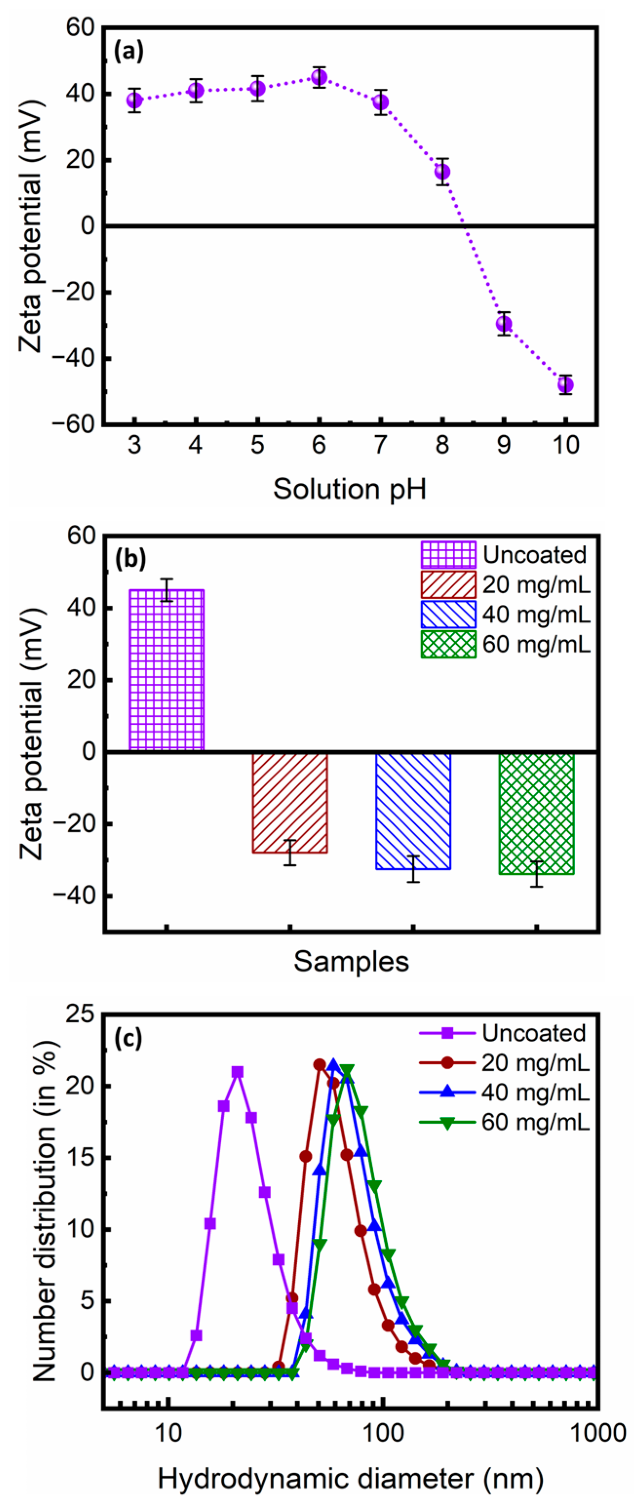
Figure 1. (a) Variation of zeta potentials with pH for the as-synthesized and acid-treated MNPs. (b) Zeta potentials for the as-synthesized and acid-treated MNPs and MNPs surface-functionalized with ~20, ~40, and ~60 mg/mL citrate concentrations. (c) Hydrodynamic diameter distributions for the uncoated MNPs and the MNPs surface-functionalized with ~20, ~40, and ~60 mg/mL citrate concentrations.
Magnetochemistry 11 00024 g001
Figure 2. Weight loss curves for the uncoated MNPs and MNPs surface-functionalized with ~20 mg/mL, ~40 mg/mL, and ~60 mg/mL citrate concentrations.
Magnetochemistry 11 00024 g002
Figure 3. FTIR spectra of the citrate-functionalized MNPs. The major absorption bands are indexed.
Magnetochemistry 11 00024 g003
Figure 4. (a) Powder XRD pattern for the citrate-functionalized MNPs at 300 K. (Inset) The variation of sin2(θ) as a function of h2 + k2 + l2 for the citrate-functionalized MNPs. (b) Magnetization as a function of the applied magnetic field for the citrate-functionalized MNPs at 300 K. (Inset) Langevin-type expression fitted to the magnetization data in the positive quadrant.
Magnetochemistry 11 00024 g004
Figure 5. (a) Typical TEM image of citrate-functionalized MNPs and (b) the corresponding size distribution.
Magnetochemistry 11 00024 g005
Figure 6. Phase contrast microscopy images of the citrate-functionalized MNPs (a) in the absence of the magnetic field, (b) under an external magnetic field (µ0H~200 G), and (c) after the removal of the magnetic field.
Magnetochemistry 11 00024 g006
Figure 7. (a) Absorbance as a function of wavelength for rhodamine B solutions of varied concentrations. (b) Standard plot for the rhodamine B dye solution showing the linear variation of absorbance with dye concentration. (c) Absorbance as a function of wavelength for the ~10 µg/mL rhodamine B dye solution and the supernatant after treatment with the citrate-functionalized MNPs. Typical photographs of the (d) rhodamine B dye solution and (e) supernatant after treatment with the citrate-functionalized MNPs. (f) Standard plot for the lead ions showing a linear variation of absorbance with concentration. Typical photographs of the (g) lead solution (supplemented with DMTD) and (h) the supernatant after treatment with the citrate-functionalized MNPs.
Magnetochemistry 11 00024 g007
Figure 8. Schematic representation of the probable mode of interaction among the citrate-functionalized MNPs, rhodamine B dye molecules, and the lead ions.
Magnetochemistry 11 00024 g008
Disclaimer/Publisher’s Note: The statements, opinions and data contained in all publications are solely those of the individual author(s) and contributor(s) and not of MDPI and/or the editor(s). MDPI and/or the editor(s) disclaim responsibility for any injury to people or property resulting from any ideas, methods, instructions or products referred to in the content.