Review

Implication of Magnetic Nanoparticles in Cancer Detection, Screening and Treatment

Department of Analytical Chemistry, Faculty of Pharmacy, Iuliu Haţieganu University of Medicine and Pharmacy, 4 Pasteur Street, 400349 Cluj-Napoca, Romania
*
Author to whom correspondence should be addressed.
These authors contributed equally to this work.
Magnetochemistry 2019, 5(4), 55; https://doi.org/10.3390/magnetochemistry5040055
Submission received: 30 August 2019 / Revised: 25 September 2019 / Accepted: 27 September 2019 / Published: 1 October 2019
(This article belongs to the Special Issue Magnetic Nanoparticles)

Abstract

:
During the last few decades, magnetic nanoparticles have been evaluated as promising materials in the field of cancer detection, screening, and treatment. Early diagnosis and screening of cancer may be achieved using magnetic nanoparticles either within the magnetic resonance imaging technique and/or sensing systems. These sensors are designed to selectively detect specific biomarkers, compounds that can be related to the onset or evolution of cancer, during and after the treatment of this widespread disease. Some of the particular properties of magnetic nanoparticles are extensively exploited in cancer therapy as drug delivery agents to selectively target the envisaged location by tailored in vivo manipulation using an external magnetic field. Furthermore, individualized treatment with antineoplastic drugs may be combined with magnetic resonance imaging to achieve an efficient therapy. This review summarizes the studies about the implications of magnetic nanoparticles in cancer diagnosis, treatment and drug delivery as well as prospects for future development and challenges of magnetic nanoparticles in the field of oncology.

1. Introduction

Material science has gained particular attention in the scientific field since the discovery of nanomaterials. In fact, nanotechnology emerged in this field of research as dealing with the fabrication of materials and technologies at length scales between 1 and 100 nm and integrating these nanoscale materials as building blocks of novel structures and devices. Furthermore, nanotechnology can offer benefits to medical applications like early diagnosis and monitoring, and due to the enhanced biocompatibility of new materials can give access to imaging and therapeutic purposes, playing an important role in disease treatment and targeted drug delivery [1].
One of the possible applications could be imagined for cancer prevention, diagnosis, and treatment where new nanomaterials could have a tremendous impact. Cancer, as defined by the World Health Organization (WHO) “is a generic term for a large group of diseases characterized by the growth of abnormal cells beyond their usual boundaries that can then invade adjoining parts of the body and/or spread to other organs” [2]. The WHO statistics show that cancer is the second leading cause of worldwide deaths and a prognostic of 29.5 million deaths was estimated by 2040. Several types of cancer are commonly found in both men and women such as lung and colorectal cancer. Stomach, liver and prostate cancers are the most common among men, while women are more likely to develop thyroid, breast, or cervical cancers.
In order to prevent metastasis, early stage cancer diagnosis is of high interest and challenging as symptoms appear only in advanced cancer stages. For this, highly accurate, fast, robust and non-invasive or minimal invasive procedures are of great importance.
In this regard, nanomaterials have already showed their medical usefulness in imaging technology applied for tumor target and visualization, allowing for early diagnosis of cancer. Another biomedicine application of nanomaterials is targeted drug delivery, where intelligent nanocarriers could improve the therapy efficacy by tailored transport of anticancer drugs to a well-established place where their release takes place without harming healthy cells.
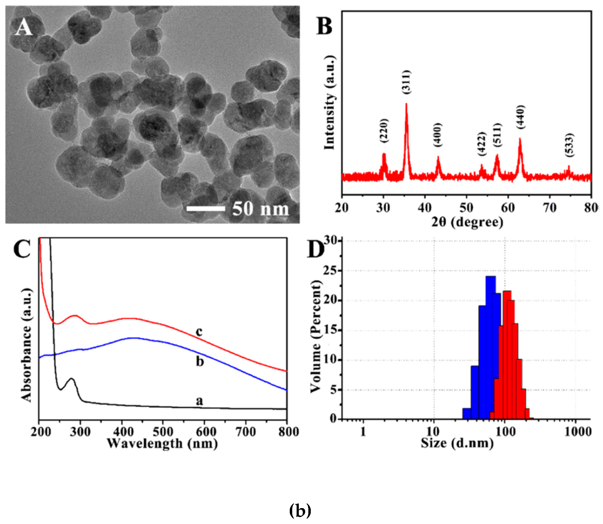
Magnetic nanoparticles (MNPs) have recently contributed to important development in oncology presenting major implications in cancer diagnosis, cancer screening, targeted drug delivery and cancer treatment. MNPs are widely applied in tumor targeting since tumor imaging technology opened the possibility for early detection of this wide-spread disease. Due to their magnetism in particular, MNPs (especially superparamagnetic iron oxide nanoparticles, so called SPIONs) have been mostly used as contrast agents in cancer screening for magnetic resonance imaging (MRI), in magneto-acoustic tomography (MAT), computed tomography (CT) and near-infrared (NIR) imaging. Moreover, drug delivery is also a hard to ignore application where the use of MNPs as drug agents (carriers) for in vivo targeted specific location can be performed by applying an external magnetic field (EMF). The specificity of MNPs is generally obtained by their functionalization with antibodies for target cells together with chemotherapeutic drugs. MNPs can be also applied in cancer treatment through magnetically induced hyperthermia (MHT), photodynamic therapy (PDT) and photothermal therapy (PTT). All these individual strategies are used in oncology, but the best therapeutic effect is usually assured by combining them since the modular design enables MNPs to perform multiple functions simultaneously. For example, MRI (Figure 1A) could be applied for early diagnosis of cancer, thus the individualized treatment (chemotherapy) may be combined with MRI, in order to achieve better and faster results [3,4,5,6].
The synthesis protocol influences the final properties of MNPs. The most important properties of MNPs that can be exploited for medical applications are superparamagnetism, high magnetic moment, magnetocaloric effect, small particle size and large specific surface area that can be easily functionalized [7,8,9,10,11,12]. The magnetic properties are related to the core of the MNPs; therefore, the superparamagnetism effect depends on the nanoparticle size and is generally observed for the MNPs with the size dimension up to 100 nm. These particles are magnetized when an EMF is applied and lose their magnetization in the absence of the field, therefore, preventing the MNPs clustering [8,9]. Magnetocaloric effect is an important property of some MNPs that are able switch their temperature depending on the existence of the EMF [11]. This feature combined with a large surface-to-volume ratio allows the efficient heat exchange with the environment, making possible the latest cancer therapy strategy, namely hyperthermia [8].
The special properties of MNPs are fully exploited in cell labeling and targeted drug delivery systems (Figure 1B), wherein the in vivo transportation of the drugs to the specific target is performed using a magnetic field positioned properly and outside the body [4,5]. MNPs may be considered a kind of intelligent magnetic material since they absorb the heat generated by the electromagnetic wave in the alternating magnetic field [3]. As already mentioned, a great advantage of MNPs is their small size, which is less or comparable to biological entities ranging from several nanometers in the case of genes and proteins, to hundreds of nanometers (viruses) up to 100 μm (cells). Such small dimensions allow for their good diffusion and distribution in tissues exactly or in the vicinity of the targeted sites [6]. Moreover, various processes to improve the magnetic properties of MNPs as well as to predict their in vivo behavior could be adjusted by modulating parameters such as size, composition, morphology or surface functionalization [5]. The most commonly used MNPs, iron oxide nanoparticles and in particular magnetite (Fe3O4) and its oxidized form maghemite (γ-Fe2O3) have attracted attention mainly due to their biocompatibility, low toxicity and cost, facile preparation, as well as their specific optical and magnetic properties that can be exploited in microsystems and medical devices’ fabrication [3,4,5].
Furthermore, MNPs applications in the field of cancer biomedicine are related to their use in MRI as contrast agents and in cancer thermotherapy, or so called hyperthermia [13], as heating mediators (Figure 1C). Additionally, novel applications of MNPs as platforms of immobilization of antibodies or aptamers for biosensors development have been proposed [14]. Affinity ligands (aptamers, hEGF, folic acid, lectin) can be immobilized at the MNPs surface to orderly direct them in the vicinity of tumors, thus enabling the MNPs to accumulate in a specific location of cells or tissues (Figure 1F) [15]. Significant progress in gene delivery and therapy has been made when a viral vector carrying a gene attached to the MNPs surface was performed. By this, rectification of genetic disorders can be enabled by gene transfection and expression with the complementary gene carried by the virus-attached MNPs [16]. This procedure is called magnetic transfection or magnetofection gene therapy and has aimed to cure lung, gastrointestinal and blood malignancies; future adaptation to non-viral transfection of biomolecules (e.g. DNA, siRNA) is yet to be envisaged.
Therefore, these strategies combined with targeted drug delivery and magnetic hyperthermia enables a synergetic effect in the efficient treatment of cancer.
MNPs could also be used for the early detection and diagnosis of cancer being building blocks of electrochemical immunoassays.
The multi-functional properties of MNPs have made them the ideal material for several applications in cancer detection, screening and treatment for: MRI, hyperthermia, drug carriers, tissue repairs, immunoassay and biosensors.
There are several reviews and book chapters describing the advances and applications of MNPs [17,18,19,20,21,22,23,24,25]; this paper aims to summarize and underline the latest findings in the field focusing on synthesis, functionalization and application of MNPs in cancer biomedicine. Cancer detection, screening and treatment within the MNPs use is presented starting with biosensing technologies, followed by the visualization of tumors within imaging techniques and the treatment of tumors through targeted delivery approaches, and lastly by hyperthermia and photodynamic therapy. Some of these applications will be detailed in the next subchapters. Finally, future perspectives and challenges yet to be solved will be presented.

2. Synthesis and Characterization of Magnetic Nanoparticles

Several ways to synthesize MNPs were proposed by researchers; many of them offer special features like shape control, monodispersity and stability and preparation on a large scale like sol-gel procedure, hydrothermal synthesis or co-precipitation. New and innovative methods were addressed: sonolysis and biosynthesis, thermal decomposition, and microemulsion [26]. Choosing the best synthesis method depends mainly on the nature of MNPs one wishes to obtain. Depending on their composition, MNPs could be classified in:
  • iron oxide nanoparticles or oxides (ferrites): hematite (α-Fe2O3), maghemite (γ-Fe2O3) and magnetite (Fe3O4); their involvement in biomedical application is based on their easy surface modification with various compounds for increased stability in aqueous media (e.g. surfactants, silica) [27];
  • metallic nanoparticles with only a metallic core: more suitable for biomedical applications due to their higher magnetic moment compared to oxides; reported drawbacks are pyrophoric property, and presence of high reactivity to oxidizing agents;
  • shell-based ferrites: chemically inert MNPs core covered by a silica shell for further functionalization through covalent bonding;
  • shell-based metallic nanoparticles: metallic core covered by a shell made of polymers, precious metals or modified surfactants [27].
Table 1 describes several methods for the synthesis of MNPs [25].
New methods of producing MNPs which try to overcome the drawbacks of the established ones are periodically reported; however, for mass production of highly controlled features, MNPs, co-precipitation and thermal/hydrothermal decomposition remain the most secure and easy to use methods.

3. Functionalization and Stabilization of Magnetic Nanoparticles

Due to the fact that biomedical applications usually need special requirements to control the MNPs’ interfaces, the functionalization of MNPs surfaces is a way of tailoring their properties. Functionalization is a useful process for enhancing the colloidal stability in complex biological environments which affect the molecular recognition. Stabilizers that prevent aggregation are generally surfactants (sodium oleate, sodium carboxymethylcellulose) or either synthetic polymers like poly(ethylene-covinyl acetate), poly(lactic-co-glycolic acid), poly(vinylpyrrolidone), polyethylene-imine, or natural ones like chitosan, gelatin, and dextran [27].
To enhance the stabilization in non-aqueous solvents, MNPs are usually covered by a hydrocarbon layer. By contrast, biomedical applications require MNPs with hydrophilic and biocompatible properties [33]. Ligand addition, ligand exchange and hydrophilic silica coating are the three most important methods for surface functionalization; organic and inorganic coatings will increase the stabilization and the resistance towards oxidation in the water or humid air [28].
For example, the process of binding different molecules at MNPs’ surfaces could be easily explained by the presence of hydroxyl groups of nanoparticles obtained by co-precipitation [34] which can be negatively or positively charged depending on the pH of the media. Although, non-peptizable particles can be synthesized at pH 7.5 when OH– ligands are free of charge, ligands still remain attached to the MNPs when the pH is kept in the range 6–10. Therefore, biomolecules can be further attached to the free hydroxyl groups at the surface of the particles. A possible biomedical application of these hydrophilic particles is represented by targeted drug delivery.
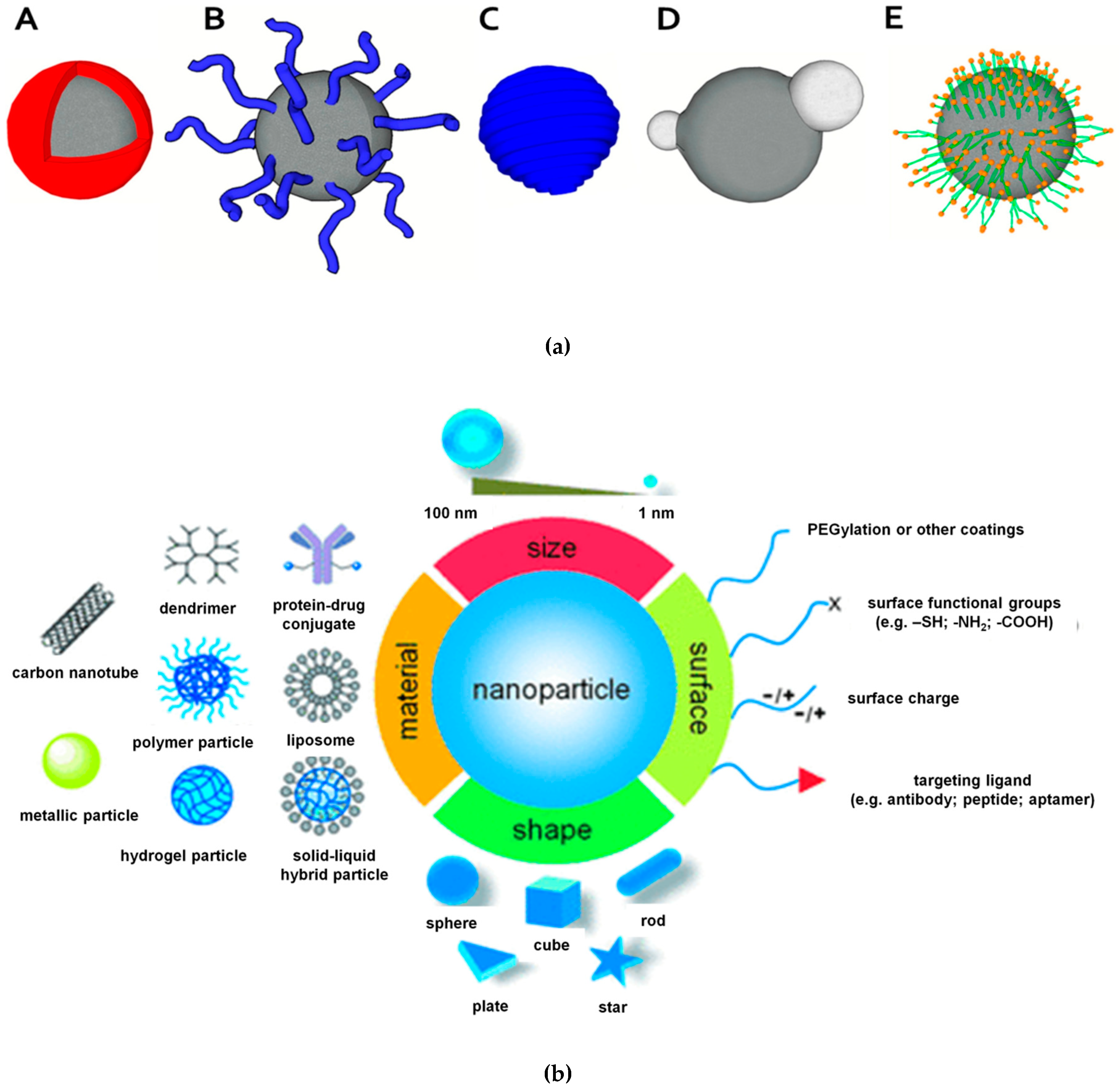
Different modifications that could be performed at MNPs surface were already revised in several papers [18,35,36] and schematically represented in Figure 2a. Furthermore, a summary of types, shapes and functionalities that have been explored for MNPs in order to be used as carriers for drug delivery in cancer therapy, together with illustrations of biophysicochemical properties, is presented in Figure 2b.
The surface modification is extremely important when MNPs are used as drug carriers due to their low drug-loading capacity and fast clearance from blood fluids. There are three approaches to overcome these drawbacks. The first approach is based on conjugation of MNPs with drugs followed by surface modification with various polymers. The next two approaches use the modification of MNPs with a pro-drug–polymer composite or either the adsorption of the drugs on the MNPs modified with polymers.
Polyethylene glycol (PEG), a frequently used polymer, is an interesting material since it owns two end-hydroxyl functional groups available for different reactions. PEG has also been used for drug conjugation, enhancing the drug solubility and bioavailability. PEG-MNPs were investigated as highly biocompatible drug carriers for antitumor medicines as curcumin [38] and doxorubicine (DOX) [34]. MNPs modified with dextrane was able to efficiently entrap indomethacine, an antiinflamatory drug, as reported by C. Jin et al. [39].
The magnetic properties of MNPs based on magnetite may decrease when subjected to acidic pH (<4) due to the fact that Fe2+ is easily oxidized to Fe3+. To answer this, coating protocols with inert oxides (e.g. silica, alumina) have been applied for enhanced magnetism stability [40]. The use of silica (SiO2) as protecting material for Fe3O4 is justified by its special properties namely chemical and magnetic stability and suitability for surface functionalization. Moreover, besides preventing degradation of MNPs, silanol groups (-SiOH) can be used as anchoring sites for further modification. Applications of silica-based MNPs are mainly reported as drug carriers, and in the field of electronics, paints, or catalysis etc.
Besides silica, decoration of MNPs with noble metals (gold or silver) enables new properties such as optical properties and enhanced bioaffinity, biocompatibility, chemical and physical properties, without affecting the magnetic features of the core. Thus, gold nanoparticles (AuNPs) are widely used for surface coverage of Fe3O4; several papers report the application of AuNPs@Fe3O4 in electrochemical (bio)sensing, separation of biological structures, targeted delivery of drugs and bioimaging [7].

4. Applications of Magnetic Nanoparticles in Cancer Biomedicine

4.1. Cancer Biomarker Detection Using Magnetic Nanoparticles

4.1.1. Biomolecules Conjugation

In the field of cancer biosensing based on MNPs, one important step in the biosensors’ development is represented by the immobilization of specific biological elements at the functionalized MNPs. Cancer biomarkers are molecules or structures involved in disease development and evolution that can offer a prognostic concerning cancer prevalence and outcome [41]. Therefore, the detection of these molecules, which can range from small elements (peptides, aptamers, DNA) to high-weighted proteins, is of crucial importance [42].
The functionalized MNPs’ surface offers linker groups to allow the binding event with the complementary biomolecules. Different bioconjugation strategies include physical interactions (e.g., electrostatic interaction, hydrophilic-hydrophobic, affinity interactions) and chemical interactions (e.g., covalent bonds) for easy immobilization on MNPs or transducers [43]. Some specific interactions between protein and their recognition elements, like biotin-avidin and antigen-antibody linking systems, have proven their utility in the design of biosensors [20].

4.1.2. Bioseparation

MNPs play a significant role in bioanalysis as biological separation represents a cost-effective and fast alternative to traditional separation methods (centrifugation and filtration) [18]. Therefore, functionalized MNPs interact with the complementary target from the pristine mixture and upon conjugation the as-formed composites can be manipulated by applying an EMF, allowing for efficient bioseparation (Figure 1E). Various types of biomolecules and cells were separated and purified using this technique as bacteria, viruses, tumor cells, T cells, monocytes [17,18].

4.1.3. Biosensing

The as-conjugated MNPs with the specific biomarkers are further used in the final step: the sensing approach. In the last decades, cancer biomarkers detection has gained significant attention and development.
Various promising sensing methods to detect the level of cancer biomarkers in plasma, blood or diseased tissues have been developed: electrophoresis, optical methods (fluorescence, electrochemiluminescence, colorimetric assay, surface plasmon resonance (SPR), surface-enhanced Raman spectroscopy (SERS), etc), immunological methods (enzyme-linked immunosorbent assay (ELISA), polymerase chain reaction (PCR), etc), microcantilevers, electrochemical assay, and others [44,45]. Biosensing events may be outlined through different labelling approaches, depending on the method (fluorescent labels, electroactive molecules, enzymes, and nano-/micro-particles, etc). Two major strategies of the integration of MNPs in the design of biosensing systems are represented by direct labelling and indirect labelling.
In the first approach, direct labelling, MNPs could be immobilized at the transducing element by affinity recognition reactions between complementary DNA sequences or streptavidin-biotin. For example, the sensor surface is modified with the magnetic particles functionalized with single-stranded DNA as capture probe, the hybridization reaction will occur as the complementary oligonucleotides are put in contact with the sensor and a physical or/and chemical response will be generated.
In the second approach, indirect labelling, the principle resembles ELISA, namely sandwich immunoassays. For example, primary antibodies complementary to the target protein are immobilized at the sensing surface followed by the affinity reaction with the solution containing the biomarker. Next, the secondary biotin-labelled antibodies are introduced into the system to enable the affinity reaction when the streptavidin-labelled MNPs solution is put in contact with the sensor surface [18]. For a more comprehensive description, we categorize the sensing approaches for cancer detection according to the detection principle as follows: electrochemical, optical, and magnetic. Inside each subsection, different detection strategies are detailed if available.
In the last decades, electrochemical biosensors have gained much attention for cancer biomarkers detection mainly due to their high accuracy and sensitivity, multiplexing and cost-effective features, and selectivity in challenging the matrix without requiring multiple sample treatments or complex protocols [46]. Currently, a wide range of analytical techniques has been integrated for the development of multiplexed immunosensing systems for cancer biomarkers. Electrochemical immunosensors have received great interest due to their high sensitivity provided by coupling the immunochemical affinity reaction (antigen-antibody) with the particular features of multifunctional electrode transduction elements [41,47].
An example of a MNPs-based sandwich immunoassay for the electrochemical determination of cancer antigen 153 (CA153) is detailed [48]. The sensing system uses disposable screen-printed carbon-based electrodes functionalized with graphene oxide (GO) and peroxidase-like magnetic silica nanoparticles/GO composites acting as labels. Firstly, the silica MNPs were functionalized with azide groups to enable the acetylene-functionalized GO via click chemistry for labelling purposes, then a monoclonal anti-CA153 antibody was immobilized at the GO-modified screen-printed carbon electrodes. The immunoassay exhibited a broad linear range (10−3 - 200 U/mL) for the determination of the cancer antigen and a limit of detection (LOD) of 2.8 × 10⁻⁴ U/mL. Another example involves the use of AuNPs-modified porous paper acting as a working electrode (Au-PWE) which is further functionalized with 1-azido undecan-11-thiol [49]. A click reaction enables the conjugation of the alkyne end-terminated capture antibody. Azide-functionalized sphere-like peroxidase magnetic silica (Fe3O4@SiO2) nanoparticles were prepared to couple alkynylated peroxidase and secondary antibodies as detection label tags in the presence of hydrogen peroxide and thionine. A sandwich immunosensor was developed for the simultaneous electrochemical detection of carcinoembryonic antigen (CEA) and alpha-fetoprotein (AFP), by the aid of multi-labelled AuNPs as signalling probes [50]. The experimental setup was fabricated by means of AuNPs conjugated with thionine and ferrocene as probes and MNPs as immobilization surface for both specific antibodies under application of an EMF. The magnetoimmunosensor enabled the simultaneous detection in the linear range of 0.05–120 ng/mL with a LOD of 0.012 ng/mL for CEA, and 0.05–100 ng/mL with a LOD of 0.018 ng/mL for AFP (S/N = 3), respectively.
Biotin-streptavidin affinity reaction is one of the most used reactions in the design of immunoassays. Guerrero and co-workers developed a new strategy for the detection of IL-13Rα2 from cells lysates and from tumor tissues extracts (sample amount 0.5 μg) by an integrated electrochemical immunosensor [51]. To this, modified screen-printed electrodes with diazonium salts and hybrid composite based on multi-walled carbon nanotubes (MWCNTs) and graphene quantum dots (GQDs) were employed as advanced nanocarriers of several enzymes and an antibodies detector to achieve signal amplification. The sensor showed a time-response of approximately 2 h and enabled the determination of IL-13Rα2 down to 0.8 ng/mL (linear range 2.7 - 100 ng/mL).
Given the knowledge gained in the last decades, aptasensing bioassays have attracted great interest because of aptamers’ ability to selectively and moreover specifically bind their target with high affinity [52]. To that end, Tian et al. designed an electrochemical aptasensor for MCF-7 circulating tumor cells (CTCs). The strategy was to use an EMF to perform a preliminary pre-concentration and separation step by inserting a magnet inside the glassy carbon electrode setup. Furthermore, an electrode modifier based on rGO/MoS2 hybrid material combined with bi-nanozyme/aptamer-functionalized Fe3O4NPs accounts for signal generation and enrichment (Figure 3). The proposed electrochemical biosensor was able to detect MCF-7 in the linear range from 15 to 45 cells/mL with a LOD of 6 cells/mL, presenting good reproducibility and stability parameters [53].
Optical techniques supply the most diverse class of biosensors due to the plethora of optical processes that can be followed during the detection step such as absorption, fluorescence, phosphorescence, refraction, dispersion, and others [45]. These methods showed promising results for use in point-of-care (POC) early cancer diagnostics and imaging, due to the fact they are fast and the signal can be often easily be seen by the naked eye (e.g., colorimetric assays), however, sometimes optical techniques require more specialized instrumentation or personnel (e.g., SPR, SERS) [44,54].
Xu and co-workers designed a DNA-based MNPs (DNA/dextran/PAA/Fe3O4 NPs) sensor working as a signal-off fluorescent assay for the sensitive determination of p53 protein expression [55]. The fluorescent sensor showed a dose-response in the linear range from 50 pM to 2 nM and detected p53 low to 8 pM (LOD). The use of MNPs allows for sensitive analysis of real samples with minimum sample treatment steps and simple instrumentation. Another MNPs-based optical sensor is described for tumour biomarker anterior gradient homolog 2 (AGR2) by means of ultraviolet–visible (UV–Vis) spectrophotometric measurements [56]. The as proposed aptasensor, which requires 3.5 h for the overall sensing operational steps and low sample volumes (20 µL), showed a LOD of 6.6 pM (linear detection range over 10–1280 pM). The synergistic effect of combining the properties of MNPs with AuNPs can be also seen in the next example.
A mucin-1 (MUC1) optical electrochemiluminescence (ECL) sensor was developed based on a sandwich-type assay and hybrid materials of luminol-decorated gold-functionalized MNPs (Lu–AuNPs@Fe3O4) [57]. The MNPs were functionalized with the synthesized ECL label via electrostatic interaction to allow the formation of the composite Lu–AuNPs@Fe3O4. The sensor was applied for the MUC1 quantification in a wide linear range from 10 fg/mL to 10 ng/mL and a very low LOD of 4.5 fg/mL MUC1 was obtained. The concept of signal enhanced Fe3O4 nanozyme was first introduced by Li and co-workers in a photoelectrochemical (PEC) immunoassay for highly sensitive determination of prostate-specific antigen (PSA) [58]. The his-Fe3O4@nanozyme allow for signal amplification much higher than that of catalytic-induced activity of the natural enzyme HRP, and hence, with lower cost fabrication, ease of preparation and modification (Figure 4). The combination of the PEC enhanced features of the ZnIn2S4/ZnO-NRs/indium tin oxide photoelectrode and the highly effective his-Fe3O4@nanozyme enabled the determination of PSA down to fg/mL range (LOD = 18 fg/mL).
Another nanohybrid composite that showed high catalytic effect was developed starting from Fe3O4 MNPs and platinum nanoparticles (PtNPs), simultaneously entrapped in the framework of GO [59]. The results showed a 30-fold increase of the maximal reaction velocity (Vmax) compared to the one obtained without GO within the colorimetric induced response of the peroxidase substrate, 3,3′,5,5′-tetramethylbenzidine (TMB). The sensor enabled fast quantification in the timeframe of 5 minutes of clinically important breast tumour cells. Gui et al. developed a charge-coupled device (CCD)-based reader to quantitatively measure the fluorescence signal of quantum dots (QDs) immobilized on lateral flow test strips for cytotoxin-associated protein (CagA) detection, which is commonly over-expressed in gastric carcinoma [60]. The sensor showed possible POC applications for CagA detection with a LOD of 20 pg/mL.
However, oftentimes sophisticated instrumentation for optical methods restricts their use for POC development in determining tumour biomarkers in real scenarios for early stage cancer diagnosis. Given this, colorimetric approaches are the most sensitive and widely employed methods due to their main advantages as simplicity, possibility for miniaturization and development of POC devices, and low costs. For example, Peng et al. proposed a magnetic colorimetric immunoassay (CIA) for human interleukin-6 (IL-6) based on Cerium oxide NPs (CeNPs)-labelling approach to enable the oxidation catalysis of the substrate into a stable yellow product [61]. Studies showed that IL-6 is overexpressed in breast and prostate cancer [62,63]. Spectrophotometric measurements showed a curve dose response in the range of 0.0001–10 ng/mL toward IL-6 and a LOD of 0.04 pg/mL was obtained. A colorimetric sandwich immunsensor based on a reverse strategy for PSA detection in biological fluids is presented [64]. To this end, hybrid nanostructured composite based on MNPs and AuNPs was synthesized and further used for the functionalization with both capture antibody (anti-PSA) and detection antibody (catalase/anti-PSA), respectively. Next, the signal generation and amplification for PSA assessment was realised by the aid of functional catalase/anti-PSA/AuNPs as enzymatic catalyst and anti-PSA-conjugated/MNPs as an optical generator. Despite classical colorimetric immunosensors, the reverse CIA technique revealed the remained quantity of H2O2 in the substrate after its depletion by the labelled-catalase. Under the optimal conditions, the immunoassay was applied for PSA sensing down to 0.03 ng/mL (S/N = 3) in a broad range of 0.05–20 ng/mL PSA.
There is a plethora of magnetic detection techniques exploited to determine the magnetic response of MNPs such as, spintronic sensors based on giant magnetoresistance (GMR), tunnel magnetoresistance (TMR), and planar Hall effect (PHE) sensors, superconducting quantum interference devices (SQUIDs), atomic magnetometers (AMs), nuclear magnetic resonance (NMR) systems, fluxgate sensors, Faraday induction coil sensors, diamond magnetometers, and domain wall-based sensors [20]. Compared to electrochemical or optical methods, the magnetic sensing techniques have proven superior performances for biomarker detection, providing high sensitivity (increased signal-to-noise ratio), high stability (fluorescent labelling presents no photo-bleaching) and feasibility for POC development [18].
The biosensing principle of magnetic sensors lies in magnetic field alterations that occur after interacting with the magnetic field of the MNPs. Therefore, a current or resistance difference of the magneto-resistive MNPs-functionalized sensor will be generated [17]. For example, DNA hybridization events can be monitored by the magnetic turbulence detected by the magnetic sensor by indirect approaches using affinity reactions (streptavidin-biotin or complementary DNA receptors) (Figure 5) [65].
The design of a lateral-flow immunoassay based on a magnetoresistive sensor combined with MNPs acting as labels is described [66]. The magnetic signal generated by the MNPs within the spin valves biosensor exhibited a detection limit of 5.5 ng/mL of the human chorionic gonadotropin hormone. Spintronic sensors and immunosensors based on giant magnetoresistance principle have also been developed. A magentosensor for S100β biomarker detection was prepared using 300 nm MNPs for signal generation and enabled a LOD of 27 pg/mL [67]. A Hall-based magnetic transduction system was applied for the sensing of a 35-base DNA-strand for a pathogenic target [68].
LOD of 364 pM and high selectivity for target DNA was obtained by optical microscopy detection using a DNA-based Hall magnetometer sensor by the aid of MNPs of 350 nm [67]. This novel strategy allows the simultaneous ultrasensitive detection (fM) of up to eight cancer biomarkers from the cytokines class with signal amplification accomplished through MNPs of about 50 nm in diameter.
The applications of MNPs used in several sensing approaches based on electrochemical, optical and magnetic readout of cancer biomarkers are summarized in Table 2 presenting the type of assay, detection method, LOD, and the type of tested real samples.

4.2. Cancer Screening Using Magnetic Nanoparticles

Magnetic Resonance Imaging (MRI)

MRI is an intense used method for cancer screening during and after chemotherapy. Despite common imaging techniques which require ionization radiation, MRI uses the magnetic properties of ions for projecting the image. When no magnetic moment is applied, protons have a randomized orientation whereas a parallel or anti-parallel arrangement occurs once the magnetic field is on. Contrast agents including those based on MNPs are used to enhance the quality of MRI images [80]. The development of reactive MNPs and magnetic colloidal particles for immobilization and easy magnetic separation of biomolecules is of great importance for early detection of diseases and consequently in therapy management and treatment of cancer in early stages. Thus, chitosan-stabilized magnetite nanoparticles were synthesized and successfully applied as negative contrast agents in MRI which further led to several biomedical applications [4].
Iron oxide MNPs are widely used in cancer screening and treatment because of their multiple advantages extensively discussed in the introduction of which we emphasize the ease distribution and functionalization. The most common functionalization of Fe3O4 particles consists in coating with antibodies. This strategy has been extensively applied for cell separation, recognition, and early diagnosis of malignancies. However, the biomedical use of Fe3O4 is restricted by the reduced biocompatibility, the remediation of this aspect being achieved by preliminary functionalization with natural compounds presenting high biocompatibility, blood compatibility, as well as microbial degradability. Therefore, a combination of magnetic Fe3O4 core particles and α-ketoglutarate chitosan shells (Fe3O4@KCTS) was applied for cancer screening through direct multi-labeling with different antibodies to sort lymphatic endothelial cells [81]. As noticed in Figure 6, firstly, the magnetic core was coated with α-ketoglutarate chitosan (KCTS) to enable the formation of Fe3O4@KCTS core-shell MNPs, followed by an activation step of the –COOH functional groups via NHS/EDC chemistry. Next, the covalent immobilization of two complementary antibodies for lymphatic endothelial cells, anti-Lyve-1 antibody and anti-podplanin antibody were bound at the Fe3O4@KCTS MNPs. A dual-targeting magnetic nanoprobe was obtained and injected into the tail vein of mice models for tumor visualization by MRI and fluorescence imaging. The results demonstrated that a dual-targeting magnetic nanoprobe was successfully developed and applied for capturing high-purity lymphatic endothelial cells from tumor tissues, providing a starting point of clinical applications based on dual-mode imaging in cancer screening.
Combining the effective features of MNPs with AuNPs some complex nanostructured materials are obtained that can be applied for cancer diagnostic and treatment by means of multiple imaging techniques and treatment strategies. These nanocomposites materials are biocompatible and can be thus further combined with other imaging and therapeutic agents (e.g., radioactive element and/or drug molecule) and biomolecules (e.g., peptide or antibody) for multimodal imaging of tumors. Therefore, a more effective treatment can be employed by the synergetic effects of combined therapy approaches [82].
Combinations between the imaging methods are also used since these dual-imaging methods may improve the accuracy of diagnosis. For instance, dual imaging of single-photon emission computed tomography (SPECT) and MRI have been applied in pancreatic and breast cancer, while MRI and optical imaging were combined for the successful diagnosis of breast cancer [3].

4.3. Cancer Treatment Using Magnetic Nanoparticles

The success in cancer treatment and the decrease in the mortality rate of patients are closely related to its diagnosis in early stages. When cancer is discovered earlier, the cure rate is greatly improved. Tumor imaging technology is used both in cancer diagnosis and treatment. Due to the high resolution and tomographic capabilities, MRI has proved to be one of the most valuable non-invasive imaging techniques. Furthermore, MNPs are the most widely researched and used contrast agents in cancer imaging. Due to the colloidal instability of MNPs, their surface modification is necessary by inducing the magnetic dipole interaction and its intrinsic surface energy. The multifunctional nanocomposite MNPs present higher potential for therapeutic and diagnostic applications [3].
MRI is related to nuclear magnetic resonance of hydrogen atoms and it usually requires the use of contrast agents for enhanced imaging. MNPs can be used as contrast agents only if they exhibit high saturation magnetization and after functionalization with compounds that increase the hydrophilicity around the Fe2O4 core [83].
Selective detection of cancer cells can be achieved by antibodies, thus many immunosensors have been developed for this purpose. Although, when using MNPs functionalized with antibodies specific for a type of tumor cell, the detection through the specific immunosensor can be combined with imaging through MRI and cancer treatment through hyperthermia. This strategy offers greatly improved survival rates among oncological patient’s response to therapy. A first approach presents MNPs based on Fe3O4, having the core diameter of about 10 nm functionalized with poly-L-lysine (PLL) [83]. This complex strategy increases the stability and biocompatibility of MNPs and allows their use for combined detection, diagnosis through MRI and cancer therapy through magnetic hyperthermia. Hence, the 3D model of MNPs accumulation in tumor was also investigated for better evaluation of selectivity and in vivo toxicity.
Another MNPs-based imagistic strategy for determining the MNPs in vivo distribution is magneto-acoustic tomography. Herein, a magnetomotive force is generated by applying a short pulsed magnetic field and then using it to promote ultrasound frequencies in SPION-labelled tumor cells. After a long period of intense study and clinical trials, it can be still stated that most forms of human cancer are yet to be cured. The main reason is that this malady has many etiologies and the different types of cancer present numerous manifestations. Furthermore, multiple individuals having the same cancer respond differently to identical therapies. Moreover, the mechanisms of tumor growth and evolution still remain unsolved. Therefore, anticancer therapy is an important topic nowadays, considering the extent of this type of disease in the world. While certain types of cancer have been successfully treated, there are still many types of neoplasms that are refractory to all modern therapies.
Three major types of antineoplastic therapies are currently used, namely: surgical oncology in which the surgeon and pathologist discriminate between tumor tissue and healthy cells based on imaging techniques; chemotherapy uses antineoplastic drugs in order to eliminate cancer cells and stop their rapid multiplication; and radiation therapy exploiting the increased sensitivity to radiation of neoplastic tissue compared with normal tissue.
Surgical oncology is applied at controlling local tumor development, while chemotherapy is able to address circulating tumor cells, being thus more suitable for metastases. However, additive or synergistic and improved therapeutic effects can be obtained combining these types of therapies and minimizing the severe side-effects on healthy cells [10].

4.3.1. Drug Delivery

Targeted drug delivery is a concept that has been developed rapidly in recent years. In the case of cancers in particular, it has become one of the most popular treatment strategies since it can lead to a substantial increase in treatment efficiency and has far fewer side effects than the conventional methods used. A promising approach in this regard refers to magnetic drug delivery systems which involves the existence of a magnetic moment. Thus, various types of materials possessing magnetic properties that can be applied in magnetic targeted drug delivery are currently used as promising tools for modern therapy of several diseases, including cancers. Ideal properties for the magnetocomplexes which are to be used for targeted drug delivery must present high magnetization activity at the operational temperature. MNPs based on iron, cobalt, and nickel, are suited for this application, but the particles’ dimensions and coating are of great importance. Thus, MNPs are constructed using biocompatible shells of polymers or metals, as well as nanocomposite mixtures consisting of MNPs encapsulated within porous polymers. Presence of the polymers or other coatings provides an opportunity to anchor various therapeutic drugs or DNA for targeted gene delivery [84,85].
Figure 7 is a suggestive representation of the multiple possibilities of MNPs’ functionalization for application in cancer detection and treatment [85].
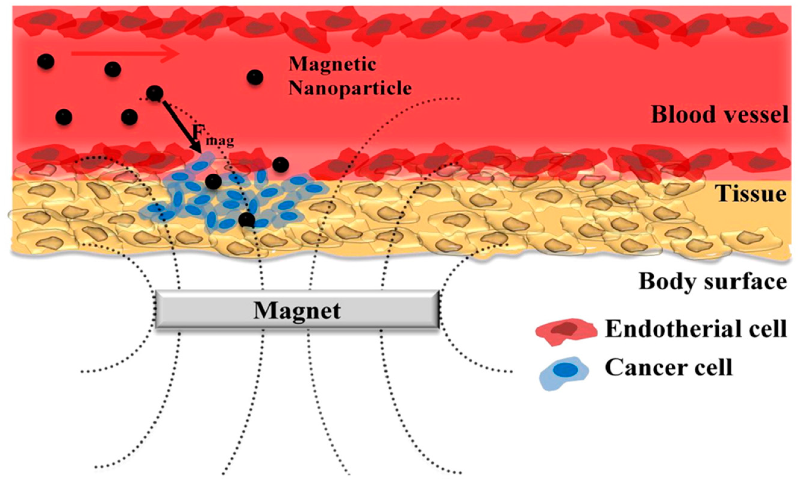
In chemotherapy, anticancer drugs are administered through the venous circulatory system, strategy that aims at the accumulation of the drugs in the tumors, where vascularization is accentuated compared to healthy tissues. Antineoplastic drugs destroy cancer cells, while poisoning healthy tissues causing severe side effects, being one major drawbacks of chemotherapy. Thus, due to the need for more efficient approaches, an explosive development of research in the field of drug carriers, including magnetic approaches, has been extensively exploited. Furthermore, drug-delivery systems are often functionalized with biomolecular recognition sites able to specifically interact with receptors located in tumors, allowing for the selective targeted orientation of the drug carriers to cancer cells. Another very popular drug-delivery system is magnetic drug targeting which involves the use of a drug delivery vehicle with magnetism properties that can be manipulated by an EMF. Both these strategies decrease the side effects caused by antineoplastic systemic treatment [6]. Most of the MNPs used in biomedical applications are the so-called SPIONs consisting of one or multiple magnetite or maghemite cores and with biocompatible shell functionalized with various modifiers. In magnetic drug delivery a therapeutic agent is coupled to a magnetic particle, a step that is followed by their injection in the blood flow and their orientation to the target location by the aid of a magnetic field. MNPs may present small magnetic moment thus being challenging to retain it at the targets while withstanding the drag of the blood flow. To overcome this phenomenon, the magnetic field and gradient have to be extremely large, or the particles will agglomerate and cause embolism of the blood vessels. Some FePd-based magnetic nanowires were developed and used in a pilot scale in vivo experiment of targeted therapy. A magnet was designed for capture of the nanowires from the blood flow in the hind leg of a rat and no negative side effects from injection of the nanowires were found. Preliminary in vivo tests performed on animals proved that FePd nanowires were non-cytotoxic and non-immunotoxic which is an essential condition for any in vivo application and promising for future use in a clinical application [86]. Although the method was successfully applied in some modeling studies and also in animals and humans, no magnetic drug delivery applications have yet clinical use.
Commonly used chemotherapy drugs include doxorubicin (DOX), paclitaxel, cisplatin, gemcitabine, methotrexate, docetaxel, sorafenib and mitomycin C [3]. The majority of anticancer drugs present limited or no targeting capacity towards specific cancer cells. A good strategy to improve the treatment efficiency and to reduce the dose of drugs used in cancer treatment is the use of targeting-based approaches. Thus, cancer cell lines derived from liver, prostate and breast was used as a model for the implementation of a targeting-based strategy. These model cancer cells overexpress the riboflavin receptors on their cell membrane and are also sensitive to the treatment with n-Butylidenephthalide (BP). Fe3O4 MNPs were functionalized with riboflavin-50-phosphate (RFMP) through Fe-phosphate chelation and successfully used as carriers for BP to treat the model cancer cells that are also sensitive to this treatment. The results demonstrated that the as-prepared functional MNPs can be used to effectively target and inhibit the cell growth, have no toxicity toward non-targeted cells, and have the potential to be used as anti-cancer agents. More in vivo tests must be further performed in order to assess the clinical relevance of this approach [87].
Even though the concept of magnetic drug delivery was used for the first time in the 1980s [6], and the first clinical cancer therapy trials in humans using magnetic microspheres filled with 4-epidoxorubcin was reported in Germany in the 1990s (being about the treatment of advanced solid liver cancer in 14 patients) [84], the development of the domain has been achieved especially in the last 10 years after the development of stronger magnets and sophisticated magnetic probes, namely the theranostic probes that allow the combination of diagnostic and treatment. In this case, the diagnostic could be achieved through MRI or magnetic particle imaging, while the therapy could be realized by approaches such as hyperthermia, drug release or magneto-drug delivery [6].
Magnetic bioprobes are intensively explored for magnetic targeting. In this case, the uptake of drugs in drug delivery systems is usually carried out through conjugation, hydrophobic interactions or physical absorption within porous structures. as in the case of the non-magnetic ones, while the release of drugs can be accomplished by mechanical forces, varying the pH of the environment, by near-infrared (NIR) irradiation, magnetic hyperthermia or chemical reduction [6,10].
Magnetic drug targeting and delivery systems are often based on the use of an EMF from electromagnetic coils or permanent magnets. Here it was proven that the most important aspects for the effective magnetic drug delivery are the geometry of the magnet and the distance between the magnet and tumors [82,88].
Three different biopolymers, hydroxyl ethylene cellulose (HEC), nanocrystalline cellulose (NCC), and a synthetic biopolymer polyvinyl pyrrolidone (PVP) were applied for the functionalization of the surfaces of superparamagnetic Fe3O4 nanoparticles, these polymers being chosen based on their ionic charges which are cationic, anionic and non-ionic, respectively. The results obtained shows that the cationic polymer used in this study is more efficient for targeted delivery applications since it completely covers particle surfaces reducing their toxicity, allows better loading efficiency for anti-tumoral drugs, and does not significantly reduce magnetization of the particles in order to be driven to the targeted site under the action of an EMF [89].
The configuration that implies the use of an EMF is difficult to be applied to target areas below 5 cm under the skin, thus the concept called delivery deep inside the body was introduced. In this case a dynamic control of magnets was proposed in order to focus magnetic carriers to deep tissue targets [6]. More recently, magnetic implants seem to be a viable alternative to the use of an EMF which may cause severe problems in the case of drug delivery to some organs. A biocompatible nanodrug delivery formulation based on poly (D, L-lactide-co-glycolic) acid (PLGA), polyethyleneglycol (PEG) and SPIONs has been developed and evaluated for the enhanced delivery of docetaxel to breast cancer cells. The higher saturation magnetization, controllable size, satisfactory drug loading, sustained release, predominant cancer cell uptake and effective cytotoxicity make this a promising and outstanding drug delivery system for breast cancer therapy [90]. A biocompatible magnetic implant scaffold made of a magnetite/poly(lactic-co-glycolic acid) nanocomposite was reported for bone cancer, providing a more accurate cancer treatment [6]. However, more in vivo tests need to be performed before these systems could be safely applied for clinical trials.
Tumor hypoxia represents the low oxygen concentration which is generally a result of disordered vasculature that leads to distinctive hypoxic microenvironments. Traditional anticancer agents cannot penetrate into these zones, thus being ineffective in cancer treatment. As can be seen in Figure 8, MNPs loaded with drugs are guided to the tumor site under the influence of EMF as a viable magnetic drug delivery system.
However, the development of this delivery system mandates that the MNPs behave magnetically only under the influence of EMF and are rendered inactive once the EMF is removed [84,91].
Additionally, cancer-targeted drug delivery particles still have many risks that can be overcame by using microrobots. These entities are constituted of an anti-cancer drug and MNPs to provide drug delivery to a cancer cell target lesion through electromagnetic actuation. However, viable solutions are still needed considering the inherent toxicity of MNPs that may remain in the body after the drug release. Various microrobots that can be manipulated by electromagnetic actuation (EMA) systems have been developed for various biomedical applications. These microrobots may be (i) shape morphing (changing their shape in response to external stimuli resulting in the delivery of the drug to target positions), (ii) shape-switching (using temperature or pH-responsive materials and can be manipulated by an EMA system to capture or release therapeutic drug particles), and (iii) controlled-drug releasing. Magnetic-actuated microrobots are envisaged as alternative to conventional drug therapy, but it should be noted that for biomedical applications, these devices should be made of biocompatible and biodegradable materials with no or little side-effects on humans. However, the MNPs used to magnetically drive microrobots are very small but lack a biodegrability property. Thus, it can be supposed that a part of MNPs will be excreted by human metabolism, while some MNPs will remain in the human body and affect cell metabolism, membrane integrity, cell death and proliferation, thus reducing treatment efficiency.
A novel microrobot was designed based on a gelatin/poly vinyl alcohol (PVA) based hydrogel, MNPs and polylactic-co-glycolic acid particles loaded with doxorubicin (PLGA–DOX). The targeted delivery of the drug as well as the retrieval of MNPs after drug release from the hydrogel microrobot are possible due to an integrated system based on electromagnetic actuation (EMA) and NIR spectroscopy. The targeted delivery of the hydrogel microrobot is based on the magnetic field of the EMA system. The subsequent NIR irradiation causes decomposition of the hydrogel microrobot. Only the PLGA–DOX drug particles remain in the target area for the generation of the therapeutic effect, while the MPs are recovered by the magnetic field of the EMA system (Figure 9). The hydrogel-based microrobot forms spherical microbeads and consists of gelatin/PVA hydrogel, PLGA–DOX drug particles and MNPs [92].

4.3.2. Therapeutic Viruses

Another strategy in cancer treatment is based on the use of therapeutic viruses. During the last few decades, therapeutic viruses have been considered as a potential cancer therapy, adenoviruses being one of the most powerful gene-delivery systems. The protection of the virus from inactivation by cells of the immune system can be achieved after its conjugation to MNPs via electrostatic interactions. This conjugation also allows targeted transportation to specific sites, this process being known as magnetofection. Furthermore, the use of supermagnetic particles for complex formation with adenovirus particles have been also applied being promising for the potential utilization in clinical treatments [80,84]. There are still few unsolved problems relating to non-specific immune responses and low efficiency before the clinically used virus-based treatments in cancer.

4.3.3. Hyperthermia

Cancer treatment via hyperthermia is one of the most important thermo-therapeutic methods since determines the death of tumoral cells by increasing their temperature to a value between 42 and 46 °C for at least 30 minutes. Although, this method drastically reduces the negative side-effects of classical treatment, any increase of the whole body temperature genuinely promotes soft to harsh damage in healthy cells as well [80]. However, magnetic hyperthermia using MNPs brings significant advantages in comparison with other hyperthermia treatments as it is possible to adjust the proper amount of MNPs and these can be delivered in the proper location [93]. During magnetic hyperthermia treatment, the MNPs are firstly injected directly into the tumor, followed by the application of a high-frequency alternating magnetic field, a process that causes local heating and finally thermal destruction of the tumor [83]. The strength and frequency of the magnetic field, the size and amount of MNPs are some important parameters that determine the efficiency of heat generation for successful cancer therapy [3].
Producing localized hyperthermia in cancer lesions, using MNPs, has the potential to destroy cancer cells or at least to enhance their susceptibility to radiation or chemotherapy. Several conditions are required in order to qualify MNP-based hyperthermia for clinical applications:
  • the MNPs need to be functionalized for increase biocompatibility and low toxicity;
  • only MNPs that are located in tumors must be heated;
  • MNPs must absorb enough power to achieve cytolytic tumor temperatures without significant heating of the surrounding cells;
  • these MNPs must be observable when in vivo using a non-invasive technique (MRI or fluorescence imaging) in order to prove their presence in the tumor;
  • the temperature variations must be monitored in real time during the hyperthermia treatment;
  • the effectiveness of the hyperthermia treatment needs to be accurately determined for optimization of all the required parameters (e.g., nanoparticle dose, administration technique, temperature, duration of the treatment);
  • functionalization of the MNPs for increased selectivity when used for metastases treatment.
The activation of MNPs using an alternating magnetic field has been intensively explored recently for targeted therapeutic heating of tumors. Superparamagnetic and ferromagnetic particles, with different coatings and functionalities have been shown to be effective in tumor therapy [10,94]. There are several routes of MNPs administration in magnetic hyperthermia, the most popular being the intra-tumoral and intravenous applications. These strategies usually lead to a decrease in the efficacy of magnetic hyperthermia and also require invasive surgery, since they often determine a broad distribution of MNPs. Therefore, alternative routes for the administration of MNPs must be found. Recently, inhalable MNPs were applied in the treatment of non-small cell lung cancer. This concerns the use of MNPs functionalized with epidermal growth factor receptor and in vivo targeted use for lung cancer cells destruction through magnetic hyperthermia. Furthermore, other research led to the synthesis of some iron oxide-based MNPs functionalized with D-mannitol that can be targeted delivered by aerosol, and that were tested in vitro for human lung A549 cancer cells [80]. Although MNPs represent an area of active development for magnetically induced hyperthermia, the in vivo anti-tumor effect under low-frequency magnetic field using MNPs has not been yet demonstrated [3].
The biomineralized bacterial magnetic nanoparticles (BMPs) are another type of MNPs that have been intensively studied for possible biomedical applications, including the in vivo magnetically targeted photothermal therapy of cancer. Compared with MNPs, the BMPs have special properties such as large production, good dispersion, high crystallinity, and close-to-bulk magnetization. The BMPs were injected directly into the tumor to mice for accumulation in tumor tissues, but this was a compromise strategy before succeeding in the local accumulation after the systemic administration. A permanent magnet was used for targeting delivery with a high retention rate of BMPs in tumor tissues of mice being observed. It was also noticed that in vivo photothermal therapy with laser irradiation determined complete tumor elimination. Thus, it can be concluded that the systematically administrated BMPs with magnetic targeting would be promising for biomedical and clinical applications [88].
Methoxy-PEGylated poly(amidoamine) (PAMAM) generation 3 dendrimers were synthesized and loaded with curcumin and SPIONs previously decorated with folic acid. A novel multifunctional nanoplatforms (FAmPEG-PAMAM G3-CUR@SPIONs) for the targeted thermo-chemotherapy of cancer cells was thus obtained and tested on two different cancer cell lines. It was proved that the nanocomplex could selectively bind to cancer cells and greatly enhance the targeted thermo-chemotherapy against the tumor upon alternating magnetic field excitation. Furthermore, the decoration of the nanocomplex with folate-targeting ligands modulated the response to thermo-chemotherapy by apoptosis. Future studies investigating MRI contrast enhancement performance of the nanocomplex are envisaged here [95].

4.3.4. Photodynamic Therapy

Photodynamic therapy (PDT) is an externally-activated and minimally invasive modality of cancer treatment. This strategy involves the systemic or local application of photosensitizing drugs, also called photosensitizers, followed by their photoexcitation in the tissue using light of the appropriate wavelength and power. These photosensitizers are excited in the presence of oxygen, thereby electrons from the ground state are delocalized to the excited state. This step is followed by activation with light of an appropriate wavelength, and an electron is transferred to nearby tissue, producing oxygen free radicals also known as reactive oxygen species (ROS), which cause cell damage, including for cancer. To enhance the effect of photosensitizers, the design of targeted drug delivery systems based on MNPs has become of interest. Thus, it was demonstrated that the use of MNPs determined remarkable and efficient photodynamic anticancer activity, and exhibited strong anti-cancer effects on human prostate cancer (PC-3), breast cancer (MDA-MB-231) and cervical (HeLa) cell lines [3].
As a light absorbent of low toxicity on skin and deep tissue penetration, NIR may directly kill cancer cells by photothermal therapy, which has recently become a highly controlled treatment method. In this treatment strategy, functionalized MNPs act as photothermal agents for solid tumor therapy and are used in combination with NIR. The photothermal effect of MNP clusters was initially reported for the in vitro and in vivo photothermal ablation of cancer cells. This strategy may determine an important increase in the NIR absorption and high cytotoxic effect against A549 cells [3].
Advances in nanomedicine determined the increased interest in the design and application of novel MNPs in cancer therapy. Thus, it has been more than 10 years since clinical trials were applied in order to find innovative methods for cancer imaging and therapy mainly through the activation of the immune response of the organism or by switching the magnetic field and producing localized heat effect at tumour cell’s level. Based on these acquirements, MNPs are increasingly used in cancer therapy, several researches being now in the stage of in vivo or even clinical trials [96]. Furthermore, the therapeutic effects of the MNPs have recently been intensively tested by using them for drug loading and transport to the target tumor, in hyperthermal and photothermal cancer therapy.
The use of MNPs for cancer treatment have proved worthy to be considered for biomedical application and especially in nanomedicine, but precautions should still be considered as their mechanism of action in the human body is not yet elucidated. With this respect, another important issue arise in cancer treatment and is represented by the therapeutic resistance.
Due to their special properties, MNPs are also suitable for gene therapy and RNA delivery. A suggestive example in this regard is the product also called “LipoMag” consisting in nanocones of magnetite covered with cationic lipid shells that was successfully applied as a gene delivery device that can be magnetically guided [97].
Magnetic hyperthermia has been approved for clinical trials in Europe since 2007, firstly for the brain, then for prostate cancer, but the problems in this area, which restrain the use of MNPs in cancer therapy, are related to the delivery route, poor transfer efficiency of magnetic MNPs and insufficient heat at the tumor level that hinders the success of treatment [98]. Thus, intratumoral, intravenous and direct intratumoral injections are few examples of delivery strategies, all of them having both advantages and disadvantages that should be considered for clinical applications.
Combined treatment strategies in cancer are more often used. Thus, magnetic-optical hybrid nanosystems were applied for magnetic-field-guided drug delivery and dual mode PTT and PDT. The system acts as both magnetic and PTT agents for amplification of heating efficiency, and presents high accumulation of MNPs in tumors with excellent tumor regression [3]. Combined therapy between magnetic hyperthermia ionizing radiation and chemotherapy has determined a synergistic effect on several tumors and is a current reality in the practice of specialized clinics all over the world. For example, radiotherapy combined with magnetic hyperthermia was approved for clinical trials almost 20 years ago and generated good results on patients suffering with glioblastoma, while chemotherapy was combined with hyperthermia as an effective treatment of advanced pelvic cancers [96].

5. Conclusions and Perspectives

Although the research in the field of magnetic particles and their use in cancer diagnosis and treatment started several decades ago, only the recent surge of progress in nanotechnology has significantly expanded this research topic. With a wide range of biomedical applications for screening, diagnosis, monitoring and treatment of severe diseases such as cancer, MNPs are excellent candidates in meeting the healthcare needs of tomorrow.
MNPs were applied as contrast agents in imaging, but exhibit significant toxicity, thereby many restrictions have been applied in their biomedical and clinical use. Surface coating and functionalization of MNPs with different organic and inorganic layers are known to improve their features, as well as to diminish the potential toxicity for the human health.
There are several challenges associated with use of MNP-based systems for clinical applications, the most important being the in vivo behavior of MNPs. For increased efficiency of MNPs, several techniques, including reduced size and biocompatible shells of polymers or metals have been employed to improve their blood circulation and reduce the time required to reach the targeted tissues.
Despite many successful studies using MNPs as a theranostic material and even though many MNP formulations have demonstrated excellent results in small animal models, there are still many challenges to overcome when being applied in clinical trials. By improving their loading capacity, and increasing their specificity and affinity to target cancer cells, MNPs may become suitable for clinical use with integrated imaging and combined therapy with a high impact on the treatment of cancer.
The use of MNPs for drug targeted delivery is still in continuous development, and synthesis of high-performance magnetic drug delivery systems and integration of multifunctional ligands are being continuously investigated. The magnetic properties of MNPs may be exploited for specific targeting of disease biomarkers by using EMF thus offering attractive means of remotely directing therapeutic drugs specifically to a disease site, while simultaneously reducing dosage and minimizing side-effects associated with non-specific uptake of cytotoxic drugs by healthy cells.
Considerable efforts have been made in medicine related to the use of MNPs for modern and efficient therapies in cancer, but there is much more to discover until these materials can be safely used to increase life expectancy and prolong cancer patient survival. However, until the obstacles such as elimination from the body and long-term toxicity are completely overcome, their clinical applications are impossible to achieve.

Author Contributions

Conceptualization, O.H., M.T., C.C.; writing—original draft preparation, M.T., O.H., C.C.; writing—review and editing, O.H., M.T., C.C.; supervision, C.C.; project administration, C.C.; funding acquisition, C.C.

Funding

This research was funded by the Romanian National Authority for Scientific Research and Innovation, CNCS/CCCDI-UEFISCDI; grant project number PN-III-P1-1.2-PCCDI-2017-0407/ 39PCCDI/2018, within PNCDI III.

Conflicts of Interest

The authors declare no conflict of interest.

References

  1. Wang, R.; Billone, P.S.; Mullett, W.M. Nanomedicine in action: An overview of cancer nanomedicine on the market and in clinical trials. J. Nanomater. 2013, 2013, 1–12. [Google Scholar] [CrossRef]
  2. WHO. Cancer; WHO: Geneva, Switzerland, 2019. [Google Scholar]
  3. Wu, M.; Huang, S. Magnetic nanoparticles in cancer diagnosis, drug delivery and treatment (Review). Mol. Clin. Oncol. 2017, 7, 738–746. [Google Scholar] [CrossRef] [PubMed] [Green Version]
  4. Khmara, I.; Strbak, O.; Zavisova, V.; Koneracka, M.; Kubovcikova, M.; Antal, I.; Kavecansky, V.; Lucanska, D.; Dobrota, D.; Kopcansky, P. Chitosan-stabilized iron oxide nanoparticles for magnetic resonance imaging. J. Magn. Magn. Mater. 2019, 474, 319–325. [Google Scholar] [CrossRef]
  5. Abu-Dief, A.M. Abdel-Mawgoud AAH. Functionalization of Magnetic Nanoparticles for Drug Delivery. SF J. Nanochem. Nanotechnol. 2018, 1, 1005. [Google Scholar]
  6. Price, P.M.; Mahmoud, W.E.; Al-Ghamdi, A.A.; Bronstein, L.M. Magnetic drug delivery: Where the field is going. Front. Chem. 2018, 6, 6. [Google Scholar] [CrossRef] [PubMed]
  7. Smith, M.; McKeague, M.; DeRosa, M.C. Synthesis, transfer, and characterization of core-shell gold-coated magnetic nanoparticles. MethodsX 2019, 6, 333–354. [Google Scholar] [CrossRef]
  8. Rikken, R.S.M.; Nolte, R.J.M.; Maan, J.C.; van Hest, J.C.M.; Wilson, D.A.; Christianen, P.C.M. Manipulation of micro- and nanostructure motion with magnetic fields. Soft Matter 2014, 10, 1295–1308. [Google Scholar] [CrossRef]
  9. Li, L.; Jiang, W.; Luo, K.; Song, H.; Lan, F.; Wu, Y.; Gu, Z. Superparamagnetic Iron Oxide Nanoparticles as MRI contrast agents for Non-invasive Stem Cell Labeling and Tracking. Theranostics 2013, 3, 595–615. [Google Scholar] [CrossRef]
  10. Giustini, A.J.; Petryk, A.A.; Cassim, S.M.; Tate, J.A.; Baker, I.; Hoopes, P.J. Magnetic Nanoparticle Hyperthermia in Cancer Treatment. Nano Life 2010, 01, 17–32. [Google Scholar] [CrossRef]
  11. Simeonidis, K.; Mourdikoudis, S.; Kaprara, E.; Mitrakas, M.; Polavarapu, L. Inorganic engineered nanoparticles in drinking water treatment: A critical review. Environ. Sci. Water Res. Technol. 2016, 2, 43–70. [Google Scholar] [CrossRef]
  12. Baresel, C.; Schaller, V.; Jonasson, C.; Johansson, C.; Bordes, R.; Chauhan, V.; Sugunan, A.; Sommertune, J.; Welling, S. Functionalized magnetic particles for water treatment. Heliyon 2019, 5, e02325. [Google Scholar] [CrossRef] [PubMed] [Green Version]
  13. Rabias, I.; Tsitrouli, D.; Karakosta, E.; Kehagias, T.; Diamantopoulos, G.; Fardis, M.; Stamopoulos, D.; Maris, T.G.; Falaras, P.; Zouridakis, N.; et al. Rapid magnetic heating treatment by highly charged maghemite nanoparticles on Wistar rats exocranial glioma tumors at microliter volume. Biomicrofluidics 2010, 4, 024111. [Google Scholar] [CrossRef] [PubMed] [Green Version]
  14. Xianyu, Y.; Wang, Q.; Chen, Y. Magnetic particles-enabled biosensors for point-of-care testing. TrAC Trends Anal. Chem. 2018, 106, 213–224. [Google Scholar] [CrossRef]
  15. Kralj, S.; Rojnik, M.; Kos, J.; Makovec, D. Targeting EGFR-overexpressed A431 cells with EGF-labeled silica-coated magnetic nanoparticles. J. Nanoparticle Res. 2013, 15, 1666. [Google Scholar] [CrossRef]
  16. Issa, B.; Obaidat, I.M.; Albiss, B.A.; Haik, Y. Magnetic nanoparticles: Surface effects and properties related to biomedicine applications. Int. J. Mol. Sci. 2013, 14, 21266–21305. [Google Scholar] [CrossRef] [PubMed]
  17. Tapeinos, C. Magnetic Nanoparticles and Their Bioapplications. In Smart Nanoparticles for Biomedicine; Elsevier: Amsterdam, The Netherlands, 2018; pp. 131–142. ISBN 9780128141564. [Google Scholar]
  18. Liu, G.; Li, R.-W.; Chen, Y. Magnetic Nanoparticle for Biomedicine Applications. Nanotechnol. Nanomed. Nanobiotechnol. 2015, 2, 1–7. [Google Scholar] [CrossRef] [Green Version]
  19. Jamshaid, T.; Neto, E.T.T.; Eissa, M.M.; Zine, N.; Kunita, M.H.; El-Salhi, A.E.; Elaissari, A. Magnetic particles: From preparation to lab-on-a-chip, biosensors, microsystems and microfluidics applications. TrAC Trends Anal. Chem. 2016, 79, 344–362. [Google Scholar] [CrossRef]
  20. Chen, Y.T.; Kolhatkar, A.G.; Zenasni, O.; Xu, S.; Lee, T.R. Biosensing using magnetic particle detection techniques. Sensors 2017, 17, 2300. [Google Scholar] [CrossRef]
  21. Revia, R.A.; Zhang, M. Magnetite nanoparticles for cancer diagnosis, treatment, and treatment monitoring: Recent advances. Mater. Today 2016, 19, 157–168. [Google Scholar] [CrossRef]
  22. Rhee, I. Medical Applications of Magnetic Nanoparticles. New Phys. Sae Mulli 2015, 65, 411–431. [Google Scholar] [CrossRef]
  23. Bucak, S.; Altan, C.L. Magnetic nanoparticles and cancer. In Nanotechnology in Cancer; Elsevier: Amsterdam, The Netherlands, 2016; pp. 105–137. ISBN 9780323390811. [Google Scholar]
  24. Hajba, L.; Guttman, A. The use of magnetic nanoparticles in cancer theranostics: Toward handheld diagnostic devices. Biotechnol. Adv. 2016, 34, 354–361. [Google Scholar] [CrossRef] [PubMed]
  25. Rocha-Santos, T.A.P. Sensors and biosensors based on magnetic nanoparticles. TrAC Trends Anal. Chem. 2014, 62, 28–36. [Google Scholar] [CrossRef]
  26. Chen, Z.; Wu, C.; Zhang, Z.; Wu, W.; Wang, X.; Yu, Z. Synthesis, functionalization, and nanomedical applications of functional magnetic nanoparticles. Chin. Chem. Lett. 2018, 29, 1601–1608. [Google Scholar] [CrossRef]
  27. Cristea, C.; Tertis, M.; Galatus, R. Magnetic nanoparticles for antibiotics detection. Nanomaterials 2017, 7, 119. [Google Scholar] [CrossRef] [PubMed]
  28. Xu, J.K.; Zhang, F.F.; Sun, J.J.; Sheng, J.; Wang, F.; Sun, M. Bio and nanomaterials based on Fe3O4. Molecules 2014, 19, 21506–21528. [Google Scholar] [CrossRef] [PubMed]
  29. Liu, C.; Zou, B.; Rondinone, A.J.; Zhang, Z.J. Reverse micelle synthesis and characterization of superparamagnetic MnFe2O4 spinel ferrite nanocrystallites. J. Phys. Chem. B 2000, 104, 1143–1145. [Google Scholar] [CrossRef]
  30. Karade, V.C.; Dongale, T.D.; Sahoo, S.C.; Kollu, P.; Chougale, A.D.; Patil, P.S.; Patil, P.B. Effect of reaction time on structural and magnetic properties of green-synthesized magnetic nanoparticles. J. Phys. Chem. Solids 2018, 120, 161–166. [Google Scholar] [CrossRef]
  31. Magdziarz, A.; Colmenares, J.C. In situ coupling of ultrasound to electro-and photo-deposition methods for materials synthesis. Molecules 2017, 22, 216. [Google Scholar] [CrossRef]
  32. Abd Elrahman, A.A.; Mansour, F.R. Targeted magnetic iron oxide nanoparticles: Preparation, functionalization and biomedical application. J. Drug Deliv. Sci. Technol. 2019, 52, 702–712. [Google Scholar] [CrossRef]
  33. Jahangirian, H.; Kalantari, K.; Izadiyan, Z.; Rafiee-Moghaddam, R.; Shameli, K.; Webster, T.J. A review of small molecules and drug delivery applications using gold and iron nanoparticles. Int. J. Nanomed. 2019, 14, 1633–1657. [Google Scholar] [CrossRef]
  34. Mehta, R.V. Synthesis of magnetic nanoparticles and their dispersions with special reference to applications in biomedicine and biotechnology. Mater. Sci. Eng. C 2017, 79, 901–916. [Google Scholar] [CrossRef] [PubMed]
  35. Kudr, J.; Haddad, Y.; Richtera, L.; Heger, Z.; Cernak, M.; Adam, V.; Zitka, O. Magnetic nanoparticles: From design and synthesis to real world applications. Nanomaterials 2017, 7, 243. [Google Scholar] [CrossRef] [PubMed]
  36. Gangwar, A.; Varghese, S.S.; Meena, S.S.; Prajapat, C.L.; Gupta, N.; Prasad, N.K. Fe 3 C nanoparticles for magnetic hyperthermia application. J. Magn. Magn. Mater. 2019, 481, 251–256. [Google Scholar] [CrossRef]
  37. Sun, T.; Zhang, Y.S.; Pang, B.; Hyun, D.C.; Yang, M.; Xia, Y. Engineered nanoparticles for drug delivery in cancer therapy. Angew. Chem. Int. Ed. 2014, 53, 12320–12364. [Google Scholar] [CrossRef] [PubMed]
  38. Ayubi, M.; Karimi, M.; Abdpour, S.; Rostamizadeh, K.; Parsa, M.; Zamani, M.; Saedi, A. Magnetic nanoparticles decorated with PEGylated curcumin as dual targeted drug delivery: Synthesis, toxicity and biocompatibility study. Mater. Sci. Eng. C 2019, 104, 109810. [Google Scholar] [CrossRef] [PubMed]
  39. Xu, Q.; Yuan, X.; Chang, J. Self-aggregates of cholic acid hydrazide-dextran conjugates as drug carriers. J. Appl. Polym. Sci. 2005, 95, 487–493. [Google Scholar] [CrossRef]
  40. de Mendonça, E.S.D.T.; de Faria, A.C.B.; Dias, S.C.L.; Aragón, F.F.H.; Mantilla, J.C.; Coaquira, J.A.H.; Dias, J.A. Effects of silica coating on the magnetic properties of magnetite nanoparticles. Surfaces Interfaces 2019, 14, 34–43. [Google Scholar] [CrossRef]
  41. Hosu, O.; Selvolini, G.; Cristea, C.; Marrazza, G. Electrochemical Immunosensors for Disease Detection and Diagnosis. Curr. Med. Chem. 2017, 25, 4119–4137. [Google Scholar] [CrossRef]
  42. Sapsford, K.E.; Algar, W.R.; Berti, L.; Gemmill, K.B.; Casey, B.J.; Oh, E.; Stewart, M.H.; Medintz, I.L. Functionalizing nanoparticles with biological molecules: Developing chemistries that facilitate nanotechnology. Chem. Rev. 2013, 113, 1904–2074. [Google Scholar] [CrossRef]
  43. Kouassi, G.K.; Irudayaraj, J. Magnetic and gold-coated magnetic nanoparticles as a DNA sensor. Anal. Chem. 2006, 78, 3234–3241. [Google Scholar] [CrossRef]
  44. Wu, L.; Qu, X. Cancer biomarker detection: Recent achievements and challenges. Chem. Soc. Rev. 2015, 44, 2963–2997. [Google Scholar] [CrossRef] [PubMed]
  45. Hosu, O.; Florea, A.; Cristea, C.; Sandulescu, R. Functionalized Advanced Hybrid Materials for Biosensing Applications. In Advanced Biosensors for Health Care Applications; Elsevier: Amsterdam, The Netherlands, 2019; pp. 171–207. ISBN 9780128157435. [Google Scholar]
  46. Soloducho, J.; Cabaj, J. Electrochemical and Optical Biosensors in Medical Applications. In Biosensors—Micro and Nanoscale Applications; Toonika, R., Ed.; IntechOpen: London, UK, 2015; pp. 321–346. ISBN 9789537619343. [Google Scholar]
  47. Hosu, O.; Selvolini, G.; Marrazza, G. Recent advances of immunosensors for detecting food allergens. Curr. Opin. Electrochem. 2018, 10, 149–156. [Google Scholar] [CrossRef]
  48. Ge, S.; Sun, M.; Liu, W.; Li, S.; Wang, X.; Chu, C.; Yan, M.; Yu, J. Disposable electrochemical immunosensor based on peroxidase-like magnetic silica-graphene oxide composites for detection of cancer antigen 153. Sens. Actuators B Chem. 2014, 192, 317–326. [Google Scholar] [CrossRef]
  49. Ge, S.; Liu, W.; Ge, L.; Yan, M.; Yan, J.; Huang, J.; Yu, J. In situ assembly of porous Au-paper electrode and functionalization of magnetic silica nanoparticles with HRP via click chemistry for Microcystin-LR immunoassay. Biosens. Bioelectron. 2013, 49, 111–117. [Google Scholar] [CrossRef] [PubMed]
  50. Alizadeh, N.; Salimi, A.; Hallaj, R. Magnetoimmunosensor for simultaneous electrochemical detection of carcinoembryonic antigen and α-fetoprotein using multifunctionalized Au nanotags. J. Electroanal. Chem. 2018, 811, 8–15. [Google Scholar] [CrossRef]
  51. Guerrero, S.; Cadano, D.; Agüí, L.; Barderas, R.; Campuzano, S.; Yáñez-Sedeño, P.; Pingarrón, J.M. Click chemistry-assisted antibodies immobilization for immunosensing of CXCL7 chemokine in serum. J. Electroanal. Chem. 2019, 837, 246–253. [Google Scholar] [CrossRef]
  52. Dadfar, S.M.; Roemhild, K.; Drude, N.I.; von Stillfried, S.; Knüchel, R.; Kiessling, F.; Lammers, T. Iron oxide nanoparticles: Diagnostic, therapeutic and theranostic applications. Adv. Drug Deliv. Rev. 2019, 138, 302–325. [Google Scholar] [CrossRef]
  53. Tian, L.; Qi, J.; Qian, K.; Oderinde, O.; Cai, Y.; Yao, C.; Song, W.; Wang, Y. An ultrasensitive electrochemical cytosensor based on the magnetic field assisted binanozymes synergistic catalysis of Fe3O4 nanozyme and reduced graphene oxide/molybdenum disulfide nanozyme. Sens. Actuators B Chem. 2018, 260, 676–684. [Google Scholar] [CrossRef]
  54. Yang, J.; Wang, K.; Xu, H.; Yan, W.; Jin, Q.; Cui, D. Detection platforms for point-of-care testing based on colorimetric, luminescent and magnetic assays: A review. Talanta 2019, 202, 96–110. [Google Scholar] [CrossRef]
  55. Xu, Q.; Liang, K.; Liu, R.Y.; Deng, L.; Zhang, M.; Shen, L.; Liu, Y.N. Highly sensitive fluorescent detection of p53 protein based on DNA functionalized Fe 3 O 4 nanoparticles. Talanta 2018, 187, 142–147. [Google Scholar] [CrossRef]
  56. Hu, Y.; Li, L.; Guo, L. The sandwich-type aptasensor based on gold nanoparticles/DNA/magnetic beads for detection of cancer biomarker protein AGR2. Sens. Actuators B Chem. 2015, 209, 846–852. [Google Scholar] [CrossRef]
  57. Wang, J.X.; Zhuo, Y.; Zhou, Y.; Yuan, R.; Chai, Y.Q. Electrochemiluminescence immunosensor based on multifunctional luminol-capped AuNPs@Fe3O4 nanocomposite for the detection of mucin-1. Biosens. Bioelectron. 2015, 71, 407–413. [Google Scholar] [CrossRef] [PubMed]
  58. Li, W.; Fan, G.C.; Gao, F.; Cui, Y.; Wang, W.; Luo, X. High-activity Fe3O4 nanozyme as signal amplifier: A simple, low-cost but efficient strategy for ultrasensitive photoelectrochemical immunoassay. Biosens. Bioelectron. 2019, 127, 64–71. [Google Scholar] [CrossRef] [PubMed]
  59. Su, D.; Teoh, C.L.; Samanta, A.; Kang, N.Y.; Park, S.J.; Chang, Y.T. The development of a highly photostable and chemically stable zwitterionic near-infrared dye for imaging applications. Chem. Commun. 2015, 51, 3989–3992. [Google Scholar] [CrossRef] [PubMed]
  60. Gui, C.; Wang, K.; Li, C.; Dai, X.; Cui, D. A CCD-based reader combined with CdS quantum dot-labeled lateral flow strips for ultrasensitive quantitative detection of CagA. Nanoscale Res. Lett. 2014, 9, 57. [Google Scholar] [CrossRef]
  61. Peng, J.; Guan, J.; Yao, H.; Jin, X. Magnetic colorimetric immunoassay for human interleukin-6 based on the oxidase activity of ceria spheres. Anal. Biochem. 2016, 492, 63–68. [Google Scholar] [CrossRef]
  62. Domingo-Domenech, J.; Oliva, C.; Rovira, A.; Codony-Servat, J.; Bosch, M.; Filella, X.; Montagut, C.; Tapia, M.; Campás, C.; Dang, L.; et al. Interleukin 6, a nuclear factor-κB target, predicts resistance to docetaxel in hormone-independent prostate cancer and nuclear factor-κB inhibition by PS-1145 enhances docetaxel antitumor activity. Clin. Cancer Res. 2006, 12, 5578–5586. [Google Scholar] [CrossRef]
  63. Lee, Y.W.; Hirani, A.A.; Kyprianou, N.; Toborek, M. Human immunodeficiency virus-1 Tat protein up-regulates interleukin-6 and interleukin-8 expression in human breast cancer cells. Inflamm. Res. 2005, 54, 380–389. [Google Scholar] [CrossRef]
  64. Gao, Z.; Xu, M.; Hou, L.; Chen, G.; Tang, D. Magnetic bead-based reverse colorimetric immunoassay strategy for sensing biomolecules. Anal. Chem. 2013, 85, 6945–6952. [Google Scholar] [CrossRef]
  65. Han, S.-J.; Wang, S. Magnetic Nanotechnology for Biodetection. J. Assoc. Lab. Autom. 2010, 15, 93–98. [Google Scholar] [CrossRef]
  66. Serrate, D.; De Teresa, J.M.; Marquina, C.; Marzo, J.; Saurel, D.; Cardoso, F.A.; Cardoso, S.; Freitas, P.P.; Ibarra, M.R. Quantitative biomolecular sensing station based on magnetoresistive patterned arrays. Biosens. Bioelectron. 2012, 35, 206–212. [Google Scholar] [CrossRef] [PubMed]
  67. Hira, S.M.; Aledealat, K.; Chen, K.S.; Field, M.; Sullivan, G.J.; Chase, P.B.; Xiong, P.; Von Molnár, S.; Strouse, G.F. Detection of target ssDNA using a microfabricated hall magnetometer with correlated optical readout. J. Biomed. Biotechnol. 2012, 2012, 1–10. [Google Scholar] [CrossRef] [PubMed]
  68. Osterfeld, S.J.; Yu, H.; Gaster, R.S.; Caramuta, S.; Xu, L.; Han, S.J.; Hall, D.A.; Wilson, R.J.; Sun, S.; White, R.L.; et al. Multiplex protein assays based on real-time magnetic nanotag sensing. Proc. Natl. Acad. Sci. USA 2008, 105, 20637–20640. [Google Scholar] [CrossRef] [PubMed] [Green Version]
  69. Xu, T.; Chi, B.; Wu, F.; Ma, S.; Zhan, S.; Yi, M.; Xu, H.; Mao, C. A sensitive label-free immunosensor for detection α-Fetoprotein in whole blood based on anticoagulating magnetic nanoparticles. Biosens. Bioelectron. 2017, 95, 87–93. [Google Scholar] [CrossRef] [PubMed]
  70. Eletxigerra, U.; Martinez-Perdiguero, J.; Merino, S.; Barderas, R.; Ruiz-Valdepeñas Montiel, V.; Villalonga, R.; Pingarrón, J.M.; Campuzano, S. Estrogen receptor α determination in serum, cell lysates and breast cancer cells using an amperometric magnetoimmunosensing platform. Sens. Bio-Sens. Res. 2016, 7, 71–76. [Google Scholar] [CrossRef]
  71. Lin, R.; Li, Y.; MacDonald, T.; Wu, H.; Provenzale, J.; Peng, X.; Huang, J.; Wang, L.; Wang, A.Y.; Yang, J.; et al. Improving sensitivity and specificity of capturing and detecting targeted cancer cells with anti-biofouling polymer coated magnetic iron oxide nanoparticles. Colloids Surfaces B Biointerfaces 2017, 150, 261–270. [Google Scholar] [CrossRef] [PubMed]
  72. Dianping, T.; Yuan, R.; Chai, Y. Ultrasensitive Electrochemical Immunosensor for Clinical Immunoassay Using Thionine-Doped Magnetic Gold Nanospheres as Labels and Horseradish Peroxidase as Enhancer. Anal. Chem. 2008, 80, 1582–1588. [Google Scholar]
  73. Tertiş, M.; Melinte, G.; Ciui, B.; Şimon, I.; Ştiufiuc, R.; Săndulescu, R.; Cristea, C. A Novel Label Free Electrochemical Magnetoimmunosensor for Human Interleukin-6 Quantification in Serum. Electroanalysis 2019, 31, 282–292. [Google Scholar] [CrossRef]
  74. Taleat, Z.; Cristea, C.; Marrazza, G.; Săndulescu, R. Electrochemical Sandwich Immunoassay for the Ultrasensitive Detection of Human MUC1 Cancer Biomarker. Int. J. Electrochem. 2013, 2013, 1–6. [Google Scholar] [CrossRef] [Green Version]
  75. Mani, V.; Chikkaveeraiah, B.V.; Patel, V.; Gutkind, J.S.; Rusling, J.F. Ultrasensitive Immunosensor for Cancer Biomarker Proteins Using Gold Nanoparticle Film Electrodes and Multienzyme-Particle Amplification. ACS Nano 2009, 3, 585–594. [Google Scholar] [CrossRef] [Green Version]
  76. Marrazza, G.; Florea, A.; Ravalli, A.; Cristea, C.; Sa, R. An Optimized Bioassay for Mucin1 Detection in Serum. Electroanalysis 2015, 27, 1594–1601. [Google Scholar]
  77. Florea, A.; Taleat, Z.; Cristea, C.; Mazloum-Ardakani, M.; Săndulescu, R. Label free MUC1 aptasensors based on electrodeposition of gold nanoparticles on screen printed electrodes. Electrochem. Commun. 2013, 33, 127–130. [Google Scholar] [CrossRef]
  78. Hong, W.; Lee, S.; Cho, Y. Dual-responsive immunosensor that combines colorimetric recognition and electrochemical response for ultrasensitive detection of cancer biomarkers. Biosens. Bioelectron. 2016, 86, 920–926. [Google Scholar] [CrossRef] [PubMed]
  79. Marques, R.C.B.; Costa-Rama, E.; Viswanathan, S.; Nouws, H.P.A.; Costa-García, A.; Delerue-Matos, C.; González-García, M.B. Voltammetric immunosensor for the simultaneous analysis of the breast cancer biomarkers CA 15-3 and HER2-ECD. Sens. Actuators B Chem. 2018, 255, 918–925. [Google Scholar] [CrossRef] [Green Version]
  80. Williams, H.M. The application of magnetic nanoparticles in the treatment and monitoring of cancer and infectious diseases. Biosci. Horizons Int. J. Student Res. 2017, 10. [Google Scholar] [CrossRef] [Green Version]
  81. Wu, S.; Liu, X.; He, J.; Wang, H.; Luo, Y.; Gong, W.; Li, Y.; Huang, Y.; Zhong, L.; Zhao, Y. A Dual Targeting Magnetic Nanoparticle for Human Cancer Detection. Nanoscale Res. Lett. 2019, 14, 228. [Google Scholar] [CrossRef]
  82. Das, P.; Fatehbasharzad, P.; Colombo, M.; Fiandra, L.; Prosperi, D. Multifunctional Magnetic Gold Nanomaterials for Cancer. Trends Biotechnol. 2019, 37, 995–1010. [Google Scholar] [CrossRef]
  83. Kubovcikova, M.; Koneracka, M.; Strbak, O.; Molcan, M.; Zavisova, V.; Antal, I.; Khmara, I.; Lucanska, D.; Tomco, L.; Barathova, M.; et al. Poly-L-lysine designed magnetic nanoparticles for combined hyperthermia, magnetic resonance imaging and cancer cell detection. J. Magn. Magn. Mater. 2019, 475, 316–326. [Google Scholar] [CrossRef]
  84. Mody, V.V.; Cox, A.; Shah, S.; Singh, A.; Bevins, W.; Parihar, H. Magnetic nanoparticle drug delivery systems for targeting tumor. Appl. Nanosci. 2014, 4, 385–392. [Google Scholar] [CrossRef]
  85. Belyanina, I.; Kolovskaya, O.; Zamay, S.; Gargaun, A.; Zamay, T.; Kichkailo, A. Targeted magnetic nanotheranostics of cancer. Molecules 2017, 22, 975. [Google Scholar] [CrossRef]
  86. Pondman, K.M.; Bunt, N.D.; Maijenburg, A.W.; Van Wezel, R.J.A.; Kishore, U.; Abelmann, L.; Ten Elshof, J.E.; Ten Haken, B. Magnetic drug delivery with FePd nanowires. J. Magn. Magn. Mater. 2015, 380, 299–306. [Google Scholar] [CrossRef] [Green Version]
  87. Wu, C.Y.; Chen, Y.C. Riboflavin immobilized Fe3O4 magnetic nanoparticles carried with n-butylidenephthalide as targeting-based anticancer agents. Artif. Cells Nanomed. Biotechnol. 2019, 47, 210–220. [Google Scholar] [CrossRef] [PubMed]
  88. Wang, F.; Chen, C.; Chen, Y.; Wang, P.; Chen, C.; Geng, D.; Li, L.; Song, T. Magnetically targeted photothemal cancer therapy in vivo with bacterial magnetic nanoparticles. Colloids Surfaces B Biointerfaces 2018, 172, 308–314. [Google Scholar] [CrossRef] [PubMed]
  89. Bekaroğlu, M.G.; Alemdar, A.; İşçi, S. Comparison of ionic polymers in the targeted drug delivery applications as the coating materials on the Fe3O4 nanoparticles. Mater. Sci. Eng. C 2019, 103, 109838. [Google Scholar] [CrossRef] [PubMed]
  90. Panda, J.; Satapathy, B.S.; Majumder, S.; Sarkar, R.; Mukherjee, B.; Tudu, B. Engineered polymeric iron oxide nanoparticles as potential drug carrier for targeted delivery of docetaxel to breast cancer cells. J. Magn. Magn. Mater. 2019, 485, 165–173. [Google Scholar] [CrossRef]
  91. Park, J.H.; Saravanakumar, G.; Kim, K.; Kwon, I.C. Targeted delivery of low molecular drugs using chitosan and its derivatives. Adv. Drug Deliv. Rev. 2010, 62, 28–41. [Google Scholar] [CrossRef] [PubMed]
  92. Kim, D.i.; Lee, H.; Kwon, S.-h.; Choi, H.; Park, S. Magnetic nano-particles retrievable biodegradable hydrogel microrobot. Sens. Actuators B Chem. 2019, 289, 65–77. [Google Scholar] [CrossRef]
  93. Roohi, R.; Emdad, H.; Jafarpur, K.; Mahmoudi, M.R. Determination of Magnetic Nanoparticles Injection Characteristics for Optimal Hyperthermia Treatment of an Arbitrary Cancerous Cells Distribution. J. Test. Eval. 2020, 48, 20170677. [Google Scholar] [CrossRef]
  94. Alomari, M.; Jermy, B.R.; Ravinayagam, V.; Akhtar, S.; Almofty, S.A.; Rehman, S.; Bahmdan, H.; AbdulAzeez, S.; Borgio, J.F. Cisplatin-functionalized three-dimensional magnetic SBA-16 for treating breast cancer cells (MCF-7). Artif. Cells Nanomed. Biotechnol. 2019, 47, 3079–3086. [Google Scholar] [CrossRef] [Green Version]
  95. Montazerabadi, A.; Beik, J.; Irajirad, R.; Attaran, N.; Khaledi, S.; Ghaznavi, H.; Shakeri-Zadeh, A. Folate-modified and curcumin-loaded dendritic magnetite nanocarriers for the targeted thermo-chemotherapy of cancer cells. Artif. Cells Nanomed. Biotechnol. 2019, 47, 330–340. [Google Scholar] [CrossRef] [Green Version]
  96. Zhang, H.; Liu, X.L.; Zhang, Y.F.; Gao, F.; Li, G.L.; He, Y.; Peng, M.L.; Fan, H.M. Magnetic nanoparticles based cancer therapy: Current status and applications. Sci. China Life Sci. 2018, 61, 400–414. [Google Scholar] [CrossRef] [PubMed]
  97. Namiki, Y.; Namiki, T.; Yoshida, H.; Ishii, Y.; Tsubota, A.; Koido, S.; Nariai, K.; Mitsunaga, M.; Yanagisawa, S.; Kashiwagi, H.; et al. A novel magnetic crystal–lipid nanostructure for magnetically guided in vivo gene delivery. Nat. Nanotechnol. 2009, 4, 598–606. [Google Scholar] [CrossRef] [PubMed]
  98. Salunkhe, A.B.; Khot, V.M.; Pawar, S.H. Magnetic Hyperthermia with Magnetic Nanoparticles: A Status Review. Curr. Top. Med. Chem. 2014, 14, 572–594. [Google Scholar] [CrossRef] [PubMed]
Figure 1. Schematic representation of possible applications of MNPs in biomedicine: (A) magnetic resonance imaging, (B) guided/targeted drug delivery, (C) magnetic hyperthermia, (D) biosensing, (E) bioseparation, and (F) tissue engineering. Reproduced with permission from Elsevier [17].
Magnetochemistry 05 00055 g001
Figure 2. (a) Different types of modification of magnetic nanoparticles (MNPs): (A) MNPs type core-shell; (B) MNPs modified with polymers; (C) MNPs entrapped in polymeric films; (D) heterodimer MNPs; (E) MNPs modified with lipids mono- and bi-layers. Reproduced with permission from MDPI (open access) [35]; (b) A summary of nanoparticles that have been explored as carriers for drug delivery in cancer therapy, together with illustrations of biophysicochemical properties. Reproduced with permission from Wiley Online Library [37].
Magnetochemistry 05 00055 g002
Figure 3. Schematic representation of bi-nanozyme/aptamer-functionalized MNPs for circulating tumor cells (CTCs) determination. Reproduced with permission from Elsevier [53].
Magnetochemistry 05 00055 g003
Figure 4. (a) Schematic representation of the photoelectrochemical (PEC) immunoassay design by means of high-activity Fe3O4 nanozyme as signal amplifier. (b) Transmission electron microscope (TEM) image (A) and X-ray diffraction (XRD) pattern (B) of the his-Fe3O4 nanoparticles; (C) ultraviolet–visible (UV–vis) absorption spectra of Ab2 (black), his-Fe3O4 (blue), and his-Fe3O4@Ab2 (red). (D) Hydrodynamic-size distribution of the his-Fe3O4 nanoparticles before (blue) and after (red) Ab2 modification. Reproduced with permission from Elsevier [58].
Magnetochemistry 05 00055 g004aMagnetochemistry 05 00055 g004b
Figure 5. Schematic representation of magnetic biosensor. (A) Immobilization of single-stranded DNA with known sequence. (B) Hybridization of complementary target DNA. (C) Capture of MNPs via streptavidin–biotin interaction. The resistance of the sensor is altered by the magnetic field generated from immobilized MNPs. Reproduced with permission from SAGE Journals (open access) [65].
Magnetochemistry 05 00055 g005
Figure 6. Schematic illustration of 2Ab/Fe3O4@KCTS MNPs synthesis, functionalization, and application for colorectal cancer imaging. Reproduced with permission from Springer (open access) [81].
Magnetochemistry 05 00055 g006
Figure 7. Schematic illustration of a multifunctional magnetic nanoparticle structure with different types of coatings, target ligands and imaging agents. Therapeutic drugs can be embedded in the coating, or conjugated on the surface. Reproduced with permission from MDPI (open access) [85].
Magnetochemistry 05 00055 g007
Figure 8. Schematic representation of a magnetic drug delivery system operated under the influence of external magnetic field. Reproduced with permission from Elsevier [91].
Magnetochemistry 05 00055 g008
Figure 9. (a) Structure of hydrogel microrobot, consisting of gelatin/polyvinyl alcohol (PVA) hydrogel, PLGA–DOX (poly(lactic-co-glycolic acid)-doxorubicin) drug nanoparticles and MNPs (magnetic nanoparticles); (b) concept of the treatment process using hydrogel microrobot, where (i) hydrogel microrobot moves to a target lesion using magnetic field, (ii) hydrogel microrobot is decomposed by NIR (near-infrared) irradiation, (iii) remaining MNPs are retrieved by magnetic field, and (iv) cancer cells are treated by the remaining PLGA–DOX drug particles. Reprinted with permission from Elsevier [92].
Magnetochemistry 05 00055 g009
Table 1. Different methods for MNPs preparation.
MethodsDetailsRef.
Co-precipitation—the most facile and efficient method for MNPs synthesis;
—iron oxides nanoparticles obtained from Fe2+/Fe3+ salts aqueous solutions;
—several parameters need to be well established like pH, Fe2+/Fe3+ ratio, temperature, nature of the solvent, etc;
[27]
Thermal
and Hydrothermal Decomposition
—synthesis in aqueous media at high pressure and high temperature;
—improves the nucleation rate and speed up the growth of the new particles;
—hydrolysis and oxidation reaction are the most commonly used;
—another route is neutralization of hybrid metal hydroxides;
—advantage: generates particles of small diameter size;
[28]
Sol-Gel Processes—hydroxylation and condensation reactions generate a sol of nanoparticles;
—condensation reaction of sol generates a three-dimensional network gel of metal oxide;
—crystallization form of the gel can be obtained by temperature-controlled treatment;
—several parameters need to be well established like pH, concentration of salts precursors and ratio, temperature, nature of the solvent, etc;
—surfactants addition influences the synthesis of the 3D gel structure;
—major drawback: coagulation of the gels may occur;
[26]
Microemulsion
and
Inverse Micelles
—specific synthesis of MFe2O4-type MNPs, where M could be Mn, Co, Ni, Cu, Zn, Mg, or Cd, etc., important magnetic materials for electronic applications;
—the size and shape of the MFe2O4 can be easily tailored depending on the parameters applied;
—major drawbacks: harsh experimental conditions (narrow working window, high solvent consumption), low yield of nanoparticles;
[29]
Biosynthesis—environment friendly method which generates biocompatible MNPs;
—biosynthesis of MNPs can be performed using reducing agents such as plant phytochemicals, microbial enzymes, bacteria and magnetotactic bacteria;
—major drawbacks: the mechanism of biological synthesis has not been yet clearly elucidated; parameters cannot be modulated for shape- and size-controlled synthesis of the nanoparticles;
[30]
Sonolysis—high intensity ultrasound-based method;
—oscillating cavities of different size can be achieved by the commutative expansive and compressive acoustic waves;
—when the oscillating cavities grow to a certain size, the ultrasonic energy can be accumulated by them;
—advantage: mild experimental conditions (pressure, temperature or reaction time);
[31]
Spray/laser Pyrolysis
—nucleation of the particles occurs through condensation after spraying an iron salt solution into a hot air or a laser beam;
—temperature assisted decomposition of the formed particles is usually followed;
—advantage: effective production of small particle size (5–60 nm);
—major drawbacks: sophisticated and expensive equipment, oxygen or other gaseous interferences;
[32]
Table 2. Sensors for diagnosis and monitoring of biomarkers involved in cancer based on MNPs.
TargetType of assayDetection methodLODSampleRef
AFPLabel-free immunosensor based on graphite electrode modified with Fe3O4-ɛ-PL-Hep nanoparticles with anti-biofouling and anticoagulating MNPsElectrochemical72 pg/mLBlood[69]
AGR2Optical aptasensor based on MNPsUV-Vis spectroscopy6.6 pMCell culture[56]
ERαSandwich immunoassay based on SPCEs modified HOOC-MNPs and HRP as labelElectrochemical19 pg/mLSerum and cell lysate[70]
D556 CTCsPolyethyleneglycol-block-ally lglycidy lether copolymer coated iron oxide nanoparticles conjugated with transferrinFlow cytometry-Cell culture
and blood
[71]
IL-13Rα2Disposable detection system based on a hybrid nanomaterial composed of MWCNTs and graphene quantum dots and enzyme labelElectrochemical0.8 ng/mLCell lysate
and
extracts from tumor tissues
[51]
p53PEDNA sensors based on DNA functionalized MNPsFluorescence8 pMSerum[55]
CagACCD-based reader combined with CdS quantum dot-labeled lateral flow stripsFluorescence20 pg/mL-[60]
LNCaPSandwich-based magnetic DNA sensorPiezoelectric0.4 ng/mLCell culture[65]
αvβ3 TMNanohybrid composite based on MNPs and platinum nanoparticles simultaneously immobilized in the framework of GOColorimetric-Cell culture[59]
hCGLateral-flow magnetoresistive immunoassays based on MNPsMagnetoresistive sensor5.5 ng/mLSerum[66]
S100βMagnetosensor based on GMROptical27 pg/mLSerum[67]
CEASandwich immunoassay based on carbon fiber microelectrode modified with thionine-doped magnetic gold nanospheres as labels and HRP as enhancerElectrochemical10 pg/mLSerum[72]
TNF-αHall-based magnetic transduction platform 35-base pathogenic DNA targetFluorescence5.7 pMSerum[68]
IL-6Colorimetric immunoassay based on CeNPsColorimetric40 fg/mLSerum[61]
Sandwich-based label free magnetoimmunosensor based on ProteinG-functionalized MNPsElectrochemical0.3 pg/mLSerum[73]
MUC1Sandwich immunosensor based on a multifunctional hybrid materials of luminol-decorated gold-functionalized MNPsElectrochemiluminescence4.5 fg/mL-[57]
Sandwich immunoassay using graphite SPEs modified with MNPs functionalized with ProteinG and HRP as labelElectrochemical1.34 ng/mLSerum[74]
PSASandwich-type immunoassay; primary Ab immobilized on MNPs; secondary Ab labelled with HRPElectrochemical0.5 ng/mLSerum[75]
PEC-based immunoassay based on ZnIn2S4/ZnO-NRs/ITO photoelectrodeUV-Vis spectroscopy18 fg/mL-[58]
Sandwich-type colorimetric immunoassay based on a reverse strategy based on two nanostructures including MNPs and AuNPsColorimetric30 pg/mLSerum[64]
CA 15-3Sandwich immunoassay built on carbon-based SPE modified with graphene oxide and peroxidase-like silica MNPs/GO composites as labelsElectrochemical2.8 × 10−4 U/mLSerum[48]
Sandwich assay; capture aptamer /Ab immobilized on MNPs modified with Protein-G and streptavidin; Detection aptamer / Ab labelled with APElectrochemical0.07 nM (aptasensor)
0.19 mM
(immunosensor)
Serum[76]
Label-free immunoassay;
aptamer immobilized on AuNPs modified graphite and Au SPEs
Electrochemical0.95 ng/mLSerum[77]
CEA
AFP
Sandwich immunoassay based on azide-functionalized sphere-like peroxidase silica MNPs and alkynylated peroxidase as labelElectrochemical12 pg/mL
18 pg/mL
Serum[50]
MCF-7 CTCsAptamer-functionalized cytosensor based on MNPs nanozyme and rGO/molybdenum disulfide immobilized on magnetic glassy carbon electrodeElectrochemical6 cells/mLCell culture[53]
PSA
CA125
CEA
Nanoroughened, biotin-doped polypyrrole immunosensor based on MNPs with HRP as labelElectrochemical and colorimetric0.7 pg/mL
0.005 U/mL
0.8 pg/mL
Plasma[78]
CA15-3
CA 125 CA19-9
Sandwich immunoassay; primary Ab immobilized on MNPs; secondary Ab labelled with PAMAM dendrimer-metal sulfide QDElectrochemical5 10−3 U/mLSerum[79]
AFP: alpha-fetoprotein; AGR2: anterior gradient homolog 2; CA15-3: cancer antigen 153; CEA: carcinoembryonic antigen; hCG: human chorionic gonadotropin hormone; IL-6: Interleukin 6; ITO: indium tin oxide; LNCaP: human prostate cancer cells; CagA: cytotoxin-associated protein; D556 CTCs: Circulating D556 tumor cells; p53PE: p53 protein expression; αvβ3 TM: integrin αvβ3 tumor marker; PSA: prostate specific antigen; SPCEs: screen-printed carbon electrodes; TNF-α tumor necrosis factor alpha; PAMAM: methoxy-PEGylated poly(amidoamine).