4.1. Co/Ag Bilayers and Nanodots
Samples of 50 nm thick Co thin films were deposited on thermally oxidized silicon wafer substrates by Ion Beam Assisted Deposition (IBAD) [
27]. During deposition, a Kaufmann ion source operating at 800 V and 7.5 mA was used to sputter the Co target. The base vacuum and deposition pressure were
Pa and
Pa, respectively. After the Co deposition, a 30 nm thick Ag capping layer was subsequently deposited without breaking the vacuum. A combination of electron beam lithography and ion milling was then used to pattern the Co/Ag thin film into a
mm
nanodot array with an individual dot diameter of about 200 nm and a pitch of 400 nm. In
Figure 3, we show scanning electron micrographs of a typical nanodot array at three different magnifications.
To better understand the nature of the VNA-FMR data, we use a continuous thin film with the same thickness profiles as for the nanodot structures. This serves as a reference from which we can compare the resonance spectra to determine the effect of nanostructuring. In
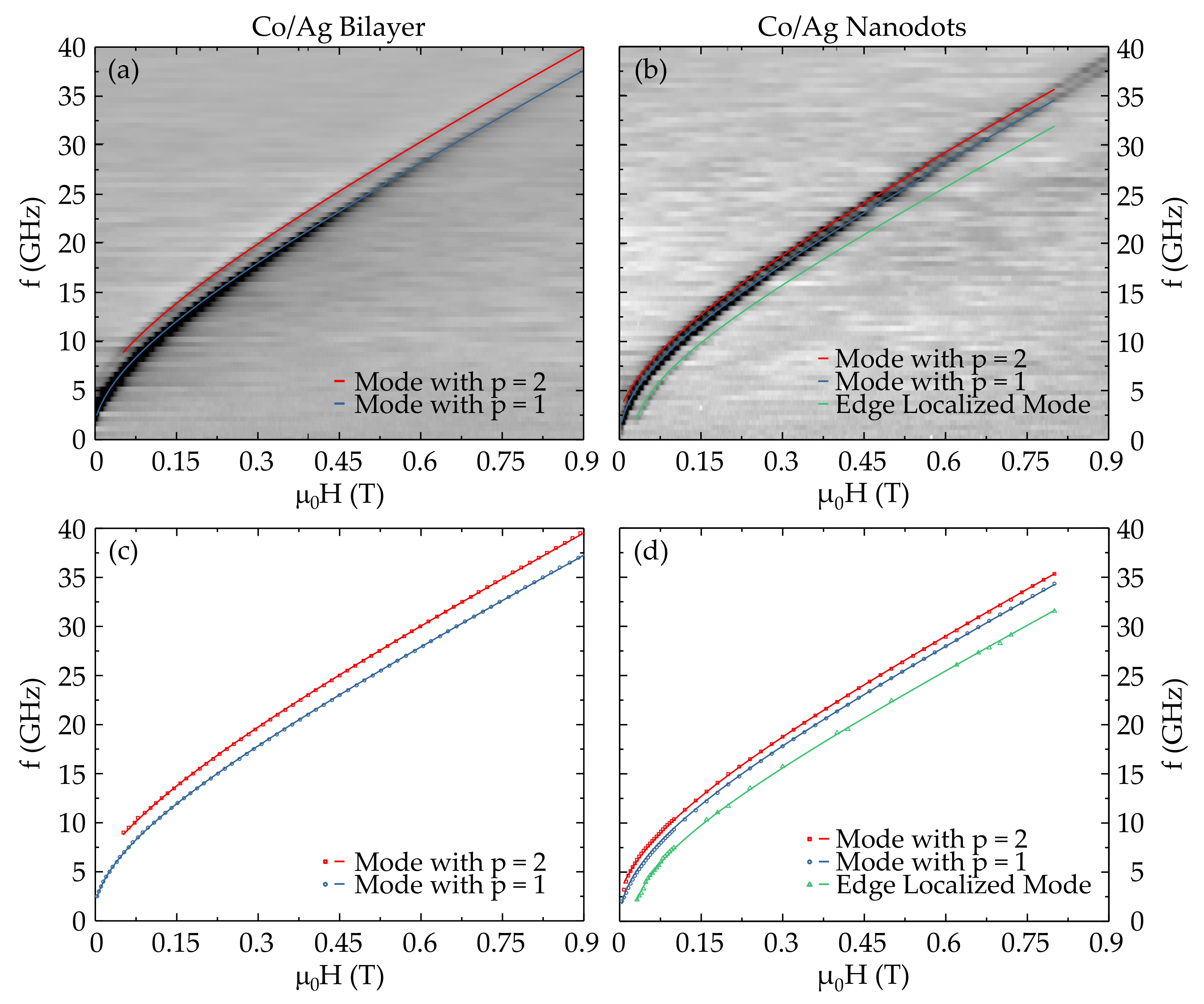
Figure 4, VNA-FMR spectra for both the Co(50 nm)/Ag(30 nm) bilayer and nanostructured dots are shown. We will analyze the bilayer sample first and then consider the nanodot sample.
For the bilayer system, we observe two clear resonance lines, which we interpret as arising from the first two perpendicular standing spin-wave (PSSW) modes, with
and
. The frequency–field characteristic and extracted data for the continuous thin film are illustrated in
Figure 4a,c, respectively. The fits to the data are performed using Equation (
10), which for our purposes takes the form:
We then use Equation (
21) in the fitting process to determine the relevant wave vectors and hence pinning parameter,
. For this we have assumed a symmetric pinning, with
. For the data shown in
Figure 4c we obtain
for
and
for
. The fitting procedure also used the following parameters:
T, a g-factor of
and an exchange stiffness constant of
J·m
[
28], which is reasonable for a 50 nm thick Co film. The fits shown in
Figure 4c are in good agreement with the data and the physical parameters are consistent with the Co film studied. With regards to the pinning conditions, we note that the value for
is close to unity and is consistent with the almost perfect pinning condition.
We now turn our attention to the results for the nanodot structures. These are shown in
Figure 4b,d for the raw and extracted data, respectively. The two resonance lines observed in the continuous thin film also appear to be present in the nanodot sample, though with modified wave vectors, these are shown in blue and red in
Figure 4c,d. In addition to these modes, there is a further resonance mode in the nanostructured sample, as seen in the spectra and illustrated by the line in green in
Figure 4d. This mode has a significantly weaker absorption, but is more clearly observed in the low frequency and low field range. The shift of resonance fields can be clearly attributed to the effect of nanostructuring. The first line that we can consider is the blue line, which appears to be very close to the corresponding blue line in the continuous film data for the
mode. The red line is somewhat shifted to smaller resonance frequencies with respect to the continuous film (
mode), but otherwise appears to be the same mode, which can be accounted for by a modified wave vector for this mode. Fitting these two lines can be based on the same principles as that for the thin film sample. In the latter, we considered that for a thin film the in-plane wave vectors should be zero since the film is effectively infinite in the film plane:
since
and thus
, see Equation (
12). For the case of our circular nanodots, the lateral dimensions, corresponding to the 200 nm diameter, can give rise to pinning conditions at the dot edges and thus we should consider the three-dimensional aspect of magnetic confinement. Using Equation (
12), we can take into account the dot geometry and the edge pinning conditions, which we can express in a similar manner to the thin film case, from which we can write:
where
denotes the edge pinning conditions and
those of the upper and lower interfaces,
is the dot diameter,
the films thickness. In considering the three-dimensional case, we need to account for the mode numbers in three directions, as represented by the integers
, and
. We note that Equation (
24) considers that the pinning is equivalent in both lateral directions and is symmetric. This is justified for the current geometry, since there is no reason to suppose that there should be any variation of the pinning at the edges of the dot structure. In our calculations, we consider that the in-plane anisotropy is weak and that inter-dot (dipole–dipole) interactions are sufficiently weak that we can neglect their effect.
In the case of the first two bulk (PSSW) modes (i.e., the blue and red lines in
Figure 4d), the resonance lines are slightly shifted towards a bulk-like uniform mode, since the origin of this curve is shifted to zero. The principal resonance line (denoted in blue) is very similar to that of the continuous film and can be expected to derive from similar boundary conditions as that of the
mode in the continuous thin film. The red line is much closer to the principal resonance (blue line) than that for the continuous film, which must be due to the modified boundary conditions for the nanodot structures. Since the lateral dimensions are almost four times greater than the perpendicular (thickness) dimension, we can expect the second term, in general, to dominate for the wave vector, Equation (
24). This will not be the case, however, for edge localized modes, since the degree of localization can be such that the in-plane wave vectors can rapidly increase for strong localization. We will discuss this issue shortly. We can express the resonance modes in terms of the quantization numbers
, and
r, from which we can write the lowest order modes as
etc. As previously, the quantization numbers are integer and commence at 1. We note that due to the circular symmetry, certain modes will be degenerated;
etc., but
. By analyzing the lowest modes, we can assess which are the ones most likely to correspond to the modes observed in the FMR spectra. Setting
and
, from Equation (
24) we can write:
Before we analyze in more detail the mode ordering, we should note that since the principal resonance line in the nanodot system (blue) can be assumed to derive from the first resonance
mode in the continuous film, the additional resonance in the nanodots (indicated in green in
Figure 4d) must therefore arise from a localized resonance mode. We conclude this from the fact that this resonance is situated at a higher magnetic field with respect to the principal resonance. This means that the wave vector is imaginary and pushes the resonance field to higher values since
. Inserting this into the resonance equation, Equation (
23), means that the resonance field is shifted up in value (or alternatively, the resonance frequencies shift down), while normal bulk or PSSW modes are situated at lower fields or higher frequencies. This can be seen from the resonance lines shown in
Figure 4. This conclusion is supported by the fact that the mode intensity is significantly weaker than the principal mode, which is to be expected since the modal intensity is proportional to the transversal dynamic magnetization. Using Equation (
23), we find a good fit to this (green) resonance for which
m
, see
Figure 4d.
To analyze the mode ordering, we first note that
m
and
m
, where we have used
d = 200 nm and
nm, respectively. We therefore see that
and can make the second terms in Equation (
24) dominate the wave vector. Since we can assume that the upper and lower pinning conditions should be close to those of the thin film, i.e.,
, we can further simplify the analysis. In effect, this means the second terms for the modes with
will effectively vanish, for example in Equations (
25), (
26) and (
28), etc. We can reasonably assign mode numbers corresponding to the wave vectors
and
or
to the first two PSSW modes, i.e., the blue and red lines shown in
Figure 4d. This is coherent with the decreased mode separations, with respect to the continuous thin film since this will depend on the first terms in the wave vectors and hence the lateral dimensions of the nanodot. Based on this analysis, we can provide excellent fits to the experimental data, as illustrated in
Figure 4d. For the fits we have used the following fit parameters:
T, a g-factor of
, and an exchange stiffness constant of
J·m
, which are in agreement with the parameters used in the fits for the continuous thin films. We note that the edge localized mode required a reduced value of the magnetization,
T. This parameter is important for setting the slope of the linear portion of the curve and may be interpreted as being due to a reduced magnetization at the edges of the nanodots.
4.2. NdCo/Al/Py Layered Structures
Trilayer samples consisting of a 64 nm thick amorphous NdCo
film with PMA and a 10 nm thick polycrystalline Py film with IMA, which are coupled through a nonmagnetic Al spacer, have been deposited via the magnetron sputtering technique. The trilayer structure itself is sandwiched between Al seed and capping layers, all of which have been grown on Si/SiO
substrates. The magnetic properties of the coupled thin films can be controlled by two independent parameters. On the one hand, varying the Co concentration (
x = 5, 7.5, and 9) in the NdCo
film allows the modification of the strength of its PMA. A maximum has been found for
5, whereas higher or lower Co concentrations lead to a gradually weaker PMA, respectively [
29,
30]. On the other hand, by adjusting the Al spacer thickness (
t = 0 nm, 2.5 nm, 5 nm, and 10 nm), the type of coupling between the two magnetic layers can be set to either direct exchange coupling (
1.5 nm) or stray field coupling (
2.5 nm). In addition to the coupled bi- and trilayers, a series of reference samples, consisting of a single 10 nm thick Py film as well as single 64 nm thick NdCo
films with varying Co concentrations
x, has also been prepared. For the remainder of this paper, the coupled trilayers will be named according to their Co concentration and Al spacer thickness as, e.g., X5T10 for a sample based on a NdCo
film and a 10 nm thick Al spacer.
The magnetic properties of the samples have been studied using magnetometry (AGM), MOKE, and FMR. In a previous work, we have analyzed the VNA-FMR data of these samples both as a function of the composition and as a function of the Al spacer thickness [
31]. Further characterization has been performed using magnetic force microscopy (MFM). Indeed, these measurements show that the remnant state of the sample has a marked stripe domain pattern, with a periodicity of around 140 nm. This stripe domain pattern has been shown to originate in the NdCo layer and is replicated in the coupled Py film. In our previous study, we presented data for the in-plane FMR with the external magnetic field
applied along the in-plane hard axis of the samples. We now present the comparison of the in-plane easy and hard axis measurements for samples with a composition value of
. The experimental FMR data are displayed in
Figure 5.
In this study, we note that the FMR signal comes solely from the Py layer and no explicit resonance is observed for the NdCo film. The FMR measurements were performed in a specific manner to enable us to correctly interpret and reproduce data. Prior to the FMR measurements, the samples were saturated with a static in-plane magnetic field of
= +0.9 T along their easy or hard axis, respectively, i.e., along the direction of the actual measurement. We then perform a full hysteresis cycle from +0.3 T to −0.3 T and then back to +0.3 T in steps of 2.5 mT. Since the spectra only show the Py FMR, we also show the FMR line for a single uncoupled Py film of the same thickness, as shown by the dash-dotted line in
Figure 5a,b. This is useful since it also gives a reference line for comparison with the FMR of the coupled Py layers. We note that the single Py film has a small in-plane anisotropy, which we estimate from the graph to give an anisotropy field of around
mT. We also note that the initial domain pattern after saturation are stripe domains, which are oriented in a direction parallel to
. Indeed, it is due to this characteristic that the sample system exhibits a reconfigurable anisotropy. We use the Py FMR as a method to probe the properties and the effect of the NdCo layer due to the magnetic coupling between the two ferromagnetic materials.
There are a number of important observations that we can make before considering a more detailed analysis. Firstly, we note that for the sample series under consideration, the non-magnetic Al spacer thickness allows us to vary the strength or the magnetic coupling between the NdCo
and Py layers. As the thickness increases, the coupling will reduce, and the Py layer characteristics will be expected to approach those of the isolated film (dashed line). This is roughly what can be observed in
Figure 5 and is more clearly seen for the easy axis orientation
Figure 5b. We have previously shown that the hysteretic behavior of the FMR line is strongly correlated to the hysteretic loop from magnetometry measurements. This allows us to find the coercive field,
, and the saturation field,
, as indicated in
Figure 5a, for the X7.5T2.5 sample. A further critical field,
, is indicated, which refers to the field at which the two branches of the hysteresis loop meet [
31]. The resonance line follows the arrows shown in
Figure 5a, where we note that the dashed arrows indicate the transition through zero-field, where the two branches (up-sweep and down-sweep) of the characteristics cross.
Figure 5c shows the corresponding raw FMR data for the X7.5T5 sample. In fact, we note that the transition through zero-field is a little more complex than the data points suggest. In the up-swept data shown, the resonance line appears to cross the zero-field and a jump in the resonance line occurs for small positive fields. This branch then increases and joins the uniform FMR line at the critical field. The down-swept line has lower values of resonance frequency than the up-swept branch in this field range. This then results in the hysteretic behavior of the resonance frequency. In this region, the resonance line appears to be rather weak. Above
, the resonance line is more pronounced and gradually joins the principal uniform resonance mode as the field reaches
.
For the hard axis measurements, the critical field appears to be independent of the spacer layer thickness, though the hysteresis is strongly influenced by the strength of the magnetic coupling of the Py layer with the NdCo underlayer. If we consider the evolution of the FMR f–H characteristics for the sample series, as t decreases and the magnetic coupling increases, the FMR branches generally shift to higher frequencies, the saturation fields increase and the hysteresis loops appears to be reduced. We also see from this that the zero-field values are strongly affected by the coupling, and increase significantly from the uncoupled Py data. In considering the free Py layer, we note that the coupled Py film in the trilayers have an induced stripe domain texture, which is responsible for these low field differences in the FMR behavior.
The easy axis data show that while the FMR shifts also follow similar trends to that observed along the hard axis, the shifts are generally smaller and the strength of the hysteresis is inverted with respect to the hard axis. Furthermore, the critical field values appear to decrease with increasing Al thickness. The easy axis data also shows that the FMR characteristics of the coupled Py layers approach those of the single Py film as the Al layer thickness increases and decoupling from the NdCo film increases.
In consideration of the FMR behavior in the regions below the coercive field, we note that FMR measurements are highly sensitive to the relative orientation of the stripe domains and the rf magnetic field. This can lead to acoustic and optical modes, due to in-phase and out-of-phase precession of the magnetization in adjacent stripe domains [
32], see
Figure 6. As simulated in [
33] for a single 200 nm thick Py film, stripe domains and rf magnetic field
(dc magnetic field
) are always perpendicular (parallel) during the entire hysteresis cycle and independent of the field sweep direction.
Above saturation, the magnetization of the coupled Py layer will be homogeneous and only the uniform FMR mode is observed. However, for applied magnetic fields lower than the saturation fields, stripe domains can give rise to two FMR modes, an acoustic mode and an optical mode, with the latter having a higher resonance frequency at the same magnetic field. The frequency difference between these modes can be substantial, though manipulation of the films can lead to a significant reduction in the separation of modes [
33,
34]. However, it should be mentioned that in the coupled trilayers, always just one type of mode is observed when sweeping the bias field
from positive to negative saturation and vice versa, with the mode order being always the following: uniform FMR mode → acoustic mode → optical mode → uniform FMR mode.
In order to understand the evolution of the resonance line, particularly in the region below saturation and between positive and negative coercive fields, we need to consider the orientation of the magnetization and the nature of the domain structure from the point of domain nucleation as the applied magnetic field
reduces from saturation to field strengths below the coercive field. This is intimately connected to the magnetization loops of the Py film. In the saturated state, the magnetization is rigorously aligned to the external field, with
. In this domain state, the FMR absorption will rigidly follow the normal uniform mode, as given by the Kittel equation, such as expressed by Equation (
23). We know that below both the saturation field and the coercive field, the magnetization can relax from the applied field orientation. In the coupled trilayers, any deviation from the Kittel equation would be indicative of a relaxation of the Py magnetization from the applied field direction, though the Py layer remains in a stripe domain state. Only below the coercive field does domain nucleation occur and the sample enters a multidomain state. For a stripe domain system, such as observed in Py coupled to NdCo, the relative sizes of the oppositely aligned domains will vary, as illustrated schematically in
Figure 7. These oppositely aligned domains will undergo FMR in either the acoustic or optical modes, as shown previously in
Figure 6, which occur at different magnetic fields (or frequencies).
From the hard axis data, see
Figure 5, we note that the resonance line continues without deviation, on passing through
, suggesting that the acoustic mode is excited (see
Figure 8a). The resonance line continues undeviated on passing through zero-field. However, once the positive coercive field is reached,
, there is an abrupt jump in the resonance line, as indicated by the red arrow in
Figure 8b. At this point in the field sweep, the sample is by definition in a state with equal volumes of the two magnetic domains. To understand the jump in the resonance line, we suggest that at this point the acoustic mode is suppressed and the optical mode becomes visible. For equal volumes of oppositely aligned domains, we expect the transverse magnetization to be zero, so at the exact value of
, the absorbed intensity will be zero. However, any further increase in
H will bring about an increase in the positive domains with respect to the negatively pointing domains, and the transverse magnetization will be non-zero and thus an optical mode can be observed (see
Figure 8a). We can resume this scenario in
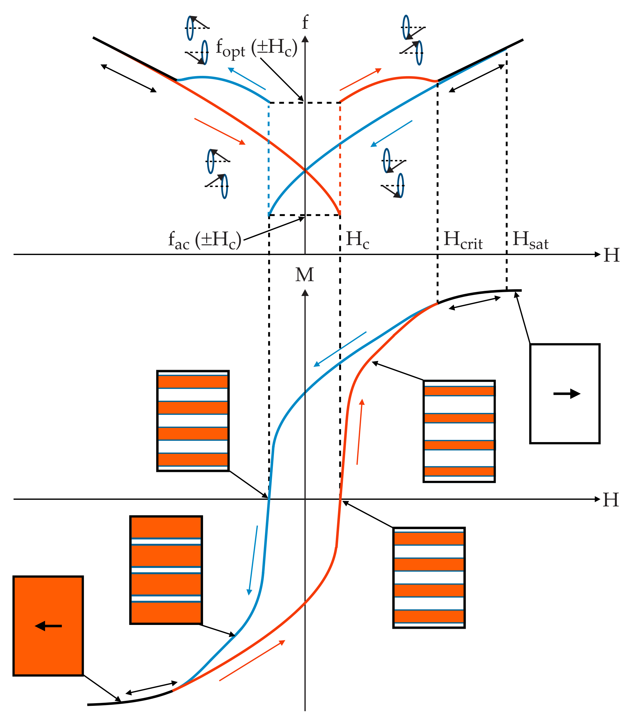
Figure 9, in which we follow the FMR hysteresis from negative to positive fields.
If we consider the resonance equation for the acoustic and optical excitations, we can adapt the FMR equation for the effective wave vectors associated with these modes. Since the in-phase, acoustic excitation follows from the uniform mode, we can consider a value of
, while the optical mode should have a
k-vector given by
, where
corresponds to the periodicity of the stripe domain texture. We can now consider the frequency shift at
as deriving from the resonance equations for these two excitations. Thus we can write:
where
and
are defined in
Figure 9. Given that the wave vector for the optical mode must physically correspond to a real value, we can express the optical mode wave vector from Equation (
30) as:
This allows us to directly assess the domain structure periodicity (at ), and if we require, its evolution with increasing field up to the critical field,. Once this field is reached, the sample becomes a single domain, at which must be zero, and . Any further increase of the applied field aligns the magnetization along this field direction. The saturation field will bring the system back to rigorous saturation and the uniform FMR mode is recovered. In fact, any difference between the uniform FMR frequency and the actual observed FMR frequency will be due to the misalignment of and .
The proposed model, schematically illustrated in
Figure 9, depicts the general form of both the magnetization and FMR hystereses. This is in agreement with previous findings [
31] and with the data presented in this paper. At this point, we note that our proposed model is made under the assumption that the imprint of the magnetic stripe domain pattern from the NdCo film into the coupled Py layer is strong. However, the strength of interaction between those two magnetic layers varies according to the thickness of the Al spacer. Synchrotron-based experiments are in progress to understand this issue in more detail.
From the fact that the second term in the square root of Equation (
31) is much larger than the first, we can simplify the expression and obtain an approximate relation between the stripe domain periodicity and the difference in frequency,
, such that:
The exchange stiffness constant for Py is of the order of
J·m
[
35], which allows us to evaluate the spin wave constant
. Using a value of
T (from fits of the in-plane VNA-FMR data of the 10 nm thick Py reference sample), we obtain
T·m
. Furthermore, we use
= 165.33 GHz/T. From the values of
GHz and
GHz for the hard axis measurement of sample X7.5T10, we calculate the stripe domain periodicity to be
nm. This is in excellent agreement with MFM images for this sample, where the periodicity was found to be around 140 nm.
There is a further element that requires explanation, which concerns the deviation of the FMR line from the uniform FMR line between the saturation and critical fields, as illustrated in
Figure 5. Indeed, this deviation is significant for samples with strong coupling between the ferromagnetic layers, i.e., with low Al interlayer thicknesses. This is particularly noticeable in the hard axis measurements, where the deviations are more significant. For the thinnest Al interlayer of
nm, there is a very large increase. From the increase of the zero-field frequency for the free Py layer, we can estimate an increase of the resonance frequency in the region of 4.2–4.5 GHz for this sample. For the sample with
nm, this value drops to around 1.0–1.5 GHz. The value of this deviation of the resonance frequency,
, should be related to the ferromagnetic coupling strength between the NdCo and Py layers. We can convert these frequencies into effective fields using a simple relation:
. Using the zero-field values for the frequencies, we estimate that the effective coupling field,
, between the NdCo and Py layers drops from about 0.4 T to about 0.1 T as the Al interlayer thickness increases from 2.5 nm to 10 nm. This represents a significant ferromagnetic coupling between the layers.


