Article

Coercivity and Exchange Bias in Ti-Doped Maghemite Nanoparticles

1
Research Institute of Medical & Health Sciences, University of Sharjah, Sharjah P.O. Box 27272, United Arab Emirates
2
Department of Physics, Sultan Qaboos University, Muscat P.O. Box 36, PC 123, Oman
3
Ioffe Physical Technical Institute, 194021 St. Petersburg, Russia
4
Department of Physics, College of Sciences, University of Sharjah, Sharjah P.O. Box 27272, United Arab Emirates
5
Department of Chemistry, United Arab Emirates University, Al-Ain P.O. Box 15551, United Arab Emirates
6
Department of Medical Diagnostic Imaging, College of Health Sciences, University of Sharjah, Sharjah P.O. Box 27272, United Arab Emirates
7
Department of Biomedical Engineering, Faculty of Engineering and Natural Sciences, Istinye University, Istanbul 34010, Turkey
8
Department of Physics, United Arab Emirates University, Al-Ain P.O. Box 15551, United Arab Emirates
*
Author to whom correspondence should be addressed.
Magnetochemistry 2022, 8(12), 165; https://doi.org/10.3390/magnetochemistry8120165
Submission received: 20 October 2022 / Revised: 12 November 2022 / Accepted: 18 November 2022 / Published: 23 November 2022
(This article belongs to the Special Issue Magnetic Nanoparticles: State of the Art and Future Perspectives)

Abstract

:
Ti-doped maghemite nanoparticles of average crystallite size 12.9 nm were synthesized using the sol–gel method. The XRD profile mainly showed the presence of maghemite phase with very small phases of TiO2 (rutile and anatase). Magnetization hysteresis loops of the nanoparticles were obtained between −4 T to +4 T at temperatures of 2, 10, 30, 50, 70, 100, 150, 200, and 300 K under field cooling (FC) of 1, 2, 3, and 4 T and zero-field cooling conditions (ZFC). The coercivity displayed nonmonotonic field dependence while it decreased sharply with temperature and vanished at 150 K at all fields. Horizontal hysteresis loop shifts were observed in the 2–150 K temperature range in both the ZFC and FC conditions. The exchange bias effect became negligible in both ZFC and FC states above 50 K. Magnetization vs. applied field measurements were conducted in both ZFC and FC cooled conditions at several temperatures in the range of 2–400 K, with spin freezing being observed below 50 K. The exchange bias effect obtained below 50 K is suggested to be attributed to the competing roles of the long-range dipolar and short-range exchange coupled interactions.

1. Introduction

Iron oxide-based hematite, maghemite, and magnetite magnetic nanoparticles (MNPs) are intensively studied due to their widespread applications in fields like magnetic storage, biomedicine, microwave devices, and catalysis [1,2,3,4]. The hematite in the antiferromagnetic phase has negligible magnetization at room temperature whereas maghemite and magnetite are ferrimagnetic in nature at room temperature [5]. The maghemite phase is the more stable phase at room temperature compared to magnetite, with slow weathering and low temperature oxidation of the magnetite leading to the formation of maghemite phase [6]. The maghemite exhibits a spinel crystal structure in which iron is present as trivalent cations as well as predominant cation vacancies with defective spinel lattice compared to the magnetite spinel lattice in which iron is occupied by Fe2+ and Fe3+ cations in tetrahedral and octahedral sites. Although the maghemite lattice has a defective lattice, it still shows excellent saturation magnetization which is essential for certain applications [7]. The maghemite phase when heated in the temperature range of 250–750 °C is unstable and transforms into rhombohedral hematite (α-Fe2O3) [8].
Tuning the magnetic properties of maghemite nanoparticles such as coercivity, remanent magnetization, saturation magnetization, and exchange bias is an important requirement to improve the efficiency of the magnetic nanoparticle in various applications [9,10,11]. These nanoscale magnetic properties are optimized by changing the particle size, surface dead layer, magnetic interface, and geometry of MNPs [12,13,14]. Using chemical methods, these properties are easily tuned by changing the synthetic conditions [15]. Fantechi et al. has reported the synthesis Co2+-doped maghemite nanoparticles with sizes 5–6 nm using the organometallic decomposition method [16]. They have shown that the doping of Co2+ to the maghemite nanoparticles can cause a nontrivial evolution of magnetic characteristics such saturation magnetization, blocking temperature, and coercive field. Saturation magnetization has strong dependence on the tetrahedral and octahedral occupation by Co2+ which significantly enhances the anisotropy constant. This in turn enhances the application of maghemite nanoparticles in magnetic hyperthermia and MRI contrast agents. Can et al. reported on the tuning of magnetic hyperthermia efficiency of Ti-doped magnetite nanoparticles, suggesting that the Ti concentration in the spinel lattice plays a role in enhancing the heating ability. SiO2-coated Ti-doped magnetite ((Fe1−xTix)3O4 x = 0.02, 0.03 and 0.05) synthesized using sol–gel method showed high heating ability with a Ti concentration of x = 0.03 [17]. Studying the exchange bias properties of the Ti-doped maghemite nanoparticles will help in understanding the enhanced heating ability in the doped magnetic nanoparticles.
The hysteresis loops have been observed to move away from the center of the loop when two different magnetic phases are brought into contact with one another. The exchange contact (coupling) between the various magnetic phases was thought to be the cause of the loop shifts. The magnitude of the shift of the hysteresis loops can be used to determine the exchange bias field, or EB, which is used to describe the exchange coupling. At the core–shell interface of MNPs, where the materials for the core and shell have various magnetic phases, exchange coupling occurs [18]. Even a minor exchange bias effect between a similar magnetic phase core and shell has been documented [19]. The exchange bias effect may also be caused by the exchange coupling between the uncompensated surface spins and the core moments [20]. The exchange anisotropy of core–shell MNPs is poorly revealed at the microscopic level. Numerous technologies, including spintronic devices, spin valves, and magneto resistive random-access memory circuits, make use of the exchange bias effect, which is a significant factor in deciding the efficiency of the nanoparticles.

2. Experimental Methods

2.1. Synthesis of Ti-Doped Maghemite Nanoparticles

Ti(IV)-doped maghemite was prepared by sol–gel method using Ti(nOBu)4 (TB) and Fe(NO3)3.9H2O (FeNt) in 2-propanol (PrOH) as the solvent and propylene oxide (PO) (Sigma Aldrich, Germany) as a gelation promoter. Solutions containing the appropriate amounts of both precursors were separately prepared and then mixed. In a typical experiment, 10.7 g (26.4 mmol) FeNt was dissolved in 50 mL 2-propanol, and 1.0 mL (2.94 mmol) TB was dissolved in 10 mL of the same solvent. The FeNt solution was added to the TB solution, and 20.4 mL (0.293 mol) PO was immediately added to the mixture. After a stirring of the mixture for three hours, it was aged for 24 h to produce a gel. Through evaporation of the solvent from the gel in a water bath at 80 °C, the gel solvent was removed. The product was then dried in an oven for one hour at 120 °C before being calcined for four hours at 350 °C.

2.2. Structure Analysis

The crystalline structure and phases of the nanoparticles were characterized from the X-ray diffraction (XRD) using a Shimadzu-6100 powder XRD diffractometer (Shimadzu, Japan) with Cu–Ka radiation source (λ = 1.542 Å). The diffraction profile of the maghemite nanoparticles was obtained in the range of 20–80 degrees at a rate of 1 degree/min. The average sizes of the nanoparticles were determined using the full width half maxima (FWHM)obtained by Gaussian fitting of the highest intensity peak (311). The average crystallite sizes were calculated using the Scherrer equation:   D P = 0.94 λ β   cos θ where D P is the average crystallite size, β is the FWHM of the highest intensity peak, λ is the X-ray wavelength, and θ is the Brags angle.

2.3. Magnetic Measurements

The Physical Properties Measurement System (PPMS, Quantum Design, San Diego, CA, USA) from Quantum Design was used to obtain the DC magnetic measurements of the nanoparticles using vibrating sample magnetometer (VSM). Field-dependence magnetization (M–H) measurements were made using the zero field cooled (ZFC) and field cooled (FC) conditions at different temperatures (2, 10, 30, 50, 70, 100, 150, 200, and 300 K) in the field range of −4 T to +4 T. The ZFC MH loops were obtained by cooling the particles in the absence of external magnetic field to the required temperature, and the field was cycled from −4 T to +4 T. Similarly, the FC cooled curves were obtained by cooling the nanoparticles under an external magnetic field of 1, 2, 3, and 4 T to the required temperature.

3. Results and Discussion

The XRD pattern of the nanoparticles synthesized is shown in Figure 1. The profile consists of the peaks corresponding to the maghemite phase (γ-Fe2O3) and a small fraction of the TiO2 phases (rutile and anatase). The maghemite peaks are indexed in the XRD profile and the small fraction of TiO2 phases are indicated by the * in Figure 1 [21]. The unit cell parameters of the nanoparticles were determined using Rietveld refinement. The diffractogram the Ti-doped maghemite nanoparticles matches with the cubic symmetry (space group P4132); Rietveld refinement was performed using FullProf open-source version 7.50. Table 1 summarizes the refined parameters, and the lattice parameter obtained was 8.3344 Å which is on lower side compared to the undoped maghemite nanoparticles. The calculated parameter was compared to the undoped (8.35673 Å) and (divalent and trivalent) doped maghemite nanoparticles reported [16,22]. The lattice parameter decreased to a great extent due to the smaller ionic radius of Ti4+ compared to the ionic radius of Fe3+. The average size of the maghemite nanoparticles determined using FWHM of the highest intensity peak (311) and Scherrer method was 12.9 nm. The WD-XRF (wavelength dispersive X-ray fluorescence) spectrum of the Ti-doped maghemite nanoparticles was obtained using a Rigaku ZSX Primus IV spectrometer (Rigaku Corporation, Japan) to perform the elemental analysis. The Ti mole fraction obtained from the intensity of the iron and titanium was 0.11.
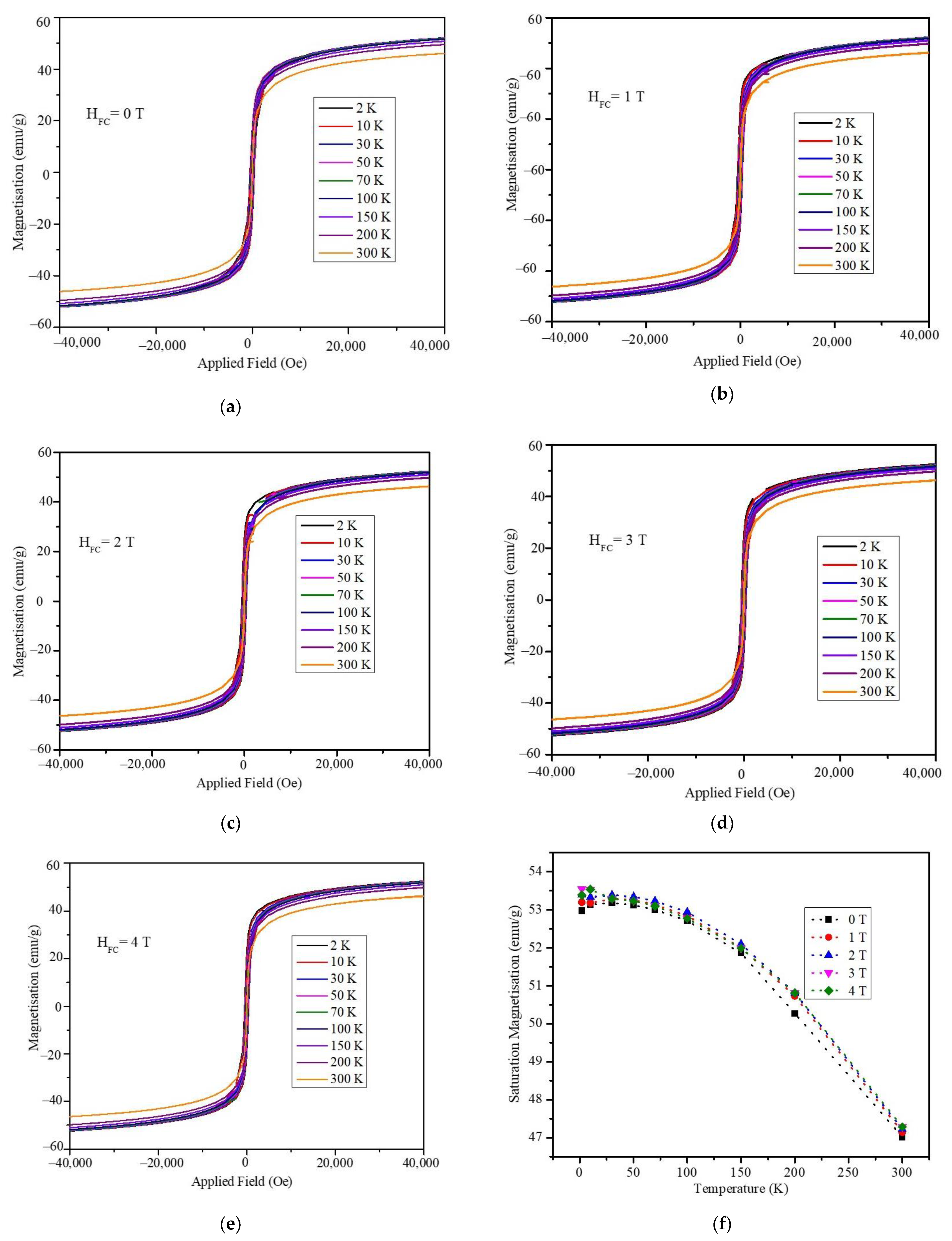
Hysteresis loops of the magnetization vs. applied magnetic field obtained in the temperature range of 2–300 K under field cooling of 0, 1, 2, 3, and 4 T are shown in Figure 2a–e. The MH loops were measured in the applied dc magnetic field range from +4 T to −4T. During the FC measurements, the particles were cooled to the required temperature in the range of 2–300 K under the influence of the magnetic field HFC, and the magnetization was measured as a function of the applied magnetic field. From the hysteresis plots, it is clear that the nanoparticles showed considerable hysteresis loop openings at temperatures below 100 K. However, the remanent magnetization and coercive field values were negligible at room temperature, indicating the superparamagnetic nature of the nanoparticles. The temperature-dependent saturation magnetization of the particles measured under all field cooling is shown in Figure 2f. The saturation magnetization of the nanoparticles showed high dependency on the temperature with the highest magnetization of 53.2 emu/g at 2 K, which decreased to 47.14 K at room temperature. The magnetization decreased gradually with the increase in the temperature from 2 to 100 K and fell sharply with the further increase in temperature to 300 K. The field cooling had little effect on the saturation magnetization values. The absolute values of Ms were slightly higher for the field cooled (1,2,3, and 4 T) conditions compared to the ZFC measurement.
The nanoparticles were cooled to 2 K in a zero magnetic field before being exposed to a tiny field of 70, 100, or 200 Oe to record the ZFC-FC magnetization measurements. To obtain the ZFC magnetization curves, the sample was heated from 2 to 400 K while the magnetization was being measured. A magnetic field of 70, 100, or 200 Oe was applied while the particles were cooled from 400 to 2 K, and the magnetization was measured as a function of temperature to produce the FC magnetization curves. Figure 3 displays the FC and ZFC plots that were produced for the maghemite nanoparticles. The blocking temperature of the maghemite nanoparticles is indicated by the arrow in each ZFC plot. The blocking temperature measured shows a dependency on the applied magnetic field used for obtaining the MT measurements as evident from the plots at 200 Oe; the blocking temperature is 187.5 K, whereas at 70 and 100 Oe, it is around 217.5 K. The broadening of the peak region in the ZFC plots indicates that the nanoparticles possess significant size distribution. The shift in the blocking temperature with respect to the magnetic field applied can be attributed to the dipolar interaction of the nanoparticles [23]. The magnetization in the FC plot increases monotonically with the decrease in the temperature down to 50 K, below which it flattens. This flattening nature of the FC curve below 50 K after bifurcation hints at the existence of surface spin freezing at low temperatures [24].
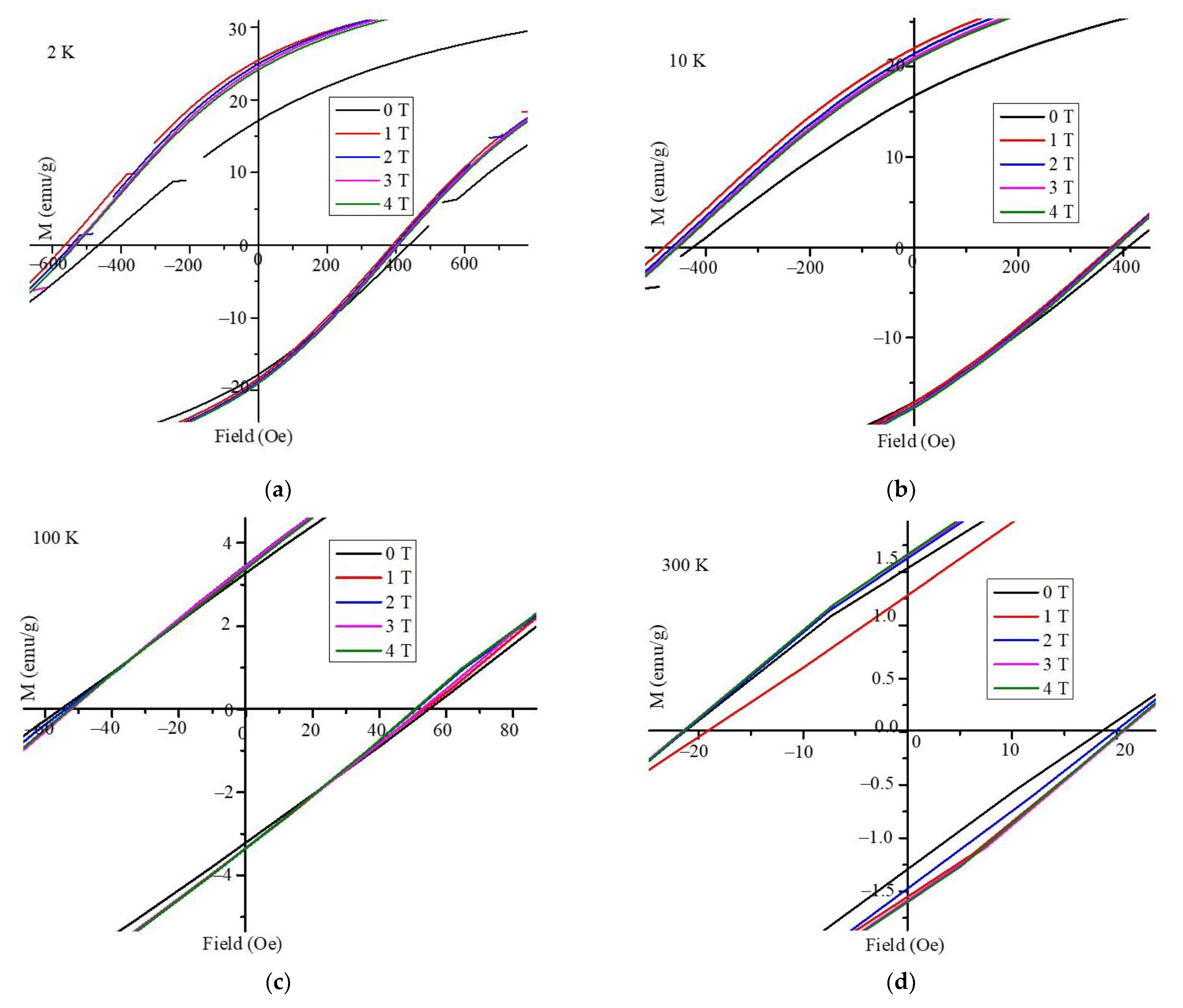
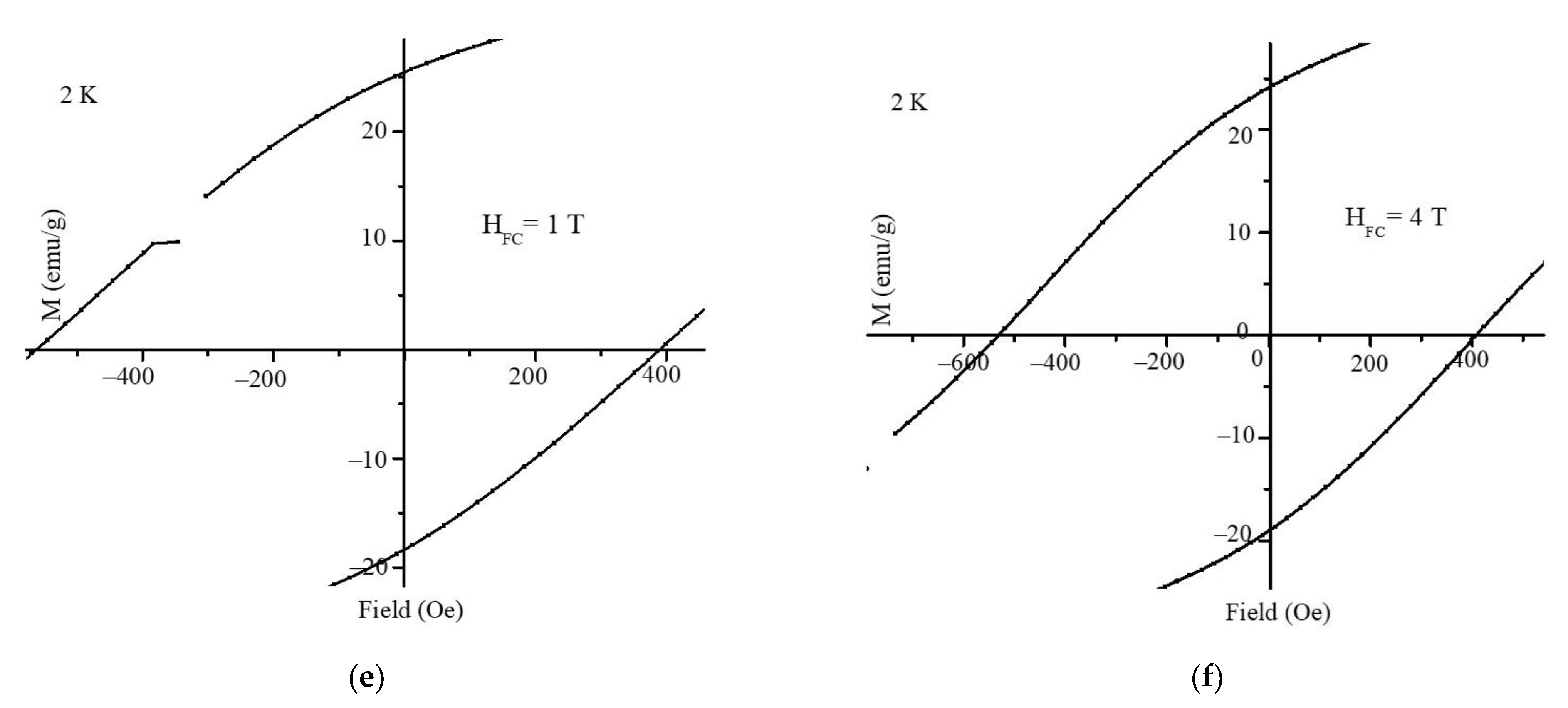
The horizontal shift in the M–H hysteresis loop away from the origin was measured to ascertain the exchange bias. Figure 4 displays the expanded M–H graphs at the hysteresis loop origin for temperatures of 2, 10, 100, and 300 K, Figure 4a–d. The shifts in the hysteresis loops depended on both the temperature and the field-cooling values, as shown in Figure 4. Figure 4e,f clearly depict the variations in the hysteresis loop at 2 K for various degrees of field cooling. The coercivity and exchange bias fields were determined from the M–H hysteresis loops. The magnetic field value at which the magnetization drops to zero during M–H measurements is known as the coercivity (or coercivity field, HC). The coercivity and exchange bias values were calculated from the hysteresis loops shown in Figure 4. The following Equation (1) was used to calculate the coercivity HC [25]:
H C = | H C 1 H C 2 | 2  
The exchange bias field, termed HEB, was used to determine the horizontal shift in the hysteresis loops:
H EB = H C 1 + H C 2 2
Here, the coercive field at the hysteresis loop descending branch is designated as HC1, and the coercive field at the ascending branch as HC2.
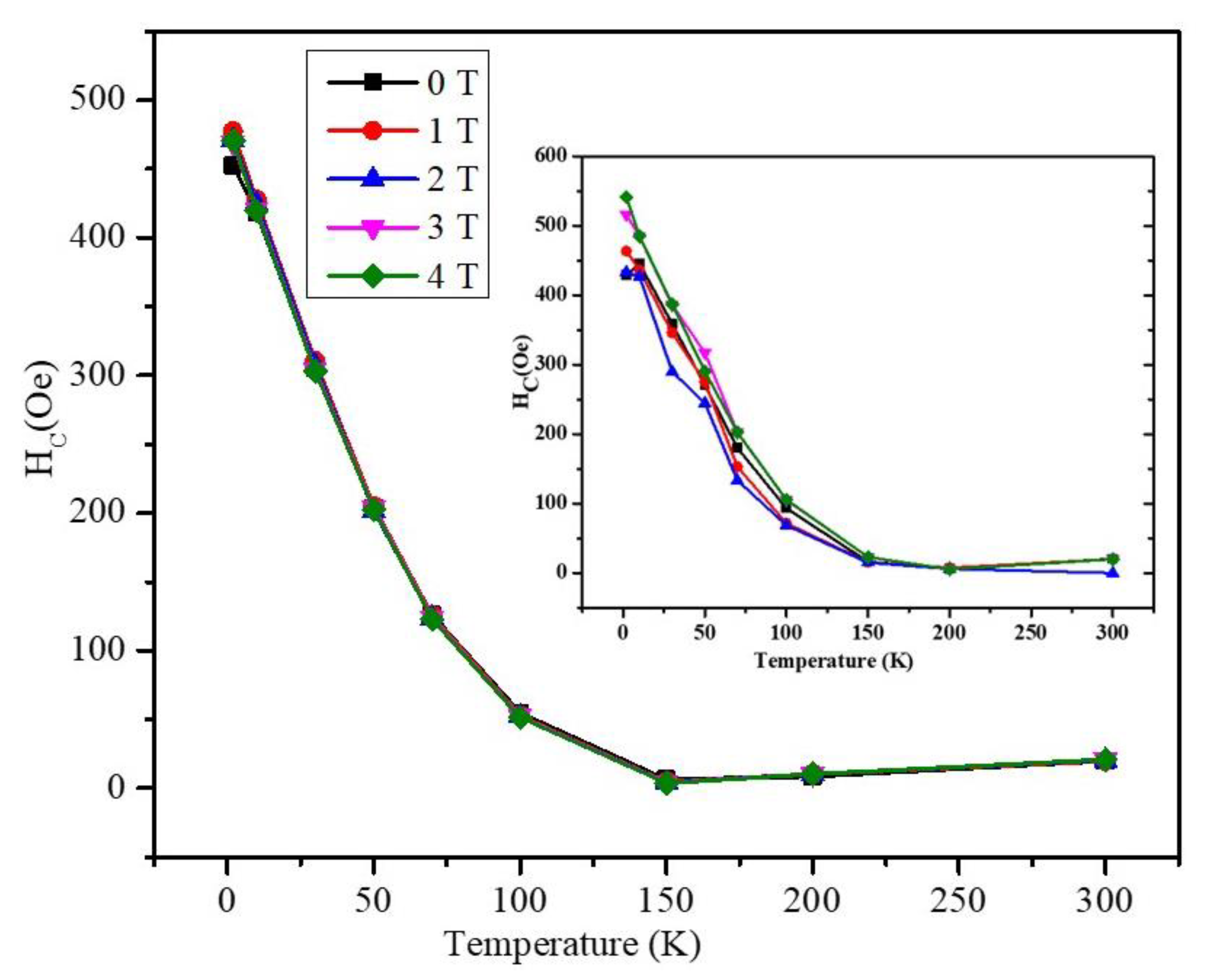
Figure 5 displays the coercivity as a function of temperature at a zero applied field (0 T) and several field cooling values (1, 2, 3, 4 T). It can be seen that all curves have similar trends indicating the absence of any effect for the cooling field on the coercivity. At all cooled fields, Hc has the highest value of 477 Oe at temperature 2 K with field cooling 1 T, and it decreases sharply with increasing temperature up to 100 K. Above 100 K, the coercivity of the nanoparticles becomes negligible at all field cooling values. The inset is the comparison of the coercivity with respect to temperature of the undoped maghemite nanoparticles reported in our earlier study [26].
Figure 6 displays the nanoparticles exchange bias field (HEB) values as a function of temperature at various field cooling settings. The FC state and the ZFC state both had negative HEB values at all field cooling values below 50 K, as shown in the Figure 6a. At all field cooling fields above 50 K, the exchange bias vanished in both the FC and ZFC states. The improved exchange coupling interaction in the Ti-doped maghemite nanoparticles included the presence of considerable HC and HEB values at low temperatures. The absolute exchange bias values of the Ti-doped nanoparticles were compared with our earlier report of undoped maghemite nanoparticles with similar size synthesized under the same conditions [26]. The saturation magnetization of the Ti-doped maghemite nanoparticles was 47 emu/g compared with only 40 emu/g for the undoped maghemite. The absolute exchange bias HEB, values of Ti-doped maghemite nanoparticles were also slightly higher compared with those of the undoped maghemite nanoparticles. We believe that the substitution of Fe cations by the smaller Ti cations decreased the lattice constant and thus increased lattice defects resulting in random (or canted) surface spins at the surfaces of the nanoparticles. The surface spins can freeze and form a spin glass-like phase at low temperatures [27,28,29]. For a specific orientation of the core magnetization, the canted surface spins could have multiple configurations leading to a reduction of magnetization [30] and an enhancement of the effective magnetic anisotropy of nanoparticles [31,32]. The exchange coupling delivers a further magnetic (exchange) anisotropy to align the surface spins in certain directions. The exchange coupling decreases with the increase of temperature and is expected to vanish above the spin freezing temperature, which was 50 K in our sample. Hence, the observed exchange bias in the Ti-doped maghemite nanoparticles could be attributed mainly to the magnetic short-range exchange coupling between the core magnetic moment with the frozen surface spins at the surface of the nanoparticle as we outlined in our previous report on nearly pure maghemite nanoparticles [26]. Referring to [26] regarding mainly maghemite nanoparticles, we might suggest that at low temperatures, spin-glass regions emerged between the maghemite nanoparticles due to the freezing of their surface spins in random orientations. We argue that the exchange bias effect is mainly due to the short-range exchange interaction between the particles core spins and the surface spins (in the spin-glass regions). The long-range dipole–dipole interactions between the magnetic moments of the nanoparticles are assumed to be small compared with the short-range spin–spin exchange couplings. The exchange bias field values were found to have monotonic dependence on temperature at all fields except at zero applied field. However, the exchange bias field had nonmonotonic field dependence at all temperatures below 50 K. The exchange bias field at zero applied field displayed both positive and negative values as shown in the inset of Figure 6b. In this study on maghemite nanoparticles with 10% Ti doping, interestingly, we obtained very similar coercivity and exchange bias values with very similar temperatures and field dependencies. One main difference is that at zero applied field, we observed negative exchange bias field at all temperatures. One other observed difference was the degree of field nonmonotonicity of the exchange bias and the decrease in coercivity, as shown in Figure 5. These observations indicate that the previously reported model of short-range spin–spin exchange coupling mainly controls the exchange bias effect. However, the introduction of Ti to the maghemite nanoparticles produced a small amount of TiO2 nanoparticles which resulted in some separations of the maghemite nanoparticles. This separation of the maghemite nanoparticles diminished the role of the short-range spin–spin exchange coupling, and thus the long-range dipolar interactions between the magnetic moments of the maghemite nanoparticles began to become significant. This long-range dipolar interaction between the magnetic moments of the maghemite nanoparticles mediated by the nonmagnetic TiO2 particles led to frozen moments at low temperatures (below 50 K) producing a super-spin-glass phase which was reflected in the almost temperature-independent magnetization below 50 K in the FC measurement (Figure 3). This super-spin-glass phase led to the small differences in exchange bias between the pure maghemite and the Ti-doped samples. Hence, in the current sample, both short range direct spin–spin exchange coupling and the long-range dipolar moment interactions coexisted and displayed the complex temperature- and field-dependence exchange bias effect. We expect that with the increase of the Ti percentage, the long-range dipolar interactions will play a bigger role in determining the exchange bias at the expense of the short-range spin–spin exchange coupling which diminishes as more maghemite nanoparticles become separated by the Ti particles. This study supports the possible tailoring of magnetic NPs to suite multifunctional applications.

4. Conclusions

The sol–gel method was used to synthesize the Ti-doped maghemite nanoparticles, which were characterized using XRD (structural) and magnetic measurements in the temperature range of 2–300 K. The nanoparticles were found to have maghemite phase with very small additional peaks of TiO2, indicating that most of the Ti atoms are present in the maghemite crystal lattice. In the ZFC and FC states, magnetic measurements were made between −4 T and +4 T at different temperatures and field chilled values. Surface spin freezing was predicted with the temperature FC behavior to occur below 50 K. At low temperatures, significant exchange bias and coercivity fields were obtained. The coercivity and exchange bias field were observed to decrease with temperature. Above the spin freezing temperature of 50 K, the exchange bias field disappeared. The competing roles of the long-range dipolar contacts and short-range spin–spin exchange couplings (between the core spins and the surface spins) are believed to be the cause of the exchange bias of the Ti-doped maghemite nanoparticles (between the cores of maghemite nanoparticles separated by TiO2 particles). These findings suggest that the magnetic characteristics of maghemite nanoparticles could be tailored for potential uses in magnetic hyperthermia and data storage.

Author Contributions

Conceptualization, I.M.O.; methodology, V.N., I.A.A.-O. and A.K; validation, B.I. and I.M.O.; formal analysis, V.N., I.A.A.-O., H.K., and A.S.K.; data curation, I.A.A.-O., A.S.K., V.N., and A.K.; writing—original draft preparation, V.N.; writing—review and editing, H.K., B.I., A.S.K., and I.M.O.; supervision, I.M.O.; project administration, I.M.O., and B.I.; funding acquisition, B.I. All authors have read and agreed to the published version of the manuscript.

Funding

University of Sharjah and Sharjah Research Academy for the Collaborative Research grant #2101050262.

Institutional Review Board Statement

Not applicable.

Informed Consent Statement

Not applicable.

Data Availability Statement

Not applicable.

Acknowledgments

The authors would like to thank the University of Sharjah and Sharjah Research Academy for the Collaborative Research (grant #2101050262). The authors also thank the Research Institute of Medical and Health Sciences (RIMHS) at the University of Sharjah.

Conflicts of Interest

The authors declare no conflict of interest.

References

  1. Wu, W.; He, Q.; Jiang, C. Magnetic Iron Oxide Nanoparticles: Synthesis and Surface Functionalization Strategies. Nanoscale Res. Lett. 2008, 3, 397–415. [Google Scholar] [CrossRef] [PubMed] [Green Version]
  2. Montiel Schneider, M.G.; Martín, M.J.; Otarola, J.; Vakarelska, E.; Simeonov, V.; Lassalle, V.; Nedyalkova, M. Biomedical Applications of Iron Oxide Nanoparticles: Current Insights Progress and Perspectives. Pharmaceutics 2022, 14, 204. [Google Scholar] [CrossRef] [PubMed]
  3. Hasany, S.F.; Abdurahman, N.H.; Sunarti, A.R.; Jose, R. Magnetic Iron Oxide Nanoparticles: Chemical Synthesis and Applications Review. Curr. Nanosci. 2013, 9, 561–575. [Google Scholar] [CrossRef]
  4. Turcheniuk, K.; Tarasevych, A.V.; Kukhar, V.P.; Boukherroub, R.; Szunerits, S. Recent Advances in Surface Chemistry Strategies for the Fabrication of Functional Iron Oxide Based Magnetic Nanoparticles. Nanoscale 2013, 5, 10729–10752. [Google Scholar] [CrossRef]
  5. Shokrollahi, H. A Review of the Magnetic Properties, Synthesis Methods and Applications of Maghemite. J. Magn. Magn. Mater. 2017, 426, 74–81. [Google Scholar] [CrossRef]
  6. Li, Z.; Chanéac, C.; Berger, G.; Delaunay, S.; Graff, A.; Lefèvre, G. Mechanism and Kinetics of Magnetite Oxidation under Hydrothermal Conditions. RSC Adv. 2019, 9, 33633–33642. [Google Scholar] [CrossRef] [Green Version]
  7. Coduri, M.; Masala, P.; Del Bianco, L.; Spizzo, F.; Ceresoli, D.; Castellano, C.; Cappelli, S.; Oliva, C.; Checchia, S.; Allieta, M.; et al. Local Structure and Magnetism of Fe2O3 Maghemite Nanocrystals: The Role of Crystal Dimension. Nanomaterials 2020, 10, 867. [Google Scholar] [CrossRef]
  8. Ponomar, V.P.; Antonenko, T.S.; Vyshnevskyi, O.A.; Brik, A.B. Thermally Induced Changes in the Magnetic Properties of Iron Oxide Nanoparticles under Reducing and Oxidizing Conditions. Adv. Powder Technol. 2020, 31, 2587–2596. [Google Scholar] [CrossRef]
  9. Yüksel, Y. Effects of the Particle Size and Shape of the Magnetic Nanoparticles on the Magnetic Hyperthermia and Exchange Bias Properties☆. Phys. B Condens. Matter 2019, 575, 411689. [Google Scholar] [CrossRef]
  10. Lu, A.-H.; Salabas, E.L.; Schüth, F. Magnetic Nanoparticles: Synthesis, Protection, Functionalization, and Application. Angew. Chem. Int. Ed. 2007, 46, 1222–1244. [Google Scholar] [CrossRef]
  11. Skumryev, V.; Stoyanov, S.; Zhang, Y.; Hadjipanayis, G.; Givord, D.; Nogués, J. Beating the Superparamagnetic Limit with Exchange Bias. Nature 2003, 423, 850–853. [Google Scholar] [CrossRef]
  12. Mercante, L.A.; Melo, W.W.M.; Granada, M.; Troiani, H.E.; Macedo, W.A.A.; Ardison, J.D.; Vaz, M.G.F.; Novak, M.A. Magnetic Properties of Nanoscale Crystalline Maghemite Obtained by a New Synthetic Route. J. Magn. Magn. Mater. 2012, 324, 3029–3033. [Google Scholar] [CrossRef] [Green Version]
  13. Andersson, M.S.; Mathieu, R.; Lee, S.S.; Normile, P.S.; Singh, G.; Nordblad, P.; Toro, J.A.D. Size-Dependent Surface Effects in Maghemite Nanoparticles and Its Impact on Interparticle Interactions in Dense Assemblies. Nanotechnology 2015, 26, 475703. [Google Scholar] [CrossRef]
  14. Di Corato, R.; Aloisi, A.; Rella, S.; Greneche, J.-M.; Pugliese, G.; Pellegrino, T.; Malitesta, C.; Rinaldi, R. Maghemite Nanoparticles with Enhanced Magnetic Properties: One-Pot Preparation and Ultrastable Dextran Shell. ACS Appl. Mater. Interfaces 2018, 10, 20271–20280. [Google Scholar] [CrossRef] [PubMed]
  15. Kolhatkar, A.G.; Jamison, A.C.; Litvinov, D.; Willson, R.C.; Lee, T.R. Tuning the Magnetic Properties of Nanoparticles. Int. J. Mol. Sci. 2013, 14, 15977–16009. [Google Scholar] [CrossRef] [Green Version]
  16. Fantechi, E.; Campo, G.; Carta, D.; Corrias, A.; de Julián Fernández, C.; Gatteschi, D.; Innocenti, C.; Pineider, F.; Rugi, F.; Sangregorio, C. Exploring the Effect of Co Doping in Fine Maghemite Nanoparticles. J. Phys. Chem. C 2012, 116, 8261–8270. [Google Scholar] [CrossRef]
  17. Can, M.M.; Bairam, C.; Aksoy, S.; Kuruca, D.S.; Kaneko, S.; Aktaş, Z.; Öncül, M.O. Effect of Ti Atoms on Néel Relaxation Mechanism at Magnetic Heating Performance of Iron Oxide Nanoparticles. Coatings 2022, 12, 481. [Google Scholar] [CrossRef]
  18. Liu, X.; Pichon, B.P.; Ulhaq, C.; Lefèvre, C.; Grenèche, J.-M.; Bégin, D.; Bégin-Colin, S. Systematic Study of Exchange Coupling in Core–Shell Fe3−δO4@CoO Nanoparticles. Chem. Mater. 2015, 27, 4073–4081. [Google Scholar] [CrossRef]
  19. Silva, F.G.; Depeyrot, J.; Raikher, Y.L.; Stepanov, V.I.; Poperechny, I.S.; Aquino, R.; Ballon, G.; Geshev, J.; Dubois, E.; Perzynski, R. Exchange-Bias and Magnetic Anisotropy Fields in Core–Shell Ferrite Nanoparticles. Sci. Rep. 2021, 11, 5474. [Google Scholar] [CrossRef]
  20. Muhammed Shameem, P.V.; Senthil Kumar, M. Training Effect of the Exchange Bias in Sputter Deposited Fe3O4 Thin Films with Varying Thickness. J. Magn. Magn. Mater. 2018, 458, 241–252. [Google Scholar] [CrossRef]
  21. Kim, W.; Suh, C.-Y.; Cho, S.-W.; Roh, K.-M.; Kwon, H.; Song, K.; Shon, I.-J. A New Method for the Identification and Quantification of Magnetite–Maghemite Mixture Using Conventional X-ray Diffraction Technique. Talanta 2012, 94, 348–352. [Google Scholar] [CrossRef] [PubMed]
  22. Cervellino, A.; Frison, R.; Cernuto, G.; Guagliardi, A.; Masciocchi, N. Lattice Parameters and Site Occupancy Factors of Magnetite–Maghemite Core–Shell Nanoparticles. A Critical Study. J. Appl. Cryst. 2014, 47, 1755–1761. [Google Scholar] [CrossRef]
  23. Lee, K.; Jang, J.; Nakano, H.; Nakagawa, S.; Paek, S.H.; Bae, S. External Magnetic Field Dependent Shift of Superparamagnetic Blocking Temperature Due to Core/Surface Disordered Spin Interactions. Nanotechnology 2017, 28, 075710. [Google Scholar] [CrossRef] [PubMed]
  24. Nayek, C.; Manna, K.; Bhattacharjee, G.; Murugavel, P.; Obaidat, I. Investigating Size- and Temperature-Dependent Coercivity and Saturation Magnetization in PEG Coated Fe3O4 Nanoparticles. Magnetochemistry 2017, 3, 19. [Google Scholar] [CrossRef] [Green Version]
  25. Obaidat, I.M.; Nayek, C.; Manna, K.; Bhattacharjee, G.; Al-Omari, I.A.; Gismelseed, A. Investigating Exchange Bias and Coercivity in Fe3O4–γ-Fe2O3 Core–Shell Nanoparticles of Fixed Core Diameter and Variable Shell Thicknesses. Nanomaterials 2017, 7, 415. [Google Scholar] [CrossRef] [Green Version]
  26. Alaabed, S.; Obaidat, I.M.; Khaleel, A.; AbuShareb, A.; Abdi, I.; Farouk, M.; Narayanaswamy, V.; Al-Omari, I.A. Positive and Negative Exchange Bias in Maghemite Nanoparticles. Mater. Today Proc. 2020, 28, 611–614. [Google Scholar] [CrossRef]
  27. Kodama, R.H.; Berkowitz, A.E.; McNiff, E.J.; Foner, S. Surface Spin Disorder in Ferrite Nanoparticles (Invited). J. Appl. Phys. 1997, 81, 5552–5557. [Google Scholar] [CrossRef]
  28. Mørup, S.; Brok, E.; Frandsen, C. Spin Structures in Magnetic Nanoparticles. J. Nanomater. 2013, 2013, e720629. [Google Scholar] [CrossRef] [Green Version]
  29. Obaidat, I.; Mohite, V.; Issa, B.; Tit, N.; Haik, Y. Predicting a Major Role of Surface Spins in the Magnetic Properties of Ferrite Nanoparticles. Cryst. Res. Technol. 2009, 44, 489–494. [Google Scholar] [CrossRef]
  30. Kodama, R.H. Magnetic Nanoparticles. J. Magn. Magn. Mater. 1999, 200, 359–372. [Google Scholar] [CrossRef]
  31. Luis, F.; Torres, J.M.; García, L.M.; Bartolomé, J.; Stankiewicz, J.; Petroff, F.; Fettar, F.; Maurice, J.-L.; Vaurès, A. Enhancement of the Magnetic Anisotropy of Nanometer-Sized Co Clusters: Influence of the Surface and of Interparticle Interactions. Phys. Rev. B 2002, 65, 094409. [Google Scholar] [CrossRef] [Green Version]
  32. Bødker, F.; Mørup, S.; Linderoth, S. Surface Effects in Metallic Iron Nanoparticles. Phys. Rev. Lett. 1994, 72, 282–285. [Google Scholar] [CrossRef] [PubMed]
Figure 1. Rietveld refinement of the XRD pattern of the Ti-doped maghemite nanoparticles. (Black—experimental data; Red—theoretical diffractogram; Blue—difference between the experimental and theoretical data). (* peaks corresponding to the anatase and rutile phases of TiO2).
Magnetochemistry 08 00165 g001
Figure 2. MH plots of the nanoparticles obtained under field cooling of (ae) 0, 1, 2, 3, and 4 T. (f) Temperature-dependent saturation magnetization of maghemite nanoparticles under FC (0–4 T).
Magnetochemistry 08 00165 g002
Figure 3. ZFC and FC plots obtained under field (a) 70, (b) 100, and (c) 200 Oe. (The arrows indicate the blocking temperature of the nanoparticles).
Magnetochemistry 08 00165 g003
Figure 4. Enlarged portion of the MH loops at temperatures of (a) 2, (b) 10, (c) 100, and (d) 300 K. Shift in the MH loop at 2 K under FC of (e) 1 T and (f) 4 T applied magnetic field.
Magnetochemistry 08 00165 g004aMagnetochemistry 08 00165 g004b
Figure 5. Temperature-dependent coercivity of the maghemite nanoparticles under field cooling of 0, 1, 2, 3, and 4 T (The inset is the coercivity as a function of temperature for undoped maghemite nanoparticles).
Magnetochemistry 08 00165 g005
Figure 6. (a) Temperature-dependent exchange bias values under 0, 1, 2, 3, and 4 T field cooling. (b) Cooling field-dependent exchange bias values in the temperature range of 2–300 K (The inset is the cooling field-dependent exchange bias field values of undoped maghemite nanoparticles).
Magnetochemistry 08 00165 g006
Table 1. Refined structural parameters of Ti doped maghemite nanoparticles.
Refinement Parameters
Lattice Constant (a = b = c) (Å)8.3344
α = β= γ (º)90.000
Density (g/cm3)4.774
V (Å3)578.917
Bragg R-factor18.6
Rf-factor28.0
Chi21.39
Publisher’s Note: MDPI stays neutral with regard to jurisdictional claims in published maps and institutional affiliations.