

Effect of the Antimicrobial Agents Peppermint Essential Oil and Silver Nanoparticles on Bone Cement Properties


 ,
,  ,
, 
.png) ,
,  and
and
Abstract
:1. Introduction
2. Materials and Methods
2.1. Energy Dispersive X-ray Diffraction (EDXRD)
2.2. Raman Spectroscopy
2.3. Mechanical Characterization
2.4. Thermogravimetric Analyses
2.5. Biocompatibility Properties
3. Results and Discussion
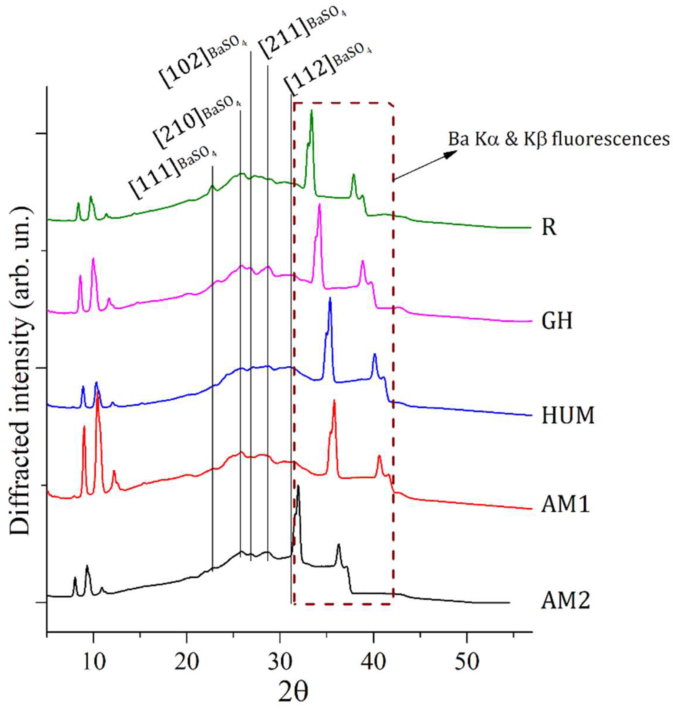
3.1. EDXRD Measurements
3.2. Raman Measurements
3.3. Thermogravimetric Analyses
3.4. Mechanical Properties
3.5. Biocompatibility Properties
4. Conclusions
Author Contributions
Funding
Institutional Review Board Statement
Informed Consent Statement
Data Availability Statement
Conflicts of Interest
References
- Robu, A.; Antoniac, A.; Grosu, E.; Vasile, E.; Raiciu, A.D.; Iordache, F.; Antoniac, V.I.; Rau, J.V.; Yankova, V.G.; Ditu, L.M.; et al. Additives Imparting Antimicrobial Properties to Acrylic Bone Cements. Materials 2021, 14, 7031. [Google Scholar] [CrossRef]
- Robu, A.; Ciocoiu, R.; Antoniac, A.; Antoniac, I.; Raiciu, A.D.; Dura, H.; Forna, N.; Cristea, M.B.; Carstoc, I.D. Bone Cements Used for Hip Prosthesis Fixation: The Influence of the Handling Procedures on Functional Properties Observed during in vitro Study. Materials 2022, 15, 2967. [Google Scholar] [CrossRef]
- CIHI. CJRR Annual Report: Hip and Knee Replacements in Canada; CIHI: Ottawa, ON, Canada, 2022; Available online: https://www.cihi.ca/en/cjrr-annual-report-hip-and-knee-replacements-in-canada (accessed on 7 June 2022).
- Hasandoost, L.; Rodriguez, O.; Alhalawani, A.; Zalzal, P.; Schemitsch, E.H.; Waldman, S.D.; Papini, M.; Towler, M.R. The Role of Poly(Methyl Methacrylate) in Management of Bone Loss and Infection in Revision Total Knee Arthroplasty: A Review. J. Funct. Biomater. 2020, 11, 25. [Google Scholar] [CrossRef] [PubMed]
- Antoniac, I.; Negrusoiu, M.; Mardare, M.; Socoliuc, C.; Zazgyva, A.; Niculescu, M. Adverse Local Tissue Reaction after 2 Revision Hip Replacements for Ceramic Liner Fracture. Medicine 2017, 96, e6687. [Google Scholar] [CrossRef]
- Rau, J.V.; Fadeeva, I.V.; Forysenkova, A.A.; Davydova, G.A.; Fosca, M.; Filippov, Y.Y.; Antoniac, I.V.; Antoniac, A.; D’Arco, A.; Di Fabrizio, M.; et al. Strontium Substituted Tricalcium Phosphate Bone Cement: Short and Long-Term Time-Resolved Studies and In Vitro Properties. Adv. Mater. Interfaces 2022, 9, 2200803. [Google Scholar] [CrossRef]
- Marinescu, R.; Antoniac, I.; Laptoiu, D.; Antoniac, A.; Grecu, D. Complications Related to Biocomposite Screw Fixation in ACL Reconstruction Based on Clinical Experience and Retrieval Analysis. Mater. Plast. 2015, 52, 340–344. [Google Scholar]
- Antoniac, I.V.; Antoniac, A.; Vasile, E.; Tecu, C.; Fosca, M.; Yankova, V.G.; Rau, J.V. In Vitro Characterization of Novel Nanostructured Collagen-Hydroxyapatite Composite Scaffolds Doped with Magnesium with Improved Biodegradation Rate for Hard Tissue Regeneration. Bioact. Mater. 2021, 6, 3383–3395. [Google Scholar] [CrossRef]
- Corobea, M.S.; Albu, M.G.; Ion, R.; Cimpean, A.; Miculescu, F.; Antoniac, I.V.; Raditoiu, V.; Sirbu, I.; Stoenescu, M.; Voicu, S.I.; et al. Modification of Titanium Surface with Collagen and Doxycycline as a New Approach in Dental Implants. J. Adhes. Sci. Technol. 2015, 29, 2537–2550. [Google Scholar] [CrossRef]
- Sarosi, C.; Biris, A.R.; Antoniac, A.; Boboia, S.; Alb, C.; Antoniac, I.; Moldovan, M. The Nanofiller Effect on Properties of Experimental Graphene Dental Nanocomposites. J. Adhes. Sci. Technol. 2016, 30, 1779–1794. [Google Scholar] [CrossRef]
- Cavalu, S.; Antoniac, I.V.; Mohan, A.; Bodog, F.; Doicin, C.; Mates, I.; Ulmeanu, M.; Murzac, R.; Semenescu, A. Nanoparticles and Nanostructured Surface Fabrication for Innovative Cranial and Maxillofacial Surgery. Materials 2020, 13, 5391. [Google Scholar] [CrossRef]
- Bistolfi, A.; Ferracini, R.; Albanese, C.; Vernè, E.; Miola, M. PMMA-Based Bone Cements and the Problem of Joint Arthroplasty Infections: Status and New Perspectives. Materials 2019, 12, 4002. [Google Scholar] [CrossRef] [Green Version]
- McLaren, A.C.; Nugent, M.; Economopoulos, K.; Kaul, H.; Vernon, B.L.; McLemore, R. Hand-Mixed and Premixed Antibiotic-Loaded Bone Cement Have Similar Homogeneity. Clin. Orthop. Relat. Res. 2009, 467, 1693–1698. [Google Scholar] [CrossRef]
- Ferraris, S.; Miola, M.; Bistolfi, A.; Fucale, G.; Crova, M.; Massé, A.; Verné, E. In Vitro Comparison between Commercially and Manually Mixed Antibiotic-Loaded Bone Cements. J. Appl. Biomater. Biomech. 2010, 8, 166–174. [Google Scholar] [CrossRef]
- Zapata, M.E.V.; Ruiz Rojas, L.M.; Mina Hernández, J.H.; Delgado-Ospina, J.; Tovar, C.D.G. Acrylic Bone Cements Modified with Graphene Oxide: Mechanical, Physical, and Antibacterial Properties. Polymers 2020, 12, 1773. [Google Scholar] [CrossRef] [PubMed]
- Arora, M.; Chan, E.K.S.; Gupta, S.; Diwan, A.D. Polymethylmethacrylate Bone Cements and Additives: A Review of the Literature. World J. Orthop. 2013, 4, 67–74. [Google Scholar] [CrossRef] [PubMed]
- de Mori, A.; di Gregorio, E.; Kao, A.P.; Tozzi, G.; Barbu, E.; Sanghani-Kerai, A.; Draheim, R.R.; Roldo, M. Antibacterial PMMA Composite Cements with Tunable Thermal and Mechanical Properties. ACS Omega 2019, 4, 19664–19675. [Google Scholar] [CrossRef]
- Wang, H.; Maeda, T.; Miyazaki, T. Preparation of Bioactive and Antibacterial PMMA-Based Bone Cement by Modification with Quaternary Ammonium and Alkoxysilane. J. Biomater. Appl. 2021, 36, 311–320. [Google Scholar] [CrossRef] [PubMed]
- Karaglani, M.; Tzitzikou, E.; Tottas, S.; Kougioumtzis, I.; Arvanitidis, K.; Kolios, G.; Chatzaki, E.; Drosos, G.I. Gentamycin Elution from Polymethylmethacrylate and Bone Graft Substitute: Comparison between Commercially Available and Home-Made Preparations. J. Orthop. 2020, 19, 9–13. [Google Scholar] [CrossRef]
- Paz, E.; Sanz-Ruiz, P.; Abenojar, J.; Vaquero-Martín, J.; Forriol, F.; del Real, J.C. Evaluation of Elution and Mechanical Properties of High-Dose Antibiotic-Loaded Bone Cement: Comparative “In Vitro” Study of the Influence of Vancomycin and Cefazolin. J. Arthroplast. 2015, 30, 1423–1429. [Google Scholar] [CrossRef] [PubMed]
- Likine, E.F.; Seligson, D. Rifampin and Tobramycin Combination with PMMA Antibiotic Cement. Eur. J. Orthop. Surg. Traumatol. 2019, 29, 499–500. [Google Scholar] [CrossRef] [PubMed]
- Abdelaziz, H.; von Förster, G.; Kühn, K.-D.; Gehrke, T.; Citak, M. Minimum 5 Years’ Follow-up after Gentamicin- and Clindamycin-Loaded PMMA Cement in Total Joint Arthroplasty. J. Med. Microbiol. 2019, 68, 475–479. [Google Scholar] [CrossRef]
- Haseeb, A.; Ajit Singh, V.; Teh, C.S.J.; Loke, M.F. Addition of Ceftaroline Fosamil or Vancomycin to PMMA: An in Vitro Comparison of Biomechanical Properties and Anti-MRSA Efficacy. J. Orthop. Surg. 2019, 27, 230949901985032. [Google Scholar] [CrossRef]
- Zupančič, Š.; Sinha-Ray, S.; Sinha-Ray, S.; Kristl, J.; Yarin, A.L. Long-Term Sustained Ciprofloxacin Release from PMMA and Hydrophilic Polymer Blended Nanofibers. Mol. Pharm. 2016, 13, 295–305. [Google Scholar] [CrossRef]
- Cara, A.; Ferry, T.; Laurent, F.; Josse, J. Prophylactic Antibiofilm Activity of Antibiotic-Loaded Bone Cements against Gram-Negative Bacteria. Antibiotics 2022, 11, 137. [Google Scholar] [CrossRef] [PubMed]
- Merkatoris, P.; Schleining, J.; Krull, A.; Borts, D.; Fajt, V. In Vitro Elution of Penicillin, Ampicillin, Tetracycline, Tulathromycin, and Florfenicol From Plaster of Paris Beads. Front. Vet. Sci. 2020, 7, 5423. [Google Scholar] [CrossRef] [PubMed]
- Anagnostakos, K.; Fürst, O.; Kelm, J. Antibiotic-Impregnated PMMA Hip Spacers: Current Status. Acta Orthop. 2006, 77, 628–637. [Google Scholar] [CrossRef] [PubMed]
- Berberich, C.E.; Josse, J.; Laurent, F.; Ferry, T. Dual Antibiotic Loaded Bone Cement in Patients at High Infection Risks in Arthroplasty: Rationale of Use for Prophylaxis and Scientific Evidence. World J. Orthop. 2021, 12, 119–128. [Google Scholar] [CrossRef]
- Ismat, A.; Walter, N.; Baertl, S.; Mika, J.; Lang, S.; Kerschbaum, M.; Alt, V.; Rupp, M. Antibiotic Cement Coating in Orthopedic Surgery: A Systematic Review of Reported Clinical Techniques. J. Orthop. Traumatol. 2021, 22, 56. [Google Scholar] [CrossRef]
- Świeczko-Żurek, B.; Zieliński, A.; Bociąga, D.; Rosińska, K.; Gajowiec, G. Influence of Different Nanometals Implemented in PMMA Bone Cement on Biological and Mechanical Properties. Nanomaterials 2022, 12, 732. [Google Scholar] [CrossRef]
- Wekwejt, M.; Michno, A.; Truchan, K.; Pałubicka, A.; Świeczko-Żurek, B.; Osyczka, A.M.; Zieliński, A. Antibacterial Activity and Cytocompatibility of Bone Cement Enriched with Antibiotic, Nanosilver, and Nanocopper for Bone Regeneration. Nanomaterials 2019, 9, 1114. [Google Scholar] [CrossRef]
- Prokopovich, P.; Köbrick, M.; Brousseau, E.; Perni, S. Potent Antimicrobial Activity of Bone Cement Encapsulating Silver Nanoparticles Capped with Oleic Acid. J. Biomed. Mater. Res. B Appl. Biomater. 2015, 103, 273–281. [Google Scholar] [CrossRef] [PubMed] [Green Version]
- Slane, J.; Vivanco, J.; Rose, W.; Ploeg, H.-L.; Squire, M. Mechanical, Material, and Antimicrobial Properties of Acrylic Bone Cement Impregnated with Silver Nanoparticles. Mater. Sci. Eng. C 2015, 48, 188–196. [Google Scholar] [CrossRef] [PubMed]
- Russo, T.; Gloria, A.; de Santis, R.; D’Amora, U.; Balato, G.; Vollaro, A.; Oliviero, O.; Improta, G.; Triassi, M.; Ambrosio, L. Preliminary Focus on the Mechanical and Antibacterial Activity of a PMMA-Based Bone Cement Loaded with Gold Nanoparticles. Bioact. Mater. 2017, 2, 156–161. [Google Scholar] [CrossRef] [PubMed]
- Phakatkar, A.H.; Shirdar, M.R.; Qi, M.; Taheri, M.M.; Narayanan, S.; Foroozan, T.; Sharifi-Asl, S.; Huang, Z.; Agrawal, M.; Lu, Y.; et al. Novel PMMA Bone Cement Nanocomposites Containing Magnesium Phosphate Nanosheets and Hydroxyapatite Nanofibers. Mater. Sci. Eng. C 2020, 109, 110497. [Google Scholar] [CrossRef]
- Tijana, A.; Valentina, V.; Nataša, T.; Miloš, H.-M.; Atlagić Suzana, G.; Milica, B.; Yoshiyuki, H.; Hironori, S.; Ivanič, A.; Rebeka, R. Mechanical Properties of New Denture Base Material Modified with Gold Nanoparticles. J. Prosthodont. Res. 2021, 65, 155–161. [Google Scholar] [CrossRef]
- Sartoratto, A.; Machado, A.L.M.; Delarmelina, C.; Figueira, G.M.; Duarte, M.C.T.; Rehder, V.L.G. Composition and Antimicrobial Activity of Essential Oils from Aromatic Plants Used in Brazil. Braz. J. Microbiol. 2004, 35, 275–280. [Google Scholar] [CrossRef]
- Unalan, I.; Slavik, B.; Buettner, A.; Goldmann, W.H.; Frank, G.; Boccaccini, A.R. Physical and Antibacterial Properties of Peppermint Essential Oil Loaded Poly (ε-Caprolactone) (PCL) Electrospun Fiber Mats for Wound Healing. Front. Bioeng. Biotechnol. 2019, 7, 346. [Google Scholar] [CrossRef]
- Thosar, N.; Basak, S.; Bahadure, R.N.; Rajurkar, M. Antimicrobial Efficacy of Five Essential Oils against Oral Pathogens: An in Vitro Study. Eur. J. Dent. 2013, 07, S071–S077. [Google Scholar] [CrossRef]
- Kasiri, N.; Fathi, M. Entrapment of Peppermint Oil Using Cellulose Nanocrystals. Cellulose 2018, 25, 319–329. [Google Scholar] [CrossRef]
- Chua, C.K.; Wong, C.H.; Yeong, W.Y. Standards, Quality Control, and Measurement Sciences in 3D Printing and Additive Manufacturing, 1st ed.; Academic Press: New York, NY, USA, 2017. [Google Scholar]
- Kuehn, K.-D.; Ege, W.; Gopp, U. Acrylic Bone Cements: Mechanical and Physical Properties. Orthop. Clin. North Am. 2005, 36, 29–39. [Google Scholar] [CrossRef]
- Lewis, G. Effect of Methylene Blue on the Fracture Toughness of Acrylic Bone Cement. Biomaterials 1994, 15, 1024–1028. [Google Scholar] [CrossRef]
- Lewis, G. Effect of Lithotriptor Treatment on the Fracture Toughness of Acrylic Bone Cement. Biomaterials 1992, 13, 225–229. [Google Scholar] [CrossRef]
- Lewis, G. Properties of Acrylic Bone Cement: State of the Art Review. J. Biomed. Mater. Res. 1997, 38, 155–182. [Google Scholar] [CrossRef]
- PCPDF-4 Diffraction Database. Available online: https://www.icdd.com/ (accessed on 14 June 2022).
- Zhou, L.; Mernagh, T.P.; Mo, B.; Wang, L.; Zhang, S.; Wang, C. Raman Study of Barite and Celestine at Various Temperatures. Minerals 2020, 10, 260. [Google Scholar] [CrossRef]
- Wang, X.; Huang, W.; Wang, Q.; Liu, C.; Wang, C.; Yang, G.; Zhao, C. Raman Hyperspectral Image Analysis of Benzoyl Peroxide Additive. J. Mol. Struct. 2017, 1138, 6–11. [Google Scholar] [CrossRef]
- Chaurasia, S.; Rao, U.; Mishra, A.K.; Sijoy, C.D.; Mishra, V. Raman Spectroscopy of Poly (Methyl Methacrylate) under Laser Shock and Static Compression. J. Raman Spectrosc. 2020, 51, 860–870. [Google Scholar] [CrossRef]
- Madkour, S.A.; Tirkes, S.; Tayfun, U. Development of Barite-Filled Acrylonitrile Butadiene Styrene Composites: Mechanical, Thermal, Melt-Flow and Morphological Characterizations. Appl. Surf. Sci. Adv. 2021, 3, 100042. [Google Scholar] [CrossRef]
- Hatipoglu, A.; Dike, A.S. Effects of Concentration and Surface Silanization of Barite on the Mechanical and Physical Properties of Poly(Lactic Acid)/Barite Composites. Polym. Polym. Compos. 2020, 28, 140–148. [Google Scholar] [CrossRef]
- DEB, S. A Review of Improvements in Acrylic Bone Cements. J. Biomater. Appl. 1999, 14. [Google Scholar] [CrossRef]
- Khaled, S.M.Z.; Charpentier, P.A.; Rizkalla, A.S. Physical and Mechanical Properties of PMMA Bone Cement Reinforced with Nano-Sized Titania Fibers. J. Biomater. Appl. 2011, 25, 515–537. [Google Scholar] [CrossRef]
- International Standard ISO 5833:2002(E). Available online: https://www.iso.org/standard/30980.html (accessed on 12 July 2022).
- ASTM F451; Standard Specification for Acrylic Bone Cement. ASTM International: West Conshohocken, PA, USA, 2021. [CrossRef]


| Samples | Components | Antimicrobial Additive |
|---|---|---|
| R | Powder 40 g (87.6% PMMA, 2.4% BPO, 10% BaSO4) + Liquid 14.4 g (85.3% MMA, 13.2% BMA, 1.5% DmpT, 20 ppm HQ) | None |
| GM | 5% gentamicin—refers to the total powder weight | |
| HUM | 5% peppermint essential oil incorporated in hydroxyapatite (HAp)—refers to the total powder weight | |
| AM1 | 2% silver nanoparticles incorporated in a ceramic glass—refers to the total powder weight | |
| AM2 | 4% silver nanoparticles incorporated in a ceramic glass—refers to the total powder weight |
| Peak Position (°) | Assignment | Relative Intensity | Component |
|---|---|---|---|
| 22.8 | [111] | 52% | BaSO4 |
| 25.8 | [210] | 100% | BaSO4 |
| 26.9 | [102] | 70% | BaSO4 |
| 28.7 | [211] | 99% | BaSO4 |
| 31.5 | [112] | 50% | BaSO4 |
| Raman Shift (cm−1) | Assignment | Component |
|---|---|---|
| 463 | M–O12 | BaSO4 |
| 487 | Out of plane deformation | PMMA |
| 603 | Deformation O–C=O | PMMA |
| 812 | Symmetric stretching vs(C–O–C) | PMMA |
| 990 | ν1 (SO4) | BaSO4 |
| 1000 | C6H6 breathing | BPO |
| 1450 | CH2 Deformation | PMMA |
| 1730 | Stretching C=O | PMMA |
| Sample | Inflection Temperature at the Highest Rate of Weight Loss (°C) | Mass Loss at Inflection Temperature (%) | T10 (°C) | T50 (°C) | Residue Content at 600 °C (%) |
|---|---|---|---|---|---|
| R | 380.03 | 39.17 | 292.83 | 372.28 | 6.41 |
| AM1 | 383.14 | 36.91 | 291.65 | 373.86 | 6.0 |
| AM2 | 382.68 | 44.83 | 294.51 | 378.12 | 18.92 |
| HUM | 383.71 | 40.92 | 285.53 | 376.96 | 10.93 |
| GH | 384.41 | 40.86 | 299.46 | 377.02 | 14.54 |
| Samples | Elastic Modulus in Flexure [GPa] | Flexural Strength [MPa] |
|---|---|---|
| R | 3.94 ± 0.15 | 56.83 ± 6.36 |
| AM1 | 4.30 ± 0.17 | 65.03 ± 9.09 |
| AM2 | 3.94 ± 0.18 | 65.46 ± 3.69 |
| HUM | 3.60 ± 0.15 | 64.44 ± 4.92 |
| GH | 3.42 ± 0.67 | 61.01 ± 7.04 |
| Samples | Elastic Modulus in Compression [GPa] | Yield Strength [MPa] |
|---|---|---|
| R | 2.90 ± 0.07 | 94.60 ± 4.34 |
| AM1 | 2.78 ± 0.06 | 87.40 ± 4.56 |
| AM2 | 2.74 ± 0.05 | 86.40 ± 3.29 |
| HUM | 2.64 ± 0.08 | 84.40 ± 5.86 |
| GH | 2.90 ± 0.06 | 83.40 ± 4.83 |
Publisher’s Note: MDPI stays neutral with regard to jurisdictional claims in published maps and institutional affiliations. |
© 2022 by the authors. Licensee MDPI, Basel, Switzerland. This article is an open access article distributed under the terms and conditions of the Creative Commons Attribution (CC BY) license (https://creativecommons.org/licenses/by/4.0/).
Share and Cite
Robu, A.; Antoniac, A.; Ciocoiu, R.; Grosu, E.; Rau, J.V.; Fosca, M.; Krasnyuk, I.I., Jr.; Pircalabioru, G.G.; Manescu, V.; Antoniac, I.; et al. Effect of the Antimicrobial Agents Peppermint Essential Oil and Silver Nanoparticles on Bone Cement Properties. Biomimetics 2022, 7, 137. https://doi.org/10.3390/biomimetics7030137
Robu A, Antoniac A, Ciocoiu R, Grosu E, Rau JV, Fosca M, Krasnyuk II Jr., Pircalabioru GG, Manescu V, Antoniac I, et al. Effect of the Antimicrobial Agents Peppermint Essential Oil and Silver Nanoparticles on Bone Cement Properties. Biomimetics. 2022; 7(3):137. https://doi.org/10.3390/biomimetics7030137
Chicago/Turabian StyleRobu, Alina, Aurora Antoniac, Robert Ciocoiu, Elena Grosu, Julietta V. Rau, Marco Fosca, Ivan I. Krasnyuk, Jr., Gratiela Gradisteanu Pircalabioru, Veronica Manescu (Paltanea), Iulian Antoniac, and et al. 2022. "Effect of the Antimicrobial Agents Peppermint Essential Oil and Silver Nanoparticles on Bone Cement Properties" Biomimetics 7, no. 3: 137. https://doi.org/10.3390/biomimetics7030137
APA StyleRobu, A., Antoniac, A., Ciocoiu, R., Grosu, E., Rau, J. V., Fosca, M., Krasnyuk, I. I., Jr., Pircalabioru, G. G., Manescu, V., Antoniac, I., & Gradinaru, S. (2022). Effect of the Antimicrobial Agents Peppermint Essential Oil and Silver Nanoparticles on Bone Cement Properties. Biomimetics, 7(3), 137. https://doi.org/10.3390/biomimetics7030137