Article

Terpenoids and Phenylpropanoids in Ligularia duciformis, L. kongkalingensis, L. nelumbifolia, and L. limprichtii

1
Department of Chemistry, Rikkyo University, Nishi-Ikebukuro, Toshima-ku, Tokyo 171-8501, Japan
2
Faculty of Pharmaceutical Sciences, Tokushima Bunri University, Yamashiro-cho, Tokushima 770-8514, Japan
3
Department of Life Science, Rikkyo University, Nishi-Ikebukuro, Toshima-ku, Tokyo 171-8501, Japan
4
Kunming Institute of Botany, Chinese Academy of Science, Kunming 650201, China
*
Author to whom correspondence should be addressed.
Molecules 2017, 22(12), 2062; https://doi.org/10.3390/molecules22122062
Submission received: 2 November 2017 / Revised: 22 November 2017 / Accepted: 23 November 2017 / Published: 25 November 2017
(This article belongs to the Special Issue Diversity of Terpenoids)

Abstract

:
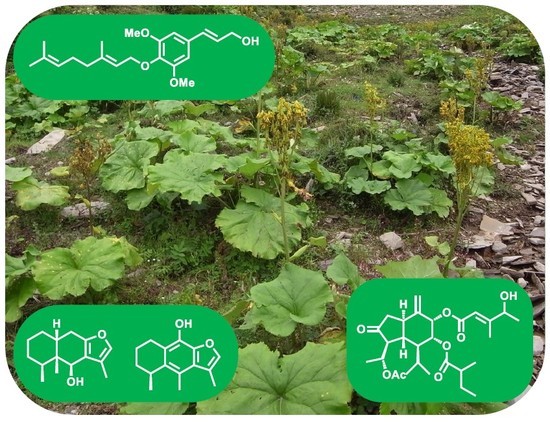
The diversity in root chemicals and evolutionally neutral DNA regions in the complex of Ligularia duciformis, L. kongkalingensis, and L. nelumbifolia (the d/k/n complex) was studied using eight samples collected in central and northern Sichuan Province of China. Cacalol (14) and epicacalone (15), rearranged eremophilanes, were isolated from the complex for the first time. Two new phenylpropanoids were also obtained. Seven of the eight samples produced phenylpropanoids and the other produced lupeol alone. Two of the seven samples also produced furanoeremophilanes or their derivatives and one produced oplopanes. The geographical distribution of the sesquiterpene-producing populations suggests that the production of sesquiterpenes evolved independently in separate regions. L. limprichtii collected in northern Sichuan was also analyzed and its chemical composition and the sequence of internal transcribed spacers (ITSs) in the ribosomal RNA gene cluster were found to be similar to that in the d/k/n complex and L. yunnanensis, which are morphologically similar.

Graphical Abstract

1. Introduction

Species belonging to the genus Ligularia are highly diversified in the Hengduan Mountains area of China and their evolution is considered to be continuing [1]. Most of major Ligularia species in the Hengduan Mountains area produce furanoeremophilanes as major root chemicals [2,3]. We previously proposed a hypothesis that furanoeremophilane-producing species or intra-specific groups are ecologically advantageous over those producing eremophilan-8-ones [2]. Eremophilan-8-ones are precursors of various furanoeremophilanes [4].
Ligularia duciformis (C. Winkl.) Hand.-Mazz., L. kongkalingensis Hand.-Mazz., and L. nelumbifolia (Bureau & Franch.) Hand.-Mazz. are abundant in the Hengduan Mountains area. The three species are morphologically close to one another and their differentiation is based on the pili on the involucres (pilose in L. kongkaligensis or glabrous in L. duciformis and L. nelumbifolia) and on the length of pappi (shorter in L. duciformis, longer in L. nelumbifolia, and intermediate in L. kongkalingensis) [5,6]. However, in our observation, morphological characters appear continuous. In addition, the three species are indistinguishable with respect to root chemicals and evolutionally neutral DNA [7], which have been used as the indices of diversity in our series of work on Ligularia [2,3]. Thus, the three species can be considered to form a complex, which we call the d/k/n complex in this report.
We previously analyzed 28 samples of the d/k/n complex and classified them into four chemotypes [7]: an eremophilane-producing type (type 1), an oplopane-producing type (type 2), a phenylpropanoid-producing type (type 3), and a type without sesquiterpenoid and phenylpropanoid (type 4). Most of the samples were of type 3. Phenylpropanoids were isolated also from types 1 and 2. Although no furanoeremophilane was detected in these 28 samples [7], a L. nelumbifolia sample, collected thereafter in Zhegushan, Hongyuan County in northern Sichuan Province, was found to produce furanoeremophilane [8]. This led us to conduct a further search in central and northern Sichuan during 2011–2015. Here we report the presence of another furanoeremophilane-producing individual as well as an individual producing cacalol, a rearranged furanoeremophilane.
A sample of L. limprichtii (Diels) Hand.-Mass. collected in 2010 in northern Sichuan was also analyzed. L. limprichtii is similar to the d/k/n complex in morphology. The major differences are in the involucres (narrowly cylindric to campanulate-cylindric in the d/k/n complex or campanulate-turbinate in L. limprichtii) and in the leaf blade (glabrous in the d/k/n complex or abaxially shortly pilose in L. limprichtii) [5]. To the best of our knowledge, its chemical composition has not been reported. Here we report the isolation of a phenylpropanoid from L. limprichtii.

2. Results

2.1. Samples

Samples of the d/k/n complex (samples 1–8) and a sample of L. limprichtii (sample 9) were collected in the field shown in Table 1 and Figure 1. The locations of three previously collected sesquiterpene-producing samples are also included in Figure 1 as samples A, B, and C for reference.

2.2. Chemical Constituents

For a characterization of the chemical composition of the samples, an EtOH extract of the fresh root of each sample was analyzed by TLC using Ehrlich’s coloring reagent, a facile method to detect furanoeremophilanes [9,10]. Sample 7 showed Ehrlich-positive spots, the most prominent at Rf = 0.65 (hexane/EtOAc 7:3), suggesting the presence of furanoeremophilanes, although the spots were weaker than for typical furanoeremophilane-producing Ligularia plants. Sample 6 showed very weak spots, and samples 1–5, 8, and 9 were negative to the test.
For the isolation of compounds, dried roots of each sample were extracted with EtOH, and the compounds therein were separated by silica-gel column chromatography and HPLC as usual. Fifteen compounds, 115, were isolated from the eight samples (Table 1 and Figure 2). From samples 1–3, 8, and 9, phenylpropanoids were isolated without terpenoids. The isolated compounds were 1 [11] and 5 [7] from sample 1; 3 [12,13], 7 [14], and lupeol (9) [15] from sample 2; 3 and 4 [11,13,14] from sample 3; 3, 4, and 8 (a mixture of known compound 8a [14]; and new compound 8b at a ratio of 7:1) from sample 8; and 3 from sample 9. From sample 4, lupeol (9) alone was isolated as in the previously reported type-4 samples [7]. Sesquiterpenes were isolated from samples 5–7. From sample 5, oplopanes 10 [7] and 11 [16] were isolated in addition to a new compound 2, coniferaldehyde (6) [17], and 9. From sample 6, cacalol (14) [18,19] and epicacalone (15) [20] were isolated together with 3, 4, 8, and 9. From sample 7, two furanoeremophilanes, ligularol (12) [21] and 13 [22], were isolated.
The structure of the new compound 2 was determined as follows. The molecular formula of 2 was determined to be C16H22O4 from the high-resolution mass spectrum (m/z 278.1510). The IR (1730 cm−1) and 13C-NMR spectra (δ 173.3) showed the presence of an ester carbonyl group. The 1H and 13C-NMR spectra showed typical signals of a coniferyl ester moiety, i.e., signals of a methoxy group [δH 3.91 (s); δC 55.9], an oxygenated methylene [δH 4.71 (dd, J = 6.6, 1.1 Hz); δC 65.0], an E-alkene [δH 6.14 (dt, J = 15.7, 6.6 Hz) and 6.58 (d, J = 15.7 Hz)], and a 1,2,4-trisubstituted aromatic ring [δH 6.87 (d, J = 7.6 Hz), 6.90 (dd, J = 7.6, 1.5 Hz), and 6.92 (d, J = 1.5 Hz)] (See Section 4.3 and Figures S1 and S2). Signals of two methyl groups were observed as a triplet (δH 0.90) and a doublet (δH 0.95) in addition to those of two methylenes and one methine (δH 1.91), the latter of which was observed in octet, suggesting that the acid part was 3-methylpentanoate. The structure was established from COSY observed for 2′-H/3′-H/4′-H/5′-H and 3′-H/6′-H and from HMBC correlations shown in Figure 3.
Compound 8 was obtained as an inseparable mixture of E- (8a) and Z-isomers (8b), the latter of which was new. Although a full characterization of 8b was not feasible due to signal overlap with 8a, its structure was deduced to be the Z isomer from its 1H-NMR spectrum, as the J-value between 2-H (δH 6.15) and 3-H (δH 7.54) was 11.4 Hz (See Section 4.4 and Figure S3). Although all 1H signals of the geranyl moiety of 8b overlapped with those of 8a except for 1′-H, the signals of the sinapylaldehyde part were separated from those of 8a. The structure was confirmed by HMBC correlations shown in Figure 3 (See Section 4.4 and Figure S4 for the 13C-NMR spectrum).
The chemical compositions of the d/k/n complex samples were compared by LC-MS analysis. Samples 1–6 and 9 showed no distinct peaks in tR = 10 to 30 min (MeOH/H2O, see Section 4.1 for the experimental conditions) where typical terpenoids and phenylpropanoids were expected (Figure S5). Large signals were observed in tR = 3 to 4 min, however, we could not identify the compounds. In contrast, ligularol (12) and 6β-ethoxyfuranoeremophilan-10β-ol (13) were detected at tR = 15.3 and 17.5 min, respectively, in the chromatogram of sample 7 (Figure 4). Compound 13 may be an artifact generated during EtOH extraction [22], and its presumed parent compound, furanoeremophilane-6β,10β-diol, is also detected at tR = 11.5 min. Sample 8 showed one major peak of O-geranylsinapyi alcohol (4) at tR = 15.4 min, which was isolated as the major component (Figure 4).

2.3. Genetic Study

To assess the genetic diversity among the samples, the DNA sequence was determined for the internal transcribed spacer 1 (ITS1)-5.8S-ITS2 region of the ribosomal RNA gene. The results are shown in Table S1. Although there are some variations, a bootstrapping analysis by the UPGMA method using MEGA6 [23] found no distinct clade among the samples. The result indicates that L. limprichtii is genetically similar to the d/k/n complex.

3. Discussion

3.1. Chemotypes

We previously grouped d/k/n complex samples, collected in western Sichuan and northwestern Yunnan, into four chemotypes: type 1 (eremophilanes), type 2 (oplopanes), type 3 (phenylpropanoids), and type 4 (no phenylpropanoid nor sesquiterpenoid) [7,8]. According to this classification, sample 7 belongs to type 1; sample 5, type 2; samples 1–3, and 8, type 3; and sample 4, type 4 (Table 1). Cacalol (14) and epicacalone (15) have a rearranged carbon skeleton generated from furanoeremophilane [18], and thus, sample 6 also belongs to type 1. While cacalol is a compound often found in Ligularia species, such as L. virgaurea [24] and L. cyathiceps [25], sample 6 is the first example of the isolation of cacalol from the d/k/n complex.
Two of our previous type-1 samples were collected in Maerkang, Sichuan (samples A and B in Figure 1) [7,8]. With the present two additions, samples 6 and 7, it can be said that type 1 is abundant in the Maerkang-Heishui area. It is interesting that this area harbors all the three evolutionary stages of eremophilane biosynthesis: non-furano-eremophilane, furanoeremophilane, and cacalol. Sample 5 is the fourth example of type 2 (oplopane). Phenylpropanoids were also obtained from the present type-1 and type-2 samples as observed previously [7]. In particular, the major component in sample 6 was O-geranylsinapyl alcohol (4), a phenylpropanoid typical of type 3.
Samples 1–3 and 8 belong to type 3. While O-geranylsinapyl alcohol (4) was the major component in sample 8 (8.3% of the extract), phenylpropanoids in samples 1–3 were small in quantity. Although lupeol (9) was the sole compound isolated from sample 4, the possibility cannot be excluded that it contained a very small quantity of phenylpropanoids. More than half of the type-3 samples in our previous study produced lupeol (9) as well as phenylpropanoids [7]. Thus, type 3 and type 4 may constitute one chemotype with a continuous composition spectrum.

3.2. Introgression and Chemical Evolution

We previously proposed that introgression might have brought the ability to produce sesquiterpenes from some other plant into type 3, resulting in types 1 and 2 [7]. This premise has found support in the discovery of hybrids between L. duciformis and L. cyathiceps [26] and hybrids between L. nelumbifolia and L. subspicata [27] and the isolation of furanoeremophilanes, the major components of both L. subspicata and L. cyathiceps, from them. Although the present DNA data did not separate samples 6 and 7 from the other samples, the few sites of multiple bases, particularly in sample 7, may be suggestive of such introgression (Table S1). Type 1 has been found in Shangrila, Yunnan, and Maerkang, Sichuan. Since these areas are distant from each other, the evolution of eremophilane production is likely to have happened independently. The location of sample 5 (type 2) was not far (ca. 30 km) from one of the previous two type-2 samples (sample C in Figure 1) but far from the other in Shangrila, Yunnan. Their evolution is also likely to have been independent.

3.3. L. limprichrii

O-Geranylsinapyl acetate (3) was isolated from L. limprichtii (sample 9). The same compound was also obtained from samples 2 and 3, as well as eight of the 18 type-3 samples in our previous study [7]. Phenylpropanoids have been isolated from taxonomically related species, L. yunnanensis (Franch.) Chang [26] and L. purdomii (Trrill) Chittenden [28], from the latter of which eremophilanes and euparin-type benzofurans were also isolated. DNA data also show the similarity as the ITS1-5.8S-ITS2 sequence of sample 9 cannot be separated from those of other samples (Table S1). The sequence of L. yunnanensis is indistinguishable from that of L. duciformis [26], therefore, L. limprichtii is similar to both the d/k/n complex and L. yunnanensis. Further study with more samples is necessary to understand the relationship among the d/k/n complex, L. limprichtii, and other taxonomically related species.

4. Materials and Methods

4.1. General Experimantal Procedure

Specific rotations were measured on a DIP-370 polarimeter (JASCO, Tokyo, Japan). NMR spectra were measured on an ECX-400 or an AL-400 (400 MHz for 1H; 100 MHz for 13C) spectrometer (JEOL, Tokyo, Japan). IR spectra were measured on a FT/IR-230 spectrometer (JASCO), and MS spectra, on a JMS-700 MStation or a CMATE II (JEOL). Kieselgel 60 F254, 0.2 mm thickness (Merck, Darmstadt, Germany) was used for analytic TLC, with either Ehrlich’s reagent (p-dimethylaminobenzaldehyde and HCl) [9,10] or p-anisaldehyde/AcOH/H2SO4 as visualizing agents. Open column chromatography (CC) was carried out on silica gel (Wakogel C-200 (Wako, Kyoto, Japan) or silica gel 60 N (Kanto, Tokyo, Japan)). HPLC with a Mightysil Si60 (10 mm × 250 mm) column (Kanto) was carried out on either an LC-20AT pump with a SPD-20A Prominence UV/VIS detector (Shimadzu, Kyoto, Japan) or a GL-7410 pump with a GL-7450 UV detector (GL Sciences, Tokyo, Japan), and with a D-2500 Chromato-Integrator (Hitachi, Tokyo, Japan) or a C-R8A Chromatopac recorder (Shimadzu). LC-MS was measured on a 1100 series LC/MSD mass spectrometer (capillary voltage 3.5 kV; corona current 4 μA; capillary exit voltage (fragmentor) 90 V; drying temperature 330 °C; drying flow 9 L/min; nebulizer pressure 50 psig) (Agilent, Santa Clara, CA, USA) with 5C18-MS-II (COSMOSIL; 4.6 mm × 150 mm; 5 μm octadecyl column (Nakarai, Kyoto, Japan)) using gradient system (MeOH/H2O; 0 min (7:3)—20 min (10:0)—35 min (10:0)—40 min (7:3)—45 min (7:3); 0.5 mL/min) as eluent. DNA was purified from dried leaves using DNeasy Plant Mini Kit (QIAGEN, Hilden, Germany). Polymerase chain reaction was carried out using L5 and L6 primers previously described [29] and HotStarTaq plus Master Mix (QIAGEN). The amplification products were separated by agarose gel electrophoresis and purified with High Pure PCR Product Purification Kit (Roche Diagnostics, Basel, Switzerland). DNA sequencing reaction was carried out using L1–L4 primers described previously [29] and BigDye Terminator v3.1 Cycle Sequencing Kit (Applied Biosystems, Waltham, MA, USA) and analyzed on a 3130xl or 3500 Genetic Analyzer (Applied Biosystems).

4.2. Plant Materials

Samples were collected in August 2010, 2011, 2012, and 2015 at locations shown in Table 1 and Figure 1. Each sample was identified by X. G. (author). Voucher specimen numbers are 2011-12, 2011-29, 2011-82, 2012-10, 2012-28, 2015-33, 2015-37, 2015-40, and 2010-32, for samples 1–9, respectively (Kunming Institute of Botany, Kunming, China).

4.3. Extraction and Purification

The dried roots of sample 1 (43.0 g) were extracted with EtOH. The extract (1799.4 mg) was subjected to silica-gel (20 g) CC using n-hexane/EtOAc (gradient) as the eluent to obtain eight fractions. Fr. 3 (eluted with n-hexane/EtOAc 96:4) was further separated by CC (silica gel, n-hexane/EtOAc) to afford seven fractions (Fr. 1–1 to 1–7). From Fr. 1–5, 5 (0.3 mg) was obtained by HPLC (Mightysil, n-hexane/EtOAc 7:3). From Fr. 1–7, 1 (6.5 mg) was obtained by HPLC (n-hexane/EtOAc 7:3).
The dried roots of sample 2 (57.4 g) were extracted with EtOH. The extract (219.1 mg) was subjected to silica-gel (6 g) CC using n-hexane/EtOAc (gradient) as the eluent to obtain seven fractions. From Fr. 3 (eluted with n-hexane/EtOAc 9:1), 9 (1.6 mg) was isolated by repeated CC and HPLC (n-hexane/EtOAc 1:1). From Fr. 4 (eluted with n-hexane/EtOAc 8:2), 7 (3.1 mg) and 3 (1.0 mg) were obtained by CC and HPLC (n-hexane/EtOAc 7:3).
The dried roots of sample 3 (13.1 g) were extracted with EtOH. The extract (218.5 mg) was subjected to silica-gel (12 g) CC using n-hexane/EtOAc (gradient) as the eluent to obtain eight fractions. From Fr. 6 (eluted with n-hexane/EtOAc 9:1 to 8:2), 3 (1.1 mg) was obtained by CC and HPLC (n-hexane/EtOAc 8:2 to 3:1). From Fr. 8 (eluted with n-hexane/EtOAc 8:2), 4 (5.1 mg) was obtained by HPLC (n-hexane/EtOAc 1:1).
The dried roots of sample 4 (48.5 g) were extracted with EtOH. The extract (3060.7 mg) was subjected to silica-gel (12 g) CC using n-hexane/EtOAc (gradient) as the eluent to obtain 10 fractions. From Fr. 3 (eluted with n-hexane), 9 (1.8 mg) was isolated by repeated CC (silica gel, n-hexane/EtOAc) and HPLC (n-hexane/Et2O 6:4).
The dried roots of sample 5 (70.3 g) were extracted with EtOH. The extract (639.4 mg) was subjected to silica-gel (12 g) CC using n-hexane/EtOAc (gradient) as the eluent to obtain eight fractions. From Fr. 4 (eluted with n-hexane/EtOAc 9:1), 9 (20.1 mg) was isolated by CC and HPLC (n-hexane/EtOAc 7:3). From Fr. 5 (eluted with n-hexane/EtOAc 85:15), 2 (0.6 mg) and 10 (2.5 mg) were obtained by CC and HPLC (n-hexane/EtOAc 7:3). From Fr. 7 (eluted with n-hexane/EtOAc 8:2 and 7:3), 6 (1.2 mg), and 11 (7.2 mg) were obtained by CC and HPLC (n-hexane/EtOAc 6:4).
The dried roots of sample 6 (45.2 g) were extracted with EtOH. The extract (838.5 mg) was subjected to silica-gel (20 g) CC using n-hexane/EtOAc (gradient) as the eluent to obtain six fractions. From Fr. 4 (eluted with n-hexane/EtOAc 9:1), 14 (2.6 mg) was isolated by repeated CC and HPLC (n-hexane/EtOAc 8:2). From Fr. 5 (eluted with n-hexane/EtOAc 8:2), 9 (5.1 mg) and 3 (1.4 mg) were obtained by CC and HPLC (n-hexane/EtOAc 7:3). From Fr. 6 (eluted with EtOAc), 8 (1.4 mg, ratio 7:2), 15 (0.1 mg), and 4 (31.3 mg) were isolated by CC and HPLC (n-hexane/EtOAc 3:2, 1:1, and 1:4 for 8, 15, and 4, respectively).
The dried roots of sample 7 (43.5 g) were extracted with EtOH. The extract (1340.9 mg) was subjected to silica-gel (20 g) CC using n-hexane/EtOAc (gradient) as the eluent to obtain eight fractions. From Fr. 4 (eluted with n-hexane/EtOAc 9:1), 13 (3.3 mg) was isolated by repeated CC and HPLC (n-hexane/EtOAc 8:2). From Fr. 5 (eluted with n-hexane/EtOAc 8:2), 12 (7.4 mg) was obtained by CC and HPLC (n-hexane/EtOAc 8:2).
The dried roots of sample 8 (54.5 g) were extracted with EtOH. The extract (2377.9 mg) was subjected to silica-gel (20 g) CC using n-hexane/EtOAc (gradient) as the eluent to obtain eight fractions. From Fr. 4 (eluted with n-hexane/EtOAc 9:1), 3 (2.8 mg) was isolated by repeated CC and HPLC (n-hexane/EtOAc 8:2). From Fr. 5 (eluted with n-hexane/EtOAc 8:2), 8 (1.6 mg; ratio 7:1) was obtained by CC and HPLC (n-hexane/EtOAc 7:3). From Fr. 6 (eluted with n-hexane/EtOAc 7:3), 8 (4.7 mg) and 4 (196.5 mg) was afforded after purification by CC and HPLC (n-hexane/EtOAc 7:3 to 6:4 for 8, 4:6 to 2:8 for 4).
The dried roots of sample 9 (L. limprichtii) (33.0 g) were extracted with EtOH. The extract (316.6 mg) was subjected to silica-gel (12 g) CC using n-hexane/EtOAc (gradient) as the eluent to obtain five fractions. From Fr. 3 (eluted with n-hexane/EtOAc 92:8), 3 (1.1 mg) was isolated after purification by HPLC (n-hexane/EtOAc 4:1).

4.4. (2E)-3-(4-Hydroxy-3-Methoxyphenyl)prop-2-en-1-yl 3-Methylpentanoate (2)

An oil; [α]D24 170 (c 0.012, MeOH); IR (neat) 3434 (OH), 1730 (C=O), 1514, 1274 cm−1; 1H-NMR (CDCl3) δ 0.90 (t, J = 7.5 Hz, 5′-H3), 0.95 (d, J = 6.5 Hz, 6′-H3), 1.18–1.44 (m, 4′-H2), 1.91 (octet, J = 6.7 Hz, 3′-H), 2.15 (dd, J = 14.8, 8.1 Hz, 2′-H), 2.35 (dd, J = 14.8, 5.1 Hz, 2′-H), 3.91 (s, OCH3), 4.71 (dd, J = 6.6, 1.1 Hz, 1-H2), 5.65 (s, OH), 6.14 (dt, J = 15.7, 6.6 Hz, 2-H), 6.58 (d, J = 15.7 Hz, 3-H), 6.87 (d, J = 7.6 Hz, 8-H), 6.90 (dd, J = 7.6, 1.5 Hz, 9-H), 6.92 (d, J = 1.5 Hz, 5-H); 13C-NMR (CDCl3) δ 11.3 (C-5′), 19.3 (C-6′), 29.4 (C-4′), 31.9 (C-3′), 41.5 (C-2′), 55.9 (OMe), 65.0 (C-1), 108.3 (C-5), 114.4 (C-8), 120.6 (C-9), 120.9 (C-2), 128.8 (C-4), 134.3 (C-3), 145.8 (C-7), 146.6 (C-6), 173.3 (C-1′); MS (CI) m/z 278 (M+, 100%), 163 (92), 41 (92); HRMS (CI) obs. m/z 278.1510, calcd for C16H22O4, M, 278.1519.

4.5. (2Z)- and (2E)- 3-[3,5-Dimethoxy-4-((2E)-3,7-Dimethylocta-2,6-Dien-1-yl)Oxyphenyl]prop-2-Enal (8a and 8b)

1H-NMR (CDCl3) δ 1.59 (br s, 9′-H3 of 8a,b), 1.66 (br s, 10′-H3 of 8a,b), 1.67 (br s, 8′-H3 of 8a,b), 1.99-2.11 (m, 4′-H2 and 5′-H2 of 8a,b), 3.87 (s, OMe of 8b), 3.90 (s, OMe of 8a), 4.59 (d, J = 7.0 Hz, 1′-H of 8b), 4.61 (d, J = 7.0 Hz, 1′-H of 8a), 5.03–5.09 (m, 6′-H of 8a,b), 5.52–5.58 (m, 2′-H2 of 8a,b), 6.15 (dd, J = 11.4, 7.7 Hz, 2-H of 8b), 6.64 (dd, J = 15.7, 7.6 Hz, 2-H of 8a), 6.60 (s, 5-H and 9-H of 8b), 6.78 (s, 5-H and 9-H of 8a), 7.40 (d, J = 15.7 Hz, 3-H of 8a), 7.54 (d, J = 11.4 Hz, 3-H of 8b), 9.68 (d, J = 7.6 Hz, 1-H of 8a), 10.03 (d, J = 7.7 Hz, 1-H of 8b); 13C-NMR (CDCl3) assigned for 8a: δ 16.4 (C-10′), 17.6 (C-9′), 25.7 (C-8′), 26.4 (C-5′), 39.6 (C-4′), 56.1 (OMe), 69.6 (C-1′), 105.6 (C-5, 9), 119.8 (C-2′), 123.9 (C-6′), 127.8 (C-2), 129.4 (C-4), 131.7 (C-7′), 139.8 (C-7), 142.0 (C-3′), 152.9 (C-3), 154.0 (C-6, 8), 193.5 (C-1); assigned for 8b: δ 16.4 (C-10′), 17.6 (C-9′), 25.7 (C-8′), 26.4 (C-5′), 39.6 (C-4′), 56.2 (OMe), 69.6 (C-1′), 107.2 (C-5, 9), 119.9 (C-2′), 123.9 (C-6′), 129.6 (C-4), 129.9 (C-2), 131.7 (C-7′), 138.5 (C-7), 142.0 (C-3′), 148.7 (C-3), 153.7 (C-6, 8), 192.6 (C-1).

5. Conclusions

Cacalol (14) and its derivative, epicacalone (15), were found in the d/k/n complex for the first time, showing that the diversity in the complex is higher than previously known. Two new phenylpropanoids (2, 8b) were also obtained. The basal chemicals in this complex appear to be phenylpropanoids and lupeol, to which sesquiterpenoids are added in some populations. The geographical separation of sesquiterpene-producing populations suggests that the production of this class of compounds evolved independently, perhaps through introgression. L. limprichtii was found to produce a phenylpropanoid, showing similarity to the d/k/n complex, which is consistent with DNA data.

Supplementary Materials

The following are available online. Table S1: Differences in the sequence of the rDNA ITS1-5.8S-ITS2 region, Figure S1: 1H-NMR spectrum of 2, Figure S2: 13C-NMR spectrum of 2, Figure S3: 1H-NMR spectrum of 8, Figure S4: 13C-NMR spectrum of 8, Figure S5: LCMS profile (total ion chromatogram) of samples 1–6 and 9.

Acknowledgments

The authors thank Takayuki Kawahara, Forestry and Forest Products Research Institute; Yoshinori Saito, Nagasaki University; and Katsuyuki Nakashima, Tokushima-Bunri University; for their help in sample collection. This work was partly supported by a Grant-in-Aid for Scientific Research from JSPS (No. 25303010 and No. 21404009), the Japan–China Scientific Cooperation Program from JSPS and NSFC, and the Strategic Research Foundation Grant-aided Projects for Private Universities from the Ministry of Education, Culture, Sports, Science, and Technology, Japan.

Author Contributions

C.K., R.H., and X.G. conceived and designed the study; R.K., A.N., Y.N., and T.I. performed the isolation of chemicals; Y.O. performed LCMS analysis; M.T. and C.K. analyzed the spectral data; R.H. analyzed DNA sequences; X.G. identified the species; C.K. and R.H. wrote the paper.

Conflicts of Interest

The authors declare no conflict of interest. The founding sponsors had no role in the design of the study; in the collection, analyses, or interpretation of data; in the writing of the manuscript, and in the decision to publish the results.

References

  1. Liu, S.W.; Deng, D.S.; Liu, J.Q. The origin, evolution and distribution of Ligularia Cass. (Compositae). Acta Phytotaxon. Sin. 1994, 32, 514–524. [Google Scholar]
  2. Kuroda, C.; Hanai, R.; Nagano, H.; Tori, M.; Gong, X. Diversity of furanoeremophilanes in major Ligularia species in the Hengduan Mountains. Nat. Prod. Commun. 2012, 7, 539–548. [Google Scholar] [PubMed]
  3. Kuroda, C.; Hanai, R.; Tori, M.; Okamoto, Y.; Saito, Y.; Nagano, H.; Ohsaki, A.; Hirota, H.; Kawahara, T.; Gong, X. Diversity in furanoeremophilane composition produced by Ligularia species (Asteraceae) in the Hengduan Mountains area of China. J. Synth. Org. Chem. Jpn. 2014, 72, 717–725. [Google Scholar] [CrossRef]
  4. Gaikwad, N.G.; Madyastha, K.M. Biosynthesis of β-substituted furan skeleton in the lower furanosesquiterpenoids: a model study. Biochem. Biophys. Res. Commun. 2002, 290, 589–594. [Google Scholar] [CrossRef] [PubMed]
  5. Liu, S. Flora Republicae Popularis Sinicae, Vol. 77, Part 2; Science Press: Beijing, China, 1989. [Google Scholar]
  6. Liu, S.; Illarionova, I.D. Ligularia. In Flora of China; Wu, Z.Y., Raven, P.H., Hong, D.Y., Eds.; Science Press: Beijing, China; Missouri Botanical Garden Press: St. Louis, MO, USA, 2011; Volume 20–21, pp. 376–415. [Google Scholar]
  7. Nagano, H.; Hanai, R.; Yamada, H.; Matsushima, M.; Miura, Y.; Hoya, T.; Ozawa, M.; Fujiwara, M.; Kodama, H.; Torihata, A.; et al. Chemical and genetic study of Ligularia duciformis and related species in Sichuan and Yunnan provinces of China. Chem. Biodivers. 2012, 9, 789–805. [Google Scholar] [CrossRef] [PubMed]
  8. Hirota, H.; Horiguchi, Y.; Kawaii, S.; Hanai, R.; Gong, X.; Kuroda, C. The first isolation of furanoeremophilane from Ligularia nelumbifolia. Nat. Prod. Commun. 2014, 9, 325–327. [Google Scholar] [PubMed]
  9. Kuroda, C.; Ueshino, T.; Nagano, H. Ehrlich’s reaction of furanoeremophilanes. Bull. Chem. Soc. Jpn. 2004, 77, 1737–1740. [Google Scholar] [CrossRef]
  10. Kuroda, C.; Nishio, E. Substituent effect in color of Ehrlich’s test of tetrahydrobenzofuran. Nat. Prod. Commun. 2007, 2, 581–585. [Google Scholar]
  11. Shibuya, H.; Takeda, Y.; Zhang, R.S.; Tong, R.X.; Kitagawa, I. Indonesian medicinal plants. III. On the constituents of the bark of Fagara rhetza (Rutaceae). (1): Alkaloids, phenylpropenoids, and acid amide. Chem. Pharm. Bull. 1992, 40, 2325–2330. [Google Scholar] [CrossRef] [PubMed]
  12. Bohlmann, F.; Zdero, C.; Natu, A.A. Weitere bisabolen-derivate und andere inhaltsstoffe aus Südafrikanischen Senecis-arten. Phytochemistry 1978, 17, 1757–1761. [Google Scholar] [CrossRef]
  13. Bohlmann, F.; Grenz, M.; Gupta, R.K.; Dhar, A.K.; Ahmed, M.; King, R.M.; Robinson, H. Eudesmane derivatives from Verbesina species. Phytochemistry 1980, 19, 2391–2397. [Google Scholar] [CrossRef]
  14. Zhao, Y.; Jia, Z.; Yang, L. Sinapyl alcohol derivatives and other constituents from Ligularia nelumbifolia. Phytochemistry 1994, 37, 1149–1152. [Google Scholar]
  15. Burns, D.; Reynolds, W.F.; Buchanan, G.; Reese, P.B.; Enriquez, R.G. Assignment of 1H and 13C spectra and investigation of hindered side-chain rotation in lupeol derivatives. Magn. Reson. Chem. 2000, 38, 488–493. [Google Scholar] [CrossRef]
  16. Tori, M.; Fujiwara, M.; Okamoto, Y.; Tanaka, M.; Gong, X.; Shen, Y.; Hanai, R.; Kuroda, C. New oplopane-type sesquiterpenoids from Ligularia duciformis. Nat. Prod. Commun. 2007, 2, 357–360. [Google Scholar]
  17. Luo, J.-R.; Ma, Q.-Y.; Zhao, Y.-X.; Yi, T.-M.; Li, C.-S.; Zhou, J. Palaeophytochemical components from the miocene-fossil wood of Pinus griffithii. J. Chin. Chem. Soc. 2009, 56, 600–605. [Google Scholar] [CrossRef]
  18. Terabe, M.; Tada, M.; Takahashi, T. Absolute stereochemistry of cacalol. Bull. Chem. Soc. Jpn. 1978, 51, 661–662. [Google Scholar] [CrossRef]
  19. Romo, J.; Joseph-Nathan, P. The constituents of Cacalia decomposita A. Gray. Structures of cacalol and cacalone. Tetrahedron 1964, 20, 2331–2337. [Google Scholar] [CrossRef]
  20. Omura, K.; Nakanishi, M.; Takai, K.; Naya, K. The sesquiterpenes of Cacalia species: 8-Oxocacalol and the stereochemistry of cacalone epimers. Chem. Lett. 1978, 1257–1260. [Google Scholar] [CrossRef]
  21. Ishii, H.; Tozyo, T.; Minato, H. Studies on sesquiterpenoids—IX. Structure of ligularol and ligularone from Ligularia sibirica Cass. Tetrahedron 1965, 21, 2605–2610. [Google Scholar] [CrossRef]
  22. Tori, M.; Watanabe, A.; Matsuo, S.; Okamoto, Y.; Tachikawa, K.; Takaoka, S.; Gong, X.; Kuroda, C.; Hanai, R. Diversity of Ligularia kanaitzensis in sesquiterpenoid composition and neutral DNA sequences. Tetrahedron 2008, 64, 4486–4495. [Google Scholar] [CrossRef]
  23. Tamura, K.; Stecher, G.; Peterson, D.; Filipski, A.; Kumar, S. MEGA6: Molecular evolutionary genetics analysis version 6.0. Mol. Biol. Evol. 2013, 30, 2725–2729. [Google Scholar] [CrossRef] [PubMed]
  24. Saito, Y.; Takashima, Y.; Kamada, A.; Suzuki, Y.; Suenaga, M.; Okamoto, Y.; Matsunaga, Y.; Hanai, R.; Kawahara, T.; Gong, X.; et al. Chemical and genetic diversity of Ligularia virgaurea collected in northern Sichuan and adjacent areas of China. Isolation of 13 new compound. Tetrahedron 2012, 68, 10011–10029. [Google Scholar] [CrossRef]
  25. Nagano, H.; Torihata, A.; Matsushima, M.; Hanai, R.; Saito, Y.; Baba, M.; Tanio, Y.; Okamoto, Y.; Takashima, Y.; Ichihara, M.; et al. Chemical and genetic study of Ligularia cyathiceps in Yunnan Province of China. Helv. Chim. Acta 2009, 92, 2071–2081. [Google Scholar] [CrossRef]
  26. Hanai, R.; Tanabe, S.; Aoyama, N.; Okamoto, Y.; Tori, M.; Zhang, N.; Gong, X.; Kuroda, C. Chemical and genetic study of two Ligularia hybrids in Shangrila County, Yunnan Province, China. Nat. Prod. Commun. 2016, 11, 1057–1060. [Google Scholar]
  27. Hanai, R.; Yamada, H.; Suzuki, Y.; Nagano, H.; Kawahara, T.; Yu, J.-J.; Gong, X.; Kuroda, C. Chemical constituents of Ligularia nelumbifolia and L. subspicata hybrid collected in Shangrila County, Yunnan Province of China. Nat. Prod. Commun. 2012, 7, 1565–1568. [Google Scholar] [PubMed]
  28. Wang, Y.-M.; Zhao, J.-Q.; Yang, J.-L.; Tao, Y.-D.; Mei, L.-J. Chemical constituents from Ligularia purdomii (Turrill) Chittenden. Biochem. Syst. Ecol. 2017, 72, 8–11. [Google Scholar] [CrossRef]
  29. Nagano, H.; Matsushima, M.; Yamada, H.; Hanai, R.; Gong, X.; Kuroda, C. Two new furanoeremophilane sesquiterpenoids from Ligularia oligonema. Nat. Prod. Commun. 2010, 5, 1–4. [Google Scholar] [PubMed]
Sample Availability: Isolated compounds are not available from the authors.
Figure 1. Locations of samples of L. duciformis, L. kongkalingensis, L. nelumbifolia, and L. limprichtii. Rectangles, samples; circles, cities; solid lines, rivers; dotted lines, boundaries of provinces. A–C are previously collected type-1 and type-2 samples within the map area [7,8].
Molecules 22 02062 g001
Figure 2. Isolated compounds from L. duciformis/L. kongkalingensis/L. nelumbifolia (samples 1–8) and L. limprichtii (sample 9). Asterisks indicate new compounds.
Molecules 22 02062 g002
Figure 3. Selected HMBC correlations of compounds 2 and 8b.
Molecules 22 02062 g003
Figure 4. LCMS profile (total ion chromatogram) of samples 7 and 8. See Figure S5 for samples 1–6 and 9.
Molecules 22 02062 g004
Table 1. Collection locality and chemical composition of L. duciformis, L. kongkalingensis, L. nelumbifolia, and L. limprichtii samples.
Sample No. 1SpeciesLocality 2Isolated CompoundsChemotype
1 (2011-12)L. duciformisJiajinshan (Baoxing)1, 53
2 (2011-29)L. nelumbifoliaBamei (Daofu)3, 7, 93
3 (2011-82)L. kongkalingensisYinduo (Xinlong)3, 43
4 (2012-10)L. kongkalingensisZheduoshan (Kangding)94
5 (2012-28)L. duciformisYulin (Kangding)2, 6, 9, 10, 112
6 (2015-33)L. nelumbifoliaZhongfengshan (Heishui)3, 4, 8, 9, 14, 151
7 (2015-37)L. duciformisZhongfengshan (Heishui)12, 131
8 (2015-40)unidentified 3Zhongfengshan (Heishui)3, 4, 83
9 (2010-32)L. limprichtiiGemo (Aba)33
1 Specimen No. in parenthesis. 2 County in parenthesis. 3 Resembling L. kongkalingensis.