
Development of a Wound-Healing Protocol for In Vitro Evaluation of Urothelial Cell Growth


Abstract
:1. Introduction

| Growth Factor | Evidence in Urethra and Wound Healing |
|---|---|
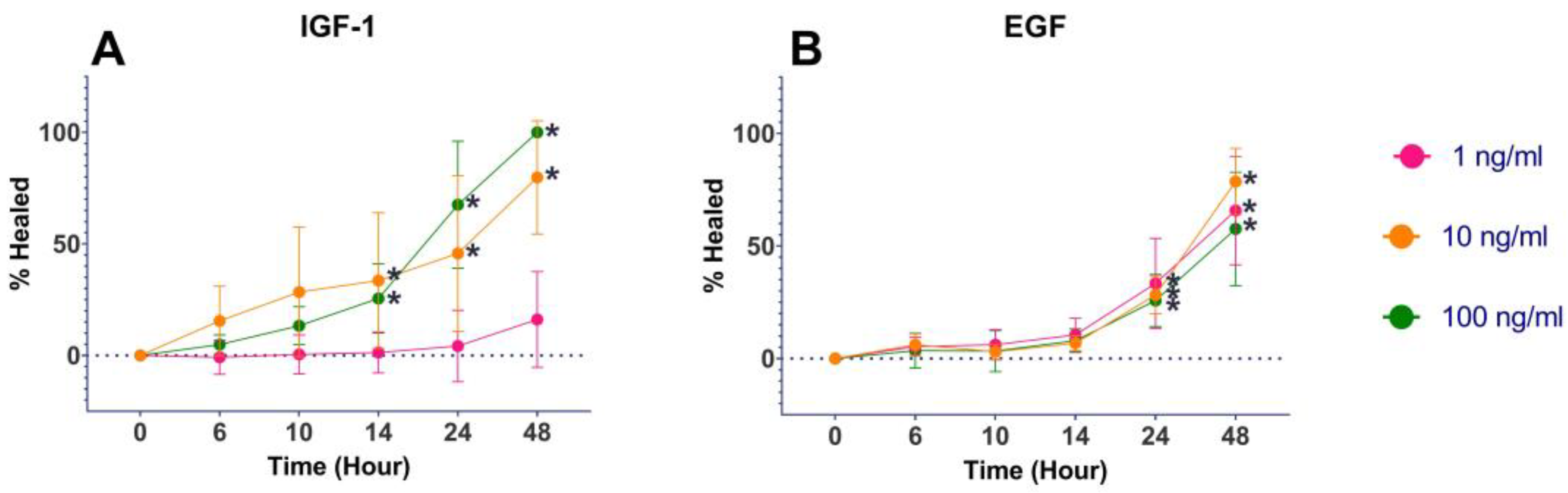
| Epidermal growth factor (EGF) | EGF receptor increases keratinocyte proliferation and cell migration leading to re-epithelialization in wound healing [11]. EGF is crucial for urethral and penile development, and is deficient in the skin adjacent to the urethra in boys with hypospadias, a congenital urethral defect [12,13,14]. The addition of EGF improves urothelial cell healing in an in vitro model of bladder injury repair [15]. |
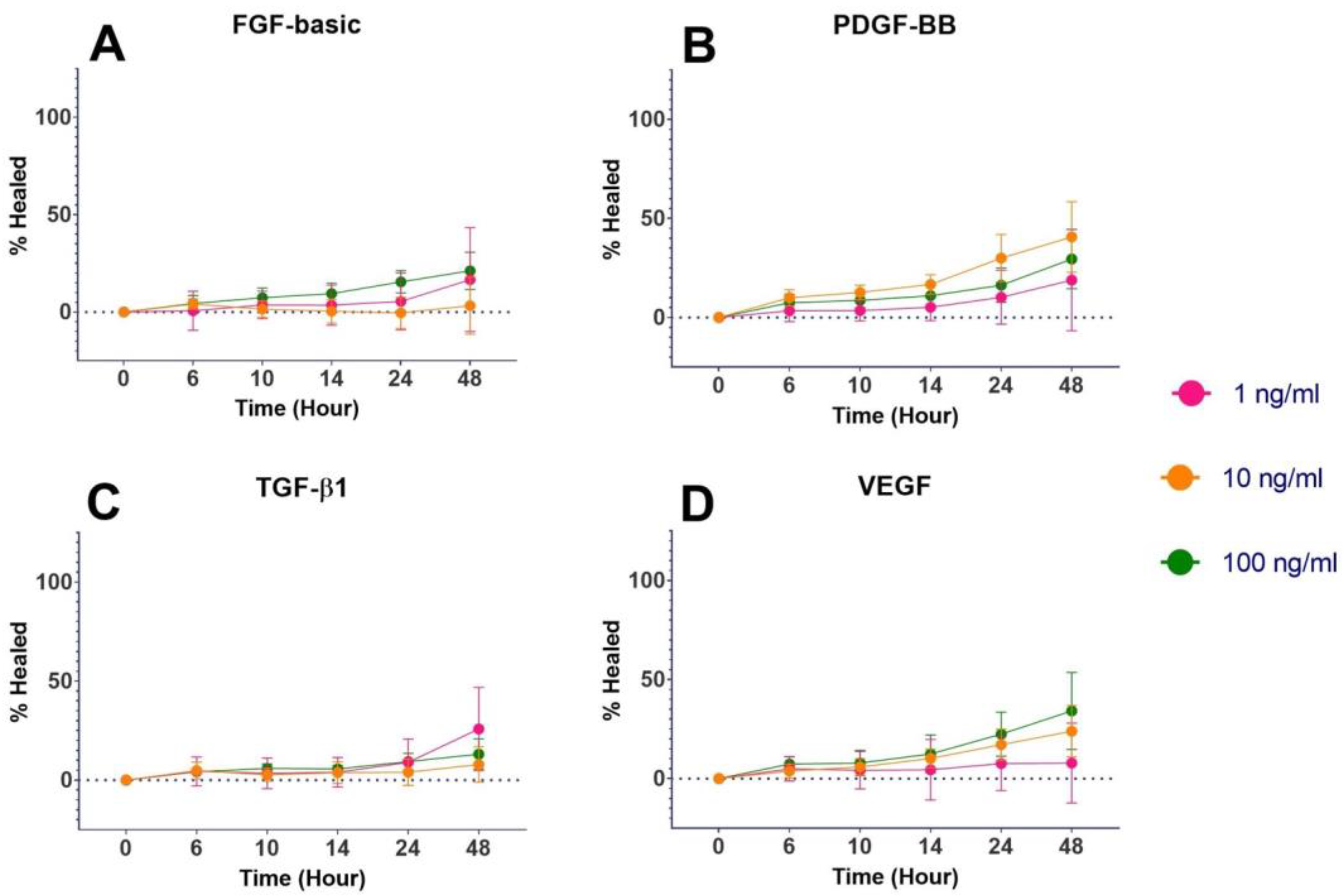
| Fibroblast growth factor-basic (FGF-basic) | FGF-basic (also known as FGF-2) also plays a role in wound healing by increasing granulation tissue formation, re-epithelialization, and tissue remodeling [11]. It is present in the developing urethra and required for proliferation of urethral progenitor cells of the epithelium [16]. |
| Insulin-like growth factor-1 (IGF-1) | IGF-1 has been linked to wound healing by increasing keratinocyte motility and promotes a proliferative response in the wound [11]. The IGF-1 receptor is prominently found in the epithelium of the rat urethra [17]. IGF-1 has been shown to promote urothelial cell proliferation resulting in improved urethral wound healing via stricture prevention [18]. |
| Platelet-derived growth factor (PDGF) | PDGF has a role in wound healing by increasing the expression of VEGF and IGF-1 to improve angiogenesis and re-epithelialization. PDGF also increases the proliferation and stimulation of fibroblasts [11]. Additionally, PDGF-BB has already been approved by the FDA for topical wound treatment in diabetic ulcers [19,20]. |
| Transforming growth factor beta (TGF-β1) | TGF-β1 has been proven to promote acceleration of healing, and to have anti-scarring and anti-fibrotic effects [11,21]. The TGF- β1 receptor is found on the mouse genital tubercle during development, and receptor expression has decreased levels after urethral injury, so increased TGF-β1 may promote urethral healing [22,23]. |
| Vascular endothelial growth factor (VEGF) | VEGF mediates angiogenesis by improving tissue ischemia and hypoxia, and limiting fibrosis and stricture [24]. VEGF receptor expression is decreased in urethral subepithelia; however, it has been shown that increased VEGF in urethral tissue supports urethral repair [22]. |
2. Materials and Methods
2.1. Cell Selection
2.2. Cell Expansion
2.3. Selection of Evaluated GFs and Controls
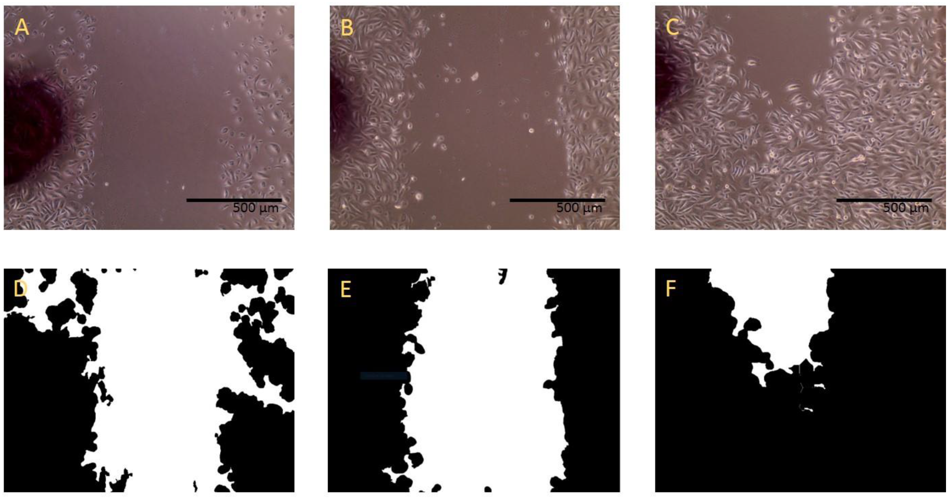
2.4. Wound-Healing Assay
2.5. Qualitative Morphology
2.6. Gene Expression
3. Results
3.1. Wound-Healing Assay
3.2. Morphology
3.3. Gene Expression
4. Discussion
5. Conclusions
Author Contributions
Funding
Institutional Review Board Statement
Informed Consent Statement
Data Availability Statement
Conflicts of Interest
References
- Bertrand, L.A.; Warren, G.J.; Voelzke, B.B.; Elliott, S.P.; Myers, J.B.; McClung, C.D.; Oleson, J.J.; Erickson, B.A. Turns Lower Urinary Tract Pain and Anterior Urethral Stricture Disease: Prevalence and Effects of Urethral Reconstruction. J. Urol. 2015, 193, 184–189. [Google Scholar] [CrossRef] [PubMed] [Green Version]
- Weese, J.R.; Raup, V.T.; Eswara, J.R.; Marshall, S.D.; Chang, A.J.; Vetter, J.; Brandes, S.B. Anterior Urethral Stricture Disease Negatively Impacts the Quality of Life of Family Members. Adv. Urol. 2016, 2016, 3582862. [Google Scholar] [CrossRef] [Green Version]
- King, C.; Rourke, K.F. Urethral Stricture is frequently a Morbid Condition: Incidence and Factors Associated with Compli-cations Related to Urethral Stricture. Urology 2019, 132, 189–194. [Google Scholar] [CrossRef]
- Hampson, L.A.; McAninch, J.W.; Breyer, B.N. Male urethral strictures and their management. Nat. Rev. Urol. 2013, 11, 43–50. [Google Scholar] [CrossRef] [PubMed] [Green Version]
- Long, C.J.; Canning, D.A. Hypospadias: Are we as good as we think when we correct proximal hypospadias? J. Pediatr. Urol. 2016, 12, 196.e1–196.e5. [Google Scholar] [CrossRef] [PubMed]
- Ninan, N.; Thomas, S.; Grohens, Y. Wound healing in urology. Adv. Drug Deliv. Rev. 2015, 82–83, 93–105. [Google Scholar] [CrossRef] [PubMed]
- Rodriguez, L.G.; Wu, X.; Guan, J.-L. Wound-Healing Assay. Methods Mol. Biol. 2005, 294, 23–29. [Google Scholar]
- Lopes, J.F.; Schned, A.; I Ellsworth, P.; Cendron, M. Histological analysis of urethral healing after tubularized incised plate urethroplasty. J. Urol. 2001, 166, 1014–1017. [Google Scholar] [CrossRef]
- Hafez, A.T.; Herz, D.; Bägli, D.; Smith, C.; McLorie, G.; Khoury, A. Healing of unstented tubularized incised plate urethroplasty: An experimental study in a rabbit model. BJU Int. 2003, 91, 84–88. [Google Scholar] [CrossRef]
- Hofer, M.D.; Cheng, E.Y.; Bury, M.I.; Park, E.; Xu, W.; Hong, S.J.; Kaplan, W.E.; Sharma, A.K. Analysis of Primary Urethral Wound Healing in the Rat. Urology 2014, 84, 246.e1–246.e7. [Google Scholar] [CrossRef]
- Barrientos, S.; Stojadinovic, O.; Golinko, M.S.; Brem, H.; Tomic-Canic, M. PERSPECTIVE ARTICLE: Growth factors and cytokines in wound healing. Wound Repair Regen. 2008, 16, 585–601. [Google Scholar] [CrossRef]
- Gupta, C. The role of epidermal growth factor receptor (EGFR) in male reproductive tract differentiation: Stimulation of EGFR expression and inhibition of Wolffian duct differentiation with anti-EGFR antibody. Endocrinology 1996, 137, 905–910. [Google Scholar] [CrossRef] [Green Version]
- Gupta, C.; Siegel, S.; Ellis, D. The role of EGF in testosterone-induced reproductive tract differentiation. Dev. Biol. 1991, 146, 106–116. [Google Scholar] [CrossRef]
- El-Galley, R.; Smith, E.; Cohen, C.; Petros, J.; Woodard, J.; Galloway, N. Epidermal growth factor (EGF) and EGF receptor in hypospadias. BJU Int. 1997, 79, 116–119. [Google Scholar] [CrossRef]
- Daher, A.; I de Boer, W.; El-Marjou, A.; van der Kwast, T.; Abbou, C.C.; Thiery, J.-P.; Radvanyi, F.; Chopin, D.K. Epidermal Growth Factor Receptor Regulates Normal Urothelial Regeneration. Lab. Investig. 2003, 83, 1333–1341. [Google Scholar] [CrossRef] [Green Version]
- Petiot, A.; Perriton, C.L.; Dickson, C.; Cohn, M.J. Development of the mammalian urethra is controlled by Fgfr2-IIIb. Development 2006, 132, 2441–2450. [Google Scholar] [CrossRef] [Green Version]
- Liu, X.; Lin, C.-S.; Spencerb, E.M.; Lue, T.F. Insulin-like Growth Factor-I Promotes Proliferation and Migration of Cavernous Smooth Muscle Cells. Biochem. Biophys. Res. Commun. 2001, 280, 1307–1315. [Google Scholar] [CrossRef]
- Shinchi, M.; Kushibiki, T.; Mayumi, Y.; Ito, K.; Asano, T.; Ishihara, M.; Horiguchi, A. Insulin-like growth factor 1 sustained-release collagen on urethral catheter prevents stricture after urethral injury in a rabbit model. Int. J. Urol. 2019, 26, 572–577. [Google Scholar] [CrossRef]
- Margolis, D.J.; Crombleholme, T.; Herlyn, M. Clinical Protocol: Phase I trial to evaluate the safety of H5.020CMV.PDGF-B for the treatment of a diabetic insensate foot ulcer. Wound Repair Regen. 2000, 8, 480–493. [Google Scholar] [CrossRef]
- Berlanga-Acosta, J.; Fernández-Montequín, J.; Valdés-Pérez, C.; Savigne-Gutiérrez, W.; Mendoza-Marí, Y.; García-Ojalvo, A.; Falcón-Cama, V.; del Barco-Herrera, D.G.; Fernández-Mayola, M.; Pérez-Saad, H.; et al. Diabetic Foot Ulcers and Epidermal Growth Factor: Revisiting the Local Delivery Route for a Successful Outcome. BioMed Res. Int. 2017, 2017, 2923759. [Google Scholar] [CrossRef] [Green Version]
- O’Kane, S.; Ferguson, M.W.J. Transforming growth factor βs and wound healing. Int. J. Biochem. Cell Biol. 1997, 29, 63–78. [Google Scholar] [CrossRef]
- Soyer, T.; Ayva, Ş.; Boybeyi, Ö.; Aslan, M.K.; Çakmak, M. The effect of platelet rich fibrin on growth factor levels in urethral repair. J. Pediatr. Surg. 2013, 48, 2545–2549. [Google Scholar] [CrossRef]
- Blaschko, S.D.; Cunha, G.R.; Baskin, L.S. Molecular mechanisms of external genitalia development. Differentiation 2012, 84, 261–268. [Google Scholar] [CrossRef] [Green Version]
- Wang, J.-H.; Xu, Y.-M.; Fu, Q.; Song, L.-J.; Li, C.; Zhang, Q.; Xie, M.-K. Continued sustained release of VEGF by PLGA nanospheres modified BAMG stent for the anterior urethral reconstruction of rabbit. Asian Pac. J. Trop. Med. 2013, 6, 481–484. [Google Scholar] [CrossRef]
- Dalghi, M.G.; Montalbetti, N.; Carattino, M.D.; Apodaca, G. The Urothelium: Life in a Liquid Environment. Physiol. Rev. 2020, 100, 1621–1705. [Google Scholar] [CrossRef]
- Georgas, K.M.; Armstrong, J.; Keast, J.R.; Larkins, C.E.; McHugh, K.M.; Southard-Smith, E.M.; Cohn, M.J.; Batourina, E.; Dan, H.; Schneider, K.; et al. An illustrated anatomical ontology of the developing mouse lower urogenital tract. Development 2015, 142, 1893–1908. [Google Scholar] [CrossRef] [Green Version]
- Nagele, U.; Maurer, S.; Feil, G.; Bock, C.; Krug, J.; Sievert, K.-D.; Stenzl, A. In Vitro Investigations of Tissue-Engineered Multilayered Urothelium Established from Bladder Washings. Eur. Urol. 2008, 54, 1414–1422. [Google Scholar] [CrossRef]
- Guan, Y.; Ou, L.; Hu, G.; Wang, H.; Xu, Y.; Chen, J.; Zhang, J.; Yu, Y.; Kong, D. Tissue Engineering of Urethra Using Human Vascular Endothelial Growth Factor Gene-Modified Bladder Urothelial Cells. Artif. Organs 2007, 32, 91–99. [Google Scholar] [CrossRef]
- El-Tabey, N.; Shokeir, A.; Barakat, N.; El-Refaie, H.; El-Hamid, M.A.; Gabr, M. Cell-seeded tubular acellular matrix for replacing a long circumferential urethral defect in a canine model: Is it clinically applicable? Arab J. Urol. 2012, 10, 192–198. [Google Scholar] [CrossRef]
- Orabi, H.; AbouShwareb, T.; Zhang, Y.; Yoo, J.J.; Atala, A. Cell-Seeded Tubularized Scaffolds for Reconstruction of Long Urethral Defects: A Preclinical Study. Eur. Urol. 2012, 63, 531–538. [Google Scholar] [CrossRef] [Green Version]
- De Filippo, R.E.; Kornitzer, B.S.; Yoo, J.J.; Atala, A. Penile urethra replacement with autologous cell-seeded tubularized collagen matrices. J. Tissue Eng. Regen. Med. 2012, 9, 257–264. [Google Scholar] [CrossRef]
- Zhang, L.; Du, A.; Li, J.; Pan, M.; Han, W.; Xiao, Y. Development of a cell-seeded modified small intestinal submucosa for urethroplasty. Heliyon 2016, 2, e00087. [Google Scholar] [CrossRef] [Green Version]
- Zhang, K.; Fu, Q.; Yoo, J.; Chen, X.; Chandra, P.; Mo, X.; Song, L.; Atala, A.; Zhao, W. 3D bioprinting of urethra with PCL/PLCL blend and dual autologous cells in fibrin hydrogel: An in vitro evaluation of biomimetic mechanical property and cell growth environment. Acta Biomater. 2017, 50, 154–164. [Google Scholar] [CrossRef]
- Möller, B.; Möller, E.B.; Glaß, M.; Misiak, D.; Posch, S. MiToBo—A Toolbox for Image Processing and Analysis. J. Open Res. Softw. 2016, 4, e17. [Google Scholar] [CrossRef] [Green Version]
- Soltys, Z.; Orzylowska-Sliwinska, O.; Zaremba, M.; Orlowski, D.; Piechota, M.; Fiedorowicz, A.; Janeczko, K.; Oderfeld-Nowak, B. Quantitative morphological study of microglial cells in the ischemic rat brain using principal component analysis. J. Neurosci. Methods 2005, 146, 50–60. [Google Scholar] [CrossRef]
- Zhu, J.; Yang, F.; He, F.; Tian, X.; Tang, S.; Chen, X. A tubular gelatin scaffold capable of the time-dependent controlled release of epidermal growth factor and mitomycin C. Colloids Surf. B Biointerfaces 2015, 135, 416–424. [Google Scholar] [CrossRef]
- Jia, W.; Tang, H.; Wu, J.; Hou, X.; Chen, B.; Chen, W.; Zhao, Y.; Shi, C.; Zhou, F.; Yu, W.; et al. Urethral tissue regeneration using collagen scaffold modified with collagen binding VEGF in a beagle model. Biomaterials 2015, 69, 45–55. [Google Scholar] [CrossRef] [PubMed]
- Wang, B.; Lv, X.; Li, Z.; Zhang, M.; Yao, J.; Sheng, N.; Lu, M.; Wang, H.; Chen, S. Urethra-inspired biomimetic scaffold: A therapeutic strategy to promote angiogenesis for urethral regeneration in a rabbit model. Acta Biomater. 2019, 102, 247–258. [Google Scholar] [CrossRef]
- Soyer, T.; Çakmak, M.; Aslan, M.K.; Şenyücel, M.F.; Kisa, Ü. Use of autologous platelet rich fibrin in urethracutaneous fistula repair: Preliminary report. Int. Wound J. 2012, 10, 345–347. [Google Scholar] [CrossRef]
- Vinter-Jensen, L.; Nielsen, K. The effects of exogenous epidermal growth factor on the developing urinary tract in rats: A stereological description. Urol. Res. 1998, 26, 105–110. [Google Scholar] [CrossRef]
- Wade, J.D.; Lun, X.-K.; Zivanovic, N.; Voit, E.O.; Bodenmiller, B. Mechanistic Model of Signaling Dynamics Across an Epithelial Mesenchymal Transition. Front. Physiol. 2020, 11, 579117. [Google Scholar] [CrossRef] [PubMed]
- Sumino, Y.; Yoshikawa, S.; Mimata, H.; Yoshimura, N. Therapeutic Effects of IGF-1 on Stress Urinary Incontinence in Rats with Simulated Childbirth Trauma. J. Urol. 2014, 191, 529–538. [Google Scholar] [CrossRef] [PubMed]
- Thiery, J.P.; Acloque, H.; Huang, R.Y.J.; Nieto, M.A. Epithelial-Mesenchymal Transitions in Development and Disease. Cell 2009, 139, 871–890. [Google Scholar] [CrossRef] [PubMed]
- Lamouille, S.; Xu, J.; Derynck, R. Molecular mechanisms of epithelial–mesenchymal transition. Nat. Rev. Mol. Cell Biol. 2014, 15, 178–196. [Google Scholar] [CrossRef] [PubMed] [Green Version]
- Kalluri, R.; Weinberg, R.A. The basics of epithelial-mesenchymal transition. J. Clin. Investig. 2009, 119, 1420–1428. [Google Scholar] [CrossRef] [Green Version]


| Area Mean (pixels2) | Area Standard Deviation | Form Factor | |
|---|---|---|---|
| GF-containing medium control | 4737 | ±1624 | 0.734 |
| GF-free medium | 3307 | ±1294 | 0.672 |
| EGF | 2722 | ±1049 | 0.612 * |
| FGF-basic | 2826 | ±1584 | 0.724 |
| IGF-1 | 3392 | ±1928 | 0.729 |
| PDGF-BB | 2730 | ±1357 | 0.799 |
| TGF-β1 | 3210 | ±1813 | 0.835 * |
Disclaimer/Publisher’s Note: The statements, opinions and data contained in all publications are solely those of the individual author(s) and contributor(s) and not of MDPI and/or the editor(s). MDPI and/or the editor(s) disclaim responsibility for any injury to people or property resulting from any ideas, methods, instructions or products referred to in the content. |
© 2023 by the authors. Licensee MDPI, Basel, Switzerland. This article is an open access article distributed under the terms and conditions of the Creative Commons Attribution (CC BY) license (https://creativecommons.org/licenses/by/4.0/).
Share and Cite
Foster, C.; Jensen, T.; Finck, C.; Rowe, C.K. Development of a Wound-Healing Protocol for In Vitro Evaluation of Urothelial Cell Growth. Methods Protoc. 2023, 6, 64. https://doi.org/10.3390/mps6040064
Foster C, Jensen T, Finck C, Rowe CK. Development of a Wound-Healing Protocol for In Vitro Evaluation of Urothelial Cell Growth. Methods and Protocols. 2023; 6(4):64. https://doi.org/10.3390/mps6040064
Chicago/Turabian StyleFoster, Christopher, Todd Jensen, Christine Finck, and Courtney K. Rowe. 2023. "Development of a Wound-Healing Protocol for In Vitro Evaluation of Urothelial Cell Growth" Methods and Protocols 6, no. 4: 64. https://doi.org/10.3390/mps6040064
APA StyleFoster, C., Jensen, T., Finck, C., & Rowe, C. K. (2023). Development of a Wound-Healing Protocol for In Vitro Evaluation of Urothelial Cell Growth. Methods and Protocols, 6(4), 64. https://doi.org/10.3390/mps6040064