
A Micropillar Array Based Microfluidic Device for Rare Cell Detection and Single-Cell Proteomics


Abstract
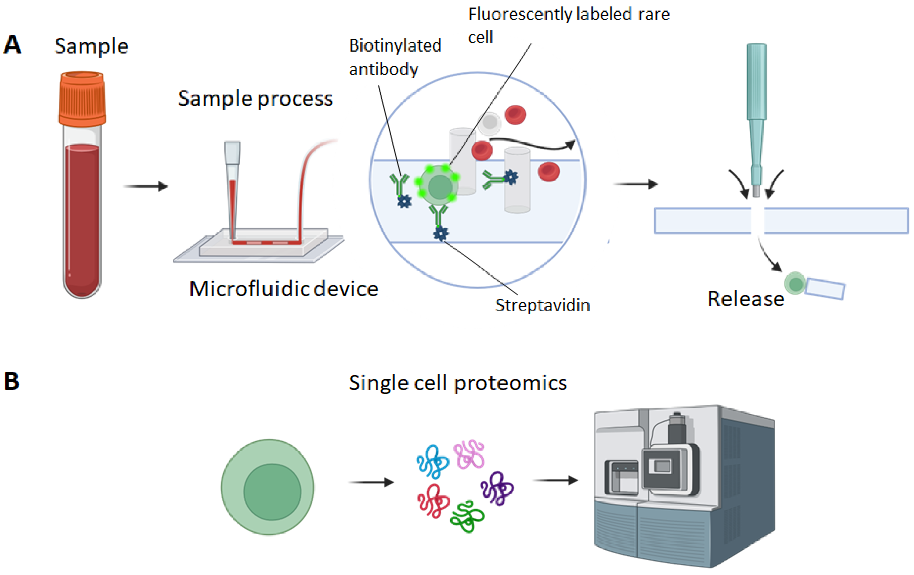
:1. Introduction
2. Experimental Design
2.1. Materials
- AZ 400k developer (MicroChemals GmbH, Ulm, Germany)
- Chromium etchant (TechniEtch Cr01, MicroChemals GmbH, Ulm, Germany)
- 5-inch chromium coated glass plate covered with positive photoresist film (Photomask Portal, Richardson, TX, USA)
- 4-inch silicon wafers (University Wafer, South Boston, MA, USA)
- Tweezers for 4-inch silicon wafer (Catalog No. 18-100-946, Fisher Scientific, Newington, NH, USA)
- Acetone (Sigma Aldrich, St Louis, MO, USA)
- Isopropanol (IPA) (Sigma Aldrich, MO, USA)
- 6-inch wide-open round beakers (Fisher Scientific, NH, USA)
- N2 dryer
- Hexamethyldisiloxane (HMDS) (SKU 440191, Sigma Aldrich, MO, USA)
- SU8 3035 photoresist (Kayaku Advanced Materials, Westborough, MA, USA)
- SU8 developer (Kayaku Advanced Materials, MA, USA)
- Polydimethylsiloxane (PDMS) Sylgard 184 kit: silicone elastomer base and curing agent (Dow Corning, Midland, MI, USA)
- Trichloro(1H,1H,2H,2H-perfluorooctyl)silane (TPOS) (SKU 448931-10G, Sigma Aldrich, MO, USA)
- Weighing dish (Catalog No. 01-549-750, Fisher Scientific, NH, USA)
- Petri dish (15 cm) (Catalog No. 09-720-500, Fisher Scientific, NH, USA)
- Weighing scale (Catalog No. 01-922-341, Fisher Scientific, NH, USA)
- Razor blade or scalpel (Catalog No. NC0134996, Fisher Scientific, NH, USA)
- PDMS punches (Catalog No. 12-460-402, 12-460-410, Fisher Scientific, NH, USA)
- Microscope slides (Catalog No. 12-550-102, Fisher Scientific, NH, USA)
- 99% ethanol (SKU 459836, Sigma Aldrich, MO, USA)
- Deionized (DI) water
- 1 mL pipet tips (Catalog No. 05412307, Fisher Scientific, NH, USA)
- Stainless steel dispensing needle with luer lock connection (Catalog No. 11-101-1671, Fisher Scientific, NH, USA)
- Cleaned and bagged high-purity white silicone rubber tubing (Catalog No. 51845K51, McMaster Carr, Elmhurst, IL, USA)
- PTFE light wall tubing (Part No. STT-21, Component Supply Co., Sparta, TN, USA)
- 3D printed supporting structure
- Sterile Luer-Lock syringes (3 mL and 5 mL) (Fisher Scientific, NH, USA)
- Phosphate-buffered saline (PBS) without Ca2+/Mg2+ ions (311-010-CL, Wisent Bioproducts, Saint-Jean-Baptiste, QC, Canada)
- Avidin (Catalog No. MP215004705, Fisher Scientific, NH, USA)
- Bovine serum albumin (BSA) (SKU A7030, Sigma Aldrich, MO, USA)
- Biotinylated anti-EpCAM (eBioscience, San Diego, CA, USA)
- Cell line (e.g., PC9 cells, ATCC)
- Cell culture medium (e.g., RPMI1640 medium,)
- Fetal bovine serum (FBS) (080-150, Wisent Bioproducts, QC, Canada)
- Trypsin-EDTA (0.25% Trypsin with EDTA 4Na) (325-042-CL, Wisent Bioproducts, QC, Canada)
- Vybrant dye DiD (Catalog No. V22887, Thermofisher, Waltham, MA, USA)
- Sterile 1.7-mL centrifuge tubes (Catalog No. 02-682-000, Fisher Scientific, NH, USA)
2.2. Equipment
- Maskless Aligner (Heidelberg uPG 501, Heidelberg Instruments Mikrotechnik GmbH, Heidelberg, Germany)
- Spin coater (H6-23, Laurell Technologies, Lansdale, PA, USA)
- Digital Programmable Stirring Hot Plates (HS40/HS40A, Torrey Pine Scientific, Carlsbad, CA, USA)
- Contact Mask Aligner (Suss MA6 Mask Aligner)
- Desiccator with a vacuum connection (Catalog No. 08-594-15A, Fisher Scientific, NH, USA)
- Programmable Oven (SKU 2958M5, Medicus Health, Kentwood, MI, USA)
- Plasma treater (SKU 12051A, Electro-Technic Products, Chicago, IL, USA)
- Programmable, dual-channel infusion and withdrawal modular syringe pump (Fusion 200-X, Chemyx, Stafford, TX, USA).
- Pipet (P10, P200, P1000) (Eppendorf) (Fisher Scientific, NH, USA)
- Serological pipet (Catalog No. 9541, Thermofisher, USA)
- CO2 incubator for cell culture (Catalog No. 11-676-604, Fisher Scientific, NH, USA)
- Biosafety hood (1300 Series, Fisher Scientific, NH, USA)
- Centrifuge (Eppendorf) (Catalog No. 05-413-112, Fisher Scientific, NH, USA)
- Mini centrifuge (Catalog No. 75002541, Fisher Scientific, NH, USA)
3. Procedure
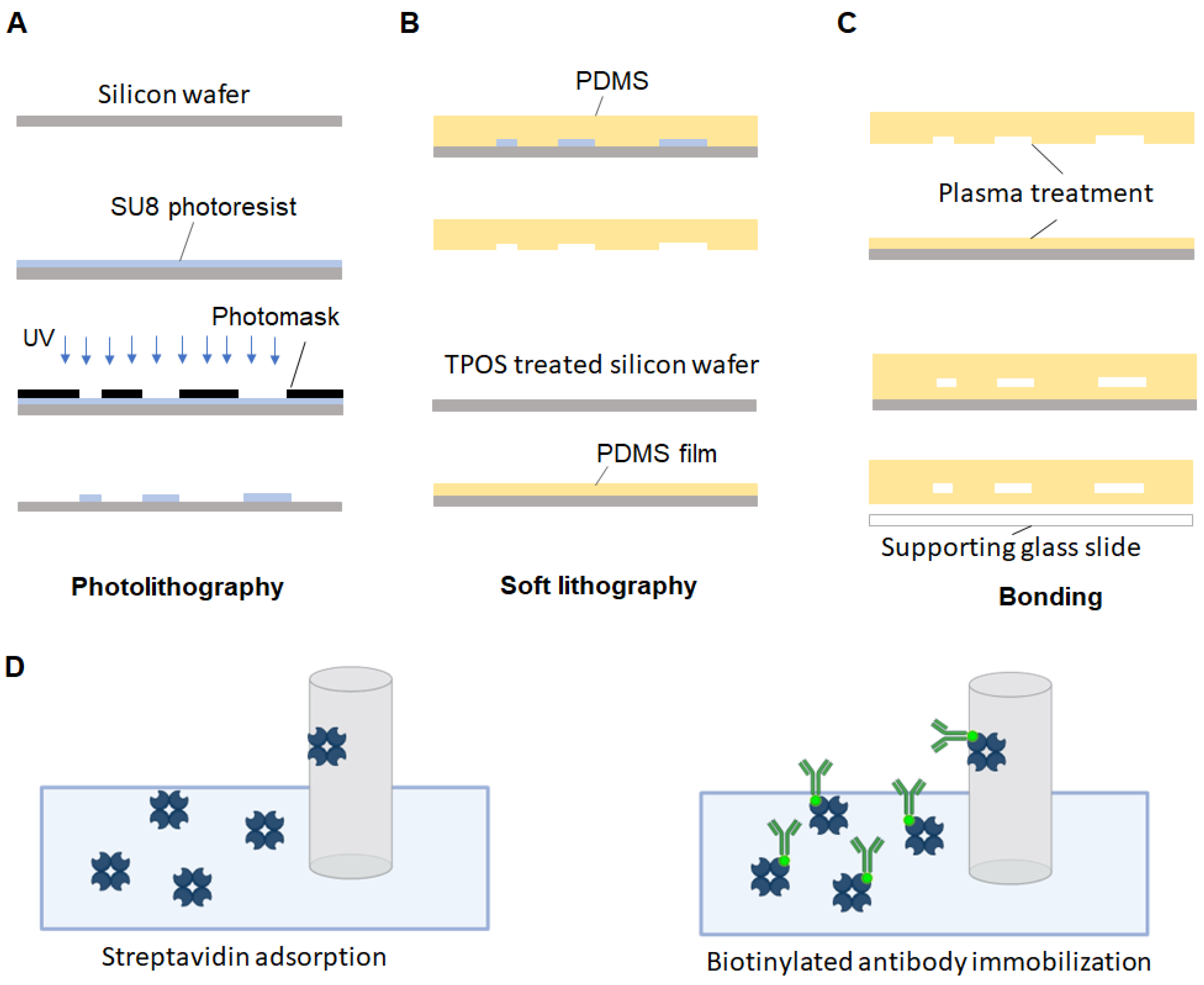
3.1. Chrome Mask Writing
- Draw the layout of the micropillar-based microfluidic device using CAD software and convert the file into DXF form.
- Load the chromium and positive photoresist coated glass plate to the maskless aligner.
- Pattern writing on the photoresist layer of the glass plate using the maskless aligner.
- Dilute the AZ 400K developer 5 times (e.g., 40 mL of AZ 400K developer and 160 mL of DI water).
- Develop the glass plate with the diluted AZ 400K developer for 20 s.
- Rinse the glass plate with running DI water for 10 s.
- Soak the glass plate in the chrome etchant for 1 min.
- Rinse the glass plate with running DI water for 10 s.
- Strip off the undeveloped photoresist in undiluted AZ 400K developer for 30 s.
- Rinse the chrome mask with running DI water for 10 s.
- Dry the chrome mask with the N2 gun.
3.2. Silicon Mold Fabrication
- 12.
- Bath a 4-inch silicon wafer in acetone for 10 min.
- 13.
- Spray wash the silicon wafer with IPA to remove acetone residues.
- 14.
- Bake the wafer at 120 °C for 10 min until fully dry and cool it down to room temperature.
- 15.
- Vaporize HMDS onto the silicon wafer to create hydrophobic surfaces.
- 16.
- Spin-coat a layer of 50 µm-thick SU-8 3035 photoresist (2000 rpm for 30 s).
- 17.
- Soft bake the wafer on a hotplate at 60 °C for 1 min.
- 18.
- Ramp up the hotplate temperature to 95° C at 450 °C/h.
- 19.
- Incubate the wafer at 95 °C for 10 min.
- 20.
- Expose the wafer using the contact mask aligner (200 mJ/cm2).
- 21.
- Post exposure bake the silicon wafer at 95 °C for 5 min.
- 22.
- Develop the silicon wafer in SU-8 developer for 7 min.
- 23.
- Hard bake the wafer at 125 °C in the oven for 15 min.
3.3. Micropillar-Based Microfluidic Device Fabrication
- 24.
- Place the silicon mold with a small weighing bowl in the vacuum desiccator.
- 25.
- Dispense 20 µL of TPOS in the weighing bowl.
- 26.
- Turn on the vacuum for 1 min to create a low-pressure environment for TPOS evaporation.
- 27.
- Incubate the silicon mold for 30 min.
- 28.
- Place the silicon mold in a 150 cm petri dish.
- 29.
- Mix 30 g of PDMS prepolymers (base: curing agent = 10:1) in a weighing boat.
- 30.
- Place the PDMS mixture in a vacuum for 15–30 min to remove bubbles.
- 31.
- Dispense PDMS mixture on the silicon mold.
- 32.
- Incubate PDMS in the oven at 75 °C for 2–4 h.
- 33.
- Cut off the polymerized PDMS substrate with fluidic feature from the silicon mold using a scalpel or razor blade.
- 34.
- Punch holes at the inlet and outlet of the PDMS substrate using a punch with inner diameter of 1.2 mm.
- 35.
- Treat an unused silicon wafer with TPOS as described from Step 24–27.
- 36.
- Apply 10 g of degassed liquid PDMS mixture on the silicon wafer.
- 37.
- Spin the wafer at 1000 rpm for 1 min.
- 38.
- Incubate the PDMS coated silicon wafer at 100 °C for 5 min.
- 39.
- Treat the PDMS substrate with an oxygen plasma treater for 1 min.
- 40.
- Treat the PDMS film on the silicon wafer with the oxygen plasma treater for 1 min.
- 41.
- Bind the PDMS substrate with the PDMS film.
- 42.
- Incubate the sandwiched layers at 100 °C for 1 min in the oven.
- 43.
- Cool the bonded device down to room temperature.
- 44.
- Gently cut off the excessive PDMS thin film using the scalpel.
- 45.
- Peel the microfluidic device off the silicon wafer.
- 46.
- Physically attach the microfluidic device on a microscope slide for sample processing.
3.4. Antibody Immobilization in the Microfludic Device
- 47.
- Prepare 2 sections of silicone rubber tubes using a scalpel or razor blade. One with a length of 5 cm. The other with a length of 10 cm.
- 48.
- Prepare 2 sections of PTFE light wall tubes with a length of 8 mm using a scalpel or razor blade.
- 49.
- Couple the silicone rubber tubes with the PTFE light wall tubes by inserting the PTFE light wall tube to one end of the silicone rubber with a depth of 4 mm.
- 50.
- Connect the short-length silicone rubber tube to the inlet of the chip by inserting the coupled PTFE light wall tube.
- 51.
- Connect the long-length silicone rubber to the outlet of the chip by inserting the coupled PTFE light wall tube.
- 52.
- Connect the short-length silicone rubber tube to a 1 mL pipet tip used as a sample reservoir. Keep the pipet tube vertical to the chip using a 3D printed hanger or fixture.
- 53.
- Fix a 5 mL Luer-locked syringe on the syringe pump.
- 54.
- Cap the syringe with a blunt-tip mixer nozzle.
- 55.
- Connect the long-length silicone rubber tube to the blunt-tip mixer nozzle.
- 56.
- Introduce 300 µL of 99% ethanol to the sample reservoir.
- 57.
- Withdraw 250 µL the ethanol at a flow rate of 2 µL/s using the syringe pump.
- 58.
- Introduce 200 µL of DI water to the sample reservoir and withdraw 200 µL at a flow rate of 2 µL/s.
- 59.
- Introduce 200 µL of PBS to the sample reservoir and withdraw 200 µL at a flow rate of 2 µL/s.
- 60.
- Introduce 100 µL of avidin to the sample reservoir and withdraw 100 µL at a flow rate of 2 µL/s.
- 61.
- Incubate for 15 min at room temperature.
- 62.
- Introduce 300 µL of PBS to the sample reservoir and withdraw 300 µL at a flow rate of 2 µL/s.
- 63.
- Dilute anti-EpCAM 25 times (4 µL of antibodies in 100 µL of PBS at a concentration of 20 µg/mL)
- 64.
- Introduce 100 µL of anti-EpCAM to the sample reservoir and withdraw 100 µL at a flow rate of 2 µL/s.
- 65.
- Incubate for 20 min at room temperature.
- 66.
- Introduce 300 µL of 1% BSA to the sample reservoir and withdraw 300 µL at a flow rate of 2 µL/s.
3.5. Capture of Rare Cells from a Spiked Sample
- 67.
- Trypsinize PC9 cancer cells by adding 5 mL of 0.25% trypsin-EDTA followed by incubating for 5 min at 37 °C.
- 68.
- Neutralize the trypsin-EDTA by adding 10 mL of whole culture medium (RPMI 1640 supplemented with 10% FBS and 1% penicillin streptomycin).
- 69.
- Spin down the cells at 1200× g for 4 min.
- 70.
- Discard the supernatant and wash the cells with 10 mL of PBS at 1200× g for 4 min.
- 71.
- Discard the supernatant and resuspend the cells in 1 mL of PBS.
- 72.
- Determine the concentration of cells using a hemocytometer.
- 73.
- Aliquot 1 mL of PC9 cells in PBS at a concentration of 1 × 10⁶ cells/mL.
- 74.
- Add 5 µL of Vybrant DID dye in the cell sample and mix cell by pipetting.
- 75.
- Incubate the mixture at 37 °C for 15 min.
- 76.
- Spin down the cells at 1200× g for 4 min.
- 77.
- Wash the cells 3 times with 1 mL of PBS.
- 78.
- Resuspend the cells in 1 mL of PBS.
- 79.
- Determine the concentration of cells using a hemocytometer.
- 80.
- Aliquot 1 mL of cell sample with a concentration of 103 cells/mL through serial dilution.
- 81.
- Add 50 µL of cells into 1 mL of whole blood sample supplemented with 1.8 mg/mL EDTA and mix well.
- 82.
- Load the spiked sample to the sample reservoir of the microfluidic device.
- 83.
- Withdraw 1.05 mL of the sample at a flow rate of 1 µL/s.
- 84.
- Wash the microfluidic device by withdrawing 300 µL of PBS at a flow rate of 2 µL/s.
- 85.
- Repeat Step 83 two times.
- 86.
- Clamp the silicone tubes of both inlet and outlet.
- 87.
- Dislodge the microfluidic device from the sample reservoir and the syringe.
- 88.
- Inspect the microfluidic device under a fluorescence microscope. Fluorescently labeled PC9 cell can be detected under the APC channel.
- 89.
- Mark the positions with captured PC9 cells using a marker.
- 90.
- Detach the microfluidic device from the microscope slide.
- 91.
- Punch though the microfluidic device of the captured PC9 position using a PDMS punch with an inner diameter of 5 mm.
- 92.
- Collect the dislodged piece of the PDMS with captured PC9 cells in a 1.7 mL centrifuge tube.
- 93.
- Fill the centrifuge tube with 1 mL of cell culture medium to keep the viability of the captured PC9 cells
4. Expected Results
Author Contributions
Funding
Institutional Review Board Statement
Informed Consent Statement
Data Availability Statement
Acknowledgments
Conflicts of Interest
References
- Chen, K.; Wang, Z. Micro-Magnetofluidic System for Rare Cell Analysis: From Principle to Translation. Chemosensors 2023, 11, 335. [Google Scholar] [CrossRef]
- Krivacic, R.T.; Ladanyi, A.; Curry, D.N.; Hsieh, H.B.; Kuhn, P.; Bergsrud, D.E.; Kepros, J.F.; Barbera, T.; Ho, M.Y.; Chen, L.B.; et al. A rare-cell detector for cancer. Proc. Natl. Acad. Sci. USA 2004, 101, 10501–10504. [Google Scholar] [CrossRef] [PubMed]
- Van Loo, P.; Voet, T. Single cell analysis of cancer genomes. Curr. Opin. Genet. Dev. 2014, 24, 82–91. [Google Scholar] [CrossRef] [PubMed]
- Powell, A.A.; Talasaz, A.H.; Zhang, H.; Coram, M.A.; Reddy, A.; Deng, G.; Telli, M.L.; Advani, R.H.; Carlson, R.W.; Mollick, J.A.; et al. Single cell profiling of circulating tumor cells: Transcriptional heterogeneity and diversity from breast cancer cell lines. PLoS ONE 2012, 7, e33788. [Google Scholar] [CrossRef] [PubMed]
- Jindal, A.; Gupta, P.; Jayadeva; Sengupta, D. Discovery of rare cells from voluminous single cell expression data. Nat. Commun. 2018, 9, 4719. [Google Scholar] [CrossRef]
- Mellman, I.; Coukos, G.; Dranoff, G. Cancer immunotherapy comes of age. Nature 2011, 480, 480–489. [Google Scholar] [CrossRef]
- Wang, Z.; Ahmed, S.; Labib, M.; Wang, H.; Hu, X.; Wei, J.; Yao, Y.; Moffat, J.; Sargent, E.H.; Kelley, S.O. Efficient recovery of potent tumour-infiltrating lymphocytes through quantitative immunomagnetic cell sorting. Nat. Biomed. Eng. 2022, 6, 108–117. [Google Scholar] [CrossRef]
- Wang, Z.; Ahmed, S.; Labib, M.; Wang, H.; Wu, L.; Bavaghar-Zaeimi, F.; Shokri, N.; Blanco, S.; Karim, S.; Czarnecka-Kujawa, K.; et al. Isolation of tumour-reactive lymphocytes from peripheral blood via microfluidic immunomagnetic cell sorting. Nat. Biomed. Eng. 2023, 4, 1–16. [Google Scholar] [CrossRef]
- Huang, N.T.; Hwong, Y.J.; Lai, R.L. A microfluidic microwell device for immunomagnetic single-cell trapping. Microfluid. Nanofluid. 2018, 22, 16. [Google Scholar] [CrossRef]
- Benham-Pyle, B.W.; Brewster, C.E.; Kent, A.M.; Mann, F.G., Jr.; Chen, S.; Scott, A.R.; Box, A.C.; Sánchez Alvarado, A. Identification of rare, transient post-mitotic cell states that are induced by injury and required for whole-body regeneration in Schmidtea mediterranea. Nat. Cell Biol. 2021, 23, 939–952. [Google Scholar] [CrossRef]
- Wang, Z.; Gagliardi, M.; Mohamadi, R.M.; Ahmed, S.U.; Labib, M.; Zhang, L.; Popescu, S.; Zhou, Y.; Sargent, E.H.; Keller, G.M. Ultrasensitive and rapid quantification of rare tumorigenic stem cells in hPSC-derived cardiomyocyte populations. Sci. Adv. 2020, 6, eaay7629. [Google Scholar] [CrossRef] [PubMed]
- Khojah, R.; Xiao, Z.; Panduranga, M.K.; Bogumil, M.; Wang, Y.; Goiriena-Goikoetxea, M.; Chopdekar, R.V.; Bokor, J.; Carman, G.P.; Candler, R.N.; et al. Single-Domain Multiferroic Array-Addressable Terfenol-D (SMArT) Micromagnets for Programmable Single-Cell Capture and Release. Adv. Mater. 2021, 33, e2006651. [Google Scholar] [CrossRef]
- Chen, K.; Dopico, P.; Varillas, J.; Zhang, J.; George, T.J.; Fan, Z.H. Integration of Lateral Filter Arrays with Immunoaffinity for Circulating-Tumor-Cell Isolation. Angew. Chem. 2019, 131, 7688–7692. [Google Scholar] [CrossRef]
- Stott, S.L.; Hsu, C.-H.; Tsukrov, D.I.; Yu, M.; Miyamoto, D.T.; Waltman, B.A.; Rothenberg, S.M.; Shah, A.M.; Smas, M.E.; Korir, G.K.; et al. Isolation of circulating tumor cells using a microvortex-generating herringbone-chip. Proc. Natl. Acad. Sci. USA 2010, 107, 18392–18397. [Google Scholar] [CrossRef] [PubMed]
- Murlidhar, V.; Zeinali, M.; Grabauskiene, S.; Ghannad-Rezaie, M.; Wicha, M.S.; Simeone, D.M.; Ramnath, N.; Reddy, R.M.; Nagrath, S. A radial flow microfluidic device for ultra-high-throughput affinity-based isolation of circulating tumor cells. Small (Weinh. Bergstr. Ger.) 2014, 10, 4895–4904. [Google Scholar] [CrossRef]
- Hur, S.C.; Henderson-MacLennan, N.K.; McCabe, E.R.; Di Carlo, D. Deformability-based cell classification and enrichment using inertial microfluidics. Lab A Chip 2011, 11, 912–920. [Google Scholar] [CrossRef]
- Cristofanilli, M.; Budd, G.T.; Ellis, M.J.; Stopeck, A.; Matera, J.; Miller, M.C.; Reuben, J.M.; Doyle, G.V.; Allard, W.J.; Terstappen, L.W.; et al. Circulating tumor cells, disease progression, and survival in metastatic breast cancer. N. Engl. J. Med. 2004, 351, 781–791. [Google Scholar] [CrossRef]
- Sheng, W.; Ogunwobi, O.O.; Chen, T.; Zhang, J.; George, T.J.; Liu, C.; Fan, Z.H. Capture, release and culture of circulating tumor cells from pancreatic cancer patients using an enhanced mixing chip. Lab A Chip 2014, 14, 89–98. [Google Scholar] [CrossRef]
- Chen, K.; Amontree, J.; Varillas, J.; Zhang, J.; George, T.J.; Fan, Z.H. Incorporation of lateral microfiltration with immunoaffinity for enhancing the capture efficiency of rare cells. Sci. Rep. 2020, 10, 14210. [Google Scholar] [CrossRef]
- Poudineh, M.; Aldridge, P.M.; Ahmed, S.; Green, B.J.; Kermanshah, L.; Nguyen, V.; Tu, C.; Mohamadi, R.M.; Nam, R.K.; Hansen, A. Tracking the dynamics of circulating tumour cell phenotypes using nanoparticle-mediated magnetic ranking. Nat. Nanotechnol. 2017, 12, 274–281. [Google Scholar] [CrossRef]
- Poudineh, M.; Labib, M.; Ahmed, S.; Nguyen, L.M.; Kermanshah, L.; Mohamadi, R.M.; Sargent, E.H.; Kelley, S.O. Profiling functional and biochemical phenotypes of circulating tumor cells using a two-dimensional sorting device. Angew. Chem. Int. Ed. 2017, 56, 163–168. [Google Scholar] [CrossRef]
- Green, B.J.; Nguyen, V.; Atenafu, E.; Weeber, P.; Duong, B.T.; Thiagalingam, P.; Labib, M.; Mohamadi, R.M.; Hansen, A.R.; Joshua, A.M. Phenotypic profiling of circulating tumor cells in metastatic prostate cancer patients using nanoparticle-mediated ranking. Anal. Chem. 2019, 91, 9348–9355. [Google Scholar] [CrossRef] [PubMed]
- Zhang, Z.; Wuethrich, A.; Wang, J.; Korbie, D.; Lin, L.L.; Trau, M. Dynamic Monitoring of EMT in CTCs as an Indicator of Cancer Metastasis. Anal. Chem. 2021, 93, 16787–16795. [Google Scholar] [CrossRef]
- Bennett, H.M.; Stephenson, W.; Rose, C.M.; Darmanis, S. Single-cell proteomics enabled by next-generation sequencing or mass spectrometry. Nat. Methods 2023, 20, 363–374. [Google Scholar] [CrossRef]
- Saliba, A.-E.; Westermann, A.J.; Gorski, S.A.; Vogel, J. Single-cell RNA-seq: Advances and future challenges. Nucleic Acids Res. 2014, 42, 8845–8860. [Google Scholar] [CrossRef] [PubMed]
- Ball, H.; Rupp, B.; Owen, S.; Smith, K.; Gunchick, V.; Keller, E.T.; Sahai, V.; Nagrath, S. Abstract 5581: Single-cell genomic analysis of patient-derived circulating tumor cells in pancreatic cancer. Cancer Res. 2023, 83, 5581. [Google Scholar] [CrossRef]
- Owen, S.; Prantzalos, E.; Gunchick, V.; Sahai, V.; Nagrath, S. Synergistic Analysis of Circulating Tumor Cells Reveals Prognostic Signatures in Pilot Study of Treatment-Naïve Metastatic Pancreatic Cancer Patients. Biomedicines 2022, 10, 146. [Google Scholar] [PubMed]
- Cao, J.; Packer, J.S.; Ramani, V.; Cusanovich, D.A.; Huynh, C.; Daza, R.; Qiu, X.; Lee, C.; Furlan, S.N.; Steemers, F.J.; et al. Comprehensive single-cell transcriptional profiling of a multicellular organism. Science 2017, 357, 661–667. [Google Scholar] [CrossRef]
- Schaum, N.; Karkanias, J.; Neff, N.F.; May, A.P.; Quake, S.R.; Wyss-Coray, T.; Darmanis, S.; Batson, J.; Botvinnik, O.; Chen, M.B.; et al. Single-cell transcriptomics of 20 mouse organs creates a Tabula Muris. Nature 2018, 562, 367–372. [Google Scholar] [CrossRef]
- The Tabula Sapiens Consortium; Jones, R.C.; Karkanias, J.; Krasnow, M.A.; Pisco, A.O.; Quake, S.R.; Salzman, J.; Yosef, N.; Bulthaup, B.; Brown, P. The Tabula Sapiens: A multiple-organ, single-cell transcriptomic atlas of humans. Science 2022, 376, eabl4896. [Google Scholar]
- Labib, M.; Kelley, S.O. Single-cell analysis targeting the proteome. Nat. Rev. Chem. 2020, 4, 143–158. [Google Scholar] [CrossRef] [PubMed]
- Wyatt Shields, C., IV; Reyes, C.D.; López, G.P. Microfluidic cell sorting: A review of the advances in the separation of cells from debulking to rare cell isolation. Lab A Chip 2015, 15, 1230–1249. [Google Scholar] [CrossRef] [PubMed]
- Nagrath, S.; Sequist, L.V.; Maheswaran, S.; Bell, D.W.; Irimia, D.; Ulkus, L.; Smith, M.R.; Kwak, E.L.; Digumarthy, S.; Muzikansky, A.; et al. Isolation of rare circulating tumour cells in cancer patients by microchip technology. Nature 2007, 450, 1235–1239. [Google Scholar] [CrossRef]
- Zborowski, M.; Chalmers, J.J. Rare cell separation and analysis by magnetic sorting. Anal. Chem. 2011, 83, 8050–8056. [Google Scholar] [CrossRef]
- Gao, Y.; Wang, Y.; He, B.; Pan, Y.; Zhou, D.; Xiong, M.; Song, Y. An Enzyme-Loaded Metal–Organic Framework-Assisted Microfluidic Platform Enables Single-Cell Metabolite Analysis. Angew. Chem. Int. Ed. 2023, 62, e202302000. [Google Scholar] [CrossRef] [PubMed]
- Chen, Y.; Fouladdel, S.; Ball, H.; Cheng, X.; Bao, L.; Serhan, H.; Liu, A.; Vandenburg, B.; Goo, L.; Merrill, N.; et al. Abstract 5592: Expansion and characterization on ALK positive NSCLC circulating tumor cells isolated using a size based inertial microfluidic Labyrinth device. Cancer Res. 2023, 83, 5592. [Google Scholar] [CrossRef]
- Sheng, W.; Chen, T.; Kamath, R.; Xiong, X.; Tan, W.; Fan, Z.H. Aptamer-Enabled Efficient Isolation of Cancer Cells from Whole Blood Using a Microfluidic Device. Anal. Chem. 2012, 84, 4199–4206. [Google Scholar] [CrossRef]
- Mishra, A.; Dubash, T.D.; Edd, J.F.; Jewett, M.K.; Garre, S.G.; Karabacak, N.M.; Rabe, D.C.; Mutlu, B.R.; Walsh, J.R.; Kapur, R.; et al. Ultrahigh-throughput magnetic sorting of large blood volumes for epitope-agnostic isolation of circulating tumor cells. Proc. Natl. Acad. Sci. USA 2020, 117, 16839–16847. [Google Scholar] [CrossRef]
- Aldridge, P.M.; Mukhopadhyay, M.; Ahmed, S.U.; Zhou, W.; Christinck, E.; Makonnen, R.; Sargent, E.H.; Kelley, S.O. Prismatic Deflection of Live Tumor Cells and Cell Clusters. ACS Nano 2018, 12, 12692–12700. [Google Scholar] [CrossRef]
- Murray, C.; Miwa, H.; Dhar, M.; Park, D.E.; Pao, E.; Martinez, J.; Kaanumale, S.; Loghin, E.; Graf, J.; Rhaddassi, K. Unsupervised capture and profiling of rare immune cells using multi-directional magnetic ratcheting. Lab A Chip 2018, 18, 2396–2409. [Google Scholar] [CrossRef] [PubMed]
- Bae, J.; Samur, M.; Richardson, P.; Munshi, N.C.; Anderson, K.C. Selective targeting of multiple myeloma by B cell maturation antigen (BCMA)-specific central memory CD8+ cytotoxic T lymphocytes: Immunotherapeutic application in vaccination and adoptive immunotherapy. Leukemia 2019, 33, 2208–2226. [Google Scholar] [CrossRef]
- Wang, Z.; Wang, H.; Lin, S.; Ahmed, S.; Angers, S.; Sargent, E.H.; Kelley, S.O. Nanoparticle Amplification Labeling for High-Performance Magnetic Cell Sorting. Nano Lett. 2022, 22, 4774–4783. [Google Scholar] [CrossRef]
- Koo, D.; Mao, Z.; Dimatteo, R.; Tsubamoto, N.; Noguchi, M.; McLaughlin, J.; Tran, W.; Lee, S.; Cheng, D.; Rutte, J.d.; et al. Defining T cell receptor repertoires using nanovial-based affinity and functional screening. bioRxiv 2023. [Google Scholar] [CrossRef]
- Miwa, H.; Dimatteo, R.; de Rutte, J.; Ghosh, R.; Di Carlo, D. Single-cell sorting based on secreted products for functionally defined cell therapies. Microsyst. Nanoeng. 2022, 8, 84. [Google Scholar] [CrossRef] [PubMed]
- Green, B.J.; Marazzini, M.; Hershey, B.; Fardin, A.; Li, Q.; Wang, Z.; Giangreco, G.; Pisati, F.; Marchesi, S.; Disanza, A. PillarX: A Microfluidic Device to Profile Circulating Tumor Cell Clusters Based on Geometry, Deformability, and Epithelial State. Small (Weinh. Bergstr. Ger.) 2022, 18, 2106097. [Google Scholar] [CrossRef] [PubMed]
- Green, B.J.; Kermanshah, L.; Labib, M.; Ahmed, S.U.; Silva, P.N.; Mahmoudian, L.; Chang, I.-H.; Mohamadi, R.M.; Rocheleau, J.V.; Kelley, S.O. Isolation of phenotypically distinct cancer cells using nanoparticle-mediated sorting. ACS Appl. Mater. Interfaces 2017, 9, 20435–20443. [Google Scholar] [CrossRef]
- Labib, M.; Green, B.; Mohamadi, R.M.; Mepham, A.; Ahmed, S.U.; Mahmoudian, L.; Chang, I.-H.; Sargent, E.H.; Kelley, S.O. Aptamer and Antisense-Mediated Two-Dimensional Isolation of Specific Cancer Cell Subpopulations. J. Am. Chem. Soc. 2016, 138, 2476–2479. [Google Scholar] [CrossRef]
- Lin, G. Magnetic particles for multidimensional in vitro bioanalysis. VIEW 2021, 2, 20200076. [Google Scholar] [CrossRef]
- Castaño, N.; Kim, S.; Martin, A.M.; Galli, S.J.; Nadeau, K.C.; Tang, S.K.Y. Exponential magnetophoretic gradient for the direct isolation of basophils from whole blood in a microfluidic system. Lab A Chip 2022, 22, 1690–1701. [Google Scholar] [CrossRef]
- Inglis, D.W.; Riehn, R.; Austin, R.H.; Sturm, J.C. Continuous microfluidic immunomagnetic cell separation. Appl. Phys. Lett. 2004, 85, 5093–5095. [Google Scholar] [CrossRef]
- Shen, Q.; Xu, L.; Zhao, L.; Wu, D.; Fan, Y.; Zhou, Y.; OuYang, W.H.; Xu, X.; Zhang, Z.; Song, M. Specific capture and release of circulating tumor cells using aptamer-modified nanosubstrates. Adv. Mater. 2013, 25, 2368–2373. [Google Scholar] [CrossRef]
- Song, Y.; Shi, Y.; Huang, M.; Wang, W.; Wang, Y.; Cheng, J.; Lei, Z.; Zhu, Z.; Yang, C. Bioinspired engineering of a multivalent aptamer-functionalized nanointerface to enhance the capture and release of circulating tumor cells. Angew. Chem. Int. Ed. 2019, 58, 2236–2240. [Google Scholar] [CrossRef]
- Hou, S.; Zhao, H.; Zhao, L.; Shen, Q.; Wei, K.S.; Suh, D.Y.; Nakao, A.; Garcia, M.A.; Song, M.; Lee, T. Capture and stimulated release of circulating tumor cells on polymer-grafted silicon nanostructures. Adv. Mater. 2013, 25, 1547–1551. [Google Scholar] [CrossRef]
- Li, W.; Reátegui, E.; Park, M.-H.; Castleberry, S.; Deng, J.Z.; Hsu, B.; Mayner, S.; Jensen, A.E.; Sequist, L.V.; Maheswaran, S. Biodegradable nano-films for capture and non-invasive release of circulating tumor cells. Biomaterials 2015, 65, 93–102. [Google Scholar] [CrossRef]
- Chen, K.; Georgiev, T.Z.; Sheng, W.; Zheng, X.; Varillas, J.I.; Zhang, J.; Hugh Fan, Z. Tumor cell capture patterns around aptamer-immobilized microposts in microfluidic devices. Biomicrofluidics 2017, 11, 054110. [Google Scholar] [CrossRef]
- Sen-Dogan, B.; Yildirim, E.; Sahin, S.; Ozgur, E.; Zorlu, O.; Kulah, H. Design of a microfluidic device for immunoaffinity-based isolation of circulating tumor cells with minimal clogging. Sens. Actuators Rep. 2023, 6, 100169. [Google Scholar] [CrossRef]
- Thege, F.I.; Lannin, T.B.; Saha, T.N.; Tsai, S.; Kochman, M.L.; Hollingsworth, M.A.; Rhim, A.D.; Kirby, B.J. Microfluidic immunocapture of circulating pancreatic cells using parallel EpCAM and MUC1 capture: Characterization, optimization and downstream analysis. Lab A Chip 2014, 14, 1775–1784. [Google Scholar] [CrossRef] [PubMed]
- Smith, J.P.; Lannin, T.B.; Syed, Y.A.; Santana, S.M.; Kirby, B.J. Parametric control of collision rates and capture rates in geometrically enhanced differential immunocapture (GEDI) microfluidic devices for rare cell capture. Biomed. Microdevices 2014, 16, 143–151. [Google Scholar] [CrossRef]
- Brunner, A.-D.; Thielert, M.; Vasilopoulou, C.; Ammar, C.; Coscia, F.; Mund, A.; Hoerning, O.B.; Bache, N.; Apalategui, A.; Lubeck, M.; et al. Ultra-high sensitivity mass spectrometry quantifies single-cell proteome changes upon perturbation. Mol. Syst. Biol. 2022, 18, e10798. [Google Scholar] [CrossRef] [PubMed]
- Specht, H.; Emmott, E.; Petelski, A.A.; Huffman, R.G.; Perlman, D.H.; Serra, M.; Kharchenko, P.; Koller, A.; Slavov, N. Single-cell proteomic and transcriptomic analysis of macrophage heterogeneity using SCoPE2. Genome Biol. 2021, 22, 50. [Google Scholar] [CrossRef]
- Woo, J.; Clair, G.C.; Williams, S.M.; Feng, S.; Tsai, C.-F.; Moore, R.J.; Chrisler, W.B.; Smith, R.D.; Kelly, R.T.; Paša-Tolić, L. Three-dimensional feature matching improves coverage for single-cell proteomics based on ion mobility filtering. Cell Syst. 2022, 13, 426–434.e424. [Google Scholar] [CrossRef] [PubMed]
- Zhu, C.; Preissl, S.; Ren, B. Single-cell multimodal omics: The power of many. Nat. Methods 2020, 17, 11–14. [Google Scholar] [CrossRef] [PubMed]


Disclaimer/Publisher’s Note: The statements, opinions and data contained in all publications are solely those of the individual author(s) and contributor(s) and not of MDPI and/or the editor(s). MDPI and/or the editor(s) disclaim responsibility for any injury to people or property resulting from any ideas, methods, instructions or products referred to in the content. |
© 2023 by the authors. Licensee MDPI, Basel, Switzerland. This article is an open access article distributed under the terms and conditions of the Creative Commons Attribution (CC BY) license (https://creativecommons.org/licenses/by/4.0/).
Share and Cite
Chen, K.; Wang, Z. A Micropillar Array Based Microfluidic Device for Rare Cell Detection and Single-Cell Proteomics. Methods Protoc. 2023, 6, 80. https://doi.org/10.3390/mps6050080
Chen K, Wang Z. A Micropillar Array Based Microfluidic Device for Rare Cell Detection and Single-Cell Proteomics. Methods and Protocols. 2023; 6(5):80. https://doi.org/10.3390/mps6050080
Chicago/Turabian StyleChen, Kangfu, and Zongjie Wang. 2023. "A Micropillar Array Based Microfluidic Device for Rare Cell Detection and Single-Cell Proteomics" Methods and Protocols 6, no. 5: 80. https://doi.org/10.3390/mps6050080