Article

No Correlation between Endo- and Exoskeletal Regenerative Capacities in Teleost Species

1
Department of Genetics, ELTE Eötvös Loránd University, 1117 Budapest, Hungary
2
Department of Cell and Developmental Biology, ELTE Eötvös Loránd University, 1117 Budapest, Hungary
3
MTA-SE “Lendület” Nephrogenetic Research Laboratory, 1083 Budapest, Hungary
*
Author to whom correspondence should be addressed.
Current address: Vienna Biocenter, 1030 Vienna, Austria.
Current address: Sars International Centre for Marine Molecular Biology, 5008 Bergen, Norway.
§
These authors contributed equally to this work.
Fishes 2019, 4(4), 51; https://doi.org/10.3390/fishes4040051
Submission received: 28 June 2019 / Revised: 18 September 2019 / Accepted: 9 October 2019 / Published: 14 October 2019
(This article belongs to the Special Issue Fish as Model Organisms for (Eco)Toxicology and Disease)

Abstract

:
The regeneration of paired appendages in certain fish and amphibian lineages is a well established and extensively studied regenerative phenomenon. The teleost fin is comprised of a proximal endoskeletal part (considered homologous to the Tetrapod limb) and a distal exoskeletal one, and these two parts form their bony elements through different ossification processes. In the past decade, a significant body of literature has been generated about the biology of exoskeletal regeneration in zebrafish. However, it is still not clear if this knowledge can be applied to the regeneration of endoskeletal parts. To address this question, we decided to compare endo- and exoskeletal regenerative capacity in zebrafish (Danio rerio) and mudskippers (Periophthalmus barbarous). In contrast to the reduced endoskeleton of zebrafish, Periophthalmus has well developed pectoral fins with a large and easily accessible endoskeleton. We performed exo- and endoskeletal amputations in both species and followed the regenerative processes. Unlike the almost flawless exoskeletal regeneration observed in zebrafish, regeneration following endoskeletal amputation is often impaired in this species. This difference is even more pronounced in Periophthalmus where we could observe no regeneration in endoskeletal structures. Therefore, regeneration is regulated differentially in the exo- and endoskeleton of teleost species.

1. Introduction

Due to its mythical overtones, appendage regeneration, the ability to regrow lost limbs has fascinated many generations of scientists. The first scientific observations of the phenomenon were made by the Italian priest and naturalist Lazzaro Spallanzani [1,2] in the 18th century, and over the next two centuries, many scientific luminaries, including Thomas Hunt Morgan [3], have contributed to the understanding of this important biological process. These early experimental studies were mostly descriptive in nature and only at the end of the 20th century, with the advent of powerful molecular techniques were scientists finally able to study the molecular processes that underpin the regeneration of appendages in different species (see [4,5,6,7] for a comprehensive review of the field).
The burst in molecular regeneration studies coincided with the emergence of zebrafish as a genetic model organism, and the expansion of the genetic toolkit in this species made it a natural subject for regeneration experiments. Due to its robustness, zebrafish fin regeneration soon became one of the better studied regeneration paradigms [8] alongside salamander limb regeneration (see references in [4,9]). Multiple signaling pathways [10,11,12,13,14,15] and other cellular phenomena [16,17] have been linked to successful fin regeneration in zebrafish and recently, even enhancers specific for fin regeneration have been identified [18,19].
On the basis of these studies, the emerging picture of fish fin regeneration is one of a typical epimorphosis process. The cells of the blastemal tissue that forms following the injury are essentially dedifferentiated cells from adjacent tissues which will start to proliferate and their progeny will ultimately redifferentiate into the original tissue type [20,21]. (While dedifferentiation appears to be the dominant regenerative process during fin regeneration, it is important to mention that alternative pathways must exist, as osteoblasts can develop in the regenerating tissue even in the absence of other osteoblasts in the injured fin [22]).
The implicit goal of these regenerative studies is to uncover universal mechanisms that could be exploited to enhance limb regeneration in other vertebrate species (such as humans) with limited regenerative capacity. Therefore, it should be noted that the majority of these experiments in zebrafish have been performed on caudal fins. While the process appears to be broadly similar in pectoral fins (which is orthologous with Tetrapod limbs), there are also some peculiarities specific for these latter structures [23,24].
The typical teleost pectoral fin is formed of a muscularized endoskeleton and a dermal fin fold that attaches to it. The endoskeleton articulates to the pectoral girdle and it is composed of proximal and distal radials, while the fin fold is supported by multiple fin rays (exoskeleton) [25]. Both the radials and the rays are bony structures, but their ossification happens through distinctly different developmental mechanisms: while the radials undergo canonical endochondrial ossification (akin to the limbs of Tetrapods), the lepidotrichia of the fin rays are formed through membranous ossification [26].
The skeletal and developmental homologies between zebrafish pectoral fins and Tetrapod limbs are still the subject of investigation, but until recently, the consensus view was that distal (endoskeletal) radials of the Teleost fin are homologous with the autopod of Tetrapods [27,28,29]. A recent study, however, suggested a common developmental origin also for fin rays and digits [30]. Although the available evidence clearly shows that similar mechanisms are activated during the development of both structures, whether such shared features reflect a true (anatomical) homology still remains to be conclusively demonstrated. The issue of anatomical homology is relevant, as almost all of the regeneration studies in zebrafish observe the process following exoskeletal amputations. Some researchers also argue that the remarkable regenerative property is a unique property of the fin folds, due to their simple structure and specific development [31,32].
To test this hypothesis, we performed endoskeletal amputations in zebrafish pectoral fins and followed the regenerative process. As zebrafish have only a small pectoral endoskeleton, we decided to extend our studies to another teleost species. We chose a mudskipper (Periophthalmus barbarous), as representatives of this family have equally well developed pectoral endo- and exoskeletons (Figure 1) [33]. Our results from both species suggest that the regenerative capacities of the Teleost endoskeleton are negligible compared with that of the exoskeleton.

2. Results

2.1. Endoskeletal Regeneration in Zebrafish

To characterize the regenerative capacity of the zebrafish endoskeleton, we performed amputations through the right pectoral fin endoskeleton of young adult females and followed their regeneration (Figure 2). Previous studies showed that exoskeletal regeneration both in caudal and pectoral fins are completed by the end of the second week post amputation [8,23]. In contrast, by the end of the second week, we were able to observe only a small fin stump in the case of the endoskeletal amputations (Figure 2b and Figure A1a), and regenerative growth was only completed by the end of the sixth week (Figure 2d). The regenerated pectoral fins had a heteromorphic character, the exoskeletal fin rays always being smaller (21/26) and often taking a twisted shape (5/26) (Figure 2d–f). In 5/26 cases, we did not observe regeneration at all. Interestingly, whenever regeneration occurred, we could observe distinct regenerated structures as soon as 2 weeks post-amputation (wpa)—the time-frame for normal exoskeletal regeneration in this species. Conversely, if by 2 wpa, no regenerated tissue formed and regeneration was not observed at all during our 6-weeks observation period (Figure A1a,b).
Pectoral fins have a well defined antero-posterior (AP) axis, and the transcriptional hallmarks of this AP pattern have been recently identified [24]. The irregular structure of the fins that develops after endoskeletal amputation could reflect a breakdown in this AP pattern. Therefore, we decided to test if there is a difference in the expression of characteristic anterior (alx4 and id4) and posterior (hand2 and hoxd11) patterning genes. We found that the relative expression differences between the anterior and posterior compartments were significantly altered in the regenerated fins for all the genes examined (Figure 2g). Our analysis suggests that the graded expression of the anterior-specific genes was essentially abolished due to a drop in gene expression levels in the anterior part, and the expression gradient of posterior-specific genes was also reduced, albeit mostly due to increased expression in the anterior compartment (Figure A1c). These results indicate that when endoskeletal regeneration occurs, it results in fins with an overall “posterior-like” character.
Importantly, unlike in the case of caudal fin exoskeletal amputations, where a practically unlimited regenerative capacity was described previously [8], repeated amputations of the endoskeleton reveal a limited regenerative capacity in the pectoral fin. Out of the three cases examined, in two cases, we observed no regeneration (Figure 2h,h’) and in one case, only a very reduced fin-stump could be seen after the second amputation during a 6-week period (Figure 2i).

2.2. Comparison of Endo- and Exoskeletal Regeneration in Mudskippers

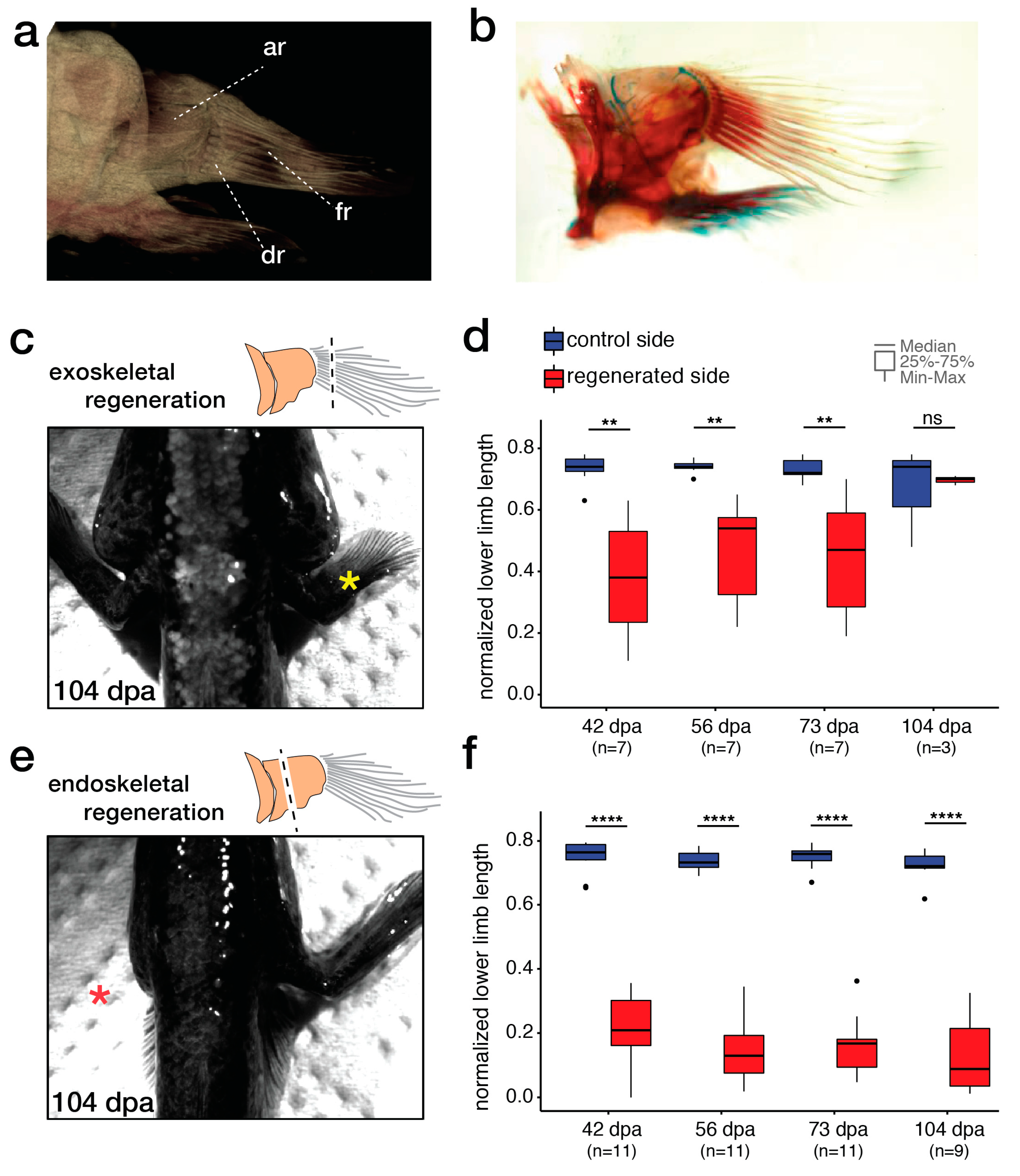
As zebrafish have a reduced endoskeleton, we decided to extend our studies to a species with well developed radials (Figure 3a,b). Using the mudskipper, Periophthalmus barbarous, we performed both exo- and endoskeletal amputations and followed the regeneration of the pectoral fins for over one hundred days (Figure 3c–f).
Exoskeletal regeneration was complete by the end of the observation period, the fins in all the observed animals recovering their original length (Figure 3c,d). In stark contrast, endoskeletal regeneration was not followed by substantive regeneration. Although blastema-like structures appeared (not shown), the amputated stumps remained essentially the same size over the whole period (Figure 3e,f).

3. Discussion

The dermal fin folds are useful during swimming, but due to their fine structure, they can get easily injured, therefore, the capacity to regenerate them has clear adaptive value. Exoskeletal regeneration has been described in many Actinopterygian species, (e.g., sturgeon (Acipenser oxyrinchus) [35], short-lived killifish (Nothobranchius furzeri) [36], sailfin molly (Poecilia latipinna) [37], mexican cavefish (Astyanax mexicanus) [38], medaka (Oryzias latipes) [39], loach (Paramisgurnus dabryanu) [40] and several others [41]), suggesting that this is an ancestral trait of ray-finned fishes.
In contrast to the numerous well-documented exoskeletal regeneration studies, there are only a few studies on endoskeletal regeneration within the Actinopterygian clade. Complete endoskeletal regeneration has been reported in the case of two Polypterid species, the bichir (Polypterus senegalus) [34,42,43] and the ropefish (Erpetoichthys calabaricus) [43]. In a recent study, Schneider and coworkers provided evidence that endoskeletal regeneration can be also observed in two other basal Actionpterygians, the paddlefish (Polyodon spathula) and the spotted gar (Lepisosteus oculatus) [44].
In the same study, the authors also examined four teleosts, the convict (Amatitlania nigrofasciata), the oscar (Astronotus ocellatus), the blue gourami (Trichogaster trichopterus) and the goldfish (Carassius auratus). They observed absent (blue gourami) or limited regeneration after pectoral endoskeletal amputations in these species, and if regeneration did occur, it often resulted in fins with a reduced and/or twisted morphology (e.g., oscar and goldfish) [44], similarly to our observations in zebrafish (Figure 2).
Earlier experiments demonstrated that zebrafish possess an almost unlimited potential to regenerate their exoskeleton [8]. In contrast, our present results demonstrate that the ability of zebrafish to regenerate the pectoral endoskeleton is much more limited and results in heteromorphic fins. Previous studies have uncovered similar differences in the regeneration capacity of the tail: whereas simple caudal fin amputations always results in fully regenerated tissue, the regeneration following tail (thus endoskeletal) amputations is successful only in about half of the cases, and when it occurs, the process is slow and results in heteromorphic fins [45]. Our results also suggest that the abnormal shape of the regenerated pectoral fins could be partially explained by changes in the expression of AP patterning genes (Figure 2 and Figure A1). Anterior-specific genes are downregulated, while posterior-specific ones are upregulated in the anterior fin rays.
We see two possible explanations for the impaired expression of the AP patterning genes, which could also account for the reduced width of the regenerated fins. First, the results might be due to incomplete (exoskeletal rather than endoskeletal) amputation of the posterior pectoral fin. Secondly, they could be attributable to a differential capacity for regeneration between the anterior and posterior parts of the endoskeleton. On the basis of our data, we cannot exclude completely the former explanation. But deeper amputation planes would cause excessive damage to the body wall, creating another confounder for the experiment. On the other hand, a differential regenerative capacity has been demonstrated during exoskeletal regeneration in male zebrafish [20], suggesting that anterior and posterior pectoral fin blastemas have unequal regenerative capacities. Overall, our results demonstrate that the endoskeletal regeneration potential of zebrafish is low.
Zebrafish, however, have a reduced endoskeletal component in their pectoral fin, which could explain their reduced regenerative capacity. Therefore, we decided to compare exo- and endoskeletal regeneration in a Teleost species with equally well-developed endo- and exoskeletal parts. Due to their lifestyle, mudskippers have pectoral fins that fit this description, which also makes them a good model to understand the evolution of terrestrial locomotion [46,47]. We followed the regeneration of exo- and endoskeletons in mudskippers for over one hundred days, and while the size of the exoskeleton recovered completely by this time, no regeneration was observed in the case of endoskeletal amputations (Figure 3).
Besides the aforementioned Actinopterygians, endoskeletal regeneration was also observed in the basal Sarcopterygian lungfish [48] and caudate amphibians (see references in [4,9]). On the basis of currently available data on endoskeletal regeneration, this feature could be an ancestral trait of the Osteichthyan group, which was parallely lost (or reduced) in most other Sarcopterygian clades and teleosts. However, it is noteworthy that finfold regeneration is absent from basal gnathostomes, like sharks [49]. Therefore, at this point, we cannot completely rule out the possibility that regeneration is a derived feature, and the differing levels of skeletal regeneration observed in certain Osteichthyan lineages reflect adaptive traits acquired by these groups. Further studies in other basal gnathostomes should help to resolve this question.
Our results show that endo- and exoskeletal regenerative capacities are not correlated in Teleosts, which suggests that the results from exoskeletal regeneration studies are not necessarily directly applicable to other species where the limbs are composed only of endoskeletal elements. Yet, the existence of the limited endoskeletal regenerative potential observed in such a well-established model species as the zebrafish suggests that there is scope for research programs that aim to understand this phenomenon better. Just as Xenopus has been used recently to study regeneration by comparing regeneration competent and incompetent stages [50,51], a comparative analysis of zebrafish exo- and endoskeletal regeneration could uncover important new factors for limb regeneration. It will be interesting to understand if the genetic program regulating teleost endoskeletal regeneration shares the commonalities of the bichir, lungfish and salamander regeneration programs [42,48] and to identify the genetic and environmental factors that can modulate this program.

4. Materials and Methods

4.1. Fish Maintenance

Wild type (ekwill) zebrafish used in this study were maintained in the zebrafish facility of ELTE Eötvös Loránd University according to standard protocols [52]. Prior to surgical procedures, fish were anesthesized using tricaine (E10521, Sigma-Aldrich, Saint Louis, MO, USA). All protocols used in this study were approved by the Hungarian National Food Chain Safety Office (Permit Number: XIV-I-001/515-4/2012).
Twenty mudskippers (ten males and ten females) were purchased from a local pet store and kept in the animal facility of ELTE Eötvös Loránd University. Fish were housed in a 30 L aquarium, where water was changed weekly. Water with a salinity of 30 ppt was prepared using Instant Ocean® Sea Salt mix dissolved in filtered water. Fish were fed once a day with medium sized crickets also purchased from a pet store. Similarly to zebrafish, mudskippers were anesthesized using 160 μg/mL tricaine before surgical procedures and documentation. All protocols were approved by the Pest County Governmental Office (Permit Number: PEI/001/1459-11/2015).

4.2. Data Acquisition and Analysis

Images of the animals were taken from dorsal and lateral perspectives. In the dorsal perspective, the limbs were positioned as they would be naturally positioned and in the lateral perspective, the limbs were straightened. The images were processed using ImageJ (https://imagej.nih.gov/ij/). The measurements were taken using the pixel length of the segmented lines within the toolbox of ImageJ. We measured the distance from the limb attachment site at the body wall to the furthest fin-ray tip or the furthest point in the callus tissue (total limb length). The distance from the limb attachment site to the start of the callus tissue was also recorded (lower limb length). Using these two measurements, we calculated the lower limb length to total limb length ratio. As a control, we also measured the contralateral side limb lengths throughout the experiment.
A statistical analysis and visualization of the data was performed in RStudio using the ggplot2 package [53]. For the statistical comparison, we used the Mann–Whitney–Wilcoxon test.

4.3. qRT-PCR Analysis

Total RNA was extracted with the TRI Reagent (Invitrogen, AM9738, Carlsbad, CA, USA), using the anteriormost and posteriormost three ray-segments of the pectoral fins, respectively. For control and 6 wpa samples material from 3-3 individuals were pooled. cDNA was synthesized from 130 ng total RNA using the SuperScript™ III Reverse Transcriptase Kit (Thermo Fisher Scientific, 18080093, Waltham, MA, USA). Quantitative real-time polymerase chain reaction (qRT-PCR) mixtures were prepared with Promega GoTaq® qPCR kit (Promega, A6001, Madison, WI, USA) and run on the Biorad CFX 384 Real Time Cycler. Primers used to amplify alx4, hand2, hoxd11, id4 and β-actin were described previously [24]. CT values of alx4, hand2, hoxd11 and id4 were normalised to β-actin using the ∆Ct method to calculate fold change.

4.4. Cartilage and Bone Stainings

For Alcian blue and Alizarin red stainings. wholemount specimens were fixed in 7% paraformaldehyde, washed with PBS, and stained in 0.01% Alcian solution (20% Acetic Acid) for 1–2 days. Specific stainings were differentiated in EtOH and then rehydratated. Alizarin-Red-S staining (in 2% KOH) was performed for 1–2 days. After dehydration, samples were stored in 87% glycerol.

4.5. Micro-CT Imaging

The pectoral girdle from one of the stained specimens was removed and placed into a scintillation vial and imaged in a SkyScan 1272 (Bruker, Kontich, Belgium), using an X-ray source of 50 kV, 200 μA. Pixel size was 26.6 × 26.6 × 26.6 μm and a total of 423 projection images were recorded for the reconstruction.

Author Contributions

Conceptualization, M.V., G.C. and N.P.; methodology, G.C. and M.V.; investigation, N.P., F.K. and M.K.; resources, T.V., M.V. and G.C.; writing—original draft preparation, visualization M.V., N.P. and F.K.; writing—review and editing by all authors; supervision, M.V. and T.V.; project administration, M.V.

Funding

M.V. is supported by grant no. NRDI-FK124230 of the Hungarian National Research, Development and Innovation Office (NRDI) and by the ELTE Excellence Program (1783-3/2018/FEKUTSRAT) sponsored by the Hungarian Ministry of Human Capacities.

Acknowledgments

We thank Natália Tőkési and Mónika Béres for helping with micro-CT imaging, and Julianna Víg and our reviewers for helpful comments on the manuscript.

Conflicts of Interest

The authors declare no conflict of interest. The funders had no role in the design of the study; in the collection, analyses, or interpretation of data; in the writing of the manuscript, or in the decision to publish the results.

Appendix A

Figure A1. Impaired regeneration of the zebrafish pectoral fin following endoskeletal amputation. (a) In individuals where a regenerated fin could be seen at 6 wpa a visible regenerate was already observable at 2 wpa (black arrowhead). (b) Absence of regenerated fin at 2 wpa was indicative for the general failure in regeneration. (c) Gene expression measured by RT-qPCR of alx4, id4, hand2 and hoxd11 in anterior and posterior parts of control and regenerated fins (Error bars show variation between technical replicates). Results suggest that regenerated fins have an overall “posterior-like” character. Data for three biological replicates are shown, expression values are normalized to β-actin.
Fishes 04 00051 g0a1

References

  1. Spallanzani, L. An Essay on Animal Reproductions, 1769.
  2. Tsonis, P.A.; Fox, T.P. Regeneration according to Spallanzani. Dev. Dyn. 2009, 238, 2357–2363. [Google Scholar] [CrossRef]
  3. Morgan, T.H. Regeneration and liability to injury. Science 1901, 14, 235–248. [Google Scholar] [CrossRef]
  4. Tanaka, E.M. The Molecular and Cellular Choreography of Appendage Regeneration. Cell 2016, 165, 1598–1608. [Google Scholar] [CrossRef] [Green Version]
  5. Mokalled, M.H.; Poss, K.D. A regeneration toolkit. Dev. Cell 2018, 47, 267–280. [Google Scholar] [CrossRef]
  6. Miller, B.M.; Johnson, K.; Whited, J.L. Common themes in tetrapod appendage regeneration: A cellular perspective. EvoDevo 2019, 10, 11. [Google Scholar] [CrossRef] [PubMed]
  7. Taghiyar, L.; Hosseini, S.; Safari, F.; Bagheri, F.; Fani, N.; Stoddart, M.J.; Alini, M.; Eslaminejad, M.B. New insight into functional limb regeneration: A to Z approaches. J. Tissue Eng. Regen. Med. 2018, 12, 1925–1943. [Google Scholar] [CrossRef] [PubMed]
  8. Azevedo, A.S.; Grotek, B.; Jacinto, A.; Weidinger, G.; Saude, L. The Regenerative Capacity of the Zebrafish Caudal Fin Is Not Affected by Repeated Amputations. PLoS ONE 2011, 6, 22820. [Google Scholar] [CrossRef] [PubMed]
  9. Nacu, E.; Tanaka, E.M. Limb Regeneration: A New Development? Annu. Rev. Cell Dev. Boil. 2011, 27, 409–440. [Google Scholar] [CrossRef] [PubMed]
  10. Blum, N.; Begemann, G. Osteoblast de- and redifferentiation are controlled by a dynamic response to retinoic acid during zebrafish fin regeneration. Development 2015, 142, 2894–2903. [Google Scholar] [CrossRef]
  11. Whitehead, G.G.; Makino, S.; Lien, C.L.; Keating, M.T. fgf20 Is Essential for Initiating Zebrafish Fin Regeneration. Science 2005, 310, 1957–1960. [Google Scholar] [CrossRef]
  12. Lee, Y.; Grill, S.; Sanchez, A.; Murphy-Ryan, M.; Poss, K.D. Fgf signaling instructs position-dependent growth rate during zebrafish fin regeneration. Development 2005, 132, 5173–5183. [Google Scholar] [CrossRef] [PubMed] [Green Version]
  13. Stoick-Cooper, C.L.; Weidinger, G.; Riehle, K.J.; Hubbert, C.; Major, M.B.; Fausto, N.; Moon, R.T. Distinct Wnt signaling pathways have opposing roles in appendage regeneration. Development 2007, 134, 479–489. [Google Scholar] [CrossRef] [PubMed] [Green Version]
  14. Wehner, D.; Cizelsky, W.; Vasudevaro, M.D.; Özhan, G.; Haase, C.; Kagermeier-Schenk, B.; Roder, A.; Dorsky, R.I.; Moro, E.; Argenton, F.; et al. Wnt/β-Catenin Signaling Defines Organizing Centers that Orchestrate Growth and Differentiation of the Regenerating Zebrafish Caudal Fin. Cell Rep. 2014, 6, 467–481. [Google Scholar] [CrossRef]
  15. Münch, J.; Gonzalez-Rajal, A.; De La Pompa, J.L. Notch regulates blastema proliferation and prevents differentiation during adult zebrafish fin regeneration. Development 2013, 140, 1402–1411. [Google Scholar] [CrossRef] [PubMed] [Green Version]
  16. Varga, M.; Sass, M.; Papp, D.; Takacs-Vellai, K.; Kobolak, J.; Dinnyes, A.; Klionsky, D.J.; Vellai, T. Autophagy is required for zebrafish caudal fin regeneration. Cell Death Differ. 2014, 21, 547–556. [Google Scholar] [CrossRef]
  17. Hasegawa, T.; Nakajima, T.; Ishida, T.; Kudo, A.; Kawakami, A. A diffusible signal derived from hematopoietic cells supports the survival and proliferation of regenerative cells during zebrafish fin fold regeneration. Dev. Biol. 2014, 399, 80–90. [Google Scholar] [CrossRef]
  18. Pfefferli, C.; Jaźwińska, A. The careg element reveals a common regulation of regeneration in the zebrafish myocardium and fin. Nat. Commun. 2017, 8, 15151. [Google Scholar] [CrossRef]
  19. Kang, J.; Hu, J.; Karra, R.; Dickson, A.L.; Tornini, V.A.; Nachtrab, G.; Gemberling, M.; Goldman, J.A.; Black, B.L.; Poss, K.D. Modulation of tissue repair by regeneration enhancer elements. Nature 2016, 532, 201–206. [Google Scholar] [CrossRef]
  20. Tu, S.; Johnson, S.L. Fate restriction in the growing and regenerating zebrafish fin. Dev. Cell 2011, 20, 725–732. [Google Scholar] [CrossRef]
  21. Knopf, F.; Hammond, C.; Chekuru, A.; Kurth, T.; Hans, S.; Weber, C.W.; Mahatma, G.; Fisher, S.; Brand, M.; Schulte-Merker, S.; et al. Bone Regenerates via Dedifferentiation of Osteoblasts in the Zebrafish Fin. Dev. Cell 2011, 20, 713–724. [Google Scholar] [CrossRef] [Green Version]
  22. Singh, S.P.; Holdway, J.E.; Poss, K.D. Regeneration of amputated zebrafish fin rays from de novo osteoblasts. Dev. Cell 2012, 22, 879–886. [Google Scholar] [CrossRef] [PubMed]
  23. Nachtrab, G.; Czerwinski, M.; Poss, K.D. Sexually dimorphic fin regeneration in zebrafish controlled by androgen/GSK3 signaling. Curr. Boil. 2011, 21, 1912–1917. [Google Scholar] [CrossRef] [PubMed]
  24. Nachtrab, G.; Kikuchi, K.; Tornini, V.A.; Poss, K.D. Transcriptional components of anteroposterior positional information during zebrafish fin regeneration. Development 2013, 140, 3754–3764. [Google Scholar] [CrossRef] [Green Version]
  25. Grandel, H.; Schulte-Merker, S. The development of the paired fins in the Zebrafish (Danio rerio). Mech. Dev. 1998, 79, 99–120. [Google Scholar] [CrossRef]
  26. Yano, T.; Tamura, K. The making of differences between fins and limbs. J. Anat. 2013, 222, 100–113. [Google Scholar] [CrossRef] [PubMed]
  27. Davis, M.C. The Deep Homology of the Autopod: Insights from Hox Gene Regulation. Integr. Comp. Boil. 2013, 53, 224–232. [Google Scholar] [CrossRef] [PubMed] [Green Version]
  28. Tanaka, M. Fins into limbs: Autopod acquisition and anterior elements reduction by modifying gene networks involving 5’Hox, Gli3, and Shh. Dev. Boil. 2016, 413, 1–7. [Google Scholar] [CrossRef]
  29. Shubin, N.; Tabin, C.; Carroll, S. Fossils, genes and the evolution of animal limbs. Nature 1997, 388, 639–648. [Google Scholar] [CrossRef]
  30. Nakamura, T.; Gehrke, A.R.; Lemberg, J.; Szymaszek, J.; Shubin, N.H. Digits and fin rays share common developmental histories. Nature 2016, 537, 225–228. [Google Scholar] [CrossRef] [Green Version]
  31. Akimenko, M.A.; Marí-Beffa, M.; Becerra, J.; Géraudie, J. Old questions, new tools, and some answers to the mystery of fin regeneration. Dev. Dyn. 2003, 226, 190–201. [Google Scholar] [CrossRef]
  32. Iovine, M.K. Conserved mechanisms regulate outgrowth in zebrafish fins. Nat. Methods 2007, 3, 613–618. [Google Scholar] [CrossRef] [PubMed]
  33. Okamoto, E.; Van Mai, H.; Ishimatsu, A.; Tanaka, M. Modification of pectoral fins occurs during the larva-to-juvenile transition in the mudskipper (Periophthalmus modestus). Zoöl. Lett. 2018, 4, 23. [Google Scholar] [CrossRef] [PubMed]
  34. Cuervo, R.; Hernández-Martínez, R.; Chimal-Monroy, J.; Merchant-Larios, H.; Covarrubias, L. Full regeneration of the tribasal Polypterus fin. Proc. Natl. Acad. Sci. USA 2012, 109, 3838–3843. [Google Scholar] [CrossRef] [PubMed]
  35. Allen, P.J.; Baumgartner, W.; Brinkman, E.; Devries, R.J.; Stewart, H.A.; Aboagye, D.L.; Ramee, S.W.; Ciaramella, M.A.; Culpepper, C.M.; Petrie-Hanson, L. Fin healing and regeneration in sturgeon. J. Fish Boil. 2018, 93, 917–930. [Google Scholar] [CrossRef]
  36. Wendler, S.; Hartmann, N.; Hoppe, B.; Englert, C. Age-dependent decline in fin regenerative capacity in the short-lived fish Nothobranchius furzeri. Aging Cell 2015, 14, 857–866. [Google Scholar] [CrossRef]
  37. Rajaram, S.; Patel, S.; Uggini, G.K.; Desai, I.; Balakrishnan, S. BMP signaling regulates the skeletal and connective tissue differentiation during caudal fin regeneration in sailfin molly (Poecilia latipinna). Dev. Growth Differ. 2017, 59, 629–638. [Google Scholar] [CrossRef]
  38. Stockdale, W.T.; Lemieux, M.E.; Killen, A.C.; Zhao, J.; Hu, Z.; Riepsaame, J.; Hamilton, N.; Kudoh, T.; Riley, P.R.; van Aerle, R.; et al. Heart regeneration in the Mexican cavefish. Cell Rep. 2018, 25, 1997–2007. [Google Scholar] [CrossRef]
  39. Katogi, R.; Nakatani, Y.; Shin-I, T.; Kohara, Y.; Inohaya, K.; Kudo, A. Large-scale analysis of the genes involved in fin regeneration and blastema formation in the medaka, Oryzias latipes. Mech. Dev. 2004, 121, 861–872. [Google Scholar] [CrossRef]
  40. Li, L.; He, J.; Wang, L.; Chen, W.; Chang, Z. Gene expression profiles of fin regeneration in loach (Paramisgurnus dabryanu). Gene Expr. Patterns 2017, 25–26, 124–130. [Google Scholar] [CrossRef]
  41. Wagner, G.P.; Misof, B.Y.; Wagner, G. Evolutionary modification of regenerative capability in vertebrates: A comparative study on teleost pectoral fin regeneration. J. Exp. Zoöl. 1992, 261, 62–78. [Google Scholar] [CrossRef]
  42. Lu, S.; Yang, L.; Jiang, H.; Chen, J.; Yu, G.; Chen, Z.; Xia, X.; He, S. Bichirs employ similar genetic pathways for limb regeneration as are used in lungfish and salamanders. Gene 2018, 690, 68–74. [Google Scholar] [CrossRef] [PubMed]
  43. Nikiforova, A.I.; Golichenkov, V.A. Characteristics of the reparative regeneration of fins in the polypterid fish (Polypteridae, Actinopterygii). Russ. J. Dev. Biol. 2012, 43, 115–120. [Google Scholar] [CrossRef]
  44. Darnet, S.; Dragalzew, A.C.; Amaral, D.B.; Sousa, J.F.; Thompson, A.W.; Cass, A.N.; Lorena, J.; Pires, E.S.; Costa, C.M.; Sousa, M.P.; et al. Deep evolutionary origin of limb and fin regeneration. Proc. Natl. Acad. Sci. USA 2019, 116, 15106–15115. [Google Scholar] [CrossRef] [PubMed] [Green Version]
  45. Shao, J.; Qian, X.; Zhang, C.; Xu, Z. Fin regeneration from tail segment with musculature, endoskeleton, and scales. J. Exp. Zoöl. Part B Mol. Dev. Evol. 2009, 312, 762–769. [Google Scholar] [CrossRef] [PubMed]
  46. Kutschera, U.; Elliott, J.M. Do mudskippers and lungfishes elucidate the early evolution of four-limbed vertebrates? Evol. Educ. Outreach 2013, 6, 8. [Google Scholar] [CrossRef]
  47. Pace, C.M.; Gibb, A.C. Sustained periodic terrestrial locomotion in air-breathing fishes. J. Fish Boil. 2014, 84, 639–660. [Google Scholar] [CrossRef]
  48. Nogueira, A.F.; Costa, C.M.; Lorena, J.; Moreira, R.N.; Frota-Lima, G.N.; Furtado, C.; Robinson, M.; Amemiya, C.T.; Darnet, S.; Schneider, I. Tetrapod limb and sarcopterygian fin regeneration share a core genetic programme. Nat. Commun. 2016, 7, 13364. [Google Scholar] [CrossRef] [Green Version]
  49. Goss, R.J. Principles of Regeneration; Academic Press, Inc.: New York, NY, USA, 1970. [Google Scholar]
  50. Lin, G.; Chen, Y.; Slack, J.M.W. Imparting regenerative capacity to limbs by progenitor cell transplantation. Dev. Cell 2013, 24, 41–51. [Google Scholar] [CrossRef]
  51. Aztekin, C.; Hiscock, T.W.; Marioni, J.C.; Gurdon, J.B.; Simons, B.D.; Jullien, J. Identification of a regeneration-organizing cell in the Xenopus tail. Science 2019, 364, 653–658. [Google Scholar] [CrossRef]
  52. Westerfield, M. The Zebrafish Book, 4th ed.; University of Oregon Press: Eugene, OR, USA, 2000. [Google Scholar]
  53. Wickham, H. ggplot2: Elegant Graphics for Data Analysis; Springer Publishing: New York, NY, USA, 2016. [Google Scholar]
Figure 1. The homology of skeletal elements in the pectoral fins and forelimbs of selected vertebrates. The figure shows the phylogenetic relationships between different taxa, their estimated time of divergence, with the hypothesized homologous regions being highlighted with similar colors. Mya: million years ago. (Based on [27,28,34]).
Fishes 04 00051 g001
Figure 2. Endoskeletal regeneration of zebrafish. (a) Blastemal tissue one week post amputation (wpa), dorsal view. (b) Small heteromorphic fin stump at 2 wpa, dorsal view. (c) Regenerated pectoral fin stump at 3 wpa, dorsal view. (d) Regenerated pectoral fin at 6 wpa, dorsal view. (e,f) Alcian Blue and Alizarin Red stainings of control and 6 wpa regenerated fins (Yellow dashed lines denote the approximate site of the original amputation). (g) Relative (anterior/posterior) gene expression of alx4, id4, hand2 and hoxd11 in control and regenerated fins suggest that AP patterning is impaired in regenerates (*: p < 0.05; **: p < 0.01). (h,h’) Lack of regeneration after second endoskeletal amputation (n = 2). (i) Regenerated fin piece after a second endoskeletal amputation.
Fishes 04 00051 g002
Figure 3. Differing exo- and endoskeletal regenerative capacities in mudskippers. (a,b) Mudskippers have well-developed endoskeletons with four large proximal radials (ar—anterior radials; dr—distal radials; fr—fin rays). (c) 104 days after exoskeletal amputation, a completely regenerated fin can be observed (The yellow asterisk denotes the regenerated fin). (d) The dynamics of the regenerative process shows gradual regeneration. (e) After endoskeletal amputation, no regeneration was observed (Red asterisk denotes the side of the amputated fin). (f) During the observation period, the size of the amputated stump did not change (**: p < 0.01; ****: p < 0.0001; ns: not significant).
Fishes 04 00051 g003