Article

Immuno-Enzymatic and Proteomic Approaches for Sexing the African Bonytongue (Heterotis niloticus Cuvier, 1829)

1
INP-HB, Département FOREN, Yamoussoukro BP 1313, Côte d’Ivoire
2
Laboratoire de Biologie des Organismes et Ecosystèmes Aquatiques (UMR 8067 BOREA), Muséum National d’Histoire Naturelle, CNRS, IRD, Sorbonne Université, Université des Antilles, Université Caen-Normandie, F-14032 Caen, France
3
Université Caen-Normandie, CNRS, UMR 8067 BOREA, F-14032 Caen, France
4
CNRS, Sorbonne Université, FR2424, ABiMS, Station Biologique, F-29680 Roscoff, France
5
Institut de Recherche pour le Développement (IRD), UMR 8067 BOREA, F-34394 Montpellier, France
*
Author to whom correspondence should be addressed.
Fishes 2022, 7(3), 106; https://doi.org/10.3390/fishes7030106
Submission received: 16 April 2022 / Revised: 2 May 2022 / Accepted: 3 May 2022 / Published: 6 May 2022

Abstract

:
Heterotis niloticus is an African species of Osteoglossiformes that presents biological peculiarities and zootechnical performances favorable for fish farming. However, the absence of a sexual dimorphism hinders the optimization of its reproduction in captivity and limits the understanding of its reproductive behavior. This study is aimed at developing a minimally invasive and reliable sexing method to detect vitellogenin (Vtg) in female plasma. A commercial sexing kit (Acobium, Montpellier, France) for Arapaima gigas—a phylogenetically sister species of H. niloticus—successfully identified only 20% of mature H. niloticus females. Enzyme-linked immunosorbent assays (ELISA) were carried out using three Vtg antibodies. The A. gigas Vtg1 antibody cross-reacted significantly with plasma dilutions of female H. niloticus ranging from 1:1000 to 1:10,000, but with relatively low intensity. The Vtg antibody from Osteoglossum bicirrhosum, another species of Osteoglossiformes, showed non-specific binding with the Vtg of H. niloticus female plasma. Finally, an antibody for H. niloticus Vtg developed in this study allowed us to differentiate the two sexes with plasma coating dilutions ranging from 1:1000 to 1:10,000. The results of the assay were validated by a proteomic approach showing that Vtg-targeted mass spectrometry analysis of H. niloticus blood protein extracts could be used to accurately determine the presence of Vtg in the plasma of mature females. The final validation of the ELISA technique using the H. niloticus Vtg antibody was confirmed by visual sexing of a significant number of blood-sampled fish gonads; 100% of the fish were correctly sexed by the ELISA method.

Graphical Abstract

1. Introduction

Heterotis niloticus belongs to the order Osteoglossiformes and is of great economic and socio-cultural importance in many African countries [1,2,3]. Its natural area of distribution is limited to the river basins of tropical Africa [4,5,6,7]. However, H. niloticus has been overexploited in the last decades, and wild populations have declined [8,9]. Breeding has been initiated in earthen ponds to cope with this decline in the natural environment, and attempts have been made to reintroduce it into its traditional habitat [10,11], but also in lakes and dams. The annual H. niloticus aquaculture production in Africa has been almost constant since 2012, reaching 6300 t. In Ivory Coast, production was estimated to be 400 t in 2019, which is second highest after tilapia Oreochromis niloticus with 3000 t annual production [12].
One of the difficulties encountered in captivity is the low reproduction rate of H. niloticus, mainly due to its complex behavior [7,13,14]. The anatomical features of its reproductive system such as Arapaima gigas (gymnovarium, lack of oviduct connected to urogenital papilla) [15,16,17] impair artificial reproductive techniques such as hypophysation or hormonal treatments used to induce oocyte maturation and spawning in other species. Reproduction can occur spontaneously following the formation of mating pairs in earthen ponds with low water depth and grassy edges from a stock of 10 or more potential breeders. The breeders of a pair build a circular nest with grass and mud, with only one entrance in the direction of open water [14]. Spawning and hatching take place in this nest, and a few days later the fry start moving in the open water under constant parent supervision because they are vulnerable to predators such as birds or other fish present in the ponds. As H. niloticus is devoid of an external sexual dimorphism [9,18], it is impossible to select male and female individuals to optimize the number of potential mating pairs introduced in each reproduction pond. Managing the number of males and females in captivity for reproduction requires prior sex determination of the breeders, but no reliable sexing method is currently available for H. niloticus [9].
Sex determination in fish that do not present external morphological characters has been approached by various invasive or non-invasive macroscopic methods, among which the most common are biopsy and ultrasonography [19,20,21,22,23]. Even if these techniques gave satisfactory results in most cases, ultrasonography is an expensive technique and sexing by cannulation biopsy requires a urogenital papilla connected to the ovary by an oviduct allowing the guidance of a catheter to the ovarian tissue. Unfortunately, in H. niloticus the ovary is not directly connected to the urogenital papilla (gymnovarium) and therefore biopsy by cannulation is not possible. Furthermore H. niloticus is an obligatory air-breathing species and the use of anesthesia for manipulation is very dangerous because of the high risk of non-recovering the airbreathing reflex. This prevents the use of sexing methods requiring the immobilization of the fish for several minutes, such as endoscopy or surgery.
This situation has also been reported for two other species devoid of a sexual dimorphism—Osteoglossum bicirrhosum and Arapaima gigas—two Amazonian phylogenetic sister species of the African bonytongue [24,25,26,27,28,29,30,31]. For A. gigas, a sexing kit on nitrocellulose membranes is now available (Acobium, Montpellier, France). This kit is based on a specific vitellogenin (Vtg) antibody developed by our team, previously tested by direct ELISA on individuals undergoing vitellogenesis [17,32]. Vtg is a phospholipoglycoprotein precursor of the yolk proteins that is deposited in oocytes during vitellogenesis in the majority of oviparous species such as teleost fish, amphibians, reptiles, birds, and most invertebrates [33,34]. Vtg is mainly synthesized and secreted by the liver under the control of 17β-estradiol and is transported by the blood where its concentration can reach tens of mg/mL [17,35]. In addition to the ovary [36,37,38], significant amounts of Vtg are also found in body mucus [39,40,41,42,43], and in the heart, kidney, spleen, skin, muscles, gills, eyes, brain, and fins [44]. The comparison of the Vtg sequences of these three closely related species shows great similarity (74.62% between H. niloticus and A. gigas (unpublished results), 70.2% between H. niloticus and O. bicirrhosum, and 71.17% between A. gigas and O. bicirrhosum, GenBank: AVI01406.1 [27], and is in accordance with phylogenetic relationship of the family Osteoglossidae [45]. As A. gigas and H. niloticus are the only two members of the Arapaiminae subfamily, they display the highest percentage of Vtg similarity. As a consequence, we hypothesized that Vtg molecules from these two closely related species would cross-react with their heterologous antibodies.
The aim of the present work is (i) develop a reliable and minimally invasive sexing method for H. niloticus based on a specific H. niloticus Vtg antibody, and (ii) assess a commercial A. gigas sexing kit (Acobium, Montpellier, France) and the O. bicirrhosum Vtg antibody (previously developed by our team) for sexing mature H. niloticus individuals.
We have also tested the feasibility of a mass spectrometry and proteomic approach based on the detection of plasma-Vtg-derived peptides as a laboratory control method of animals sexed by ELISA. The final step of this work is the validation of the ELISA technique by direct observation of a significant number of H. niloticus gonads.

2. Materials and Methods

2.1. Testing of the Arapaima Sexing Kit

The commercial Arapaima sexing kit (Acobium, Montpellier, France) is based on the ability of a Vtg antibody to specifically bind to the Vtg present in maturing females, e.g., females undergoing vitellogenesis. The test can detect low concentrations of Vtg in the blood plasma (0.04 mg/mL). We have tested this kit on 24 mature H. niloticus individuals weighing more than 1.5 kg and at least 50 cm in length (sexual maturity is reported to be reached from around 0.8 to 1 kg body weight [14] during the breeding season (November). The fish raised in ponds came from four fish farms in central and western Ivory Coast (Yamoussoukro: 5°08.71′ W; 6°50.93′ N, Daloa: 6°26.67′ W; 6°49.79′ N, Gnanangonfla: 6°17.45′ W; 7° 07.12′ N, Oupohio: 5°58.96′ W; 5° 50.23′ N). The tests were carried out on-site at ambient temperature. The entire procedure lasted about 2 h and 30 min on each location.
Net-captured fish were size-selected, immobilized in a wet tissue bag, and a 0.5 mL blood sample was taken from the caudal vein of each fish with a 2.5 mL syringe and a 23 gauge × L size needle. Each sample was stored in a tube containing 10 µL of heparin. When sacrificed for direct sex determination, fish were deeply anesthetized with eugenol (0.1 mL/L) according to the procedure described for rainbow trout [46], and then decapitated. The whole blood sampling process took less than one minute, and the non-sacrificed fish were immediately returned to the pond. No mortality was observed in the following days after this short handling time. For the A. gigas sexing kit, we followed the manufacturer’s manual. Briefly, the working sample was prepared by adding 20 µL of the blood sample to 10 mL of dilution buffer and placed in a 15 mL tube, then 1 mL of diluted blood was deposited on a 15 × 15 mm Mini Array nitrocellulose membrane (Whatman BA85) placed carefully in a microplate well using plastic forceps. The microplates were incubated and shaken manually every 10 min for 1 h at ambient temperature (30 ± 2 °C). Two series of membrane washes were carried out with 2 mL of washing buffer. To reveal Vtg, 1 mL of revealing antibody solution was added to the membrane, followed by a second incubation under the same conditions as the first one. The revelation antibody solution was removed, and the membrane was washed twice as explained above, and the washing buffer was removed. Vtg was detected by colorimetry with 1 mL of the revelation solution deposited in each well for 5 to 10 min depending on the color development speed, and then 1 mL of stop buffer was added when color development was clearly visible in the control spot. The appearance of color in the test spot revealed the presence of Vtg in the corresponding sample. Only the control spot was revealed in the male samples.

2.2. Induction of Heterotis Vtg by 17β-Estradiol Treatment in Immature Individuals and Purification

2.2.1. Vtg Induction in Immature Fish with 17β-Estradiol

Twelve immature H. niloticus individuals weighing 600 ± 20 g each were placed in a 4 m3 tanks and induced with a mixture of 2 mg/mL of 17β-estradiol (Sigma) in 20 mM PBS (phosphate buffer saline) emulsified with 1% peanut oil. Each fish was identified using a 1.5 × 12 mm passive integrated transponder tag (PIT-Tag) inserted in the dorsal anterior part of the dorsal musculature with a specific syringe. Each fish received a cumulated dose of 2 mg of 17β-Estradiol divided in 7 injections (285 µL of the emulsified 17β-estradiol mixture/injection) given in the medio-dorsal musculature at 48 h intervals. Two mL of blood were taken from each fish before induction (control) and 48 h after the 7th injection. The blood was collected in tubes containing 25 µL of heparin and 20 µL of 10 mM PMSF (phenylmethylsulfonyl fluoride), and centrifuged at 3000× g at room temperature (25 °C) for 3 min. One hundred µL aliquots were stored at −20 °C.

2.2.2. Vtg Purification

Sodium dodecyl sulfate polyacrylamide gel electrophoresis (SDS-PAGE) was performed with the 12 estradiol-induced fish plasmas on 0.75-mm thick gels, with 0.1% SDS and 12% polyacrylamide at 80 V for 100 min.
To check the identity of the proteins, the major band corresponding to the MW (molecular weight) of Heterotis Vtg (142 kDa) was cut out and submitted to digestion at 37 °C overnight using 0.25 μg of porcine trypsin (Promega, Madison, WI, USA). The digestate was desalted and concentrated on a ZipTip µC18 Omix (Agilent, Santa Clara, CA, USA) before analysis by MALDI-TOF/TOF.
Once the identification of Heterotis Vtg was confirmed, all bands corresponding to Vtg were cut out in the remaining migration lanes, homogenized with a piston potter and stored at −80 °C in 100 μg aliquots. Vtg quantification was performed according to the method described by Bradford [47].

2.3. Development of a Heterotis Vtg Antibody

H. niloticus Vtg antibodies were produced in the facilities of PROVETSUR laboratory, Universidad Autónoma Gabriel René Moreno (UAGRM), Bolivia during a collaborative research program between IRD and UAGRM.
Two rabbits were immunized with emulsified solutions of SDS-PAGE-purified Vtg (50% Vtg at 200 mg·mL−1 in 9% NaCl, 50% complete Freund’s adjuvant (Sigma, Saint Louis, MO, USA)) according to the procedure described for A. gigas Vtg [17]. Each rabbit received one injection per week for 4 weeks, followed by 2 injections of Vtg solutions and Freund’s incomplete adjuvant every 2 weeks after 2 resting weeks from the 4th injection. One week after each injection, 1 mL of blood was taken before immunization for antibody production tests. One week after the last injection, both serums obtained after 6 h of blood coagulation at 20 °C were tested by direct ELISA. One hundred µL aliquots of these H. niloticus Vtg antibodies were stored at −20 °C.

2.4. Arapaima, Osteoglossum, and Heterotis Vtg Antibody Tests

Plasmas of mature male and female H. niloticus and A. gigas sampled during the breeding season were used to test Vtg1 antibodies from A. gigas, O. bicirrhosum and H. niloticus using direct ELISA. A gigas and O. bicirrhosum antibodies were developed earlier by our team [17,48]. The steps described for A. gigas [17] and O. niloticus [35] Vtg were carried out with some minor modifications as follows:
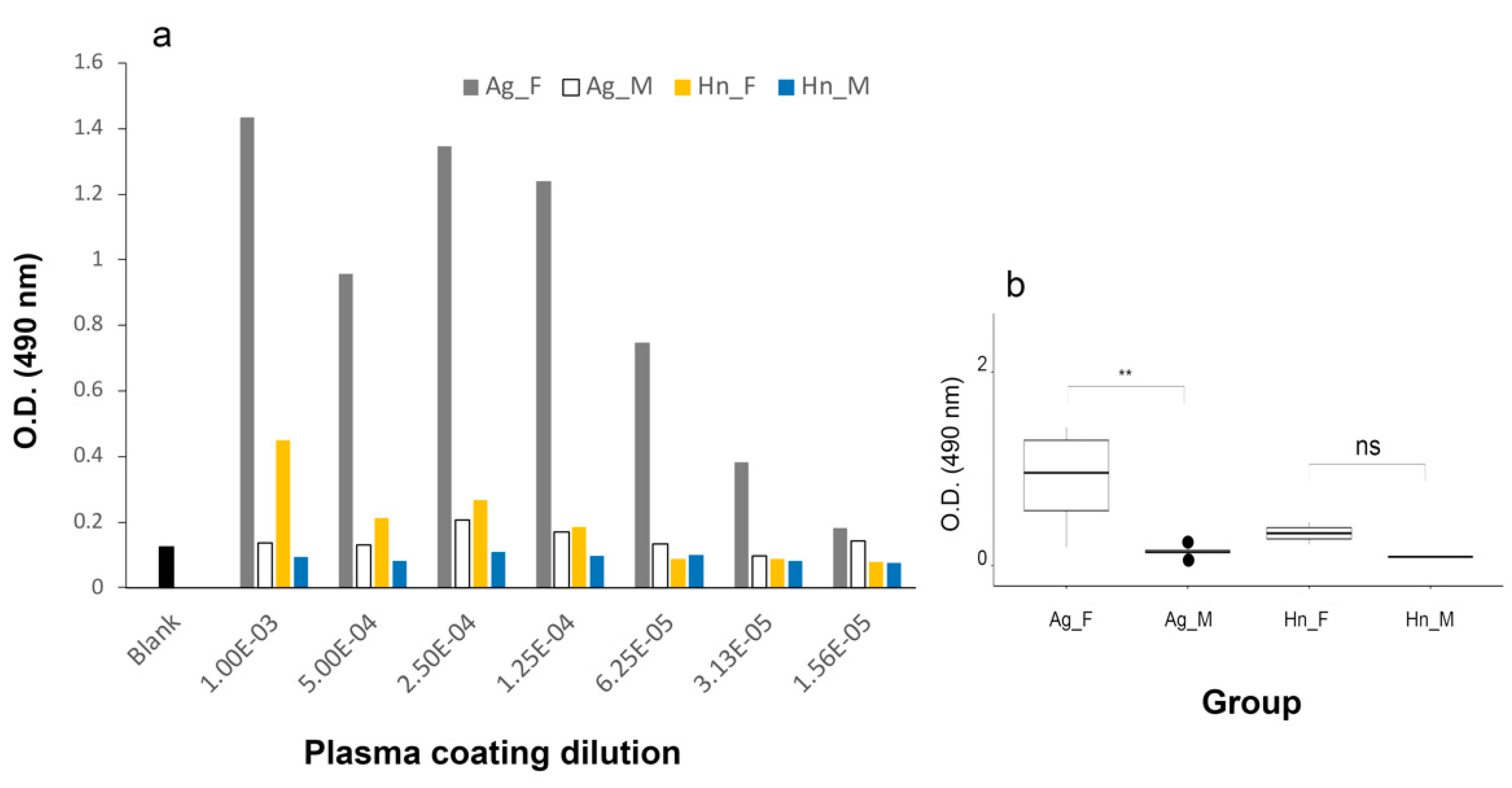
  • Antigen coating: a series of 100 µL plasma dilutions (1:1000, 1:2000, 1:4000, 1:8000, 1:16,000, 1:32,000 and 1:64,000) in carbonate buffer (0.05 M; pH 9.6) were distributed in a 96-well plate and incubated at 4 °C overnight.
  • Saturation: 100 μL of phosphate buffer saline with 1% Tween 20 (Sigma) and 2% normal pig serum (PBS-T-NPS) were added to each well, and the plate was incubated at 37 °C for 30 min. The contents of the wells were emptied, and each well was rinsed three times with 100 μL of PBS-T.
  • First incubation of the Vtg antibody: 100 µL of 1:10,000 diluted Vtg antibody in PBS-T-NPS was added to each well. Then, the plate was incubated at 37 °C for 90 min or at room temperature (25 ± 2 °C) for 4 h, its contents were discarded, and each well was rinsed 3 times with 100 μL of PBS-T.
  • Second incubation of the anti-rabbit IgG-peroxidase complex: 100 µL of anti-rabbit IgG labeled with peroxidase (Sigma) and diluted 1:3000 in PBS-T-NPS was added to each well, and the plate was incubated at 37 °C for 1 h. Then, each well was rinsed 3 times with 100 μL of PBS-T.
  • Color development: peroxidase activity was revealed in the dark by adding 100 μL of a solution containing 20 mL of citrate buffer (0.2 M; pH 5.0) + 10 mg of o-phenylenediamine + 30 µL of 30% H2O2 (Sigma) to each well. The reaction was stopped after 30 min by adding 50 µL of 4 M H2SO4 to each well.
  • The absorbance of each well was measured using a microplate reader at 490 nm. A blank well was added to aggregate the plate + reagents background; it contained all reagents except those of the coating step. The blank values were deduced from male plasma ODs to determine non-specific binding for a given antibody dilution.
Statistical analyses of OD values were performed with R [49], and the rstatix package (https://rpkgs.datanovia.com/rstatix, accessed on 15 November 2021).

2.5. Proteomic Approach

Samples for mass spectrometry protein analysis were prepared from 1 mL of frozen H. niloticus plasma submitted to an extraction medium composed of 200 mM PBS, 0.1 mM EDTA (ethylenediaminetetraacetic acid, Sigma), 1 mM DTT (dithiothreitol, Sigma), and 400 mM PMSF. The samples were stored at −80 °C until extraction.
For proteomic analysis, the samples were first thawed in cold water (5 ± 1 °C) and centrifuged at 20,000× g for 5 min. The supernatants containing solubilized proteins were precipitated by adding 80 μL of refrigerated (5 °C) acetone, and evaporated to 20 μL of sample. After one night at −20 °C, the samples were centrifuged at 20,000× g for 20 min and completely evaporated, and then the protein pellets were resuspended in 150 µL of ammonium bicarbonate buffer (50 mM, pH 7.0). Protein yield was 30 µg × 100 mL−1 for plasma. The proteins were digested at 37 °C overnight using 0.25 μg/mL porcine trypsin (Promega, Madison, WI, USA) in 25 μL of bicarbonate buffer. The digestates were desalted and concentrated on a ZipTip µC18 Omix (Agilent) before analysis.

2.5.1. NanoLC-MS/MS

The chromatography step was carried out on an ultra-high-pressure nano-chromatography system (NanoElute, Bruker Daltonics, Billerica, MA, USA). Peptides were concentrated on a C18 pepmap 100 (5 mm × 300 µm) precolumn (Thermo Scientific, Waltham, MA, USA) and separated at 50 °C on a Reprosil reverse phase column (25 cm × 75 µm, 1.6 µm, C18) (Ionopticks, Fitzroy, VIC, Australia). The mobile phases consisted of 0.1% formic acid in 99.9% water (v/v) (A) and 0.1% formic acid in 99.9% acetonitrile (v/v) (B). The flow rate was set at 400 nL/min, and the gradient profile was as follows: 2 to 15% B in 60 min, followed by an increase to 25% B in 30 min and then to 37% B in 10 min, followed by a 95% B wash step and 2% B rebalance.
MS analyses were carried out on a TIMS-TOF mass spectrometer (Bruker Daltonics) with a modified nano electrospray ion source (CaptiveSpray, Bruker Daltonics). The system was calibrated weekly, and the accuracy of mass measurements was better than 1 ppm. A capillary voltage of 1400 volts was used for ionization. MS spectra were acquired in positive mode in the 100–1700 m/z mass range. The mass spectrometer was in parallel accumulation-serial fragmentation (PASEF) mode. Single-charged peptides were excluded. Ten PASEF MS/MS scans were performed in 1.25 s from the 2–5 charge range.

2.5.2. Peptide Sequencing and Protein Identification

Two local H. niloticus databases were used: an ovarian protein database (437,476 entries) and a liver protein database (244,234 entries), both constructed from the corresponding transcriptomes [27]. The fragmentation spectrum was used to determine the peptide sequences. The database search was performed using the Mascot 2.6.1 program (Matrix Science). The Mascot score for a protein is the total score of individual peptides, i.e., the peptide masses and ion masses of the peptide fragments for each of the peptides corresponding to a given protein. The Mascot score (http://www.matrixscience.com/, accessed on 17 March 2021) must be above the 95% confidence level for positive protein identification, and the 95% confidence level corresponds to a score of ~90 for a search in the NCBI non-redundant protein database [50,51,52].
The following modifications of the variables were allowed: C-carbamidomethyl, K-acetylation, oxidation and di-oxidation of methionine. Mass accuracy was set at 30 ppm and 0.05 Da for the MS and MS/MS modes, respectively. Mascot data were transferred to the Proline validation software program (http://www.profiproteomics.fr/proline/, accessed on 17 March 2021) for data filtering according to a significance level ≤ 0.05 and to eliminate redundant proteins.
We have used this ~90 threshold as a criterion for determining a successful Vtg-peptide detection score among the sampled individuals. Samples with a Mascot score >90 were considered as females, and those with scores <90 were considered as males. All blood-sampled sexually mature test fish (59 individuals) were sacrificed to compare MS/MS and ELISA results with those of visual macroscopic sexing of fish gonads (Figure 1).

3. Results

3.1. Sexing of H. niloticus with the A. gigas Sexing Kit

Twenty-four mature H. niloticus individuals in their reproductive period (November) were used to test the efficiency of the sexing kit developed for A. gigas. Vtg was only detected in two spots, with weaker staining than in the control spot (Figure 2a,b). After all 24 fish were sacrificed to be sexed visually, the batch turned out to include 10 females. Therefore, the success rate of the kit was 20%. We concluded that it was not sensitive enough for the sexing of H. niloticus mature females.

3.2. Sexing of H. niloticus with Heterologous Vtg Antibodies by Direct ELISA

3.2.1. A. gigas Vtg1 Antibodies

The A. gigas Vtg1 antibody diluted (1:10,000) was tested on H. niloticus and A. gigas plasma samples taken from mature male and female individuals in their reproductive period (Figure 3). Samples from males of both species displayed a maximum OD of 0.206 for the lowest 1:1000 plasma coating dilution, while the plate + buffer blank had an average OD of 0.123. Samples from H. niloticus and A. gigas females showed maximum ODs of 0.449 and 1.433 for plasma dilution coatings of 1:1000, respectively. ANOVA of female vs. male OD values over dilution range (1:1000 to 1:32,000) indicated significant differences between the sexes of A. gigas plasmas (p = 0.003), while differences between the two sexes were not significant for the same coating dilutions range of H. niloticus plasmas (p = 0.87).

3.2.2. O. bicirrhosum Vtg Antibody

The maximum OD value reached only 0.09 and 0.140 for 1:1000-diluted A. gigas and H. niloticus female plasma samples, respectively, similar to that of males from both species (0.078 for A. gigas; 0.098 for H. niloticus) (results not shown), indicating a total lack of immunological cross-reactivity.

3.3. Purification of H. niloticus Vtg

The treatment of immature H. niloticus with 17β-estradiol induced the secretion of a blood protein observed by 12% SDS-PAGE (Figure 4). This major 142 kDa band was present in two “induced” fish plasmas and absent from two “uninduced” ones. This band was used to produce the H. niloticus Vtg antibody.

3.4. Sexing of H. niloticus with the Developed H. niloticus Vtg Antibody

After 8 weeks, the H. niloticus Vtg antibody produced by one of the two rabbits was selected because it showed the best reactions with plasma Vtg and weaker non-specific binding with the plasma of male individuals (results not shown).
The H. niloticus Vtg antibody diluted (1:80,000) was tested on male and female plasmas of mature H. niloticus and A. gigas sampled during the breeding season. The absorbance measurements of each well (Figure 5) showed that female H. niloticus reached a maximum OD of 0.565. Female A. gigas showed a maximum OD of 0.205, with a plate + buffer blank of 0.075. The OD values for H. niloticus plasma with H. niloticus Vtg antibody were above the values obtained with the A. gigas Vtg1 antibody within the same dilution range, and a significant difference was observed for the OD values of H. niloticus male vs. female plasma coating dilutions ranging from 1:1000 to 1:32,000 (p = 0.024).

3.5. Mass Spectrometry Analysis Results

To test the sexing capabilities of mass spectrometry analysis of mature H. niloticus individuals, tryptic peptides of Vtg or their cleavage products (Vtg-p) were investigated in protein extracts from male and female plasma samples. Vtg-p were detected in the plasma of 44 fish out of 59 (Figure 6), and Vtg Mascot scores ranged from 0 to 11,610 (Figure 7). Based on these results, 21 fish were considered as males (mascot score < 90) and 38 as females (mascot score > 90). Mascot scores ranged from 0 to 50 in putative males, and from 96 to 11,610 in putative females. The number of Vtg-p was always ≥2 in females, while it never exceeded 1 in males (6 males), and 15 males out of 21 had zero Vtg-p, hence also a Mascot score of 0. Thus, the Vtg-p Mascot scores for males never reached the threshold value of 90. The 100% correspondence of sex identification (i) by Vtg-p and their mascot scores by MS/MS, (ii) by direct Vtg ELISA and (iii) by macroscopic sexing after fish sacrifice, was confirmed for all 59 sampled fish.

4. Discussion

We have tested minimally invasive sexing methods based on the detection of Vtg in mature African bonytongue (H. niloticus) individuals. Vtg had previously been used as a female-sex marker in a few species such as Siberian sturgeon [53], yellowfin tuna [54], and sea trout [55]. The sex of mature individuals is generally identifiable from external characters in most species, but no external characteristic is available to distinguish sexes in H. niloticus. In some cases, biopsy or endoscopy on anesthetized animals allowed sex identification, even outside the breeding season or in immature fish [18,56,57,58]. The African bonytongue runs a very high risk of mortality during anesthesia because it is an obligate air breather. Bare and exposed ovarian lamellae in their body cavity do not allow easy sex determination by cannulation because H. niloticus females do not have an oviduct that could guide the cannula, as other species do [3,9,14,16,17,59]. In addition, a sexing technique based on morphometric data has given unreliable results [9]. The long evolutionary history of A. gigas and H. niloticus (±100 My) has led to a significant divergence of their chromosomal formula [60] with 2n = 56 for A. gigas and 2n = 40 for H. niloticus. However, owing to their phylogenetic proximity and relatively high percentages of Vtg sequence homology (>70%), we hypothesized that the Vtgs of the two species would cross-react with their heterologous Vtg antibodies.
Despite a high percentage of Vtg sequence conservation between A. gigas and H. niloticus, the A. gigas sexing kit used on H. niloticus females gave only 20% of positive sex determination. When the A. gigas Vtg antibody was used for H. niloticus sexing by direct ELISA, it consistently showed an OD < 0.25 for males regardless of the plasma coating dilution, whereas only dilutions lower than 1:10,000 allowed reliable sexing of H. niloticus females (OD > 0.25). However, for H. niloticus females the maximum OD values (OD range 0.268–0.449) obtained with the A. gigas Vtg antibody were lower than those obtained with equivalent dilutions of A. gigas female plasma in this study (OD range 0.383–1.43) and a previous study [17] with 1:10,000-diluted plasma and 1:80,000-diluted antibodies (OD range 1.4–2.95). These results show a noticeable affinity of H. niloticus Vtg for the A. gigas Vtg antibody, probably due to the phylogenetic closeness of these two species [24,25,26,27,28,29,30,31,61]. The direct ELISA test with A. gigas Vtg antibodies appeared to be more sensitive than the A. gigas sexing kit. Similar observations have been reported in yellowfin tuna (Thunnus albacares) [54] and sole (Solea vulgaris) [62], where immuno-diffusion detection techniques were also less sensitive than direct ELISA to detect Vtg. However, taking our results with the A. gigas Vtg antibody into account, direct ELISA still remains insufficient for the sexing of mature H. niloticus individuals: the OD signal obtained at the lowest plasma dilution (1:1000) was too weak, and could lead to false negative results in females with low Vtg levels. Lower plasma dilutions are not recommended because they tend to increase non-specific binding significantly (results not shown). Vtg antibodies from O. bicirrhosum, another Osteoglossiformes species, provided OD values similar to non-specific binding (OD < 0.141) for H. niloticus and A. gigas male and female plasmas. Therefore, we can also hypothesize that given the greater phylogenetic distance between A. gigas and O. bicirrhosum [45], their Vtg molecules are more dissimilar, and H. niloticus and A. gigas Vtg are not recognized by O. bicirrhosum Vtg antibodies. The H. niloticus Vtg antibody developed in this study showed non-specific binding (OD < 0.21) with plasmas from male H. niloticus and A. gigas individuals. We expected a positive reaction of this antibody with the plasma of A. gigas females, such as the reaction of the A. gigas Vtg antibody with the plasma of H. niloticus females, but the result was similar to that of male individuals (OD < 0.205), i.e., non-specific. The lack of specificity of the H. niloticus Vtg antibody for A. gigas Vtg is surprising. However, we used polyclonal antibodies, so we can hypothesize a difference in Vtg epitope selection for H. niloticus and A. gigas during immunization, as reported for other closely related fish species [63].
For H. niloticus females, OD values in response to the H. niloticus Vtg antibody were greater than 0.25 with 1:1000, 1:5000, and 1:10,000 plasma coating dilutions: they ranged between 0.336 and 0.565, indicating positive results with an OD threshold >0.25 as described for A. gigas [17]. Therefore, the antibody developed in this work is specific to H. niloticus Vtg, and can be used for sex determination of mature individuals of this species. This specificity is attested by the 100% correspondence of the results obtained by direct sex determination on 59 fish gonads and blood samples of the same fish tested by direct ELISA.
Mass spectrometry analysis of protein extracts from the plasma of mature H. niloticus females gave Mascot scores greater than 90, while the Mascot scores of males were less than 90. This Vtg identification threshold [50] was used to confirm the presence of Vtg-peptides in H. niloticus breeders (59 animals). The sacrifice of all 59 fish confirmed 100% of the sexing results of the Mascot score threshold. Proteomic analyses are increasingly used to identify proteins or peptides of interest [64,65] involved in various processes. For example, a similar procedure was applied to Epinephelus lanceolatus blood samples and was also found effective for sex determination [66]. The reproductive status and the degree of sexual maturation of several oviparous species have also been evaluated through proteomic studies as a function of the plasma Vtg levels [67]. Mascot scores could also be used to detect variations likely to reflect the degree of maturation of sexed females, as Vtg levels are correlated with the oocyte development stages during the reproductive cycle [35,62,68]. Nevertheless, the correlation between Mascot scores and Vtg plasma levels has not been evaluated yet in H. niloticus.

5. Conclusions

Minimally invasive methods—from small blood samples—based on the detection of plasma Vtg are now available to sex mature H. niloticus. The existing A. gigas sexing kit gave mixed results with H. niloticus samples, and the O. bicirrhosum Vtg antibody was not effective to sex H. niloticus or A. gigas. The A. gigas Vtg antibody sexed only 20% of H. niloticus individuals, but ELISA tests using the H. niloticus Vtg antibody developed in this study and a proteomic approach (Mascot scores) successfully sexed sexually mature H. niloticus. Direct ELISA is cheap and could be easily implemented in African research centers involved in H. niloticus aquaculture development plans. Depending on local demand, a field sexing kit could be developed, as it is for A. gigas in South America.

Author Contributions

Conceptualization, J.H. and J.N.; Data curation, E.C. and B.B.; Investigation, N.D.K., J.H. and J.N.; Methodology, N.D.K., J.H. and J.N.; Resources, N.D.K. and J.N.; Software, N.D.K., E.C., J.P. and B.B.; Supervision, J.N.; Visualization, N.D.K.; Writing—original draft, N.D.K.; Writing—review and editing, J.H. and J.N. All authors have read and agreed to the published version of the manuscript.

Funding

The authors are thankful for the financial support of AFD-C2D/ESA-INPHB N° CCI 1201 01 X, LMI EDIA, and South-South collaboration research program funded by IRD.

Institutional Review Board Statement

As there is no ethics committee in Côte d’Ivoire, the study was conducted in accordance with the guidelines on animals’ protection used for scientific purposes from the European Directive 2010/63/EU.

Acknowledgments

We wish to thank Sabbat Affif and Silué Drissa, respectively, owner of the Yamoussoukro fish farm and the chairman of the board of directors of the NGO APDRA-CI managing the Daloa fish farm and the owners of local fish farms where samples were collected for this study. Special thanks to all the staff of the PROVETSUR laboratory at UAGRM in Santa Cruz (Bolivia) for their collaboration in the development of the H. niloticus Vtg antibody and the ELISA tests.

Conflicts of Interest

The authors declare no conflict of interest. The authors declare that they have no known competing financial interest or personal relationship that could have appeared to influence the work reported in this paper.

References

  1. Ezekiel, E.N.; Abowei, J.F.N. Length-Weight Relationship and Condition Factor of Heterotis niloticus from Amassoma flood plain, Niger Delta, Nigeria. Appl. Sci. Rep. 2013, 4, 164–172. [Google Scholar]
  2. Odo, G.E.; Nwamba, H.O.; Eyo, J.E. Aspects of the biology of Heterotis niloticus, Cuvier 1829 (Osteoglossiformes: Osteoglossidae) in the Anambra flood river system, Nigeria. Anim. Res. Int. 2009, 6, 994–1002. [Google Scholar] [CrossRef]
  3. Adite, A.; Winemiller, K.O.; Fiogbé, E.D. Population structure and reproduction of the African bonytongue Heterotis niloticus in the Sô River-floodplain system (West Africa): Implications for management. Ecol. Freshw. Fish 2006, 15, 30–39. [Google Scholar] [CrossRef]
  4. Bard, J. Les poissons de la famille des Osteoglossidae et la pisciculture. Bois For. Trop. 1973, 147, 63–73. [Google Scholar]
  5. Lemasson, L. Chronique piscicole. Bois For. Trop. 1957, 54, 53–55. [Google Scholar]
  6. Reizer, C. Comportement et reproduction d’Heterotis niloticus en petits étangs. Bois For. Trop. 1964, 95, 49–60. [Google Scholar]
  7. Moreau, J.; Moreau, I. Etude du cycle annuel de la gamétogenèse chez Heterotis niloticus au lac Ivakoina (zone des Pangalanes) Madagascar. Rev. Hydrobiol. Trop. 1982, 15, 271–280. [Google Scholar]
  8. Bauchot, R.; Ridet, J.-M.; Diagne, M. The epibranchial organ, its innervations and its probable functioning in Heterotis niloticus (Pisces, Teleostei, Osteoglosssidae). Environ. Biol. Fishes 1993, 37, 307–315. [Google Scholar] [CrossRef]
  9. Oladosu, G.A.; Obi, A.; Oladosu, O.O. Sex determination in Heterotis niloticus (Cuvier 1829) Based on morphometric features. ASSET Ser. B 2007, 6, 22–30. [Google Scholar]
  10. Kimou, N.B.; Koumi, R.A.; Koffi, M.K.; Atsé, C.B.; Ouattara, I.N.; Kouamé, P.L. Utilisation des sous-produits agroalimentaires dans l’alimentation des poissons d’élevage en Côte d’Ivoire. Cah. Agric. 2016, 25, 25006. [Google Scholar] [CrossRef] [Green Version]
  11. Yao, A.H.; Koumi, A.R.; Atse, B.C.; Kouamelan, E.P. Etat de connaissance sur la pisciculture en Côte d’Ivoire. Agron. Afr. 2017, 29, 227–244. [Google Scholar]
  12. FAO. FishStatJ. 2021. Available online: https://www.fao.org/fishery/statistics/software/fishstatj/en (accessed on 17 March 2021).
  13. Monentcham, S.-E.E.; Kouam, J.; Pouomogne, V.; Kestemont, P. Biology and prospect for aquaculture of African bonytongue, Heterotis niloticus (Cuvier, 1829): A review. Aquaculture 2009, 289, 191–198. [Google Scholar] [CrossRef]
  14. Moreau, J. Exposé synoptique des données biologiques sur Heterotis niloticus (Cuvier, 1829). FAO Synop. Pêches 1982, 131, 45. [Google Scholar]
  15. Torati, L.S.; Varges, A.P.S.; Galvão, J.A.S.; Mesquita, P.E.C.; Migaud, H. Endoscopy application in broodstock management of Arapaima gigas (Schinz, 1822). J. Appl. Ichthyol. 2016, 32, 353–355. [Google Scholar] [CrossRef]
  16. Godinho, H.P.; Santos, J.E.; Formagio, P.S.; Guimaraes-Cruz, R.J. Gonadal morphology and reproductive traits of the Amazonian fish Arapaima gigas (Schinz, 1822). Acta Zool. 2005, 86, 289–294. [Google Scholar] [CrossRef]
  17. Chu-Koo, F.; Dugué, R.; Alván Aguilar, M.; Casanova Daza, A.; Alcántara Bocanegra, F.; Chávez Veintemilla, C.; Duponchelle, F.; Renno, J.F.; Tello, S.; Nuñez, J. Gender determination in the Paiche or Pirarucu (Arapaima gigas) using plasma vitellogenin, 17β-estradiol, and 11-ketotestosterone levels. Fish Physiol. Biochem. 2009, 35, 125–136. [Google Scholar] [CrossRef]
  18. Carreiro, C.R.P.; Furtado-Neto, M.A.d.A.; Mesquita, P.E.C.; Bezerra, T.A. Sex determination in the Giant fish of Amazon Basin, Arapaima gigas (Osteoglossiformes, Arapaimatidae), using laparoscopy. Acta Amaz. 2011, 41, 415–419. [Google Scholar] [CrossRef]
  19. Bureau du Colombier, S.; Jacobs, L.; Gesset, C.; Elie, P.; Lambert, P. Ultrasonography as a non-invasive tool for sex determination and maturation monitoring in silver eels. Fish. Res. 2015, 164, 50–58. [Google Scholar] [CrossRef]
  20. Kucharczyk, D.; Malinovskyi, O.; Nowosad, J.; Kowalska, A.; Cejko, B.I. Comparison of responses to artificial spawning of ruffe (Gymnocephalus cernua) specimens captured from their natural habitat to those produced in cultured conditions. Anim. Reprod. Sci. 2021, 225, 106684. [Google Scholar] [CrossRef]
  21. Kujawa, R.; Nowosad, J.; Biegaj, M.; Cejko, B.I.; Kucharczyk, D. Use of ultrasonography to determine sex in sexually immature European river lamprey Lampetra fluviatilis (L.). Anim. Reprod. Sci. 2019, 204, 95–100. [Google Scholar] [CrossRef]
  22. Mansur, V.F.R.; Melo, N.; Di Chiacchio, I.M.; de Lima Assis, I.; Machado, G.J.; Paiva, I.M.; de Carvalho, A.F.S.; Pereira, R.N.; Solis Murgas, L.D. Sex identification of the ornamental amazon fish Astronotus ocellatus by videoceloscopy and gonadal biopsy. Anim. Reprod. Sci. 2021, 230, 106780. [Google Scholar] [CrossRef] [PubMed]
  23. Moghim, M.; Vajhi, A.; Veshkini, A.; Masoudifard, M. Determination of sex and maturity in Acipenser stellatus by using ultrasonography. J. Appl. Ichthyol. 2002, 18, 325–328. [Google Scholar] [CrossRef] [Green Version]
  24. Betancur, R.R.; Wiley, E.O.; Arratia, G.; Acero, A.; Bailly, N.; Miya, M.; Lecointre, G.; Ortí, G. Phylogenetic classification of bony fishes. BMC Evol. Biol. 2017, 17, 162. [Google Scholar] [CrossRef] [Green Version]
  25. Guo-Qing, L.; Wilson, M.V.H. The discovery of Heterotidinae (Teleostei: Osteoglossidae) from the Paleocene Paskapoo Formation of Alberta, Canada. J. Vertebr. Paleontol. 1996, 16, 198–209. [Google Scholar] [CrossRef]
  26. Hilton, E.J. Tongue Bite Apparatus of Osteoglossomorph Fishes: Variation of a Character Complex. Copeia 2001, 2001, 372–381. [Google Scholar] [CrossRef]
  27. Koua, N.Z.D.; Núñez-Rodriguez, J.; Orjuela, J.; Zatylny-Gaudin, C.; Dubos, M.-P.; Bernay, B.; Pontin, J.; Corre, E.; Henry, J. Identification and structural characterization of the factors involved in vitellogenesis and its regulation in the African Osteoglossiforme of aquacultural interest Heterotis niloticus (Cuvier, 1829). Gen. Comp. Endocrinol. 2020, 296, 113532. [Google Scholar] [CrossRef]
  28. Lavoué, S. Was Gondwanan breakup the cause of the intercontinental distribution of Osteoglossiformes? A time-calibrated phylogenetic test combining molecular, morphological, and paleontological evidence. Mol. Phylogenet. Evol. 2016, 99, 34–43. [Google Scholar]
  29. Lavoué, S.; Sullivan, J.P. Simultaneous analysis of five molecular markers provides a well-supported phylogenetic hypothesis for the living bony-tongue fishes (Osteoglossomorpha: Teleostei). Mol. Phylogenet. Evol. 2004, 33, 171–185. [Google Scholar] [CrossRef]
  30. Nelson, G.J. Gill arches of teleostean fishes of the division Osteoglossomorpha. J. Linn. Soc. Lond. Zool. 1968, 47, 261–277. [Google Scholar] [CrossRef]
  31. Nelson, G.J. Infraorbital bones and their bearing on the phylogeny and geography of Osteoglossomorph fishes. Am. Mus. Novit. 1969, 2394, 1–37. [Google Scholar]
  32. Dugué, R.; Atta Shikema, R.K.; Corcuy Arana, N.; Legendre, M.; Duponchelle, F.; Renno, J.F.; Nuñez, J. Seasonality of reproduction in Piaractus brachypomus in South Bolivia. Cybium 2009, 32 (Suppl. 2), 241. [Google Scholar]
  33. Wallace, R.A.; Hoch, K.L.; Carnevali, O. Placement of small lipovitellin subunits within the vitellogenin precursor in Xenopus laevis. J. Mol. Biol. 1990, 213, 407–409. [Google Scholar] [CrossRef]
  34. Zhang, S.; Dong, Y.; Cui, P. Vitellogenin is an immunocompetent molecule for mother and offspring in fish. Fish Shellfish Immunol. 2015, 46, 710–715. [Google Scholar] [CrossRef] [PubMed]
  35. Ndiaye, P.; Forgue, J.; Lamothe, V.; Cauty, C.; Tacon, P.; Lafon, P.; Davail, B.; Fostier, A.; Le Menn, F.; Núñez, J. Tilapia (Oreochromis niloticus) vitellogenins: Development of homologous and heterologous ELISAs and analysis of vitellogenin pathway through the ovarian follicle. J. Exp. Zool. A 2006, 305A, 576–593. [Google Scholar] [CrossRef]
  36. Kong, H.J.; Kim, J.L.; Moon, J.Y.; Kim, W.J.; Kim, H.S.; Park, J.Y.; Cho, H.K.; An, C.M. Characterization, expression profile, and promoter analysis of the Rhodeus uyekii vitellogenin Ao1 gene. Int. J. Mol. Sci. 2014, 15, 18804–18818. [Google Scholar] [CrossRef] [Green Version]
  37. Wang, H.; Tan, J.T.T.; Emelyanov, A.; Korzh, V.; Gong, Z. Hepatic and extrahepatic expression of vitellogenin genes in the zebrafish, Danio rerio. Gene 2005, 356, 91–100. [Google Scholar] [CrossRef]
  38. Wang, R.; Gao, Y.; Zhang, L.; Zhang, Y.; Fang, Z.; He, J.; Zhang, W.; Ma, G. Cloning, expression, and induction by 17-β estradiol (E2) of a vitellogenin gene in the white cloud mountain minnow Tanichthys albonubes. Fish Physiol. Biochem. 2010, 36, 157–164. [Google Scholar] [CrossRef]
  39. Arukwe, A.; Røe, K. Molecular and cellular detection of expression of vitellogenin and zona radiata protein in liver and skin of juvenile salmon (Salmo salar) exposed to nonylphenol. Cell Tissue Res. 2008, 331, 701–712. [Google Scholar] [CrossRef]
  40. Genovese, G.; Regueira, M.; Da Cuña, R.H.; Ferreira, M.F.; Varela, M.L.; Lo Nostro, F.L. Nonmonotonic response of vitellogenin and estrogen receptor α gene expression after octylphenol exposure of Cichlasoma dimerus (Perciformes, Cichlidae). Aquat. Toxicol. 2014, 156, 30–40. [Google Scholar] [CrossRef]
  41. Gordon, M.R.; Owen, T.G.; Ternan, T.A.; Hildebrand, L.D. Measurement of a sex-specific protein in skin mucus of premature coho salmon (Oncorhynchus kisutch). Aquaculture 1984, 43, 333–339. [Google Scholar] [CrossRef]
  42. Meucci, V.; Arukwe, A. Detection of vitellogenin and zona radiata protein expressions in surface mucus of immature juvenile Atlantic salmon (Salmo salar) exposed to waterborne nonylphenol. Aquat. Toxicol. 2005, 73, 1–10. [Google Scholar] [CrossRef]
  43. Moncaut, N.; Nostro, F.L.; Maggese, M.C. Vitellogenin detection in surface mucus of the South American cichlid fish Cichlasoma dimerus (Heckel, 1840) induced by estradiol-17beta. Effects on liver and gonads. Aquat. Toxicol. 2003, 63, 127–137. [Google Scholar] [CrossRef]
  44. Zhong, L.; Yuan, L.; Rao, Y.; Li, Z.; Zhang, X.; Liao, T.; Xu, Y.; Dai, H. Distribution of vitellogenin in zebrafish (Danio rerio) tissues for biomarker analysis. Aquat. Toxicol. 2014, 149, 1–7. [Google Scholar] [CrossRef] [PubMed]
  45. Mu, X.D.; Wang, X.J.; Song, H.M.; Yang, Y.X.; Luo, D.; Gu, D.E.; Xu, M.; Liu, C.; Luo, J.R.; Hu, Y.c. Mitochondrial DNA as effective molecular markers for the genetic variation and phylogeny of the family Osteoglossidae. Gene 2012, 511, 320–325. [Google Scholar] [CrossRef] [PubMed]
  46. Anderson, G.W.; McKinley, S.R.; Colavecchia, M. The use of clove oil as an anesthetic for Rainbow trout and its effects on swimming performance. N. Am. J. Fish. Manag. 1997, 17, 301–307. [Google Scholar] [CrossRef]
  47. Bradford, M.M. A rapid and sensitive method for the quantitation of microgram quantities of protein utilizing the principle of protein-dye binding. Anal. Biochem. 1976, 72, 248–254. [Google Scholar] [CrossRef]
  48. Dugué, R.; Chu-Koo, F.; Alcantara, F.; Duponchelle, F.; Renno, J.-F.; Nuñez, J. Purification and assay of Arapaima gigas vitellogenin: Potential use for sex determination. Cybium 2008, 32 (Suppl. 2), 111. [Google Scholar]
  49. R Core Team. R: A Language and Environment for Statistical Computing; R Foundation for Statistical Computing: Vienna, Austria, 2013. [Google Scholar]
  50. Hamidi, Z.; Tejero, E.; Schmidt, R.; Tucker, R.; Pedro, A. Identification of potential blood-derived extracellular vesicles biomarkers to diagnose and predict distant metastases in ER+ breast cancer patients. bioRxiv 2017. [Google Scholar] [CrossRef] [Green Version]
  51. Koenig, T.; Menze, B.H.; Kirchner, M.; Monigatti, F.; Parker, K.C.; Patterson, T.; Steen, J.J.; Hamprecht, F.A.; Steen, H. Robust prediction of the MASCOT score for an improved quality assessment in mass spectrometric proteomics. J. Proteome Res. 2008, 7, 3708–3717. [Google Scholar] [CrossRef]
  52. Perkins, D.N.; Pappin, D.J.C.; Creasy, D.M.; Cottrell, J.S. Probability-based protein identification by searching sequence databases using mass spectrometry data. Electrophoresis 1999, 20, 3551–3567. [Google Scholar] [CrossRef]
  53. Ceapa, C.; Williot, P.; Le Menn, F.; Davail-Cuisset, B. Plasma sex steroids and vitellogenin levels in stellate sturgeon (Acipenser stellatus Pallas) during spawning migration in the Danube River. J. Appl. Ichthyol. 2002, 18, 391–396. [Google Scholar] [CrossRef]
  54. Takemura, A.; Oka, M. Immunochemical sexing of living yellowfin tuna, Thunnus albacares (Bonnaterre), using a vitellogenin- like protein. Aquac. Res. 1998, 29, 245–249. [Google Scholar] [CrossRef]
  55. Pottinger, T.G.; Pulman, K.G.T.; Carrick, T.R.; Scott, A.P. Evaluation of biochemical methods for the non-destructive identification of sex in upstream migrating salmon and sea trout. J. Fish Biol. 2005, 67, 1514–1533. [Google Scholar] [CrossRef] [Green Version]
  56. Bryan, J.L.; Wildhaber, M.L.; Papoulias, D.M.; Delonay, A.J.; Tillitt, D.E.; Annis, M.L. Estimation of gonad volume, fecundity, and reproductive stage of shovelnose sturgeon using sonography and endoscopy with application to the endangered pallid sturgeon. J. Appl. Ichthyol. 2007, 23, 411–419. [Google Scholar] [CrossRef]
  57. Kynard, B.; Kieffer, M. Use of a borescope to determine the sex and egg maturity stage of sturgeons and the effect of borescope use on reproductive structures. J. Appl. Ichthyol. 2002, 18, 505–508. [Google Scholar] [CrossRef]
  58. Swenson, E.A.; Rosenberger, A.E.; Howell, P.J. Validation of endoscopy for determination of maturity in small salmonids and sex of mature individuals. Trans. Am. Fish. Soc. 2007, 136, 994–998. [Google Scholar] [CrossRef]
  59. Bard, J.; Imbiriba, E.P. Piscicultura do pirarucu, Arapaima gigas. Ser. Doc. Circ. Téc. 1986, 52, 17. [Google Scholar]
  60. De Oliveira, E.A.; Bertollo, L.A.C.; Rab, P.; Ezaz, T.; Yano, C.F.; Hatanaka, T.; Jegede, O.I.; Tanomtong, A.; Liehr, T.; Sember, A.; et al. Cytogenetics, genomics and biodiversity of the South American and African Arapaimidae fish family (Teleostei, Osteoglossiformes). PLoS ONE 2019, 14, e0214225. [Google Scholar]
  61. Hilton, E.J. Comparative osteology and phylogenetic systematics of fossil and living bony-tongue fishes (Actinopterygii, Teleostei, Osteoglossomorpha). Zool. J. Linn. Soc. 2003, 137, 1–100. [Google Scholar] [CrossRef] [Green Version]
  62. Nunez Rodriguez, J.; Dugué, R.; Otemé, Z.J.; Hem, S.; Le Menn, F. Vitellogenin plasma levels in two cultured African catfish species, Chrysichthys nigrodigitatus (Claroteidae) and Heterobranchus longifilis (Clariidae). Aquat. Living Resour. 1997, 10, 231–238. [Google Scholar] [CrossRef]
  63. Watts, M.; Pankhurst, N.W.; Pryce, A.; Sun, B. Vitellogenin isolation, purification and antigenic cross-reactivity in three teleost species. Comp. Biochem. Physiol. Part B Biochem. Mol. Biol. 2003, 134, 467–476. [Google Scholar] [CrossRef] [Green Version]
  64. López, J.L.; Marina, A.; Álvarez, G.; Vázquez, J. Application of proteomics for fast identification of species-specific peptides from marine species. Proteomics 2002, 2, 1658–1665. [Google Scholar] [CrossRef]
  65. Sun, B.; Pankhurst, N.W.; Watts, M. Development of an enzyme-linked immunosorbent assay (ELISA) for vitellogenin measurement in greenback flounder Rhombosolea tapirina. Fish Physiol. Biochem. 2003, 29, 13–21. [Google Scholar] [CrossRef]
  66. Om, A.D.; Jasmani, S.; Ismail, N.; Yeong, S.Y.; Abol-Munafi, A.B. Application MALDI TOF on protein identification of vitellogenin in giant grouper (Epinephelus lanceolatus). Fish Physiol. Biochem. 2013, 39, 1277–1286. [Google Scholar] [CrossRef] [PubMed]
  67. Cohen, A.M.; Banoub, J.H. Detection of Biological Agents for the Prevention of Bioterrorism; Banoub, J., Ed.; Springer: Berlin/Heidelberg, Germany, 2011; pp. 301–318. [Google Scholar]
  68. Nuñez-Rodriguez, J.; Kah, O.; Geffard, M.; Le Menn, F. Enzyme Linked Immunosorbent Assay (ELISA) for sole (Solea vulgaris) vitellogenin. Comp. Biochem. Physiol. Part B Comp. Biochem. 1989, 92, 741–746. [Google Scholar] [CrossRef]
Figure 1. Macroscopic aspects of the testis (a) and ovary (b) of sexually mature male and female African bonytongue, Heterotis niloticus, respectively. Scale bars, 1 cm.
Fishes 07 00106 g001
Figure 2. Test of the Pirarucu, Arapaima gigas, sexing kit on four African bonytongues, Heterotis niloticus, individuals—three females undergoing vitellogenesis and one control male. The positive Vtg detection spot was observed for two females (a,b) with low intensity for female (a), but the other female (d) was negative. The control male was negative as expected (c).
Fishes 07 00106 g002
Figure 3. Dilution series of male and female Pirarucu, Arapaima gigas, and African bonytongue, Heterotis niloticus, plasmas revealed with Arapaima gigas Vtg1 antibodies in ELISA, ODs at 490 nm. Blank (plate + reagents background) received all reagents except the coating one (a). Box plots of Statistical analysis of ODs by ANOVA, rstatix (b). ** indicates significant statistical differences between male and female coating dilutions range (p = 0.003), ns indicates non-significant difference (p = 0.87), Tukey HSD with p adjust. Ag_F: Arapaima gigas female; Ag_m: Arapaima gigas male; Hn_F: Heterotis niloticus female; Hn_M: Heterotis niloticus male.
Fishes 07 00106 g003
Figure 4. SDS-PAGE of two African bonytongues, Heterotis niloticus, plasma samples before induction (BI) and after 17-β Estradiol treatment (I). One major band at 142 kDa molecular mass indicates the presence of Vtg in the treated plasmas.
Fishes 07 00106 g004
Figure 5. Dilution series of male and female Pirarucu, Arapaima gigas, and African bonytongue, Heterotis niloticus, plasmas revealed with Heterotis niloticus Vtg antibodies in ELISA, ODs at 490 nm. Blank (plate + reagents background) received all reagents except the coating one (a). Box plots of Statistical analysis of ODs by ANOVA, rstatix (b). * indicates significant statistical differences between male and female coating dilutions range (p = 0.024); ns indicates non-significant difference (p = 0.93), Tukey HSD with p adjust. Ag_F: A. gigas female; Ag_m: Arapaima gigas male; Hn_F: Heterotis niloticus female; Hn_M: Heterotis niloticus male.
Fishes 07 00106 g005
Figure 6. Detection of Vtg and Vtg cleavage products by mass spectrometry from plasma of sexually mature African bonytongue, Heterotis niloticus, (log-scale Y-axis). The fish with a number of Vtg-specific peptides ≥2 were considered as females (gray vertical bars), those with a number of Vtg-specific peptides = 0 (indicated by * for 15 out of 21 fish) or 1 (solid vertical bars for 6 out of 21 fish) were considered as males (bold IDs on X-axis). The male/female threshold value is represented as a blue line at number of Vtg tryptic peptides = 1.
Fishes 07 00106 g006
Figure 7. Vtg and Vtg cleavage products Mascot scores determined by mass spectrometry from plasma of sexually mature African bonytongue, Heterotis niloticus, (same individuals as in Figure 6). The fish with a Vtg Mascot score >90 were considered as females (gray vertical bars), those with a Vtg Mascot score ranging from 0 to 90 were considered as males (black vertical bars and * with bold IDs on the X-axis). The male/female threshold value is represented as a blue line at Mascot score = 90 (log-scale Y-axis).
Fishes 07 00106 g007
Publisher’s Note: MDPI stays neutral with regard to jurisdictional claims in published maps and institutional affiliations.

