Article

Sporosarcina aquimarina MS4 Regulates the Digestive Enzyme Activities, Body Wall Nutrients, Gut Microbiota, and Metabolites of Apostichopus japonicus

1
School of Biological Engineering, Dalian Polytechnic University, Dalian 116034, China
2
School of Food Science and Engineering, Dalian Ocean University, Dalian 116023, China
3
Key Laboratory of Biotechnology and Bioresources Utilization, Dalian Minzu University, Dalian 116600, China
4
Engineering Research Center of Health Food Design & Nutrition Regulation, School of Chemical Engineering and Energy Technology, Dongguan University of Technology, Dongguan 523808, China
*
Authors to whom correspondence should be addressed.
Fishes 2022, 7(3), 134; https://doi.org/10.3390/fishes7030134
Submission received: 27 April 2022 / Revised: 5 June 2022 / Accepted: 6 June 2022 / Published: 7 June 2022
(This article belongs to the Section Nutrition and Feeding)

Abstract

:
Sporosarcina aquimarina MS4 is a microecological preparation for overwintering Apostichopus japonicus, which has an immune regulation function, but its role in the nutritional regulation of A. japonicus is not clear. This study aimed to describe the effects of S. aquimarina MS4 on the growth, digestion, and body wall nutrition of A. japonicus through feeding experiments and to discuss the potential mechanism of S. aquimarina MS4 regulating gut function through the detection of gut microbiota and metabolites. After 60 days of culture, the growth performance of A. japonicus fed S. aquimarina MS4 (108 cfu/g) significantly improved, and the content of polysaccharide, leucine, phenylalanine, lysine, and docosahexaenoic acid in the body wall significantly increased. Gut microbiota analysis showed that although Proteobacteria, Verrucomicrobia, Firmicutes, and Bacteroidetes were the predominant phyla in all the sea cucumbers, Haloferula and Rubritalea showed significant difference between the group fed with or without S. aquimarina MS4. Metabolomics analysis showed that differential metabolites in the gut were mainly enriched in amino acid metabolism and lipid metabolism. The association analysis of differential metabolites and microbiota showed that the production of some differential metabolites was significantly related to differential microorganisms, which improved the understanding of the function of microorganisms and their roles in the gut of A. japonicus. This study reveals the life activities such as growth and metabolism of A. japonicus, and it provides support for the functional study of the gut microbiome of A. japonicus.

1. Introduction

Sea cucumbers are traditional high-value marine food in China, with an annual output of approximately 200,000 tons and a mariculture area of more than 2400 square kilometers. The main reason they are popular among consumers is their rich nutritional value and their health care function [1,2]. Apostichopus japonicus has gradually become the main variety of sea cucumber culture because it is rich in polysaccharides, saponins, and other nutrients [3]. However, with the development of large-scale intensive farming, in order to avoid diseases in aquaculture, antibiotics and other chemical drugs began to be abused, seriously affecting the quality and the food safety of the sea cucumber [4].
Probiotics are currently feasible alternatives to antibiotics. By regulating the microecology of the culture environment and the gut microbiota of animals, they have good effects on animal weight gain, immunity enhancement, and nutritional quality improvement [5,6]. The yeast Rhodotorula benthica could increase growth performance and some digestive enzyme activities of juvenile A. japonicus [7]. Lactic acid bacteria isolated from marine fish had positive effects on the immune response of A. japonicus [8]. In addition, probiotic fermented feed plays a positive role in the growth, digestion, and immunity of A. japonicus [9,10]. Probiotics can not only regulate growth and immunity but also improve the nutritional value of A. japonicus [6,10,11].
In our previous work, a low-temperature-tolerant probiotic Sporosarcina aquimarina MS4 suitable for the cultivation of A. japonicus in winter was developed, which improved the immunity of A. japonicus and prevented infection of Vibrio splendidus [12]. However, the effect of S. aquimarina MS4 on digestion and body wall nutrient composition is unclear. This study intends to solve the above problems through aquaculture experiments, and to understand the mechanism of S. aquimarina MS4 affecting the digestion of A. japonicus through the analysis of gut microbiota and metabolome.

2. Materials and Methods

2.1. Animal Ethics

The experimental animals and protocols used in this study were approved for animal ethics by the Animal Experiment Ethics Committee of Dalian Polytechnic University.

2.2. Strains, Medium and Feed

The S. aquimarina MS4 used in this study was isolated and screened in our laboratory, and its culture medium and culture conditions were described in previous studies [12]. The basic feed (particle size 0.038–0.075 mm) used in this study was purchased from Dalian Shengtai Aquatic Feed Co., Ltd., Dalian, China. The main ingredients are shown in Table 1. The cultured S. aquimarina MS4 was added into the basic feed at 108 cfu/g to prepare the experimental feed in the current experiment.

2.3. Culture Trial

The sea cucumber A. japonicus were purchased from Dalian Boshiao Biotechnology Co., Ltd., Dalian, China. The feeding period was 60 days, and the pre-feeding period was 10 days before the formal experiment. The experiment was divided into the control group (group C) and the experimental group (group M), each group was set up with three tanks (tank size 50 L), totaling six tanks. The healthy A. japonicus were screened and divided into groups, with 40 in each tank. The average weight of A. japonicus were 1.88 g and 1.95 g in group C and M, respectively. The control group was fed the basic diet, and the experimental group was fed with the experimental feed. Each tank of the sea cucumber was provided with the diet at 5% of the total weight every day. Seawater (salinity level 35 g/L) was replaced every 5 days. The seawater temperature was controlled at 8 ± 1 °C, and air was continuously supplied into the tank during the period.
At the end of the experiment, three sea cucumbers were randomly selected from each tank for dissection, the gut was used for the detection of digestive enzyme activity, and the body wall was used for the detection of body wall nutrients after freeze-drying. Six sea cucumbers were randomly selected from each tank, and the guts were frozen with liquid nitrogen for gut microbiota and metabolites detection. At the time of sampling, three sea cucumber samples in each tank were mixed and used as a biological replicate. Six replicates were used for gut metabolites and three replicates were used for other assays.

2.4. Growth Performance

During the feeding period, the body weight and the feeding level of A. japonicus were recorded regularly, and the weight gain rate (WGR), specific growth rate (SGR), and feed conversion rate (FCR) were calculated according to the initial body weight (W0), final body weight (Wt), and feeding level (F). At the end of the experiment, the number of dead sea cucumbers (D) was recorded, and the natural mortality was calculated.
WGR = 100 × (Wt − W0)/W0
SGR = 100 × (lnWt − lnW0)/60
FCR = F/(Wt − W0)
Natural mortality = 100 × D/40

2.5. Digestive Enzyme Activities

The protease activity, cellulase activity, and amylase activity were measured by a protease assay kit, cellulase assay kit, and an amylase assay kit of the Nanjing Jiancheng Bioengineering Institute. The operation steps followed the kits manual.

2.6. Nutrient Compositions

The determination methods of moisture, crude protein, crude fat, and ash refer to the method of Haider et al. [13]. Total sugar was determined by phenol sulphuric acid method [14].
Amino acids: 1 g sample was weighed and placed in an anaerobic hydrolysis tube, and 5 mL of 6 mol/L hydrochloric acid was added and mixed. The solution was frozen in liquid nitrogen until solidified and then vacuumized. It was hydrolyzed at 110 °C for 24 h. After cooling, distilled water was added to the hydrolyzate to 10 mL and filtered by a 0.45 μm water filtration membrane; 0.5 mL filtrate was taken for vacuum drying. The residue was dissolved with 1 mL deionized water and then dried and repeated twice. The sample was dissolved with a buffer solution and filtered with a 0.22 μm water filtered membrane. An amino acid automatic analyzer (S-433D, Sykam GmbH, Munich, Germany) was used for analysis.
Fatty acids: 200 mg of A. japonicus body wall powder was added to 1.5 mL of chloroform–methanol (2:1). After grinding, it was fully homogenized for 1 min. A total of 1 mL of chloroform was added and vortexed for 1 min. Then, 1 mL of water was added and mixed. After layering for 30 min, the mixture was centrifuged for 5 min at 4 °C, 5000× g. The lower layer solution was taken, and 1/4 volume of methanol–water (1:1) was added. The impurities in the upper and the middle layers were removed after layering for 5 min. After drying with nitrogen, 30 μL of chloroform was added to dissolve. A total of 1 mL of NaOH-CH3OH solution (8/100 w/v) was added and maintained at 100 °C for 30 min. A total of 1.5 mL of boron trifluoride-methanol was added and maintained at 100 °C for 10 min. After cooling, 1 mL heptane was added and an ultrasound was performed for 5 min. A total of 1 mL saturated NaCl solution was added for washing and ultrasonication for 1 min. The solution was allowed to stand at 4 °C for 30 min and the layers were separated. The upper layer solution was filtered by a 0.22 μm filter and then analyzed by a gas chromatography mass spectrometer (Agilent 5975C, Agilent Technologies, Inc., Santa Clara, CA, USA). The gas chromatography conditions were HPTM-2560 quartz capillary column (100 m × 0.25 mm × 0.2 μm), high-purity He as carrier gas, constant pressure mode, split ratio of 30:10, injector temperature of 230 °C, and detector temperature of 250 °C. The column temperature was maintained at 140 °C for 5 min, increased to 240 °C at a rate of 4 °C/min, maintained for 15 min, and the entire analysis process was 51 min. The mass spectrometry conditions were GC/MS interface temperature of 250 °C, EI ion source temperature of 250 °C, and quadrupole temperature of 230 °C.

2.7. Gut Microbiota

The gut samples were sequenced by 16S amplicon in Beijing Novogene Co., Ltd., Beijing, China. to study the composition and the structure of gut microbiota. The experimental process was briefly described as follows: the genomic DNA of the sample was extracted by CTAB method, and the diluted genomic DNA was used as the template to amplify 16S rDNA. A TruSeq DNA PCR Free Sample Preparation Kit was used for library construction. The constructed library was quantified by Qubit and Q-PCR. After the library was qualified, NovaSeq6000 was used for sequencing. The method of bioinformatics analysis refers to the work of Liu et al. [15].

2.8. Gut Untargeted Metabolomics Assay

The gut samples were sent to Beijing Novogene Co., Ltd., Beijing, China. for untargeted gut metabolomics detection. The extraction of gut metabolites and the detection of metabolomics followed a previous work with small modifications [16]. A total of 100 mg of the gut samples ground in liquid nitrogen were added into 500 μL of 80% methanol. After vortexing, the samples were melted on ice for 5 min. After centrifugation at 15,000× g and 4 °C for 20 min, a certain amount of supernatant was taken and diluted with water to a methanol content of 53%. The supernatant was collected by centrifugation at 15,000× g for 20 min at 4 °C and then injected into LC-MS (Vanquish UHPLC coupled with Q ExactiveTM HF, Thermo Fisher Scientific Inc., Waltham, MA, USA) for analysis. The chromatographic and the mass spectrometry conditions referred to Yuan’s method [17]. The data processing and the analysis were described in Liu’s work [16].

2.9. Statistical Analysis

The data in this study are shown as the mean ± standard deviation. Statistical significance was performed on growth performance, nutrient content, and digestive enzyme activity. A t-test was used to compare the significance of the two groups of data.

3. Results

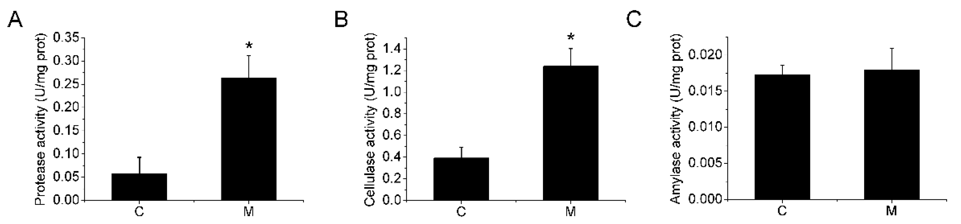
3.1. Growth Performance and Digestive Enzyme Activities

After 60 days of the feeding experiment, the body weight and the feed intake of A. japonicus were monitored, and the growth performance was evaluated by WGR, SGR, and FCR. As shown in Table 2, compared with group C, group M showed no significant difference in initial and final body weight, but a significant increase in WGR. The SGR (0.19%/d) of group M was significantly higher than that of the control group (0.09%/d), while the FCR (4.89) was significantly lower than that of the control group (6.14). No natural death occurred in group M, while the control group had a natural mortality rate of 4.26%. The lower mortality rates may be caused by individual differences. In summary, feeding S. aquimarina MS4 promoted the growth of A. japonicus, significantly increased WGR and SGR, and significantly decreased FCR, which was consistent with the results of our previous study.
As shown in Figure 1, after 60 days of feeding, the gut protease activity and the cellulase activity in group M significantly increased, but the amylase activity did not significantly change compared with the control group.

3.2. Body Wall Nutrients

Table 3 shows the nutritional composition of the sea cucumber body wall. There was no significant difference in the contents of total protein, fat, and ash between the sea cucumber fed with S. aquimarina MS4 and the control group. The crude polysaccharide content of group M was 7.34%, which was significantly higher than that of group C (6.49%). The two groups of sea cucumbers showed differences in amino acid and fatty acid composition (Table 4 and Table 5). In terms of amino acid composition, the main amino acids of the two groups were aspartate, glutamate, and glycine (with the content above 10%). Compared with the control group, the contents of leucine, phenylalanine, and lysine in the amino acid composition of sea cucumbers in group M were significantly increased. However, the total amino acid content was not changed significantly. There were no significant differences in the content of saturated, monounsaturated, and polyunsaturated fatty acids between groups M and C, but they differed in specific fatty acid composition (Table 5). Among the saturated fatty acids, group M contained less pentadecanoic acid and more heptacosanoic acid. Among the differences in monounsaturated fatty acids, group C only had 9-hexadecenoic acid and 9-octadecenoic acid, while group M mainly contained 7-hexadecenoic acid, 12-octadecenoic acid, and 13-octadecenoic acid. Among the polyunsaturated fatty acids, the DHA content of group M reached 9.27%, which was significantly higher than the that in group C (2.06).

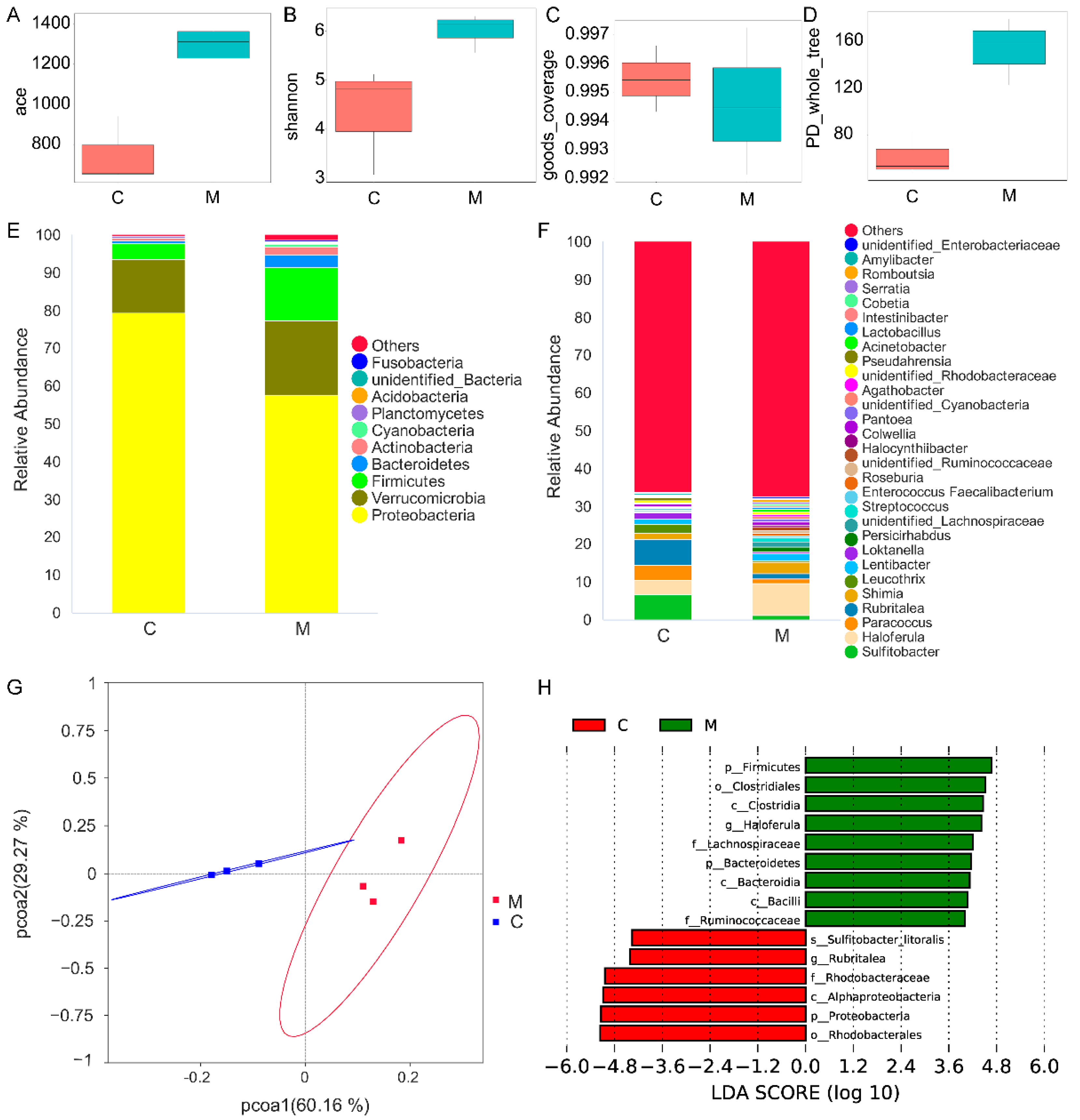
3.3. Gut Microbiota Analysis

Feeding probiotics can affect digestion, immunity, and other functions by regulating gut microbiota. This work examined the differences in gut microbiota between groups M and C. Operational Taxonomic Units clustering and species classification were shown in Table S1. As shown in Figure 2, no significant differences were observed in community richness (ACE, Figure 2A), community diversity (Shannon, Figure 2B), sequencing depth (Good’s coverage, Figure 2C), and phylogenetic diversity (PD whole tree, Figure 2D). It indicated that feeding S. aquimarina MS4 did not affect the alpha-diversity of A. japonicus gut microbiota.
Figure 2E,F showed the top 10 phyla and top 30 genera in the gut microbiota. Both groups were mainly composed of Proteobacteria, Verrucomicrobia, Firmicutes, and Bacteroidetes. Among them, Proteobacteria was the dominant phylum in the gut of sea cucumber, with a relative abundance of more than 50%. At the genus level, the relative abundances of Sulfitobacter, Haloferula, Paracoccus, and Rubritalea in group C were higher (>3%), while those in group M were mainly Haloferula and Shimia. Further PCoA analysis found that the same group of samples tended to cluster together, and the two groups of samples could be separated, indicating that the gut community structure of the two groups was different (Figure 2G). Species with significant differences in abundance between different groups were detected by LEfSe, and the results are shown in Figure 2H. The relative abundance of Firmicutes and Bacteroidetes, Lachnospiraceae and Ruminococcaceae, and Haloferula in group M were significantly higher than those in group C at the phylum, family, and genus levels, respectively. The relative abundance of Proteobacteria, Rhodobacteraceae, and Rubritalea in group C were significantly higher than those in group M at the phylum, family, and genus levels, respectively. These results indicated that the structure of the gut microbiota of A. japonicus was altered after feeding with S. aquimarina MS4.

3.4. Differential Metabolites in the Gut

Alterations in gut microbiota may directly affect gut metabolites. We used untargeted metabolomics to detect the gut metabolites of the two groups of sea cucumbers. As shown in Figure 3A–D, PLS-DA was used to model the relationship between the metabolite expression level and the sample. Good separation of metabolites was achieved between groups M and C; the model was not over-fitting; and it could describe the sample well. After screening, 517 metabolites were detected in positive ion mode (POS) and 195 metabolites were detected in negative ion mode (NEG). Compared with group C, group M was screened to obtain 10 significantly up-regulated metabolites, 8 significantly down-regulated metabolites in POS (Table S2), and 16 significantly down-regulated metabolites in NEG (Table S3).
Hierarchical clustering analysis was performed on the obtained differential metabolites in each group, and the differences in the metabolic expression patterns between the two groups and within the group were obtained for the same comparison. The results are shown in Figure 4A,B. The most important biochemical metabolic pathways and signal transduction pathways involved in differential metabolites were determined by KEGG pathway enrichment (Figure 4C,D). In POS, 13 pathways including lysine degradation were enriched, of which the lysine degradation pathway was significantly enriched. In NEG, 25 pathways such as beta-alanine metabolism were enriched. Further analysis found that the enriched pathways were mainly concentrated in amino acid metabolism (Pipecolic acid, N6-Acetyl-L-lysine, L-Argininosuccinate, D-Proline, and L-Histidine as the main differential metabolites), lipid metabolism (Turmerone, lysophosphatidylserine 22:6, lysodiacylglyceryltrimethyl homoserines 17:0, lysophosphatidylethanolamine 12:0, lysophosphatidylcholine 22:6, lysophosphatidylethanolamine 22:6, monoacylglyceride 18:2, and deoxycorticosterone as the main differential metabolites), amino sugar and nucleotide sugar metabolism (UDP-N-acetylglucosamine as the main differential metabolite), and ascorbate and aldarate metabolism (L-Ascorbate as the main differential metabolite). These results suggested that the levels of amino acids, lipids, amino sugars, and vitamins played crucial roles in the gut metabolism of A. japonicus.

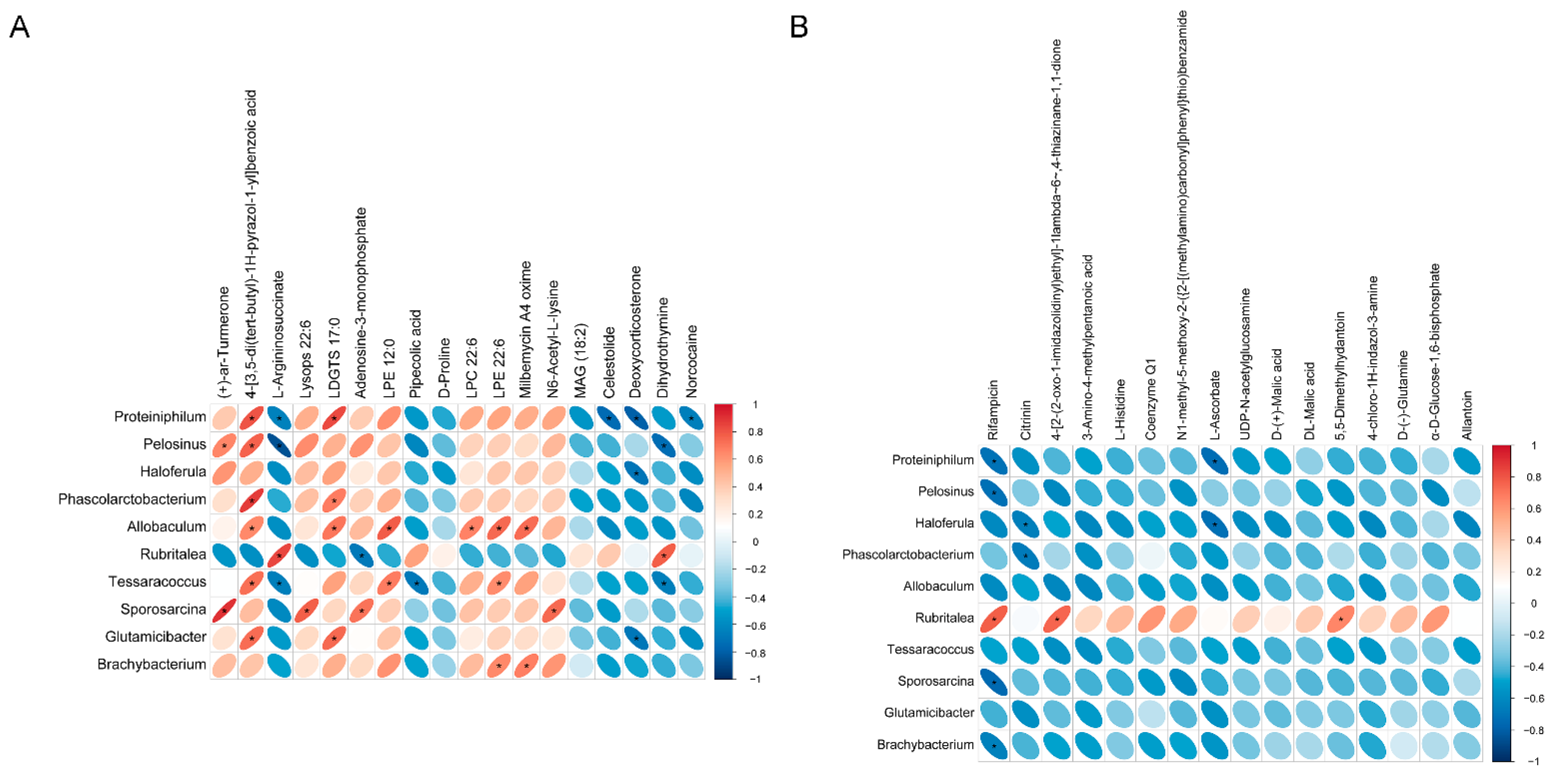
3.5. Correlation Analysis

In order to study the phenotypic changes that may be caused by changes in the structure of the gut microbiota, it is necessary to perform an association analysis between the metabolomics and the microbiota. The genera with significant differences at the genus level obtained by t-test analysis and the metabolites with significant differences obtained by metabolomics analysis were correlated based on the Pearson correlation coefficient and a heat map was obtained. As shown in Figure 5, multiple differential species were significantly associated with amino acid metabolism and lipid metabolism. For example, Sporosarcina was positively correlated with LysoPs and N6-Acetyl-L-lysine, Allobaculum was positively correlated with LDGTS, LPE, and LPC, Rubritalea was positively correlated with L-argininosuccinate, Haloferula was negatively correlated with deoxycorticosterone, and Tessaracoccus was negatively correlated with L-argininosuccinate and pipecolic acid.

4. Discussion

S. aquimarina MS4 is a microecological preparation for winter aquaculture of sea cucumbers, which can promote the growth of sea cucumbers and regulate immunity at a low dose [12]. The present work is to investigate the effects of S. aquimarina MS4 on sea cucumber growth, digestion, and gut health at a high dose (108 cfu/g). Although S. aquimarina MS4 significantly improves the growth performance of the sea cucumber compared with the control group, the weight gain is still very slow, which is related to the growth stagnation of sea cucumbers at low water temperature [18,19]. Interestingly, feeding S. aquimarina MS4 significantly reduced the natural mortality of sea cucumbers, which is similar to some studies on probiotics used in sea cucumber aquaculture [7]. In addition to individual differences, the improvement of sea cucumber immunity by probiotics may be the main reason for reducing its mortality [12,20]. The gut is the main digestive organ of the sea cucumber. Feeding S. aquimarina MS4 directly affects the gut digestive function and it further affects the growth performance and nutritional quality of the sea cucumber. Digestive enzyme activities are important indexes to evaluate the gut digestive function of sea cucumbers [21]. Since the sea cucumber feed contains a lot of high-starch, high-cellulose, and high-protein components such as cornmeal, seaweed and fish meal, amylase, cellulase, and protease degrade, the macromolecular polysaccharides and protein are processed into small molecular sugars and amino acids that are easily absorbed and utilized. Feeding S. aquimarina MS4 significantly increased cellulase and protease activities in the gut and improved the digestive performance of the sea cucumber, and it was beneficial to improve growth performance (Table 2).
The content of polysaccharide in the body wall of sea cucumbers fed with S. aquimarina MS4 significantly increased, and the composition of amino acids and fatty acids changed (Table 3, Table 4 and Table 5). Sea cucumber polysaccharides are important functional components of the body wall, and they have biological effects such as improving immunity, anti-cancer, anti-SARS-CoV-2, and improving metabolic syndrome [22,23,24]. In terms of amino acid composition, three essential amino acids, lysine, leucine, and phenylalanine significantly increased. Lysine has a positive nutritional significance in promoting human growth and development and enhancing immunity [25]. Leucine, a branched-chain amino acid, helps to repair muscle and to control blood sugar [26]. Phenylalanine is a flavor amino acid, which can improve the taste of the sea cucumber. From the change of amino acid composition, feeding S. aquimarina MS4 increased the nutritional value of the sea cucumber. In fatty acids, the double bond position of monounsaturated fatty acid in group M changed. Among the polyunsaturated fatty acids, the content of DHA significantly increased. DHA is essential for human brain development, along with other health benefits [27]. Overall, feeding S. aquimarina MS4 increased the nutritional value of the sea cucumber body wall.
The mechanism by which S. aquimarina MS4 affects the growth, digestion, immunity, and body wall components of A. japonicus is systematic and complex, but as a microbial feed additive, it directly interacts with the digestive organ—the gut—and directly affects the gut microecology. Similar to other studies on the gut microbiota of A. japonicus, the gut microbiota of the two groups were mainly composed of Proteobacteria, Verrucomicrobia and Firmicutes [28,29]. Among the microbiota differences, more Bacteroidetes and Firmicutes were produced after feeding S. aquimarina MS4. These two phyla are common in the gut of A. japonicus and include probiotics such as Flaviramulus, Flavobacterium, Bacillus, and Lactobacillus [30]. At the family level, the relative abundances of Lachnospiraceae and Ruminococcaceae significantly increased in group M, while at the genus level, Haloferula significantly increased. The family Lachnospiraceae and Ruminococcaceae and the genus Haloferula are common bacteria that can be found in marine environments and marine animals [28,31,32]. Although many studies have shown that Lachnospiraceae and Ruminococcaceae play important roles in the maintenance of intestinal homeostasis in humans [33], the role of these microorganisms in the gut of marine animals is poorly understood. Large-scale studies should be carried out to reveal the role played by the gut microbiota of different marine animals. Results show significant differences in microbial community composition between groups C and M, and they indicate that feeding S. aquimarina MS4 plays an important role in shaping the gut microbiota.
The gut microbiota often affects their hosts through its metabolites [34]. Understanding the metabolic communication between microorganisms and A. japonicus will provide us with an opportunity to find new methods for animal nutrition and health regulation. The gut microbiota will ferment the macromolecular carbohydrates that cannot be digested and absorbed by the host and generate small-molecule easily absorbed metabolites, linking the microbiota with the host’s physiology [35]. In this study, differential metabolites were enriched in amino acid metabolism after feeding S. aquimarina MS4, combined with a significant increase in protease activity in the gut. It is speculated that the difference in microbiota structure may cause the change in protease activity, which in turn affects the composition of some amino acids in the gut. In lipid metabolism, a variety of biologically active lysophospholipids were significantly increased, such as lysophosphatidylserine, lysophosphatidylcholine, and lysophosphatidylethanolamine. Lysophospholipids can improve the digestion and the absorption efficiency of lipids mainly through emulsification and the formation of micelle [36]. A meta-analysis of lysophospholipid application experiments in 33 different regions found that if additional lysophospholipids were added to broiler diets, the FCR were significantly reduced [37]. Similarly, in this study, after feeding S. aquimarina MS4, gut lysophospholipid increased significantly and FCR decreased significantly. The association analysis of differential metabolites and microbiota showed that the production of some differential metabolites was significantly related to differential microorganisms, which improved the understanding of the function of microorganisms and their roles in the gut of A. japonicus.

5. Conclusions

In conclusion, feeding S. aquimarina MS4 significantly increased the growth performance of A. japonicus and the enzyme activities of protease and cellulase, and it enhanced the nutritional composition of the body wall. Feeding S. aquimarina MS4 altered the structure of the gut microbiota of A. japonicus, resulting in changes in amino acid metabolism and lipid metabolism in the gut. These results provide some support for the microecological regulation mechanism of A. japonicus nutrition.

Supplementary Materials

The following supporting information can be downloaded at: https://www.mdpi.com/article/10.3390/fishes7030134/s1, Table S1: The Operational Taxonomic Units clustering and species classification; Table S2: The data of differential metabolites in positive ion mode; Table S3: The data of differential metabolites in negative ion mode.

Author Contributions

Conceptualization, B.L.; methodology, B.L.; software, H.Z.; validation, H.Y.; formal analysis, Q.B.; investigation, H.Y.; resources, H.Z.; data curation, Q.B.; writing—original draft preparation, B.L.; writing—review and editing, Y.L. (Ying Li); visualization, Y.L. (Yujia Liu); supervision, B.L.; project administration, J.W.; funding acquisition, J.W. and Y.L. (Ying Li). All authors have read and agreed to the published version of the manuscript.

Funding

This work was supported by the Dalian Science and Technology Bureau (2019RQ099 & 2018J11CY028), Department of Education of Liaoning Province (J2020097), and Open Fund of Key Laboratory of Biotechnology and Bioresources Utilization (Dalian Minzu University), Ministry of Education (KF2022007), China.

Institutional Review Board Statement

The animal study protocol was approved by the Animal Experiment Ethics Committee of Dalian Polytechnic University.

Informed Consent Statement

Not applicable.

Data Availability Statement

The data obtained in this study has been presented “as is” on at least one of the figures or tables embedded in the manuscript. Nevertheless, the data of microbiota and metabolomics presented in this study are available in the Supplementary Material Tables S1–S3.

Conflicts of Interest

The authors declare no conflict of interest. The funders had no role in the design of the study; in the collection, analyses, or interpretation of data; in the writing of the manuscript, or in the decision to publish the results.

References

  1. Bordbar, S.; Anwar, F.; Saari, N. High-value components and bioactives from sea cucumbers for functional foods—A review. Mar. Drugs 2011, 9, 1761–1805. [Google Scholar] [CrossRef] [PubMed] [Green Version]
  2. Pangestuti, R.; Arifin, Z. Medicinal and health benefit effects of functional sea cucumbers. J. Tradit. Complement. Med. 2018, 8, 341–351. [Google Scholar] [CrossRef] [PubMed]
  3. Xing, L.; Sun, L.; Liu, S.; Zhang, L.; Sun, J.; Yang, H. Metabolomic analysis of white, green and purple morphs of sea cucumber Apostichopus japonicus during body color pigmentation process. Comp. Biochem. Physiol. Part D Genom. Proteom. 2021, 39, 100827. [Google Scholar] [CrossRef]
  4. Romero, J.; Feijoo, C.G.; Navarrete, P. Antibiotics in aquaculture—Use, abuse and alternatives. In Health and Environment in Aquaculture; Carvalho, E.D., David, G.S., Silva, R.J., Eds.; IntechOpen: Rijeka, Croatia, 2012; pp. 159–198. [Google Scholar]
  5. Xia, Y.; Wang, M.; Gao, F.; Lu, M.; Chen, G. Effects of dietary probiotic supplementation on the growth, gut health and disease resistance of juvenile Nile tilapia (Oreochromis niloticus). Anim. Nutr. 2020, 6, 69–79. [Google Scholar] [CrossRef]
  6. Wang, G.; Jiang, Y.; Jiang, J.; Chen, L. Effects of feed supplemented with probiotics on the culture of sea cucumber Apostichopus japonicus. Aquac. Nutr. 2021, 27, 2703–2711. [Google Scholar] [CrossRef]
  7. Wang, J.H.; Zhao, L.Q.; Liu, J.F.; Wang, H.; Xiao, S. Effect of potential probiotic Rhodotorula benthica D30 on the growth performance, digestive enzyme activity and immunity in juvenile sea cucumber Apostichopus japonicus. Fish Shellfish Immunol. 2015, 43, 330–336. [Google Scholar] [CrossRef] [PubMed]
  8. Li, C.; Ren, Y.; Jiang, S.; Zhou, S.; Zhao, J.; Wang, R.; Li, Y. Effects of dietary supplementation of four strains of lactic acid bacteria on growth, immune-related response and genes expression of the juvenile sea cucumber Apostichopus japonicus Selenka. Fish Shellfish Immunol. 2018, 74, 69–75. [Google Scholar] [CrossRef]
  9. Feng, Z.; Song, X.; Zhao, L.; Zhu, W. Isolation of probiotics and their effects on growth, antioxidant and non-specific immunity of sea cucumber Apostichopus japonicus. Fish Shellfish Immunol. 2020, 106, 1087–1094. [Google Scholar] [CrossRef] [PubMed]
  10. Wang, J.-h.; Guo, H.; Zhang, T.-r.; Wang, H.; Liu, B.-n.; Xiao, S. Growth performance and digestion improvement of juvenile sea cucumber Apostichopus japonicus fed by solid-state fermentation diet. Aquac. Nutr. 2017, 23, 1312–1318. [Google Scholar] [CrossRef]
  11. Ning, Y.C.; Wu, X.Y.; Zhou, X.H.; Ding, J.; Chang, Y.Q.; Yang, Z.L.; Huang, Z.Q.; Zuo, R.T. An evaluation on the selenium yeast supplementation in the practical diets of early juvenile sea cucumber (Apostichopus japonicus): Growth performance, digestive enzyme activities, immune and antioxidant capacity, and body composition. Aquac. Nutr. 2021, 27, 2142–2153. [Google Scholar] [CrossRef]
  12. Zhou, W.; Liu, B.; Li, Y.; Zhan, H.; Liu, Y.; Xiao, S.; Zhao, Q.; Wang, J. Dietary supplementation with Sporosarcina aquimarina MS4 enhances juvenile sea cucumber (Apostichopus japonicus) growth, immunity and disease resistance against Vibrio splendidus infection at low temperature. Aquac. Nutr. 2021, 27, 918–926. [Google Scholar] [CrossRef]
  13. Haide, M.S.; Sultana, R.; Jamil, K.; Tarar, O.M.; Afzal, W. A study on proximate composition, amino acid profile, fatty acid profile and some mineral contents in two species of sea cucumber. J. Anim. Plant Sci. 2015, 25, 168–175. [Google Scholar]
  14. DuBois, M.; Gilles, K.A.; Hamilton, J.K.; Rebers, P.A.; Smith, F. Colorimetric method for determination of sugars and related substances. Anal. Chem. 1956, 28, 350–356. [Google Scholar] [CrossRef]
  15. Liu, X.; Liu, L.; Leng, P.; Hu, Z. Feasible and effective reuse of municipal sludge for vegetation restoration: Physiochemical characteristics and microbial diversity. Sci. Rep. 2019, 9, 879. [Google Scholar] [CrossRef] [Green Version]
  16. Liu, S.; Yi, H.; Zhan, H.; Wang, L.; Wang, J.; Li, Y.; Liu, B. Gibberellic acid-induced fatty acid metabolism and ABC transporters promote astaxanthin production in Phaffia rhodozyma. J. Appl. Microbiol. 2022, 132, 390–400. [Google Scholar] [CrossRef]
  17. Yuan, M.; Breitkopf, S.B.; Yang, X.; Asara, J.M. A positive/negative ion-switching, targeted mass spectrometry-based metabolomics platform for bodily fluids, cells, and fresh and fixed tissue. Nat. Protoc. 2012, 7, 872–881. [Google Scholar] [CrossRef] [PubMed] [Green Version]
  18. Ren, J.S.; Stenton-Dozey, J.; Zhang, J. Parameterisation and application of dynamic energy budget model to sea cucumber Apostichopus japonicus. Aquac. Environ. Interact. 2017, 9, 1–8. [Google Scholar] [CrossRef] [Green Version]
  19. Jiang, J.; Zhou, Z.; Dong, Y.; Jiang, B.; Chen, Z.; Gao, S.; Guan, X.; Han, L. Seasonal variations of immune parameters in the coelomic fluid of sea cucumber Apostichopus japonicus cultured in pond. Aquac. Res. 2017, 48, 1677–1687. [Google Scholar] [CrossRef]
  20. El-Saadony, M.T.; Alagawany, M.; Patra, A.K.; Kar, I.; Tiwari, R.; Dawood, M.A.O.; Dhama, K.; Abdel-Latif, H.M.R. The functionality of probiotics in aquaculture: An overview. Fish Shellfish Immunol. 2021, 117, 36–52. [Google Scholar] [CrossRef]
  21. Sun, J.; Zhang, L.; Pan, Y.; Lin, C.; Wang, F.; Kan, R.; Yang, H. Feeding behavior and digestive physiology in sea cucumber Apostichopus japonicus. Physiol. Behav. 2015, 139, 336–343. [Google Scholar] [CrossRef]
  22. Song, S.; Peng, H.; Wang, Q.; Liu, Z.; Dong, X.; Wen, C.; Ai, C.; Zhang, Y.; Wang, Z.; Zhu, B. Inhibitory activities of marine sulfated polysaccharides against SARS-CoV-2. Food Funct. 2020, 11, 7415–7420. [Google Scholar] [CrossRef]
  23. Li, Y.; Qin, J.; Cheng, Y.; Lv, D.; Li, M.; Qi, Y.; Lan, J.; Zhao, Q.; Li, Z. Marine sulfated polysaccharides: Preventive and therapeutic effects on metabolic syndrome: A review. Mar. Drugs 2021, 19, 608. [Google Scholar] [CrossRef] [PubMed]
  24. Xu, H.; Zhou, Q.; Liu, B.; Chen, F.; Wang, M. Holothurian fucosylated chondroitin sulfates and their potential benefits for human health: Structures and biological activities. Carbohydr. Polym. 2022, 275, 118691. [Google Scholar] [CrossRef] [PubMed]
  25. Benevenga, N.J.; Blemings, K.P. Unique aspects of lysine nutrition and metabolism. J. Nutr. 2007, 137, 1610s–1615s. [Google Scholar] [CrossRef] [Green Version]
  26. Plotkin, D.L.; Delcastillo, K.; Van Every, D.W.; Tipton, K.D.; Aragon, A.A.; Schoenfeld, B.J. Isolated leucine and branched-chain amino acid supplementation for enhancing muscular strength and hypertrophy: A narrative review. Int. J. Sport Nutr. Exerc. Metab. 2021, 31, 292–301. [Google Scholar] [CrossRef]
  27. Cardoso, C.; Afonso, C.; Bandarra, N.M. Dietary DHA and health: Cognitive function ageing. Nutr. Res. Rev. 2016, 29, 281–294. [Google Scholar] [CrossRef]
  28. Zeng, F.; Rabbi, M.H.; Hu, Y.; Li, Z.; Ren, X.; Han, Y.; Ren, T. Synergistic effects of dietary selenomethionine and vitamin C on the immunity, antioxidant status, and intestinal microbiota in sea cucumber (Apostichopus japonicus). Biol. Trace Elem. Res. 2021, 199, 3905–3917. [Google Scholar] [CrossRef] [PubMed]
  29. Kim, T.Y.; Lee, J.J.; Kim, B.S.; Choi, S.H. Whole-body microbiota of sea cucumber (Apostichopus japonicus) from south korea for improved seafood management. J. Microbiol. Biotechnol. 2017, 27, 1753–1762. [Google Scholar] [CrossRef] [PubMed] [Green Version]
  30. Wang, A.; Ran, C.; Wang, Y.; Zhang, Z.; Ding, Q.; Yang, Y.; Olsen, R.E.; Ringø, E.; Bindelle, J.; Zhou, Z. Use of probiotics in aquaculture of China—A review of the past decade. Fish Shellfish Immunol. 2019, 86, 734–755. [Google Scholar] [CrossRef]
  31. Bai, S.; Zhang, P.; Zhang, C.; Du, J.; Du, X.; Zhu, C.; Liu, J.; Xie, P.; Li, S. Comparative study of the gut microbiota among four different marine mammals in an aquarium. Front. Microbiol. 2021, 12, 769012. [Google Scholar] [CrossRef]
  32. Kang, H.; Traiwan, J.; Weerawongwiwat, V.; Jung, M.Y.; Jeong, J.H.; Myung, S.C.; Lee, K.C.; Lee, J.S.; Kim, W. Haloferula chungangensis sp. nov., isolated from marine sediment. Int. J. Syst. Evol. Microbiol. 2013, 63, 19–24. [Google Scholar] [CrossRef] [PubMed] [Green Version]
  33. Maukonen, J.; Ouwehand, A.C. Changes in the microbiota composition and function in relation to aging. In Comprehensive Gut Microbiota; Glibetic, M., Ed.; Elsevier: Oxford, UK, 2022; pp. 85–96. [Google Scholar] [CrossRef]
  34. McCarville, J.L.; Chen, G.Y.; Cuevas, V.D.; Troha, K.; Ayres, J.S. Microbiota metabolites in health and disease. Annu. Rev. Immunol. 2020, 38, 147–170. [Google Scholar] [CrossRef]
  35. Debnath, N.; Kumar, R.; Kumar, A.; Mehta, P.K.; Yadav, A.K. Gut-microbiota derived bioactive metabolites and their functions in host physiology. Biotechnol. Genet. Eng. Rev. 2021, 37, 105–153. [Google Scholar] [CrossRef] [PubMed]
  36. Kontara, E.K.; Djunaidah, I.S.; Coutteau, P.; Sorgeloos, P. Comparison of native, lyso and hydrogenated soybean phosphatidylcholine as phospholipid source in the diet of postlarval Penaeus japonicus bate. Arch. Anim. Nutr. 1998, 51, 1–19. [Google Scholar] [CrossRef] [PubMed]
  37. Wealleans, A.L.; Jansen, M.; di Benedetto, M. The addition of lysolecithin to broiler diets improves growth performance across fat levels and sources: A meta-analysis of 33 trials. Br. Poult. Sci. 2020, 61, 51–56. [Google Scholar] [CrossRef]
Figure 1. Digestive enzyme activities of the gut in groups M and C. (A) Protease activity; (B) Cellulose activity; (C) Amylase activity. Asterisks indicate significant differences compared to the control group (p < 0.05).
Fishes 07 00134 g001
Figure 2. Gut microbiota analysis. Alpha diversity ((A), ace; (B), Shannon; (C), goods coverage; (D), PD whole tree) reflects the richness and diversity of the microbial community within the sample. Species relative abundance display at the phylum (E) and genus (F) levels. Beta diversity comparative analysis of the microbial community composition of each group. (G), Principal Co-ordinates Analysis (PCoA), each point in the figure represents a sample, and samples of the same group are represented by the same color; (H), LDA Effect Size (LEfSe), species with significantly different abundances in different groups (LDA Score > 4) are shown in the figure.
Fishes 07 00134 g002
Figure 3. Partial least squares discrimination analysis (PLS-DA) of groups M and C ((A), POS & (C), NEG). Different colored dots represent samples from different experimental groups, and the ellipse is a 95% confidence interval. R2Y represents the interpretation rate of the model, Q2Y is used to evaluate the predictive ability of the PLS-DA model, and when R2Y is greater than Q2Y, the model is well established. Sorting verification of PLS-DA score of group M and C ((B), POS & (D), NEG). When R2 is greater than Q2 and the intercept between the Q2 regression line and the Y axis is less than 0, it indicates that the model is not “overfitting”.
Fishes 07 00134 g003
Figure 4. Cluster analysis of differential metabolites ((A), POS & (B), NEG) and KEGG enrichment pathways ((C), POS & (D), NEG). The color of the dot represents the p-value, and the size of the dot represents the number of differential metabolites in the corresponding pathway.
Fishes 07 00134 g004
Figure 5. Heatmap of the correlation between gut microbiota and metabolites ((A), POS & (B), NEG). The legend on the right is the correlation coefficient, where red indicates a positive correlation and blue indicates a negative correlation. Asterisks indicate statistical significance (p < 0.05).
Fishes 07 00134 g005
Table 1. Feed nutrients.
IngredientsContent (g/100 g)
Degumming kelp powder 32
Sargasso powder20
Ulva powder 20
Cornmeal6
Wall-broken yeast 5
Fermented soybean meal 4
Shrimp meal 4
Multidimensional premix4
Fish meal 3
Scallop edge powder2
Analyzed nutrients
Moisture5.13 ± 0.12
Crude protein14.96 ± 0.55
Total sugar33.40 ± 0.63
Crude fat1.52 ± 0.03
Ash43.64 ± 1.21
Table 2. Growth performance of the sea cucumber after 60 days feeding of control diet and diet with S. aquimarina MS4.
Growth ParametersGroup CGroup M
Initial weight (g)1.88 ± 0.02 a1.95 ± 0.13 a
Final weight (g)1.98 ± 0.05 a2.19 ± 0.10 a
Natural mortality (%)4.17 ± 1.18 a0.00 ± 0.00 b
WGR (%)5.48 ± 1.85 a12.22 ± 2.35 b
SGR (%/d)0.09 ± 0.03 a0.19 ± 0.03 b
FCR6.14 ± 0.17 a4.89 ± 0.56 b
a,b Different letters in each row indicate statistically significant variations between groups (p < 0.05).
Table 3. Body wall nutrients.
Component Group C (g/100 g)Group M (g/100 g)
Moisture9.60 ± 0.21 a9.27 ± 0.24 a
Protein49.40 ± 0.53 a50.65 ± 0.49 a
Polysaccharide6.49 ± 0.14 a7.34 ± 0.24 b
Fat0.82 ± 0.04 a0.89 ± 0.08 a
Ash33.47 ± 1.01 a31.70 ± 1.35 a
a,b Different letters in each row indicate statistically significant variations between groups (p < 0.05).
Table 4. Amino acids of the body wall.
Amino AcidsGroup C (mg/g)Group M (mg/g)
Asp40.29 ± 2.18 43.09 ± 1.65
Thr19.74 ± 0.91 21.42 ± 0.86
Ser18.69 ± 0.90 19.37 ± 0.38
Glu60.37 ± 2.29 62.73 ± 1.08
Gly38.87 ± 3.35 40.42 ± 3.21
Ala23.53 ± 1.85 24.67 ± 1.53
Cys5.00 ± 0.37 4.53 ± 1.65
Val16.18 ± 0.86 17.64 ± 1.09
Met6.10 ± 0.23 6.34 ± 0.35
Ile13.03 ± 0.66 14.54 ± 0.43
Leu *21.61 ± 0.85 23.65 ± 0.50
Tyr11.32 ± 0.65 12.37 ± 0.24
Phe *13.81 ± 0.67 15.51 ± 0.41
His13.21 ± 0.38 14.11 ± 0.56
Lys *18.97 ± 0.3320.23 ± 0.34
Arg24.59 ± 2.18 26.99 ± 1.19
Pro19.06 ± 1.4220.21 ± 1.59
Total364.37 ± 19.46387.82 ± 15.50
* Asterisks indicate that the amino acid content was significantly different between the two groups (p < 0.05).
Table 5. Fatty acids of the body wall.
Fatty AcidsControlMS1
Saturated fatty acidsTridecanoic acid (C13:0)1.72 ± 0.24-
Tetradecanoic acid (C14:0)-2.81 ± 0.13
Pentadecanoic acid (C15:0) *9.08 ± 3.051.36 ± 0.05
Hexadecanoic acid (C16:0)0.38 ± 0.113.10 ± 1.39
Stearate (C18:0)5.94 ± 0.665.49 ± 0.37
Nonadecanoic acid (C19:0)0.75 ± 0.351.19 ± 0.32
Eicosanoic acid (C20:0)2.19 ± 0.551.20 ± 0.17
Heneicosanoic acid (C21:0)1.94 ± 0.302.00 ± 0.21
Docosanoic acid (C22:0)0.97 ± 0.451.83 ± 0.39
Heptacosanoic acid (C27:0) *0.61 ± 0.092.51 ± 0.20
Subtotal 23.58 ± 4.0721.48 ± 0.63
Monounsaturated fatty acids7-Hexadecenoic acid (C16:1)-5.02 ± 0.73
9-Hexadecenoic acid (16:1) *11.62 ± 0.910.55 ± 0.16
9-Octadecenoic acid (C18:1) *15.93 ± 3.210.25 ± 0.03
12-Octadecenoic acid (C18:1)-8.89 ± 1.96
13-Octadecenoic acid (C18:1)-7.44 ± 0.58
cis-11-Eicosenoic acid (C20:1)11.75 ± 0.7011.18 ± 2.21
Total13-Docosenoic acid (C22:1)1.21 ± 1.052.26 ± 0.88
15-Tetracosenoic acid (C24:1)2.60 ± 0.682.42 ± 1.03
Subtotal
Polyunsaturated fatty acids
43.11 ± 2.0538.01 ± 4.95
9,12-Octadecadienoic acid (C18:2)4.91 ± 0.364.33 ± 0.23
11,14-Eicosadienoic acid (C20:2)3.83 ± 0.363.71 ± 0.47
5,8,11,14-Eicosatetraenoic acid (C20:4)16.75 ± 1.4418.34 ± 3.82
5,8,11,14,17-Eicosapentaenoic acid, methyl ester (C20:5)5.76 ± 4.044.87 ± 0.52
4,7,10,13,16,19-Docosahexaenoic acid (C22:6) *2.06 ± 0.799.27 ± 1.78
Subtotal 33.31 ± 6.0940.51 ± 5.39
* Asterisks indicate that the fatty acid content was significantly different between the two groups (p < 0.05). “-” means not detected.
Publisher’s Note: MDPI stays neutral with regard to jurisdictional claims in published maps and institutional affiliations.

