Article

The Responses of the Ovary and Eyestalk in Exopalaemon carinicauda under Low Salinity Stress

1
Key Laboratory of Sustainable Development of Marine Fisheries, Ministry of Agriculture and Rural Affairs, Yellow Sea Fisheries Research Institute, Chinese Academy of Fishery Sciences, Qingdao 266071, China
2
Laboratory for Marine Fisheries Science and Food Production Processes, Pilot National Laboratory for Marine Science and Technology, Qingdao 266071, China
3
Pilot National Laboratory for Marine Science and Technology, Qingdao 266237, China
*
Author to whom correspondence should be addressed.
These authors have contributed equally to this work.
Fishes 2022, 7(6), 365; https://doi.org/10.3390/fishes7060365
Submission received: 2 November 2022 / Revised: 24 November 2022 / Accepted: 28 November 2022 / Published: 30 November 2022

Abstract

:
As a euryhaline shrimp, the ridgetail white prawn Exopalaemon carinicauda is strongly adaptable to salinity. Exploring the effect of long-term low salinity stress on ovarian development in E. carinicauda is essential to promote its culture in a non-marine environment. In this study, we performed biochemical assays and ovary histology analysis, finding that the E. carinicauda can adapt to low salinity stress through osmotic adjustment, and there was no substantial damage to the ovary of E. carinicauda under low salinity stress. Then, the ovarian development of E. carinicauda under low salt stress was further explored by RNA sequencing of eyestalk and ovarian tissues. A total of 389 differentially expressed genes (DEGs) in ovary tissue were identified under low salinity stress, and the 16 important DEGs were associated with ovarian development. The majority of the DEGs were enriched in ECM-receptor interaction, folate biosynthesis, arginine biosynthesis, insect hormone biosynthesis and lysosome which were involved in the ovarian development of E. carinicauda. A total of 1223 DEGs were identified in eyestalk tissue under low salinity stress, and the 18 important DEGs were associated with ovarian development. KEGG enrichment analysis found that ECM-receptor interaction, folate biosynthesis, lysosome, arginine biosynthesis and retinol metabolism may be involved in the ovarian development under low salinity stress. Our results provided new insights and revealed new genes and pathways involved in ovarian development of E. carinicauda under long-term low salinity stress.

1. Introduction

Salinity is one of the most important environmental factors affecting the survival, growth and reproduction of aquatic animals [1,2]. It is reported that changes in salinity might affect the transmembrane ion/water transport, disrupt the osmotic balance [3,4] and also affect molting, oogenesis, embryogenesis and larval quality [5,6]. Mohanty et al. [6] reported that the salinity exposure significantly affected the gonadosomatic index, ovary histology and morphometric features of oocytes in stenohaline freshwater catfish. It was also reported that low salinity stress could maximally inhibit steroid mediated gonadal recurrence in an euryhaline fish [7]. Although many crustaceans, such as Penaeus monodon [8], Scylla serrata [9], Eriocheir sinensis [10] and Litopenaeus vannamei [11], can survive in a wide range of salinity environments, there are few reports on their reproduction under low salinity stress. Long et al. [12] found that salinity plays a key role in ovarian development, osmotic regulation and metabolism during the reproductive migration of female E. sinensis; the increase of salinity from a fresh water to a brackish water environment led to the reduction of metabolism, accelerated ovarian development, and produced non-mating spawning crabs when female E. sinensis molted after puberty.
The ridgetail white prawn Exopalaemon carinicauda (Arthropoda, Crustacea, Decapoda and Exopalaemon), is widely distributed in the Yellow Sea and Bohai Sea and is one of the most important commercial shrimps in China [13,14,15]. Due to multiple merits of fast growth, high reproductive performance and strong environmental adaptability, the culture area of E. carinicauda in China expanded in recent years [16,17]. According to incomplete statistics in 2016, the breeding area of E. carinicauda is about 4000 hectares, with an annual output of about 100,000 tons [18]. The E. carinicauda can adapt to salinity in a wide range [19], and can live in water bodies with salinity ranging from 4.3 to 35. It can even live in fresh water after desalting. Furthermore, E. carinicauda has been successfully cultured and bred in the saline-alkaline ponds (approximate salinity 5–8) at Dongying City, Shandong province, China, suggesting that they have a high tolerance to saline-alkaline stress [14,20]. However, there are few reports on the effects of low salinity on the reproduction of white shrimps. Only Liang et al. [21] reported that the gonads of the E. carinicauda can develop and mature under salinity 2–30. However, we still do not have a thorough grasp of the mechanism of low salinity stress on the reproduction of E. carinicauda.
Recently, RNA sequencing (RNA-seq) transcriptomics has been widely used to study the differential expression and molecular pathways of genes under specific environmental stresses [22]. For example, RNA-seq was used to compare the transcriptomic responses of L. vannamei to changes in salinity [23], and Li et al. (2014) used transcriptome sequencing to reveal the genes and pathways related to salt stress in E. sinensis [24]. However, most of the studies on the effect of low salinity on aquatic organisms focus on osmoregulation, and the research on gonad development is relatively few. The eyestalk is part of the X-organ sinus gland and an important endocrine organ for crustaceans; it is thought to play a key role in various physiological activities, including ovarian maturation [25]. Studies found that removal of the eyestalk can induce ovarian maturation and oviposition in many crustaceans [26,27]. Although the mechanism by which eyestalk ablation leads to ovarian maturation is still uncertain, some genes expressed in the eyestalk regulate ovarian development. For example, in polychaete-fed female Penaeus monodon, eyestalk ablation led to ovarian maturation, and it was found that genes in several key pathways were up-regulated, namely the gonadotropin-releasing hormone (GnRH) signal transduction pathway, the calcium signal pathway and the progesterone mediated oocyte maturation pathway [28]. Therefore, in this study, the transcriptome of the E. carinicauda ovary and eyestalk was sequenced by using RNA-seq technology for the first time. We compared and analyzed the transcriptome data between the control group and the low salinity group to determine the genes and pathways related to ovarian development. The findings of this study will help to clarify the ovarian development mechanism of E. carinicauda in its adaptation to salinity challenges.

2. Materials and Methods

2.1. Animals

Adult female shrimps were collected from Haichen Aquaculture Co. Ltd. in Rizhao city, Shandong province, China. The experiment was carried out in a 200 L PVC barrel, and the shrimps were domesticated in the laboratory environment (25 °C) for two weeks before experiment. One hundred and eighty shrimps were randomly sorted into two groups, including the low salinity group (salinity 5 ppt), and a control group (salinity 25 ppt). Each group had three replicates with 30 shrimps. During the experimental periods, the shrimps were fed according to 3–5% of their body weight twice a day (8:00 and 18:00). The water was aerated and 30% was changed daily with the adjusted seawater in order to maintain the original salinity. Natural illumination was used during the experiment, and water quality was maintained at a temperature of 25 ± 0.5 °C, pH of 8.2 ± 0.1 and a dissolved oxygen level of 7.4 ± 0.3 mg L−1. The experiment lasts for 60 days.

2.2. Sample Collection

After 60 days, eighteen female shrimps (3 individuals × 3 replicates × 2 groups) were used for biochemical assays, twelve female shrimps (2 individuals × 3 replicates ×2 groups) were used for histological sections, and thirty-six female shrimps (6 individuals × 3 replicates ×2 groups) were used for Illumina RNA-seq. The ovary and eyestalk tissues were collected and rapidly frozen in liquid nitrogen, then stored at −80 °C until RNA isolation, respectively. The ovary samples in the low salinity group are labeled LS_O, eyestalk samples in the low salinity group are labeled LS_E, the ovary samples in the control group are labeled CG_O and the eyestalk samples in the control group are labeled CG_E.

2.3. Biochemical Assays

Na+/K+-ATPase and carbonic anhydrase activities in the tissue were determined using Detection Kit (Suzhou Keming Biotechnology Co., Ltd. Suzhou, China). We accurately weighed 0.1 g of hepatopancreas, gill and muscle tissue, added the extract according to the weight volume ratio of 1:10, and performed ice bath homogenization with a 8000 × g centrifuge at 4 °C for 10 min, collected the supernatant and placed it on ice. We then measured according to the instructions of the kit and used the microplate reader to test and read.

2.4. Ovary Histology

The ovaries were fixed in 4% paraformaldehyde for 24 h before washing with 1 × PBS and dehydrating using a graded ethanol series (80% ethanol for 1 h, followed by 95% ethanol for 1 h, followed by 100% ethanol for 1 h). Transparency was improved using xylene (pure ethanol: xylene (1:1) for 1 h, then xylene for 1 h). Samples were infiltrated with paraffin (xylene: paraffin (1:1) at 62 °C for 1 h, then paraffin at 62 °C for 2 h) and processed for paraffin embedding. Sections were cut to 6 µm before staining with hematoxylin and eosin. Samples were scanned using a microscope slide scanner (Pannoramic MIDI, Budapest, Hungary).

2.5. RNA Isolation, Library Construction and Illumine Sequencing

Total RNA extraction from each of the collected samples was performed using a TRIzol® reagent (Invitrogen, San Diego, CA, USA) according to the manufacturer’s instructions. The DNase I was used to process total RNA for DNA digestion and obtain pure RNA products. Finally, the RNA purity and concentration were then examined using NanoDrop 2000 and the RNA integrity and quantity were measured using the Agilent 2100/4200 system. (Agilent Technologies, Santa Clara, CA, USA). Equal amounts of RNA from different individuals in the same group were pooled for library construction. Next generation sequencing library preparations were constructed according to the manufacturer’s protocol (NEBNext® Ultra™ RNA Library Prep Kit for Illumina®, NEB, Ipswich, MA, USA). After the mRNA library passed the quality inspection, PE150 sequencing was performed using Illumina Novaseq 6000 platform.

2.6. Basic Analysis of Sequencing Data

In order to remove technical sequences, including adapters, polymerase chain reaction (PCR) primers, or fragments thereof, and bases with a quality lower than 20, the pass filter data in the FASRQ format were processed by Trimmomatic (v0.30, http://www.usadellab.org/cms/?page=trimmomatic accessed on 15 November 2022) to provide high-quality, clean data. Firstly, the whole genome sequence of E. carinicauda assembled by our research group was taken as the reference genome (the data have not been published yet). Secondly, Hisat2 (v2.0.1, http://ccb.jhu.edu/software/hisat2/index.shtml accessed on 15 November 2022) was used to index the reference genome sequence. Finally, we mapped the clean reads to the silva database to remove the rRNA. All the downstream analyses were based on the clean data without rRNA.

2.7. Differential Expression Genes (DEGs) Analysis and Enrichment Analysis

The DESeq2 and edgeR [29] methods were used to perform the differential expression analysis. The fragments per kilobase per million reads (FPKM) method was used to normalized data, which can eliminate the influence of gene length and sequencing amount on the calculated gene expression, and the calculated gene expression level can be directly used to compare the expression differences between different genes. After adjusting using the Benjamini and Hochberg’s approach for controlling the false discovery rate, the p-values < 0.05 and |log2FC| ≥ 1 were set to detect significant differentially expressed genes. Gene Ontology (GO) and Kyoto Encyclopedia of Genes and Genome (KEGG) enrichment analyses of differentially expressed gene sets were implemented by the topG and KOBAS package 3.0 (http://kobas.cbi.pku.edu.cn/ accessed on 15 November 2022) [30], respectively.

2.8. RNA-Seq Data Validation by Real-Time Quantitative PCR and Statistical Analysis

To validate and measure the differential expression of mRNAs by high-throughput sequencing, ten differentially expressed mRNAs were randomly selected for real-time quantitative PCR (qPCR) analysis. The qPCR assay was performed using SYBR Green PCR Master Mix (life Technologies, USA) in the 7500 fast Real-Time PCR system (Applied Biosystems, Foster, California) according to the manufacturer’s agreement. The 18S rRNA of E. carinicauda was used as the internal reference [31]. All primer sequences and 18S rRNA sequences were listed in Table S1. The relative expression of target genes was calculated with 2−∆∆CT methods. The one-way ANOVA method and Duncan’s test in the statistical software SPSS 22.0 (SPSS, Chicago, IL, USA) were used for statistical analysis. The results are presented as the mean ± standard error, and differences in gene expression were considered statistically significant at P < 0.05.

3. Results

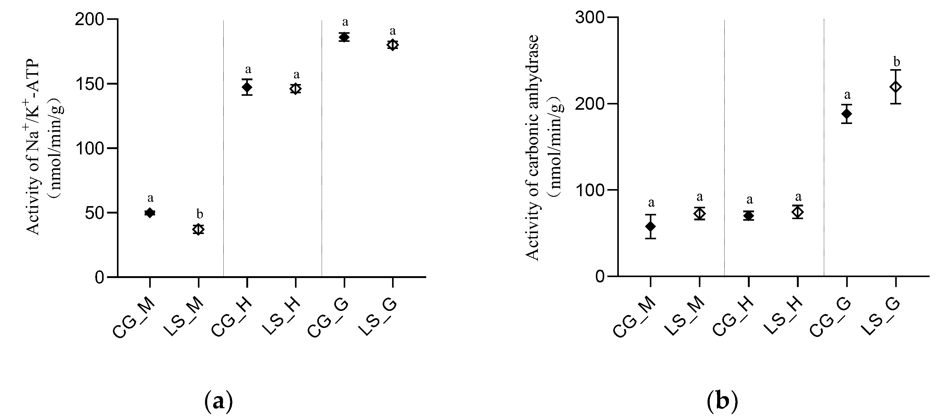
3.1. Variation of CARBONIC anhydrase and Na+/K+-ATPase Activity in the Tissue

Na+/K+-ATPase and carbonic anhydrase activity in the different tissue (muscle, hepatopancreas and gill) were determined using Detection Kit (Figure 1), and the highest activities of both enzymes were found in gill. In comparison with the CG group, the Na+/K+-ATPase activity of the LS group was significantly lower only in muscle, and no significant change in other tissues was identified. Compared with the CG group, the carbonic anhydrase activity of LS group was significantly higher only in the gill, and there was no significant change in other tissues.

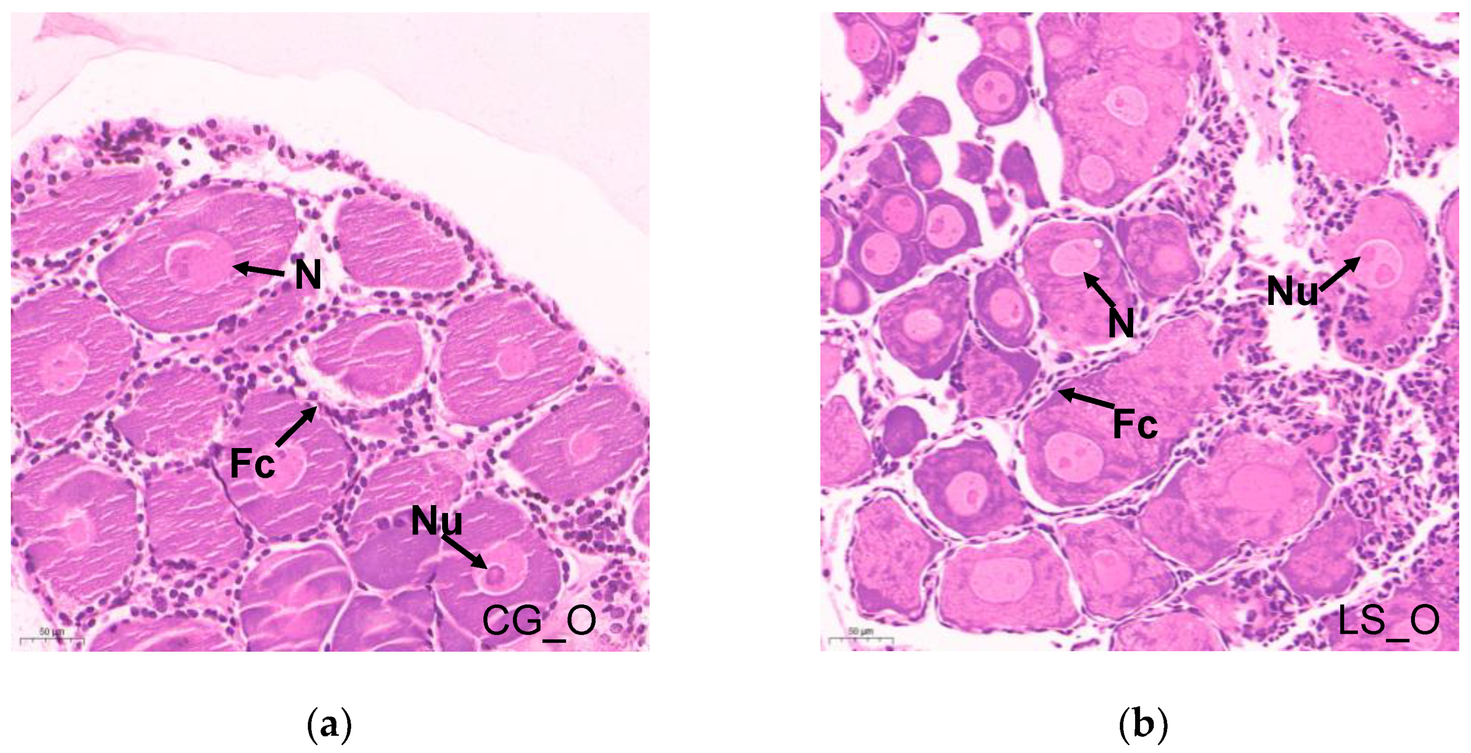
3.2. Histopathology of Ovary

Normal reproductive characteristics can be observed in the histological sections of the ovary in Figure 2. The E. carinicauda ovary was in Phase I. The oogonia were oval and proliferative, and the nuclei were round. Most cells had one nucleolus, while a few had two nucleoli. The nucleolus stained the deepest color. The cells of the oogonia were closely arranged. A single layer of follicles was closely arranged around the oogonia (Figure 2a).
After under low salinity stress for 60 days, the development characteristics were similar to those of the control group. No substantial damage was observed in the histological sections; only the oogonia were loosely arranged, and the surrounding follicular cells were loosely arranged with a small number (Figure 2b).

3.3. Summary of the RNA-seq Data

In order to identify the underlying molecular signaling pathways of low salinity on ovarian development in E. carinicauda, six mRNA libraries were constructed from ovary and eyestalk, respectively. The raw data were submitted to the NCBI with the accession numbers PRJNA881755 and PRJNA881756. A total of 270,444,484 clean reads were obtained from ovaries, with Q30 (%) varying from 92.69% to 93.21%. Of these reads, 70.69%–72.74% were mapped to the reference genome of E. carinicauda. A total of 253,177,112 clean reads were obtained from eyestalks, with Q30 (%) varying from 91.72% to 92.67%. Of these reads, 79.38%–83.02% were mapped to the reference genome (Table S2).

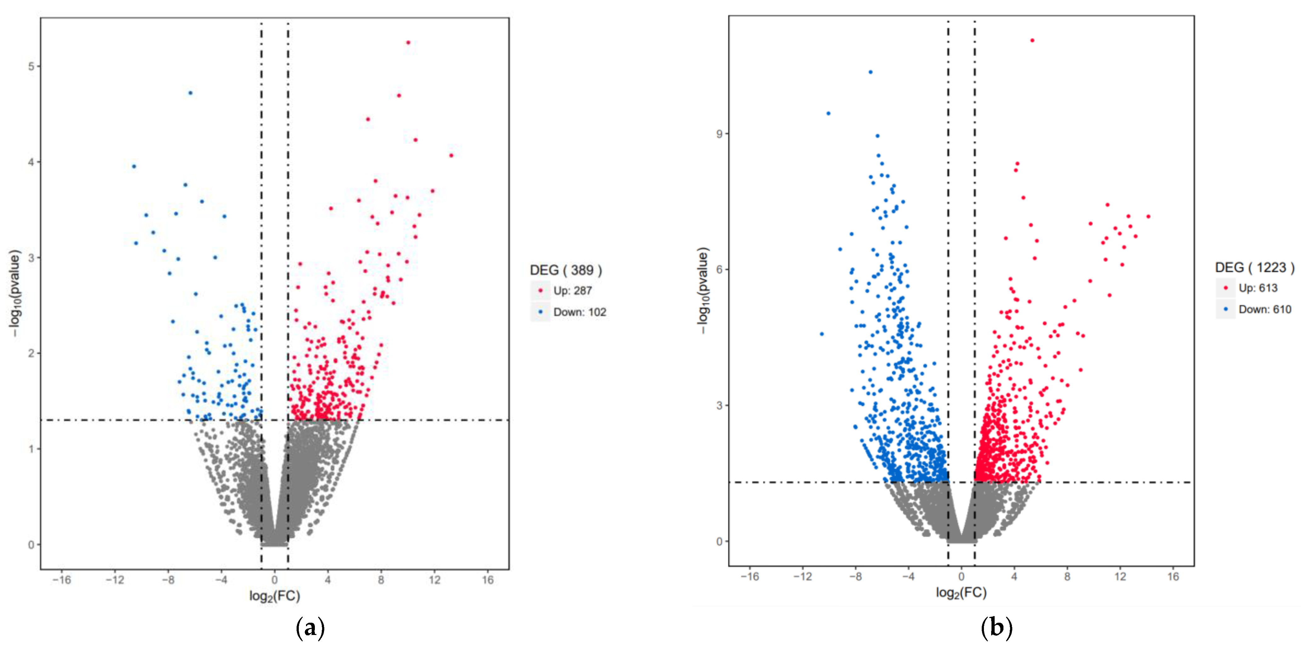
3.4. Differential Expression Genes (DEGs) Analysis

In total, 389 significant differentially expressed genes (DEGs) were identified the between LS_O and CG_O groups. Compared to the CG_O group, 287 up-regulated genes and 102 down-regulated genes were expressed in the LS_O group (Figure 3a). The same experimental method was applied to eyestalk transcriptome analysis. Compared to the CG_E group, 1,223 significant DEGs (613 up-regulated genes and 610 down-regulated genes) were identified in the LS_E group (Figure 3b).

3.5. Gene Ontology (GO) Analysis of Significant DEGs

The DEGs comparison of LS_O and CG_O were classified into biological process, cellular component, and molecular function. Compared with CG_O group, up-regulated genes in the LS_O group had enriched larval chitin-based cuticle development, ecdysteroid biosynthetic process, ecdysteroid metabolic process, sterol transport and so on. Conversely, the down-regulated genes in the LS_O group had enriched negative regulation of neurological system process, desensitization of G protein coupled receptor, negative regulation of G protein coupled receptor signaling pathway and so on (Table 1).
The DEGs comparison of LS_E and CG_E were classified into biological process, cellular component and molecular function. Compared with CG_E group, up-regulated genes in the LS_E group had enriched regulation of ovulation, embryonic liver development, luteinization, positive regulation of ovulation and so on. The down-regulated genes in the LS_E group had an enriched molting cycle, response to estrogen, molting cycle process, sterol homeostasis and so on (Table 2).

3.6. Kyoto Encyclopedia of Genes and Genomes (KEGG) Analysis

By matching with the KEGG pathway database, the possible functions of significant DEGs were analyzed to further understand the ovarian development of E. carinicauda under low salinity stress.
For low salinity stress, 27 KEGG pathways (19 up-regulated pathways and 8 down-regulated pathways) were significantly enriched in the LS_O group (Figure 4). Among these KEGG pathways, some were associated with ovarian development, such as ECM-receptor interaction, folate biosynthesis, Arginine biosynthesis and the FoxO signaling, lysosome and metabolic pathways.
In the comparison of the LS_E and CG_E group, 37 KEGG pathways (22 up-regulated and 15 down-regulated pathways) were significantly enriched (Figure 5). Concurrently, the insect hormone biosynthesis, phototransduction, ECM-receptor interaction, folate biosynthesis, lysosome and metabolic pathways, arginine biosynthesis and retinol metabolism were associated with ovarian development.

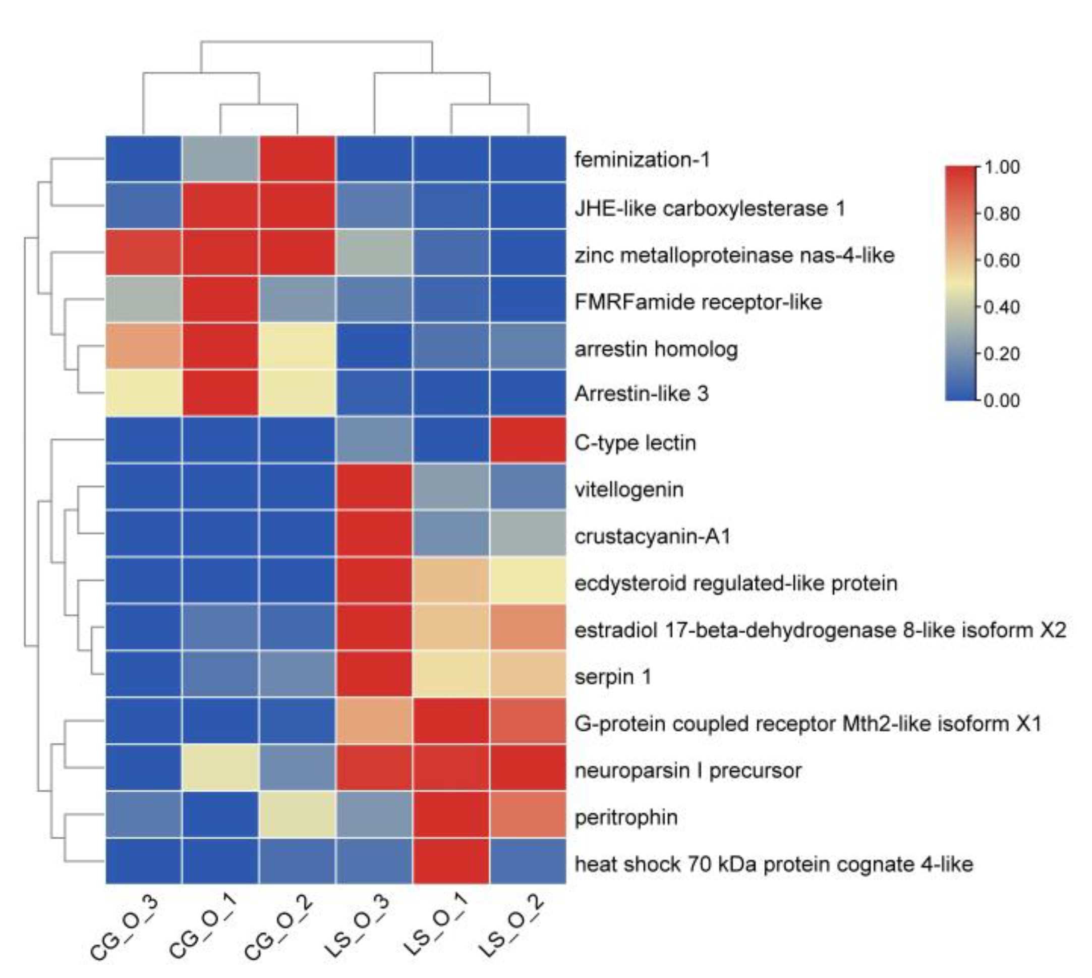
3.7. DEGs Involved in Ovarian Development

In the ovary, some DEGs related to ovarian development were selected in the low salinity group, such as the FMRF amide receptor, feminization 1, JHE-like carboxylesterase 1, heat shock 70 kDa protein, G protein-coupled receptor, C-type lectin, ecdysteroid regulated-like protein, estradiol 17-beta-dehydrogenase 8 and vitellogenin. A clustering heatmap of these results is provided (Figure 6).
In the eyestalk, some DEGs related to ovarian development were selected low salinity stress, such as the vitelline membrane outer layer protein 1, estradiol 17-beta-dehydrogenase, pigment dispersing hormone 1, insulin-like growth factor binding protein, insulin receptor-related protein, heat shock 70 kDa protein, neuronal acetylcholine receptor, neuroligin 2 and JHE-like carboxylesterase 1. A clustering heatmap of these results is provided (Figure 7).

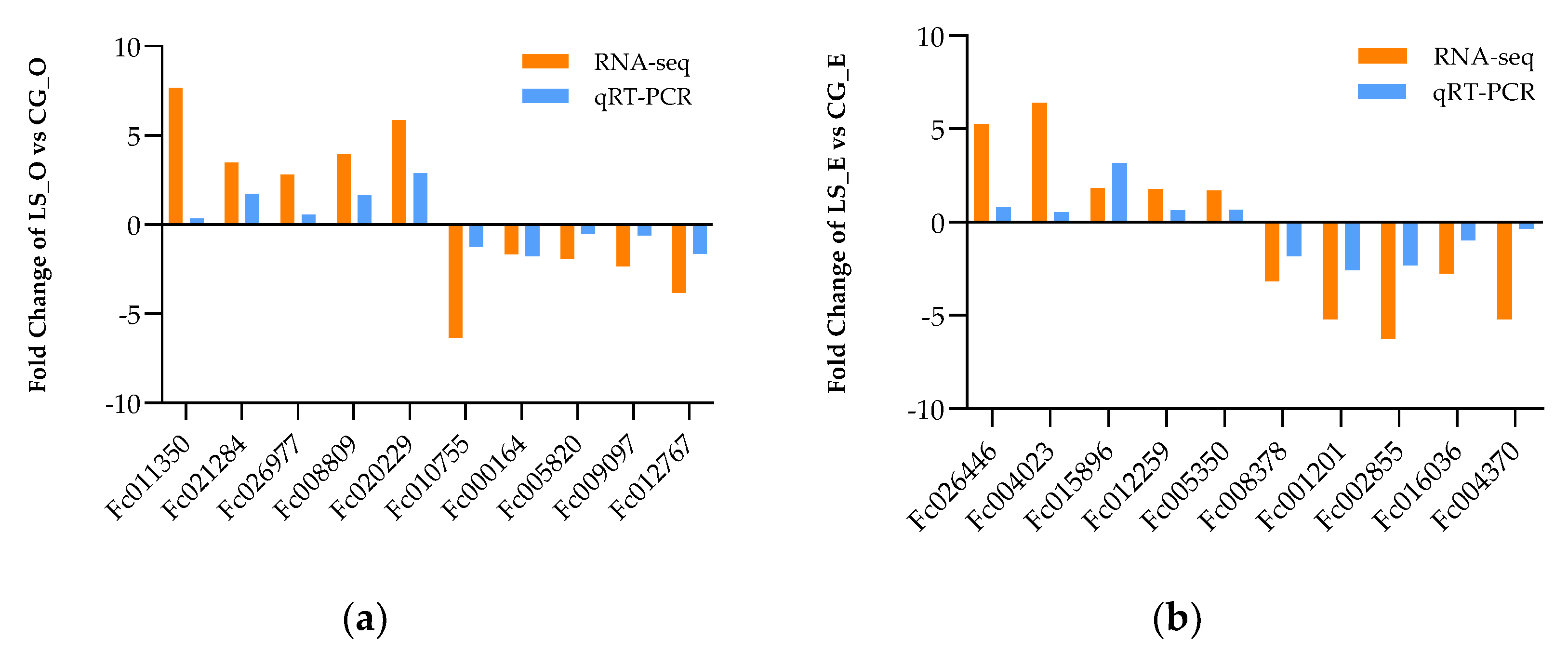
3.8. The Validation of Differently Expressional Genes by qRT-PCR

To further validate the reliability of DEGs identified by RNA-Seq, ten DEGs were randomly selected from two comparisons, LS_O vs. CG_O, LS_E vs. CG_E, respectively. The results of qRT-PCR were consistent with RNA-seq, indicating that the RNA-Seq data were accurate. (Figure 8).

4. Discussion

Na+/K+-ATPase and carbonic anhydrase are key enzymes for maintaining osmotic regulation in crustaceans [32,33]. For a euryhaline aquatic animal, in the adaptation process of L. vannamei from low salinity mutation to high salinity, Na+/K+-ATPase plays a leading role and carbonic anhydrase plays a supporting role [34]. In the process of gradually decreasing salinity, the Trachidermus fasciatus can maintain a stable physiological level, and the branchial Na+/K+-ATPase presents no significant difference [35]. In this study, we found that the Na+/K+-ATPase activity of LS group was significantly lower than that of the CG group only in muscle, and the carbonic anhydrase activity of LS group was significantly higher than that of the CG group only in the gill, and there were no significant change in other tissues. Furthermore, in the early stress process, we found that salinity had no lethal effect on the growth of E. carinicauda, similar to the study of Ren et al. [19], which confirmed that the E. carinicauda can cope with low salinity stress through osmotic adjustment. In addition, histological sections give further results; we found that there was no substantial damage to the ovary of the E. carinicauda, but the number of follicular cells was relatively small. The occurrence of crustacean eggs is closely related to follicular cells. Many scholars generally believe that follicular cells play an important role in the accumulation of exogenous yolk substances in oocytes. Li et al. [36] found that both follicular cells and egg cells were differentiated from the reproductive epithelium in the center of the ovary by observing the ovaries of E. carinicauda at different stages. The follicular cells differentiated faster and moved between the egg cells to provide nutrition for the development of the egg cells. When the egg cells developed to the endogenous yolk synthesis stage, the follicular cells began to wrap the egg cells to form a follicular cavity, which could better provide nutrition for the large volume of egg cells and further verified our results.
Ovarian development is a very important physiological process for animal reproduction [37] which requires the regulation of large sets of genes to ensure proper oocyte development. The ovary and eyestalk, as important organs for the development of crustacean ovaries, expressed a large number of key genes possibly involved in ovarian development in the transcriptome sequencing data of the E. carinicauda in response to ground salt stress. Although the mechanism of eyestalk ablation leading to ovarian maturation is still inconclusive, some genes expressed in the eyestalk regulate ovarian development.
In this study, a large number of key genes possibly involved in ovarian development were detected in the transcriptome sequencing data of ovarian tissue. Vitellogenin is an important precursor of egg yolk in nearly all oviparous animals’ it provides carbohydrates, lipids, amino acids, vitamins, phosphorus, sulfur, various metal ions and other nutritional and functional substances for the development of embryos and larvae [38,39]. In this study, low salinity stress upregulates vitellogenin expression, which seems to be consistent with our previous discovery that the E. carinicauda can develop in low salinity seawater [21]. As an important sex steroid hormone, estradiol is widely distributed in the hepatopancreas, ovary, hemolymph and other tissues, and participates in the regulation of ovarian development in crustaceans [40]. It is reported that 17β-estradiol induces Vg synthesis and oocyte development in immature shrimp ovaries [41,42]. In this study, we found that 17β-estradiol expressed significantly under low salinity stress. Therefore, we speculate that 17β-estradiol may have a similar function in E. carinicauda, but its specific role needs further study. In addition, another DEG, heat shock proteins, ubiquitously distribute and highly conserve, behaving as molecular chaperones and involved in protein folding, degradation and transport. In addition, expression level is regulated by sex steroid hormones [43,44]. It has been proved that heat shock protein 70 is involved in estrogen nuclear initiated steroid signal transduction [41]. Chan et al. [45,46] confirmed that heat shock cognate 70 negatively regulated the expression of vitellogenin. Furthermore, Liu et al. found that the decreased expression of Hsp70 may be related to the promotion of vitellogenesis by estradiol [47], which is similar to our findings. In this study, the differential expression of the above genes in the ovarian development of white shrimps under low salinity stress indicates that vitellogenin and other regulatory genes are indeed involved in the ovarian development of E. carinicauda.
Methyl farnesate (MF) secreted by the mandibular organs of crustaceans has a significant stimulating effect on Vg synthesis in various decapods [48,49,50]. Although the methyl farnesate biosynthetic pathway has been well established in arthropods [51], little is known about its degradation in crustaceans. Juvenile hormone esterase (JHE) is a key enzyme for insects, playing an important role in the regulation of insect growth, development, diapause and reproduction [5]. In insects, juvenile hormone esterase is a carboxylesterase, which specially degrade JHs by converting JH to JH acid (JHa) and catalyzing the conversion of JH diol (JHd) into JH acid diol (JHad) [52]. The expression levels of JHE-like carboxylesterase 1, JHE like carboxylesterase 2 and juvenile hormone esterase like protein in the hepatopancreas and brain ganglion of P. trituberculatus were up-regulated, which promoted the inactivation of methyl farnesate in the two tissues [47]. In addition, methyl farnesate is metabolized to farnesic acid in vitro through esterase existing in crustacean tissues [53]. In this study, JHE-like carboxylesterase 1 down-regulation can negatively regulate methyl farnesate, thus promoting ovarian development under low salinity stress.
Crustacean ovarian development is regulated by various hormone factors [54]. The crustacean eyestalk is known to regulate reproduction, molting and energy metabolism [25,55]. Eyestalk-derived neuropeptides regulate vitellogenesis in crustaceans. The pigment-dispersing hormone (PDH) participates in the regulation of ovarian maturation in crustaceans [56]. The PDH may participate in vitellogenesis according to their spatiotemporal expression patterns which maintained a high level from the pre vitellogenesis stage and decreased significantly in the mature stage in Scylla paramamosain [57,58]. The study of Wei et al., 2021 provided the evidence for the inductive effect of PDH on the oocyte meiotic maturation in E. sinensis [59]. In this study, the PDH was up-regulated in the LS_E group, suggesting that it may be involved in the ovarian development of E. carinicauda under low salinity stress.
Retinol and its derivatives play key roles in the initiating meiosis in germ cells of mammalian fetal ovaries [60], follicular development [61], ovarian steroidogenesis [62], and oocyte maturation [63]. The retinol dehydrogenase (RDH), as a member of the short-chain dehydrogenase/reductase (SDR) superfamily, which includes three RDHs, RDH11, RDH12 and RDH13, were in the transcriptome sequences. RDH13 shows significantly higher expression levels in vitellogenic ovaries than in a non-vitellogenic ovaries in zebrafish [64]. Knockdown of RDH11 resulted in decreased transcription of vitellogenin and vitellogenin receptor in Procambarus clarkia [37], which suggested that RDH11 might have a function in the synthesis and conveyance of vitellogenin in crustacean. Our previous study also revealed that RDH11 is critical for ovarian development in E. carinicauda [65]. In this study, RDH13 which was related to ovarian development in E. carinicauda was significantly expressed in response to unfavorable environmental stress.
KEGG pathway enrichment can identify the main biochemical metabolic pathways and signal transduction pathways involved in genes. Lysosome was the most significant pathway and contained the larger number of DEGs between the LS_O and CG_O groups, and also between LS_E and CG_E groups. The lysosome is important for intracellular trafficking, metabolic signaling, lipid metabolism and immune response [66]. Lysosomes are implicated in the preparation of free cholesterol for steroidogenesis and degradation of regulators of steroidogenesis and follicle rupture during ovulation in the ovary of vertebrates [67]. As the central digestive organ of cells, various macromolecules are sent to the lysosome for degradation. Vitellogenin is an important precursor of egg yolk in nearly all oviparous animals [39]. Lysosomes play a key role in the degradation of the vitellogenin internalized by endocytosis [38]. The lysosomes are related to the hydrolysis of vitellogenin and energy demand during Macrobrachium nipponense ovarian maturation [68]. The lysosomal enzymes, especially cathepsin B and L, are associated with ovarian development in crustaceans [69,70]. However, the DEGs enriched by the lysosomal pathway in different tissues are different. The Lysosomal pathway was significantly up-regulated in the ovaries and significantly down−regulated in the eyestalks. Therefore, we speculated that this change in lysosomes is closely related to the ovarian development of E. carinicauda under carbonate alkalinity stress. However, the specific mechanism is unknown.
As an essential amino acid of aquatic animals, arginine plays an important physiological role in growth and development. Arginine is decomposed into nitric oxide under the action of nitric oxide synthase (NOS). It has been proved that nitric oxide, the metabolite of arginine, can promote the secretion of GnRH [71], luteinizing hormone releasing hormone (LHRH) [72] and regulate the secretion of gonadotropins by the pituitary. It is reported that intraperitoneal injection of L-Arg, the precursor of NO, significantly increased the content of follicle stimulating hormone (FSH) and luteinizing hormone (LH) in rat serum. In shrimp and crab, preliminary studies have found that NO can activate dependent protein kinase by regulating the second messenger cGMP, and then affect the expression of molt inhibiting hormone (MIH) [73], while MH can promote the synthesis of vitellogenin [74]. Arginine is the only NO donor in the animal body. Therefore, arginine plays an important role in regulating the secretion of pituitary hormones. As the only NO donor in animals, it plays an irreplaceable role in ovarian development. In this study, we found that the arginine pathway was significantly up-regulated in the ovary, and also in the eyestalk, which seemed to indicate that arginine and its product NO played a very important role in the ovarian development of the E. carinicauda in response to low salinity stress. However, the specific mechanism of the effect remains to be studied.

5. Conclusions

In the present study, we studied the ovarian development of E. carinicauda under low salinity stress for the first time. Biochemical indicators confirmed that white shrimps can adapt to low salinity stress by regulating osmotic regulation. Ovarian tissue sections provided us with preliminary results of ovarian development. Transcriptome analysis of ovaries and eyestalks was used to study the effects of low salinity on genes and signal pathways related to ovarian development. 16 and 18 DEGs were identified in ovary and eyestalk tissues, respectively. These key genes were identified as participating in folate biosynthesis, insect hormone biosynthesis, lysosome and retinol metabolism, which play important roles in the response of the ovaries and eyestalks of E. carinicauda to low salinity stress. This study provides new insights into the ovarian development of E. carinicauda under low salinity stress, which could be useful for non-marine aquaculture and related studies on the reproduction of crustaceans.

Supplementary Materials

The following supporting information can be downloaded at: https://www.mdpi.com/article/10.3390/fishes7060365/s1, Table S1: Primers of qRT-PCR designed for validation experiment of DEGs; Table S2: Number of high-throughput clean reads and mapped clean reads generated from E. carinicauda ovary and eyestalk mRNA library.

Author Contributions

X.Z., J.W. and J.L. (Jitao Li) conceived and designed the research; X.Z., J.W., C.W., W.L., Q.G. and Z.Q. performed the experiments and analyzed the data; J.L (Jian Li). provided research ideas for the experiments. All authors were involved in drafting and revising the manuscript. All authors have read and agreed to the published version of the manuscript.

Funding

This research was funded by National Key R & D Program of China (2018YFD0901302), National Natural Science Foundation of China (32072974), China Agriculture Research System of MOF and MARA (CARS-48) and Central Public-Interest Scientific Institution Basal Research Fund, CAFS (2020TD46).

Institutional Review Board Statement

The animal study protocol was approved by the Institutional Animal Care and Use Committee of Yellow Sea Research Institute, Chinese Academy of Fishery Sciences (approval code YSFRI-20220332, approved on 4 November 2022).

Data Availability Statement

The datasets presented in this study can be found in online repositories. The names of the repository/repositories and accession number(s) can be found below: Center for Biotechnology Information (NCBI) with the accession numbers PRJNA881755 and PRJNA881756.

Conflicts of Interest

The authors declare that they have no known competing financial interests or personal relationships that could have appeared to influence the work reported in this paper.

References

  1. McFarland, K.; Donaghy, L.; Volety, A. Effect of acute salinity changes on hemolymph osmolality and clearance rate of the non-native mussel, Perna viridis, and the native oyster, Crassostrea virginica, in Southwest Florida. Aquat. Invasions 2013, 8, 299–310. [Google Scholar]
  2. Aguilar, C.; Raina, J.B.; Fôret, S.; Hayward, D.C.; Lapeyre, B.; Bourne, D.G.; Miller, D.J. Transcriptomic analysis reveals protein homeostasis breakdown in the coral Acropora millepora during hypo-saline stress. BMC Genom. 2019, 20, 148. [Google Scholar]
  3. Yang, W.K.; Chung, C.H.; Cheng, H.C.; Tang, C.H.; Lee, T.H. Different expression patterns of renal Na+/K+-ATPase α-isoform-like proteins between tilapia and milkfish following salinity challenges. Comp. Biochem. Physiol. B. Biochem. Mol. Biol. 2016, 202, 23–30. [Google Scholar]
  4. Wang, H.; Tang, L.; Wei, H.L.; Lu, J.K.; Mu, C.K.; Wang, C.L. Transcriptomic analysis of adaptive mechanisms in response to sudden salinity drop in the mud crab, Scylla paramamosain. BMC Genom. 2018, 19, 421. [Google Scholar]
  5. Huang, C.; Wu, Q.; Jiang, C.Y.; Xing, L.S.; Shi, G.L.; Zhang, B.; Qian, W.Q.; Li, Y.Z.; Xi, Y.; Yang, N.W.; et al. Identification and developmental expression of putative gene encoding juvenile hormone esterase (CpJHE-like) in codling moth, Cydia pomonella (L.). J. Integr. Agric. 2019, 18, 1624–1633. [Google Scholar]
  6. Mohanty, B.; Gupta, K.; Babu, G.; Pal, H.; Verma, P. Exposure to salinity stress cause ovarian disruption in a stenohaline freshwater teleost, Heteropneustes fossilis (Bloch, 1794). Aquac. Res. 2020, 51, 1964–1972. [Google Scholar]
  7. Mandal, B.; Sawant, P.B.; Dasgupta, S.; Chadha, N.K.; Sundaray, J.K.; Sawant, B.T.; Bera, A. Deviation of habitat salinity during seasonal gonad recrudescence affects plasma sex steroid levels and suppresses gonadal maturation in an euryhaline fish Etroplus suratensis. Aquac. Res. 2017, 48, 5973–5983. [Google Scholar]
  8. Kaeodee, M.; Pongsomboon, S.; Tassanakajon, A. Expression analysis and response of Penaeus monodon 14-3-3 genes to salinity stress. Comp. Biochem. Physiol. B Biochem. Mol. Biol. 2011, 159, 244–251. [Google Scholar]
  9. Romano, N.; Wu, X.G.; Zeng, C.S.; Genodepa, J.; Elliman, J. Growth, osmoregulatory responses and changes to the lipid and fatty acid composition of organs from the mud crab, Scylla serrata, over a broad salinity range. Mar. Biol. Res. 2014, 10, 460–471. [Google Scholar]
  10. Lu, S.K.; Li, R.H.; Zheng, W.B.; Chen, L.; Ren, Z.M.; Mu, C.K.; Song, W.W.; Wang, C.L. Long-term low-salinity stress affects growth, survival and osmotic related gamma-aminobutyric acid regulation in the swimming crab Portunus trituberculatus. Aquac. Res. 2019, 50, 2888–2895. [Google Scholar]
  11. Hu, D.X.; Pan, L.Q.; Zhao, Q.; Ren, Q. Transcriptomic response to low salinity stress in gills of the Pacific white shrimp, Litopenaeus vannamei. Mar. Genom. 2015, 24, 297–304. [Google Scholar]
  12. Long, X.W.; Wu, X.G.; Lei, Z.; Ye, H.H.; Cheng, Y.X.; Zeng, C.S. Physiological responses and ovarian development of female Chinese mitten crab Eriocheir sinensis subjected to different salinity conditions. Front. Physiol. 2017, 8, 1072. [Google Scholar]
  13. Feng, Y.Y.; Zhai, Q.Q.; Wang, J.J.; Li, J.T.; Li, J. Comparison of florfenicol pharmacokinetics in Exopalaemon carinicauda at different temperatures and administration routes. J. Vet. Pharmacol. Ther. 2019, 42, 230–238. [Google Scholar]
  14. Ge, Q.Q.; Li, J.; Wang, J.J.; Li, Z.D.; Li, J.T. Characterization, functional analysis, and expression levels of three carbonic anhydrases in response to pH and saline-alkaline stresses in the ridgetail white prawn Exopalaemon carinicauda. Cell Stress Chaperon. 2019, 24, 503–515. [Google Scholar]
  15. Qin, Z.; Ge, Q.Q.; Wang, J.J.; Li, M.D.; Liu, P.; Li, J.; Li, J.T. Comparative transcriptomic and proteomic analysis of Exopalaemon carinicauda in response to alkalinity stress. Front. Mar. Sci. 2021, 8, 1523. [Google Scholar]
  16. Xu, W.J.; Xie, J.J.; Shi, H.; Li, C.W. Hematodinium infections in cultured ridgetail white prawns, Exopalaemon carinicauda, in eastern China. Aquaculture 2010, 300, 25–31. [Google Scholar]
  17. Li, J.T.; Ma, P.; Liu, P.; Chen, P.; Li, J. The roles of Na+/K+-ATPase alpha-subunit gene from the ridgetail white prawn Exopalaemon carinicauda in response to salinity stresses. Fish Shellfish Immunol. 2015, 42, 264–271. [Google Scholar]
  18. Zhang, C.S.; Su, F.; Li, S.H.; Yu, Y.; Xiang, J.H.; Liu, J.G.; Li, F.H. Isolation and identification of the main carotenoid pigment from a new variety of the ridgetail white prawn Exopalaemon carinicauda. Food Chem. 2018, 269, 450–454. [Google Scholar]
  19. Ren, H.; Li, J.; Li, J.T.; Ying, Y.; Ge, H.X.; Li, D.L.; Yu, T.J. Cloning of catalase and expression patterns of catalase and selenium-dependent glutathione peroxidase from Exopalaemon carinicauda in response to low salinity stress. Acta Oceanol. Sin. 2015, 34, 52–61. [Google Scholar]
  20. Chang, Z.Q.; Neori, A.; He, Y.Y.; Li, J.T.; Qiao, L.; Preston, S.I.; Liu, P.; Li, J. Development and current state of seawater shrimp farming, with an emphasis on integrated multi-trophic pond aquaculture farms, in China-a review. Rev. Aquac. 2020, 12, 2544–2558. [Google Scholar]
  21. Liang, J.P.; Li, J.; Li, J.T.; Liu, P.; Zhao, F.Z.; Nie, G.X.; Li, X.J.; Kong, X.H. Effects of salinity on spawning and larval development of Exopalaemon carinicauda. Chin. J. Appl. Ecol. 2014, 25, 2105–2113. (In Chinese) [Google Scholar]
  22. Teets, N.M.; Peyton, J.T.; Colinet, H.; Renault, D.; Denlinger, D.L. Gene expression changes governing extreme dehydration tolerance in an Antarctic insect. Proc. Natl. Acad. Sci. USA 2013, 109, 20744–20749. [Google Scholar]
  23. Chen, K.; Li, E.C.; Li, T.Y.; Xu, C.; Wang, X.D.; Lin, H.Z.; Qin, J.G.; Chen, L.Q. Transcriptome and molecular pathway analysis of the hepatopancreas in the pacific white shrimp Litopenaeus vannamei under chronic low-salinity stress. PLoS ONE 2015, 10, e131505. [Google Scholar]
  24. Li, E.; Wang, S.; Li, C.; Wang, X.; Chen, K.; Chen, L. Transcriptome sequencing revealed the genes and pathways involved in salinity stress of Chinese mitten crab, Eriocheir sinensis. Physiol. Genom. 2014, 46, 177–190. [Google Scholar]
  25. Brady, P.; Elizur, A.; Williams, R.; Cummins, S.F.; Knibb, W. Gene expression profiling of the cephalothorax and eyestalk in Penaeus Monodon during ovarian maturation. Int. J. Biol. Sci. 2012, 8, 328–343. [Google Scholar]
  26. Qiao, H.; Xiong, Y.W.; Zhang, W.Y.; Fu, H.T.; Jiang, S.F.; Sun, S.M.; Bai, H.K.; Jin, S.B.; Gong, Y.S. Characterization, expression, and function analysis of gonad-inhibiting hormone in oriental river prawn, Macrobrachium nipponense and its induced expression by temperature. Comp. Biochem. Physiol. A-Mol. Integr. Physiol. 2015, 185, 1–8. [Google Scholar]
  27. Koshio, S.; Teshima, S.I.; Kanazawa, A. Effects of Unilateral Eyestalk Ablation and Feeding Frequencies on Growth, Survival, and Body Compositions of Juvenile Freshwater Prawn Macrobrachium rosenbergii. Nippon Suisan Gakkaishi 1992, 58, 1419–1425. [Google Scholar]
  28. Uawisetwathana, U.; Leelatanawit, R.; Klanchui, A.; Prommoon, J.; Klinbunga, S.; Karoonuthaisiri, N. Insights into eyestalk ablation mechanism to induce ovarian maturation in the black tiger shrimp. PLoS ONE 2011, 6, e24427. [Google Scholar]
  29. Robinson, M.D.; Mccarthy, D.J.; Smyth, G.K. edgeR: A Bioconductor package for differential expression analysis of digital gene expression data. Biogeosciences 2010, 26, 139–140. [Google Scholar]
  30. Bu, D.H.; Luo, H.T.; Huo, P.P.; Wang, Z.H.; Zhang, S.; He, Z.H.; Wu, Y.; Zhao, L.H.; Liu, J.J.; Guo, J.C.; et al. KOBAS-i: Intelligent prioritization and exploratory visualization of biological functions for gene enrichment analysis. Nucleic Acids Res. 2021, 49, 317–325. [Google Scholar]
  31. Duan, Y.F.; Liu, P.; Li, J.T.; Li, J.; Gao, B.Q.; Chen, P. cDNA cloning, characterization and expression analysis of peroxiredoxin 5 gene in the ridgetail white prawn Exopalaemon carinicauda. Mol. Biol. Rep. 2013, 40, 6569–6577. [Google Scholar]
  32. Geers, C.; Gros, G. Carbon dioxide transport and carbonic anhydrase in blood and muscle. Physiol. Rev. 2000, 80, 681–715. [Google Scholar]
  33. Lucu, C.; Towle, D.W. Na++K+-ATPase in gills of aquatic crustacea. Comp. Biochem. Physiol. A-Mol. Integr. Physiol. 2003, 135, 195–214. [Google Scholar]
  34. Henry, R.P.; Gehnrich, S.; Weihrauch, D.; Towle, D.W. Salinity-mediated carbonic anhydrase induction in the gills of the euryhaline green crab, Carcinus maenas. Comp. Biochem. Physiol. A-Mol. Integr. Physiol. 2003, 136, 243–258. [Google Scholar]
  35. Ma, Q.; Kuang, J.; Liu, X.; Li, A.; Feng, W.; Zhuang, Z. Effects of osmotic stress on Na+/K+-ATPase, caspase 3/7 activity, and the expression profiling of sirt1, hsf1, and hsp70 in the roughskin sculpin (Trachidermus fasciatus). Fish Physiol. Biochem. 2020, 46, 135–144. [Google Scholar]
  36. Li, Z.G.; Zhang, C.S.; Li, F.H.; Xiang, J.H. Histological study on the gonadal development of Exopalaemon carinicauda (Holthuis, 1950). J. Fish. China 2014, 38, 362–370. (In Chinese) [Google Scholar]
  37. Kang, P.F.; Mao, B.; Fan, C.; Wang, Y.F. Transcriptomic information from the ovaries of red swamp crayfish (Procambarus clarkii) provides new insights into development of ovaries and embryos. Aquaculture 2019, 505, 333–343. [Google Scholar]
  38. Carnevali, O.; Cionna, C.; Tosti, L.; Lubzens, E.; Maradonna, F. Role of cathepsins in ovarian follicle growth and maturation. Gen. Comp. Endocrinol. 2006, 146, 195–203. [Google Scholar]
  39. Harwood, G.; Amdam, G. Vitellogenin in the honey bee midgut. Apidologie 2021, 52, 837–847. [Google Scholar]
  40. Rodriguez, E.M.; Medesani, D.A.; Greco, L.; Fingerman, M. Effects of some steroids and other compounds on ovarian growth of the red swamp crayfish, Procambarus clarkii, during early vitellogenesis. J. Exp. Zool. 2002, 292, 82–87. [Google Scholar]
  41. Quackenbush, L.S. Yolk synthesis in the marine shrimp, Penaeus vannamei. Comp. Biochem. Phys. A 1992, 103, 711–714. [Google Scholar]
  42. Yano, I.; Hoshino, R. Effects of 17 β-estradiol on the vitellogenin synthesis and oocyte development in the ovary of kuruma prawn (Marsupenaeus japonicus). Comp. Biochem. Phys. A 2006, 144, 18–23. [Google Scholar]
  43. Mayer, M.P.; Bukau, B. Hsp70 chaperones: Cellular functions and molecular mechanism. Cell. Mol. Life Sci. 2005, 62, 670–684. [Google Scholar]
  44. Romani, W.A.; Russ, D.W. Acute effects of sex-specific sex hormones on heat shock proteins in fast muscle of male and female rats. Eur. J. Appl. Physiol. 2013, 113, 2503–2510. [Google Scholar]
  45. Chan, S.F.; He, J.G.; Chu, K.H.; Sun, C.B. The shrimp heat shock cognate 70 functions as a negative regulator in vitellogenin gene expression. Biol. Reprod. 2014, 91, 1–11. [Google Scholar]
  46. Dhamad, A.E.; Zhou, Z.Q.; Zhou, J.H.; Du, Y.C. Systematic proteomic identification of the heat shock proteins (Hsp) that interact with estrogen receptor alpha (ER alpha) and biochemical characterization of the ER alpha-Hsp70 Interaction. PLoS ONE 2016, 11, e160312. [Google Scholar]
  47. Liu, M.; Pan, J.; Dong, Z.; Cheng, Y.; Gong, X. Comparative transcriptome reveals the potential modulation mechanisms of estradiol affecting ovarian development of female Portunus trituberculatus. PLoS ONE 2019, 14, e226698. [Google Scholar]
  48. Mak, A.S.C.; Choi, C.L.; Tiu, S.H.K.; Hui, J.H.L.; He, J.G.; Tobe, S.S.; Chan, S.M. Vitellogenesis in the red crab Charybdis feriatus: Hepatopancreas-specific expression and farnesoic acid stimulation of vitellogenin gene expression. Mol. Reprod. Dev. 2005, 70, 288–300. [Google Scholar]
  49. Yang, Y.; Ye, H.H.; Huang, H.Y.; Jin, Z.X.; Li, S.J. Cloning, expression and functional analysis of farnesoic acid O-methyltransferase (FAMeT) in the mud crab, Scylla paramamosain. Mar. Freshw. Behav. Phy. 2012, 45, 209–222. [Google Scholar]
  50. Miyakawa, H.; Toyota, K.; Sumiya, E.; Iguchi, T. Comparison of JH signaling in insects and crustaceans. Curr. Opin. Insect Sci. 2014, 1, 81–87. [Google Scholar]
  51. Borst, D.W.; Ogan, J.; Tsukimura, B.; Claerhout, T.; Holford, K.C. Regulation of the crustacean mandibular organ. Am. Zool. 2001, 41, 430–441. [Google Scholar]
  52. Liu, S.H.; Yangb, B.J.; Go, J.H.; Yao, X.M.; Zhang, Y.X.; Song, F.; Liu, Z.W. Molecular cloning and characterization of a juvenile hormone esterase gene from brown planthopper, Nilaparvata lugens. J. Insect Physiol. 2008, 54, 1495–1502. [Google Scholar]
  53. Tobe, S.S.; Young, D.A.; Khoo, H.W.; Baker, F.C. Farnesoic acid as a major product of release from crustacean mandibular organs In vitro. J. Exp. Zool. Part A 1989, 249, 165–171. [Google Scholar]
  54. Nagaraju, G. Reproductive regulators in decapod crustaceans: An overview. J. Exp. Biol. 2011, 214, 3–16. [Google Scholar]
  55. Jung, H.T.; Lyons, R.E.; Hurwood, D.A.; Mather, P. Genes and growth performance in crustacean species: A review of relevant genomic studies in crustaceans and other taxa. Rev. Aquacult. 2013, 5, 77–110. [Google Scholar]
  56. Rotllant, G.; Nguyen, T.V.; Aizen, J.; Suwansa-ard, S.; Ventura, T. Toward the identification of female gonad-stimulating factors in crustaceans. Hydrobiologia 2018, 825, 91–119. [Google Scholar]
  57. Huang, X.; Ye, H.; Huang, H.; Yu, K.; Huang, Y. Two beta-pigment-dispersing hormone (β-PDH) isoforms in the mud crab, Scylla paramamosain: Implication for regulation of ovarian maturation and a photoperiod-related daily rhythmicity. Anim. Reprod. Sci. 2014, 150, 139–147. [Google Scholar]
  58. Bao, C.; Yang, Y.; Huang, H.; Ye, H. Neuropeptides in the cerebral ganglia of the mud crab, Scylla paramamosain: Transcriptomic analysis and expression profiles during vitellogenesis. Sci. Rep. 2015, 5, 17055. [Google Scholar]
  59. Wei, L.L.; Chen, T.T.; Luo, B.Y.; Qiu, G.F. Evidences for red pigment concentrating hormone (RPCH) and beta-pigment dispersing hormone (beta-PDH) inducing oocyte meiotic maturation in the Chinese mitten crab, Eriocheir sinensis. Front. Endocrinol. 2021, 12, 802768. [Google Scholar]
  60. Jiang, Y.W.; Li, C.J.; Chen, L.; Wang, F.G.; Zhou, X. Potential role of retinoids in ovarian physiology and pathogenesis of polycystic ovary syndrome. Clin. Chim. Acta 2017, 469, 87–93. [Google Scholar]
  61. Kawai, T.; Yanaka, N.; Richards, J.S.; Shimada, M. De novo-synthesized retinoic acid in ovarian antral follicles enhances FSH-mediated ovarian follicular cell differentiation and female fertility. Endocrinology 2016, 157, 2160–2172. [Google Scholar]
  62. Damdimopoulou, P.; Chiang, C.; Flaws, J.A. Retinoic acid signaling in ovarian folliculogenesis and steroidogenesis. Reprod. Toxicol. 2019, 87, 32–41. [Google Scholar]
  63. Tahaei, L.S.; Eimani, H.; Yazdi, P.E.; Ebrahimi, B.; Fathi, R. Effects of retinoic acid on maturation of immature mouse oocytes in the presence and absence of a granulosa cell co-culture system. J. Assist. Reprod. Genet. 2011, 28, 553–558. [Google Scholar]
  64. Levi, L.; Ziv, T.; Admon, A.; Levavi-Sivan, B.; Lubzens, E. Insight into molecular pathways of retinal metabolism, associated with vitellogenesis in zebrafish. Am. J. Physiol.-Endocrinol. Metab. 2012, 302, 626–644. [Google Scholar]
  65. Wang, J.J.; Li, J.T.; Ge, Q.Q.; Li, W.Y.; Li, J. Full-Length transcriptome sequencing and comparative transcriptomic analysis provide insights into the ovarian maturation of Exopalaemon carinicauda. Front. Mar. Sci. 2022, 9, 906730. [Google Scholar]
  66. Lamming, D.W.; Bar-Peled, L. Lysosome: The metabolic signaling hub. Traffic 2019, 20, 27–38. [Google Scholar]
  67. Li, Y.; Wang, Z.; Andersen, C.L.; Ye, X. Functions of lysosomes in mammalian female reproductive system. Reprod. Dev. Med. 2020, 4, 109–122. [Google Scholar]
  68. Zhang, Y.N.; Jiang, S.F.; Qiao, H.; Xiong, Y.W.; Fu, H.T.; Zhang, W.Y.; Gong, Y.S.; Jin, S.B.; Wu, Y. Transcriptome analysis of five ovarian stages reveals gonad maturation in female updates Macrobrachium nipponense. BMC Genom. 2021, 22, 100868. [Google Scholar]
  69. Wu, P.; Qi, D.; Chen, L.Q.; Zhang, H.; Zhang, X.W.; Qin, J.G.; Hu, S.N. Gene discovery from an ovary cDNA library of oriental river prawn Macrobrachium nipponense by ESTs annotation. Comp. Biochem. Phys. D 2009, 4, 111–120. [Google Scholar]
  70. Luo, B.Y.; Qian, H.L.; Jiang, H.C.; Xiong, X.Y.; Ye, B.Q.; Liu, X.; Guo, Z.Q.; Ma, K.Y. Transcriptional changes revealed water acidification leads to the immune response and ovary maturation delay in the Chinese mitten crab Eriocheir sinensis. Comp. Biochem. Phys. D 2021, 39, 100863. [Google Scholar]
  71. Rettori, V.; McCann, S.M. Role of nitric oxide and alcohol on gonadotropin release in vitro and in vivo. Ann. N. Y. Acad. Sci. 1998, 840, 185–193. [Google Scholar]
  72. Lopez, F.J.; Moretto, M.; Merchenthaler, I.; Negro-Vilar, A. Nitric oxide is involved in the genesis of pulsatile LHRH secretion from immortalized LHRH neurons. J. Neuroendocrinol. 1997, 9, 647–654. [Google Scholar]
  73. Kim, H.W.; Batista, L.A.; Hoppes, J.L.; Lee, K.J.; Mykles, D.L. A crustacean nitric oxide synthase expressed in nerve ganglia, Y-organ, gill and gonad of the tropical land crab, Gecarcinus lateralis. J. Exp. Biol. 2004, 207, 2845–2857. [Google Scholar]
  74. Subramoniam, T. Mechanisms and control of vitellogenesis in crustaceans. Fish. Sci. 2011, 77, 1–21. [Google Scholar]
Figure 1. Enzyme activity in the different tissue of E. carinicauda in response to low salinity stress. (a) Carbonic anhydrase activity in the different tissue of E. carinicauda in response to low salinity stress; (b) Na+/K+-ATPase activity in the different tissue of E. carinicauda in response to low salinity stress. Different lowercase letters in the same tissue indicates that there is significant difference between groups (p > 0.05). Abbreviations: M, muscle; H, hepatopancreas; G, gill.
Fishes 07 00365 g001
Figure 2. Histological sections of the ovarian status of E. carinicauda: (a) CG_O group; (b) LS_O group. Bar: 50 µm. Abbreviations: N, Nucleus; Nu, nucleolus; Fc, follicular cell.
Fishes 07 00365 g002
Figure 3. The number of differentially expressed genes between different groups: (a) LS_O vs. CG_O group; (b) LS_E vs. CG_E group. Significantly up-regulated and down-regulated genes are indicated in red and blue, respectively, and those not significantly different are in gray.
Fishes 07 00365 g003
Figure 4. The KEGG significant enrichment pathway in LS_O vs. CG_O group. (a) Up-regulation pathways in LS_O vs. CG_O group.; (b) down-regulation pathways in LS_O vs. CG_O group.
Fishes 07 00365 g004
Figure 5. The KEGG significant enrichment pathway in LS_E vs. CG_E group. (a) Up-regulation pathways in LS_E vs. CG_E group; (b) down-regulation pathways in LS_E vs. CG_E group.
Fishes 07 00365 g005
Figure 6. The heatmap of DEGs related to ovarian development in the LS_O vs. CG_O group. The columns and rows indicate individuals and genes, respectively. The color scale represents FPKM after standard normalization, the same below.
Fishes 07 00365 g006
Figure 7. The heatmap of DEGs related to ovarian development in the LS_E vs. CG_E group.
Fishes 07 00365 g007
Figure 8. The results were verified by qRT-PCR. (a) Relative fold change of DEGs between qRT-PCR and RNA-seq results in LS_O vs. CG_O group; (b) relative fold change of DEGs between qRT-PCR and RNA-seq results in LS_E vs. CG_E group. Relative expression levels from the RNA-seq results were calculated as log2FC values: Fc011350 ecdysteroid regulated; Fc021284 legumain; Fc026977 lysosome-associated membrane glycoprotein 1; Fc008809 baculoviral IAP repeat-containing; Fc020229 N-acetylated-alpha-linked acidic dipeptidase; Fc010755 high-affinity choline transporter 1; Fc000164 tripartite motif-containing protein 3; Fc005820 reelin 3; Fc009097 arrestin; Fc012767 facilitated trehalose transporter; Fc026446 neroligin 2; Fc004023 alpha 1 inhibitor 3; Fc015896 insulin receptor-related; Fc012259 serpin 1; Fc005350 UDP-glucosyltransferase 2; Fc008378 crustacyanin C1; Fc001201 legumain; Fc002855 cathepsin L; Fc016036 multidrug resistance-associated protein.;Fc004370 macrophage mannose receptor 1.
Fishes 07 00365 g008
Table 1. The GO term of related to ovarian development in the LS_O vs. CG_O group.
TermSignificant/AnnotatedUp/DownIDp-Value
Larval chitin-based cuticle development4/23up00083630.00039
Ecdysteroid biosynthetic process4/27up00454560.00073
Ecdysteroid metabolic process4/37up00454550.00244
Sterol transport6/103up00159180.00517
Molting cycle8/216up00423030.01884
Steroid biosynthetic process6/150up00066940.02876
Regulation of gastrulation3/50up00104700.04185
Sterol transporter activity5/21up00152482.30×10−5
Sterol binding6/48up00329340.00016
Negative regulation of neurological system process3/52down00316450.00439
Desensitization of G protein coupled receptor
Signaling pathway
2/18down00020290.00574
Negative regulation of G protein coupled receptor
Signaling pathway
2/26down00457440.01181
Positive regulation of reproductive process4/160down20002430.01891
DNA/RNA helicase activity1/6down00336770.04114
Table 2. The GO term of related to ovarian development in the LS_E vs. CG_E group.
TermSignificant/AnnotatedUp/DownIDp-Value
Regulation of ovulation3/12up00602780.00537
Embryonic liver development3/12up19904020.00537
Luteinization3/22up00015530.02989
Positive regulation of ovulation2/10up00602790.03703
Intracellular sterol transport4/42up00323660.04113
Sterol metabolic process8/126up00161250.04343
Molting cycle20/216down00423035.20 × 10−5
Response to estrogen10/82down00436270.00047
Molting cycle process13/131down00224040.00055
Sterol homeostasis7/65down00550920.00663
Estrogen secretion2/4down00359370.00672
Positive regulation of estrogen secretion2/4down20008630.00672
Sterol transport8/103down00159180.025
Placenta development7/93down00018900.04033
Publisher’s Note: MDPI stays neutral with regard to jurisdictional claims in published maps and institutional affiliations.

