Article

A Novel Method for Sensitive Detection of Vibrio alginolyticus Based on Aptamer and Hybridization Chain Reaction in Aquaculture

1
State Key Laboratory for Managing Biotic and Chemical Threats to the Quality and Safety of Agro-Products, Ningbo University, Ningbo 315211, China
2
Laboratory of Biochemistry and Molecular Biology, School of Marine Sciences, Ningbo University, Ningbo 315211, China
3
Key Laboratory of Marine Biotechnology of Zhejiang Province, Ningbo University, Ningbo 315211, China
4
College of Food and Pharmaceutical Sciences, Ningbo University, Ningbo 315211, China
5
Ningbo Boao Biological Engineering Limited Company, Ningbo 315211, China
*
Authors to whom correspondence should be addressed.
Fishes 2023, 8(10), 477; https://doi.org/10.3390/fishes8100477
Submission received: 21 August 2023 / Revised: 17 September 2023 / Accepted: 22 September 2023 / Published: 24 September 2023
(This article belongs to the Special Issue Disease Control in Fish and Shrimp Aquaculture)

Abstract

:
In this study, we developed a novel method for the detection of Vibrio alginolyticus, utilizing the specific recognition of an aptamer for V. alginolyticus and signal amplification via hybridization chain reaction (HCR) and horseradish-peroxidase-conjugated streptavidin. The proposed HCR-based multivalent aptamer (multi-Apt) amplifier allows for sensitive detection of V. alginolyticus in a linear range from 10 to 107 CFU/mL. The linear equation is y = 747.5x + 126.2, R2 = 0.986, and the limit of detection (LOD) is 3 CFU/mL. Seawater and freshwater samples were utilized in the spike recovery experiment, yieldng a recovery rates ranging from 94.3% to 108.8%. The relative standard deviation (RSD) for all samples is below 6.73%. Taken together, the proposed method has great potential for application in monitoring of V. alginolyticus in aquaculture environments.
Key Contribution: This study developed a multivalent aptamer (multi-Apt) amplifier for the sensitive detection of Vibrio alginolyticus by combining the benefits of high binding affinity of multi-Apt and the effective signal amplification of a hybridization chain reaction.

1. Introduction

Vibrio alginolyticus, a Gram-negative bacterium, is widely distributed in ocean, coastal, and estuarine areas [1,2]. V. alginolyticus infection causes high mortality in aquaculture animals, including shrimps, shellfish, fish, and sea horses [2]. V. alginolyticus can also pose a threat to public health. Of the various Vibrio species that cause human diseases, V. alginolyticus is particularly concerning due to its significant impact on morbidity and mortality [3]. Consumption of raw seafood contaminated with V. alginolyticus or exposure to water containing this bacterium could result in bacterial infections. When V. alginolyticus infects humans, it can cause diseases such as gastroenteritis, otitis media, and chronic diarrhea [3,4]. Diseases caused by V. alginolyticus infection have been found worldwide, including in Europe, Asia, North America, and South America [5,6,7]. To address this issue, antibiotics are widely used, the abuse of which can lead to an increase in bacterial antibiotic resistance [8]. Rapid detection is essential to reduce the loss of infection with bacteria [9]. Therefore, a rapid, sensitive, and accurate method to detect V. alginolyticus is urgently required.
In order to achieve monitoring of V. alginolyticus, various detection methods have been developed. The use of a specialized plate culture medium for bacterial detection is the gold-standard method for V. alginolyticus detection. This method has high accuracy and could be used for single-cell-level detection of V. alginolyticus. However, the long process and cumbersome operation cannot meet the requirements of rapid detection [10]. Several molecular biological techniques are also used to detect V. alginolyticus, such as polymerase chain reaction (PCR), loop-mediated isothermal amplification (LAMP), recombinase polymerase amplification (RPA) based on molecular amplification technology, enzyme-linked immunosorbent assay (ELISA), and immunofluorescence assay (IFA) based on immunology [11,12,13]. The molecular amplification-based detection method, which involves DNA extraction and amplification, enables highly sensitive detection down to the single-cell level. However, this approach is associated with a lengthy detection time and complex operational procedures. Moreover, the presence of challenging-to-remove pollutants such as aerosols results in non-specific amplification and false-positive results, limiting the accuracy of this method [10]. Immunology-based detection methods utilize antigen–antibody interactions for bacterial detection, followed by signal collection through fluorescence or enzymatic reactions. This approach offers a notable advantage in terms of its high specificity, owing to the antigen recognition antibodies. Moreover, it effectively addresses the complexities arising from DNA extraction and aerosol pollutants [14]. However, the complexity of the preparation process and the fragility of the biological activity of monoclonal antibodies make these methods cost-intensive and vulnerable to unstable results [15]. The requirement for expensive specialized equipment and the need for sufficient expertise have also restricted their widespread applications. Thus, there is a need to establish new methods for detecting V. alginolyticus.
Aptamer is short, single-stranded DNA or RNA screened by exponential enrichment systems that can bind to targets with high affinity and specificity [16]. In recent years, aptamer has become a powerful tool for bacterial detection due to its small size, easy synthesis and labeling, strong affinity and specificity, and low production cost [17]. As a promising recognition element, aptamers have been widely used in the development of water pathogen detection. For instance, using a specific aptamer, a specific, sensitive, and easy-operation ELISA method for monitoring V. alginolyticus was established. The developed ELISA demonstrated the ability to detect V. alginolyticus at a concentration as low as 5 × 10⁴ CFU/mL. An important achievement was the substantial reduction in the incubation time to just 1 min while maintaining accurate results even at a high incubation temperature of 45 °C [18]. A differential fluorescence method for Vibrio anguillarum detection in seawater and tissue samples was developed based on aptamer, and the limit of detection (LOD) of V. anguillarum reached 102 CFU/mL, with a good linear relationship was found in the detection range of 102~108 CFU/mL [19]. A method for rapid detection of Vibrio vulnificus was developed based on aptamer-functionalized magnetic nanoparticles. The aptamer (Vapt2) selected for this study exhibited a notable affinity towards V. vulnificus, with a dissociation constant (Kd) of 26.8 ± 5.3 nM. Importantly, Vapt2 demonstrated a high level of selectivity by specifically binding to V. vulnificus, even in the presence of other pathogenic bacteria. The LOD of V. vulnificus reached 8 CFU/mL, and a good linear relationship was found in the detection range of 10~107 CFU/mL [20]. However, most aptamers are limited in practical applications due to the complex matrix affecting affinity, complex preparation, and modification of nanomaterials [21].
To solve the impact of affinity impairment, the signal amplification strategy is often used in aptamer-based detection methods, including novel nanomaterials and DNA programming multivalent aptamer (multi-Apt) [21,22]. In multivalency, the initial binding of the first ligand to the target can facilitate the subsequent binding of adjacent ligands. This process effectively reduces the entropic penalty associated with binding events, thereby enhancing the binding affinity of the ligand to its target [23]. Compared with other polymers, nucleic acid materials can obtain various rigid three-dimensional structures through the sequence-programmed self-assembly of component chains [22]. Recently, an increasing number of studies have used nucleic acid structures for multivalent ligand presentation to enhance the affinity between ligand and aptamer [24]. For example, a novel biosensor was fabricated using a personal glucose meter (PGM) platform and HCR strategy for the specific detection of Staphylococcus aureus. By harnessing the cascade signal amplification capability of HCR, this biosensor allows for highly sensitive detection of S. aureus on a PGM platform, with a remarkable LOD as low as 2 CFU/mL [25]. In our previous study, we developed a multi-Apt based on a hybridization chain reaction (HCR), which was prepared as an effective signal amplifier for V. alginolyticus detection. The proposed multi-Apt amplifier exhibited remarkable sensitivity with the capability to detect Salmonella as low as 7 CFU/mL, with a broad detection range of 10 to 107 CFU/mL [26].
Here, we developed an efficient detection method for V. alginolyticus. The HCR scaffold serves as a carrier for the aptamer, enabling assembly into multi-Apt to enhance its binding affinity towards V. alginolyticus. Simultaneously, the biotin–aptamer conjugate binds to streptavidin on horseradish-peroxidase-conjugated streptavidin (SA-HRP), facilitating the catalytic reaction of ECL reagent and generating chemiluminescent (CL) signals. By combining the benefits of dual signal amplification, a highly sensitive and specific detection method of V. alginolyticus is realized using a multi-Apt amplifier based on HCR.

2. Materials and Methods

2.1. Materials and Reagents

V. alginolyticus (ATCC17749), Vibrio parahaemolyticus (ATCC33847), Vibrio harveyi (ATCC33866), V. vulnificus (ATCC27562), S. aureus (ATCC26538), Escherichia coli (ATCC25922), Pseudomonas plecoglossicida (NZBD9), Edwardsiella tarda (MCCC235), and Aeromonas veronii (ATCC35624) were kept in our lab. Phosphate-buffered solution (PBS), bovine serum albumin (BSA), trypticase soy broth (TSB), and Luria–Bertani broth (LB) were procured from Soblarbio (Beijing, China), and the enhanced chemiluminescence reagent (ECL) was obtained from Beyotime (Hangzhou, China). The aptamer used in this study was identified in a previous study [6]. SA-HRP or 6-carboxyfluorescein (FAM)-linked aptamer was synthesized by Sangon Biotech (Shanghai, China), with the sequences listed in Table 1.

2.2. Bacterial Culture

V. alginolyticus, V. parahaemolyticus, V. harveyi, and V. vulnificus were cultured in trypticase soy broth (TSB) medium (Solarbio, Beijing, China), while S. aureus, E. coli, P. plecoglossicida, E. tarda, and A. veronii were cultured in Luria–Bertani (LB) medium (Solarbio) [27]. V. alginolyticus, V. parahaemolyticus, V. harveyi, V. vulnificus, P. plecoglossicida E. tarda, and A. veronii were cultured at 28 °C, while S. aureus and E. coli were cultured at 37 °C. After culturing at 28 °C or 37 °C for 12 h, the bacteria solution was separated from the broth by centrifugation at 3000 rpm for 10 min, then resuspended in phosphate-buffered solution (PBS). Quantification of bacteria was achieved using the plate-counting method. The bacteria were then resuspended in PBS to obtain samples with concentrations ranging from 10 to 109 CFU/mL.
For plate counting, the bacteria were resuspended with PBS, allowing them to be fully dispersed into individual cells. The bacteria were then diluted in a tenfold gradient, and 100 μL of the diluted suspension was evenly plated onto a plate culture medium. Following incubation at a constant temperature, discernible colonies were observed and counted. By considering the dilution ratio and the volume of the inoculated sample, the bacterial content in the original sample was calculated.

2.3. Preparation of HCR-Based Multi-Apt Structure

The DNA sequence was dissolved in PBS to a final concentration of 50 μM and denatured at 95 °C for 10 min. Following denaturation, the sequence was immediately cooled on ice for 15 min to form an optimal reaction structure. The trigger (0.1 μM), H1 (5 μM), and H2 (5 μM) were mixed with PBS at 37 °C overnight, initiating the HCR. To generate a multi-Apt, the biotin-modified aptamer (6 μM) was mixed with HCR and incubated at 37 °C for 2 h. Ultrafiltration was subsequently employed to remove unbound hairpins and aptamers, resulting in the purification of pure multi-Apt based on the HCR scaffold. The process and preparation process of multi-Apt based on FAM fluorescence were similar to those of the biotin-modified multi-Apt, except that the biotin-modified aptamer was replaced by the FAM-modified aptamer.

2.4. ELISA Assays

ELISA was employed to detect V. alginolyticus and assess the aptamer–bacteria binding affinity. V. alginolyticus (100 μL) was initially added to high-adsorption 96-well ELISA plates (Sangon) and incubated at 37 °C for 3 h. Subsequently, the plates were washed three times with PBST (phosphate-buffered saline containing tween), followed by a 40 min blocking with 2% BSA (150 μL). After another three washes with PBST, multi-Apt (50 μL, 0.1 μM) was added and incubated at 37 °C for 30 min. Then, three additional PBST washes were performed, followed by the addition of SA-HRP (50 μL) and a 25 min incubation. Finally, 80 μL of ECL reagent was added, and the luminescent signal was detected with an ultrasensitive microbial chemiluminescence detection instrument (Boao Biological, Ningbo, China).

2.5. Binding Affinity between the Aptamer and V. alginolyticus

The equilibrium dissociation constant (Kd) is negatively correlated with affinity [28]. Kd was used to assess the affinity between aptamers and V. alginolyticus. For ELISA detection of V. alginolyticus at a concentration of 105 CFU/mL, the multi-Apt concentration was diluted to 1–400 nM. Due to the weak signal of mono-Apt, the concentration was increased to 200–3200 nM to ensure sufficient signal strength during detection. The Kd value was subsequently determined using the formula Y = Bmax X/(Kd + X), where X denotes the aptamer concentration, Y represents the CL intensity (obtained by subtracting the CL value of the blank group from the ELISA detection), and Bmax corresponds to the maximum Y observed during detection [16,29].

2.6. Fluorescence Microscopy

Fluorescence microscopy was utilized to investigate the binding affinity between aptamers and V. alginolyticus. Briefly, 100 μL V. alginolyticus (109 CFU/mL) was incubated with 1 μM FAM-modified multi-Apt or mono-Apt (2.5 μL) at 37 °C for 2 h to form the V. alginolyticus–aptamer complex. Subsequently, unbound aptamers were removed by centrifugation at 6000 rpm for 5 min. The resulting complex was then washed twice with PBST (PBS with 0.1% Tween 20) to remove the non-specific binding aptamers. Following resuspension in PBS and subsequent dilution to a concentration of 107 CFU/mL, mono-Apt or multi-Apt labeled V. alginolyticus cells were deposited onto a glass slide as a 100 μL droplet. The droplet was then subjected to drying at 60 °C. Finally, the neutral resin was applied to the slide and covered with a cover glass. The fluorescence images were obtained using a fluorescence microscope (Nikon, Tokyo, Japan).

2.7. Detection of the Spiked Sample

A volume of 1 L seawater or fresh water was collected from an aquaculture farm in Ningbo city (Zhejiang, China). The water samples were filtered with 100 mesh nylon mesh to remove impurities, and different concentrations of V. alginolyticus were added to the filtered samples to produce a bacterial solution (103–105 CFU/mL). The water samples were filtered through a 0.22 μm membrane, and the solid samples obtained from the filter membrane were resuspended in 1 mL of PBS. Subsequently, the resulting suspensions were introduced into a 96-well plate for ELISA. Finally, the samples were subjected to detection utilizing the ELISA method described in Section 2.4.

2.8. Statistical Analysis

All data are presented as means of the standard deviations (SD) and were analyzed by one-way ANOVA using SPSS 22.0 (IBM, Chicago, IL, USA). A p value < 0.05 was considered statistically significant.

3. Results and Discussion

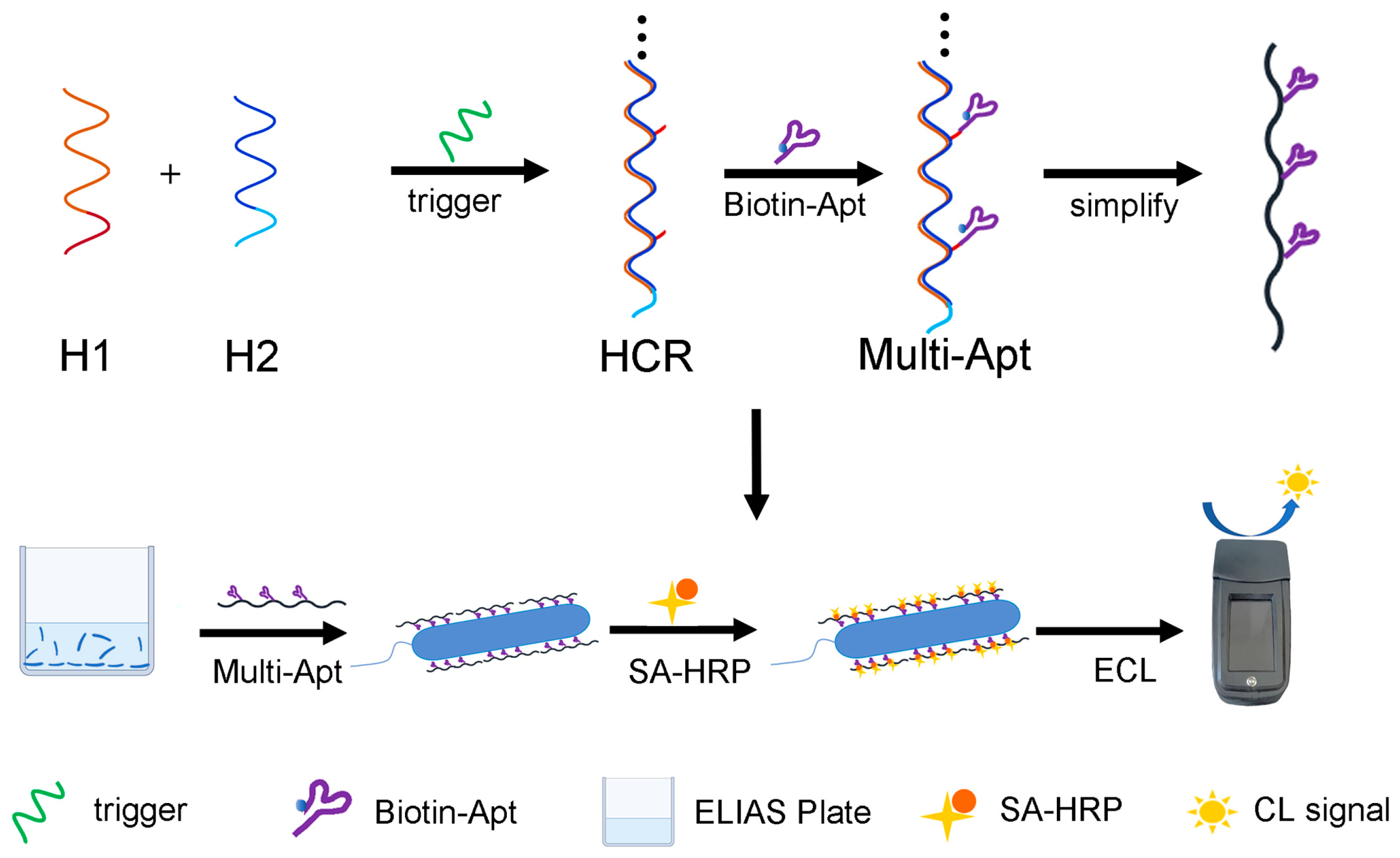
3.1. Principle of the HCR-Based Multi-Apt

In a previous study, we developed an HCR-based multi-Apt amplifier for the detection of Salmonella [26]. Here, to enhance the accuracy of detection, we upgraded the detection signal from absorbance to CL and applied this technology to detect V. alginolyticus. The detection process is illustrated in Scheme 1. In the presence of trigger DNA, H1 and H2 generate an HCR scaffold. H1 contains a prominent terminal that can hybridize with aptamer. The 5’ end of the specific aptamer of V. alginolyticus hybridizes with the prominent terminal of H1 and attaches to the HCR scaffold, forming a multi-Apt structure.
After preparation of the HCR-based multi-Apt, it can be utilized for CL-based detection of V. alginolyticus. The detection process involves the initial binding of V. alginolyticus to a high-affinity ELISA plate, followed by binding to the biotin-modified aptamer. Subsequently, SA-HRP that can bind to the aptamer is added, facilitating the catalysis of luminol ECL by SA-HRP. This catalytic reaction generates a CL signal, which can be measured to quantify the quantity of V. alginolyticus in the sample.

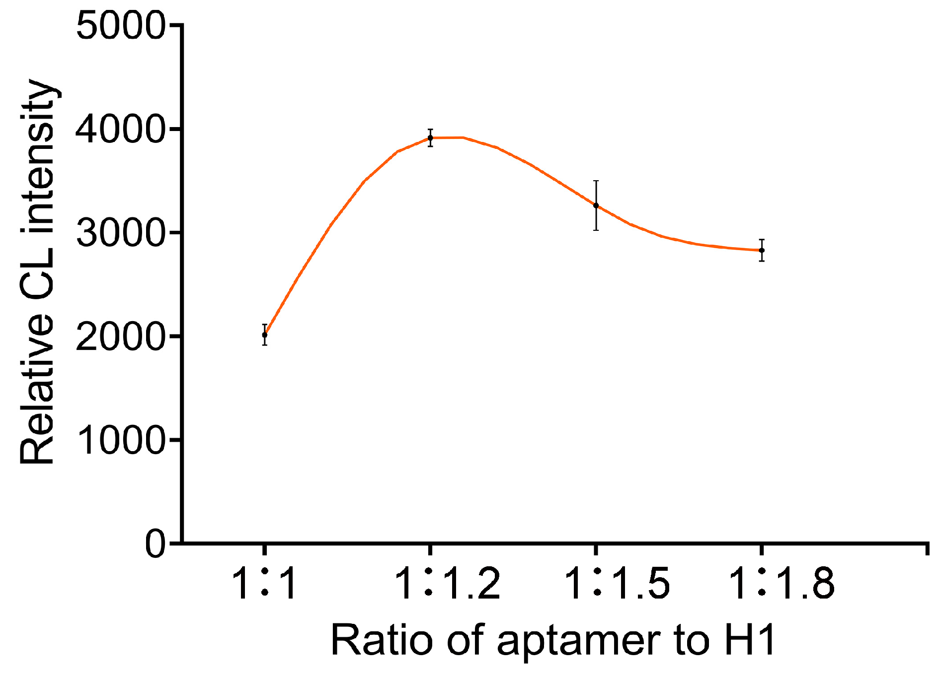
3.2. Optimization of the Aptamer-to-H1 Ratio

Adaptor valence refers to the number of ligands that can attach a receptor to an entity. Previous reports have highlighted that increasing the valence state of ligands on a multivalent scaffold significantly enhances the binding affinity of the multi-Apt [18]. Based on our previous investigation, the optimal binding avidity of multi-Apt was observed when the neighboring aptamer distance was set at 72 bp, with a trigger hairpin ratio of 50:1 [26]. We subsequently optimized the ratio of aptamers to H1. As shown in Figure 1, the CL signal initially increased with the ratio of aptamer to H1 and reached its peak at a ratio of 1.2 before gradually decreasing. The results indicate that efficient multi-Apt was obtained when the level of aptamer was appropriately higher than that of H1. Consequently, an aptamer-to-H1 ratio of 1.2 was selected for subsequent investigations.

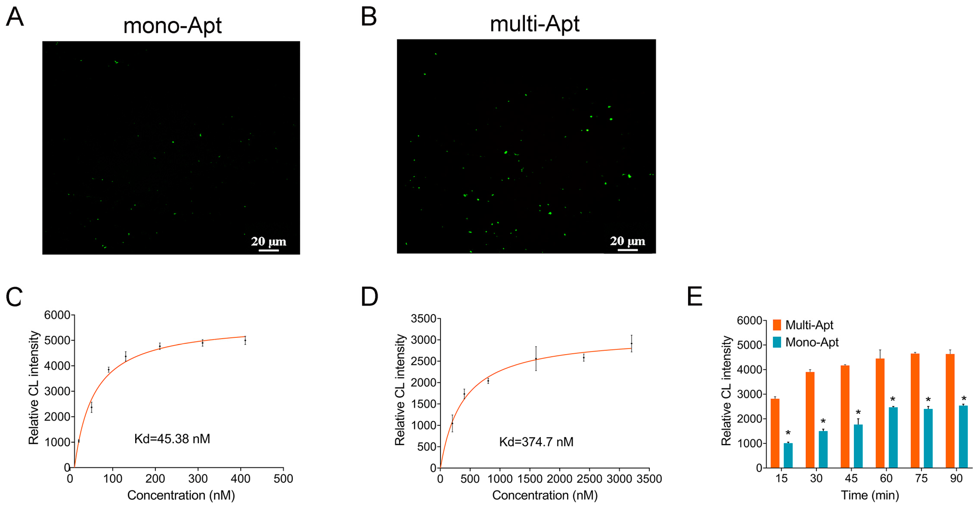
3.3. The Affinity of V. alginolyticus and Multi-Apt

The H1 terminal sequence on the HCR skeleton enhances the binding efficiency of aptamers towards the target by enabling multiple-aptamer binding. To evaluate the increased affinity of the multi-Apt structure, FAM-labeled multi-Apt was employed for fluorescence microscopy imaging of V. alginolyticus. Figure 2A,B demonstrated prominent green fluorescence signals from both multi- and mono-Apt. Importantly, the multi-Apt-treated group exhibited significantly stronger fluorescence intensity compared to the mono-Apt group, indicating the superior affinity provided by the multi-Apt.
In general, multi-Apt has better targeting affinity than mono-Apt [21]. The binding capacity of the aptamer was assessed by the affinity curve, enabling the determination of the optimal quantity of multivalent aptamers for ELISA detection [30]. In this study, ELISA was utilized to assess the affinity curve between different concentrations of biotin-labeled aptamer and V. alginolyticus. Figure 2C,D demonstrate that the multi-Apt group exhibited a higher CL signal compared to the mono-Apt group at lower concentrations (<400 nM). The CL signal for the mono-Apt group at these lower concentrations was too weak to be measured. Consequently, the concentration of the mono-Apt was elevated to 200–3200 nM. However, even with this increased concentration, the CL signal remained considerably weaker than that of the multi-Apt group (Figure 2D). The calculated Kd values for the multi-Apt and mono-Apt were determined as 45.38 nM and 374.7 nM, respectively. This result is consistent with previous studies, reaffirming the strong affinity exhibited by multi-Apt [23,26].
The duration of the detection process is a crucial metric for evaluating effectiveness, predominantly influenced by the reaction time between the aptamer and target, thus determining the overall detection time [31]. In this study, we evaluated the binding efficiency of aptamers and V. alginolyticus by measuring the effects of various incubation times on CL signal intensity. As shown in Fig 3E, during the designated incubation period, the CL signal in the multi-Apt group was significantly higher than that in the mono-Apt group. In the mono-Apt group, the appearance of numerous CL signals was observed after 15 min of incubation, which gradually intensified until reaching its peak at 60 min. In contrast, the CL signal of the multi-Apt group achieved a comparably high value after 30 min of incubation, with no further increase noted as the incubation time was extended. These findings underscore the superior binding efficiency of multi-Apt in contrast to that of mono-Apt, consistent with prior studies [22,24,26]. As a result, a 30 min incubation time was chosen for detection of V. alginolyticus using multi-Apt.

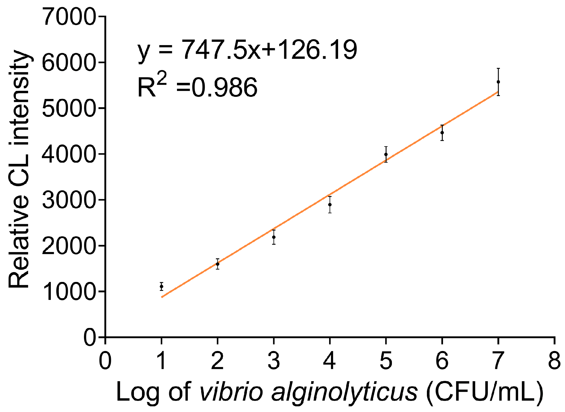
3.4. Sensitivity Investigation of Multi-Apt Amplifier

To evaluate the performance of multi-Apt quantitative detection of V. alginolyticus, we assessed the CL signal intensity at different concentrations of V. alginolyticus. The resulting standard curve showed an increasing CL signal with higher V. alginolyticus concentrations (up to 1.0 × 10⁷ CFU/mL) (Figure 3). This can be attributed to the binding of multi-Apt to V. alginolyticus, leading to enriched HRP and subsequent signal amplification by ECL. Within the concentration range of 10–107 CFU/mL, the regression equation y = 747.5x + 126.2 was derived with a high correlation coefficient (R2 = 0.986). The LOD, determined as three times the standard deviation of the CL signal of the blank sample, was found to be 3 CFU/mL. The LOD achieved in this study exhibits strong competitiveness when compared to previously reported methods. For instance, the LOD for aptamer-based detection of V. alginolyticus using magnetic nanoparticles and gold nanoparticles was reported as 2.4 CFU/mL [32], while the LOD for aptamer-functionalized magnetic relaxation switch sensor detection of V. alginolyticus was documented as 26 CFU/mL [17]. Furthermore, the LOD for V. alginolyticus detection employing a potentiometric aptasensing assay using signal amplification and magnetic separation strategies was reported to be 10 CFU/mL [30]. It is noteworthy that the LOD obtained in this study surpasses the LOD of 7 CFU/mL observed in our previously established Salmonella detection method [26]. These excellent detection capabilities are primarily due to the high binding affinity of multi-Apt and the use of highly sensitive ECL for detection of HRP immobilized on an HCR scaffold.

3.5. Specificity Investigation of the Multi-Apt

Next, we evaluated the specificity of multi-Apt for V. alginolyticus and some other pathogenic bacteria, including S. aureus, E. coli, P. plecoglossicida, E. tarda, A. veronii, V. parahaemolyticus, V. harveyi, and V. vulnificus, at a concentration of 105 CFU/mL under identical detection conditions. As shown in Figure 4, the CL signals generated by the other bacterial species were similar to that of the negative control, indicating minimal binding affinity. In contrast, V. alginolyticus exhibited a significantly higher CL signal compared to the other bacterial species, suggesting a strong binding affinity with the multi-Apt. The results confirm the high selectivity of the proposed multi-Apt towards V. alginolyticus. Importantly, the detection of V. alginolyticus was unaffected by the presence of other pathogenic bacteria, demonstrating the specificity of multi-Apt to V. alginolyticus.

3.6. Application of the Developed Method in Spiked Water Samples

Finally, to determine the practical applicability of the developed multi-Apt amplifier, we conducted a V. alginolyticus recovery test in seawater and fresh water, since water is the main carrier of V. alginolyticus. To mimic real water samples containing V. alginolyticus, it was added at specific concentrations of 1.0 × 10³, 1.0 × 10⁴, and 1.0 × 10⁵ CFU/mL to seawater or fresh water. As shown in Table 2, the recovery rate of V. alginolyticus in freshwater samples ranged from 97% to 103.2%, with an average recovery rate of 99.28%, while the recovery rate in seawater samples ranged from 94.3% to 108.8%, with an average recovery rate of 99.28%. All relative standard deviation (RSD) values were less than 6.73%. These data cover V. alginolyticus from seawater and fresh water, indicating the promising potential of the proposed HCR-based multi-Apt amplifier for practical applications.

4. Conclusions

In summary, we developed a multi-Apt amplifier for the sensitive detection of V. alginolyticus by combining the benefits of the high binding affinity of multi-Apt and the effective signal amplification of HCR. The HCR scaffold was synthesized using H1, H2, and trigger DNA, followed by the addition of a biotin-labeled aptamer to initiate the assembly of the multi-Apt amplifier. Once successfully prepared, the multi-Apt amplifier was employed for CL-based detection of V. alginolyticus. The developed HCR-based multi-Apt amplifier allowed for sensitive detection of V. alginolyticus in a linear range from 10 to 107 CFU/mL with an LOD of 3 CFU/mL. The proposed method has great potential for application in monitoring of V. alginolyticus in aquaculture environments.

Author Contributions

Conceptualization, funding acquisition, and writing—original draft preparation, J.L.; project administration, resources, and funding acquisition, J.C.; methodology, investigation, and software, Y.Z. and S.L.; validation and supervision, Z.Q., J.F. and Q.Z. All authors have read and agreed to the published version of the manuscript.

Funding

This research was funded by the Key Research and Development Project of Zhejiang Province (grant number 2021C02062), the Natural Science Foundation of Zhejiang Province (grant number LY23C190001), the Program of the Science and Technology Department of Ningbo City (grant number 2022S156), the One Health Interdisciplinary Research Project of Ningbo University (grant number HZ202201), and the National Natural Science Foundation of China (grant number 32001773).

Institutional Review Board Statement

Not applicable.

Informed Consent Statement

Not applicable.

Data Availability Statement

Relevant information is included in the article.

Acknowledgments

We thank Tong Xu and our colleagues for their support.

Conflicts of Interest

The authors declare no conflict of interest.

References

  1. Loo, K.Y.; Law, J.W.F.; Tan, L.T.H.; Pusparajah, P.; Letchumanan, V.; Lee, L.H. Diagnostic techniques for rapid detection of Vibrio species. Aquaculture 2022, 561, 738628. [Google Scholar] [CrossRef]
  2. Manchanayake, T.; Salleh, A.; Amal, M.N.A.; Yasin, I.S.M.; Zamri-Saad, M. Pathology and pathogenesis of Vibrio infection in fish: A review. Aquacult. Rep. 2023, 28, 101459. [Google Scholar] [CrossRef]
  3. Jacobs Slifka, K.M.; Newton, A.E.; Mahon, B.E. Vibrio alginolyticus infections in the USA, 1988–2012. Epidemiol. Infect. 2017, 145, 1491–1499. [Google Scholar] [CrossRef] [PubMed]
  4. Wang, J.; Ding, Q.; Yang, Q.; Fan, H.; Yu, G.; Liu, F.; Bello, B.K.; Zhang, X.; Zhang, T.; Dong, J.; et al. Vibrio alginolyticus triggers inflammatory response in mouse peritoneal macrophages via activation of NLRP3 inflammasome. Front. Cell. Infect. Mi. 2021, 11, 769777. [Google Scholar] [CrossRef] [PubMed]
  5. Jin, T.C.; Lu, J.F.; Luo, S.; Wang, L.C.; Lu, X.J.; Chen, J. Characterization of large yellow croaker (larimichthys crocea) osteoprotegerin and its role in the innate immune response against to Vibrio alginolyticus. Comp. Biochem. Phys. Part B 2022, 258, 110680. [Google Scholar] [CrossRef]
  6. Yu, Q.; Liu, M.; Su, H.; Xiao, H.; Wu, S.; Qin, X.; Li, S.; Mi, H.; Lu, Z.; Shi, D.; et al. Selection and characterization of ssDNA aptamers specifically recognizing pathogenic Vibrio alginolyticus. J. Fish Dis. 2019, 42, 851–858. [Google Scholar] [CrossRef]
  7. Sheikh, H.; John, A.; Musa, N.; Abdulrazzak, L.A.; Alfatama, M.; Fadhlina, A. Vibrio spp. and their vibriocin as a vibriosis control measure in aquaculture. Appl. Biochem. Biotech. 2022, 194, 4477–4491. [Google Scholar] [CrossRef]
  8. Naudet, J.; d’Orbcastel, E.R.; Bouvier, T.; Godreuil, S.; Dyall, S.; Bouvy, S.; Rieuvilleneuve, F.; Restrepo-Ortiz, C.X.; Bettarel, Y.; Auguet, J.C. Identifying macroplastic pathobiomes and antibiotic resistance in a subtropical fish farm. Mar. Pollut. Bull. 2023, 194, 115267. [Google Scholar] [CrossRef]
  9. Siddique, M.P.; Jang, W.J.; Lee, J.M.; Hasan, M.T.; Kim, C.H.; Kong, I.S. Detection of Vibrio anguillarum and Vibrio alginolyticus by singleplex and duplex loop-mediated isothermal amplification (LAMP) assays targeted to GROEL and FKLB genes. Int. microbiol. 2019, 22, 501–509. [Google Scholar] [CrossRef]
  10. Yin, J.F.; Wang, M.Y.; Chen, Y.J.; Yin, H.Q.; Wang, Y.; Lin, M.Q.; Liu, A.Y.; Hu, C.J. Direct detection of Vibrio vulnificus, Vibrio parahaemolyticus, and Vibrio alginolyticus from clinical and environmental samples by a multiplex touchdown polymerase chain reaction assay. Surg. Infect. 2018, 19, 48–53. [Google Scholar] [CrossRef]
  11. Tian, X.; Hu, J.; Wei, T.; Ding, W.; Miao, Q.; Ning, Z.; Fan, S.; Wu, H.; Lu, J.; Lyu, M.; et al. Fast and sensitive graphene oxide-dnazyme-based biosensor for Vibrio alginolyticus detection. J. Fish Dis. 2022, 45, 687–697. [Google Scholar] [CrossRef] [PubMed]
  12. Saad, M.; Faucher, S.P. Aptamers and aptamer-coupled biosensors to detect water-borne pathogens. Front. Microbiol. 2021, 12, 643797. [Google Scholar] [CrossRef] [PubMed]
  13. Liu, H.M.; Hao, J.M.; Xu, J.; Yan, Q.P.; Gao, H.; Zheng, J. Selection and identification of common aptamers against both Vibrio harveyi and Vibrio alginolyticus. Chinese J. Anal. Chem. 2020, 48, 623–631. [Google Scholar] [CrossRef]
  14. Mishra, S.S. Use of dot immunoassay for rapid detection of pathogenic bacteria Vibrio alginolyticus and Aeromonas hydrophila from shrimps and fishes. Indian J. Mar. Sci. 1998, 27, 222–226. [Google Scholar]
  15. Hayrapetyan, H.; Tran, T.; Tellez-Corrales, E.; Madiraju, C. Enzyme-Linked Immunosorbent Assay: Types and Applications. Methods. Mol. Biol. 2023, 2612, 1–17. [Google Scholar]
  16. Zhou, Q.Q.; Xu, Z.G.; Liu, Z.M. Molecularly Imprinting-Aptamer Techniques and Their Applications in Molecular Recognition. Biosensors 2022, 12, 576. [Google Scholar] [CrossRef]
  17. Wang, X.; Ni, S.; Wang, Y. An aptamer-functionalized magnetic relaxation switch sensor for the rapid detection of Vibrio alginolyticus in water. Appl. Magn. Reson. 2021, 52, 1561–1580. [Google Scholar] [CrossRef]
  18. Yu, Q.; Liu, M.; Xiao, H.; Wu, S.; Qin, X.; Ke, K.; Li, S.; Mi, H.; Shi, D.; Li, P. Development of novel aptamer-based enzyme-linked apta-sorbent assay (elasa) for rapid detection of mariculture pathogen Vibrio alginolyticus. J. Fish Dis. 2019, 42, 1523–1529. [Google Scholar] [CrossRef]
  19. Fan, Y.T.; Zheng, J.; Bo, J.; Jiang, X.L.; Liu, H.M.; Hang, j.y. Detection of Vibrio anguillarum by differential fluorescence method using aptamer. Oceanologia Limnologia Sinica 2023, 54, 7. [Google Scholar]
  20. Yan, W.L.; Gu, L.; Liu, S.; Ren, W.; Lyu, M.S.; Wang, S.J. Identification of a highly specific DNA aptamer for Vibrio vulnificus using systematic evolution of ligands by exponential enrichment coupled with asymmetric PCR. J. Fish Dis. 2018, 41, 1821–1829. [Google Scholar] [CrossRef]
  21. Wang, Y.M.; Wu, Z.; Liu, S.J.; Chu, X. Structure-switching aptamer triggering hybridization chain reaction on the cell surface for activatable theranostics. Anal. Chem. 2015, 87, 6470–6474. [Google Scholar] [CrossRef] [PubMed]
  22. Dirks, R.M.; Pierce, N.A. Triggered amplification by hybridization chain reaction. Proc. Natl. Acad. Sci. USA. 2004, 101, 15275–15278. [Google Scholar] [CrossRef] [PubMed]
  23. Jia, L.P.; Zhao, R.N.; Wang, L.J.; Ma, R.N.; Zhang, W.; Shang, L.; Wang, H.S. Aptamer based electrochemical assay for protein kinase activity by coupling hybridization chain reaction. Biosens. Bioelectron. 2018, 117, 690–695. [Google Scholar] [CrossRef]
  24. Yeldell, S.B.; Seitz, O. Nucleic acid constructs for the interrogation of multivalent protein interactions. Chem. Soc. Rev. 2020, 49, 6848–6865. [Google Scholar] [CrossRef] [PubMed]
  25. Yang, Y.; Wu, T.; Xu, L.P.; Zhang, X. Portable detection of Staphylococcus aureus using personal glucose meter based on hybridization chain reaction strategy. Talanta 2021, 226, 122132. [Google Scholar] [CrossRef] [PubMed]
  26. Sun, M.N.; Ma, N.; Shi, H.X.; Cheong, L.Z.; Yang, W.G.; Qiao, Z.H. A hcr based multivalent aptamer amplifier for ultrasensitive detection of Salmonella. Sensor. Actuat. B Chem. 2023, 375, 132860. [Google Scholar] [CrossRef]
  27. Luo, S.; Wang, L.C.; Shuai, Z.H.; Yang, G.J.; Lu, J.F.; Chen, J. A short peptidoglycan recognition protein protects boleophthalmus pectinirostris against bacterial infection via inhibiting bacterial activity. Fish. Shellfish. Immun. 2022, 127, 119–128. [Google Scholar] [CrossRef]
  28. Li, A.; Zuo, P.; Ye, B.C. An aptamer biosensor based dual signal amplification system for the detection of Salmonella typhimurium. Anal. Biochem. 2021, 615, 114050. [Google Scholar] [CrossRef]
  29. Ren, D.; Sun, C.; Huang, Z.; Luo, Z.; Zhou, C.; Li, Y. A novel fret biosensor based on four-way branch migration hcr for Vibrio parahaemolyticus detection. Sensor. Actuat. B Chem. 2019, 296, 126577. [Google Scholar] [CrossRef]
  30. Zhao, G.; Ding, J.; Yu, H.; Yin, T.; Qin, W. Potentiometric aptasensing of Vibrio alginolyticus based on DNA nanostructure-modified magnetic beads. Sensors 2016, 16, 2052. [Google Scholar] [CrossRef]
  31. Xu, J.; Zhang, X.; Yan, C.; Qin, P.; Yao, L.; Wang, Q.; Chen, W. Trigging isothermal circular amplification-based tuning of rigorous fluorescence quenching into complete restoration on a multivalent aptamer probe enables ultrasensitive detection of Salmonella. Anal. Chem. 2022, 94, 1357–1364. [Google Scholar] [CrossRef] [PubMed]
  32. Sadsri, V.; Trakulsujaritchok, T.; Tangwattanachuleeporn, M.; Hoven, V.P.; Na Nongkhai, P. Simple Colorimetric Assay for Vibrio parahaemolyticus Detection Using Aptamer-Functionalized Nanoparticles. ACS Omega 2020, 5, 21437–21442. [Google Scholar] [CrossRef] [PubMed]
Scheme 1. Schematic illustration of the method employed for the determination of Vibrio alginolyticus.
Fishes 08 00477 sch001
Figure 1. The impact of the aptamer-to-H1 ratio on the detection outcomes was investigated using ELISA. The ratios tested ranged from 1:1 to 1:1.8, with a fixed concentration of V. alginolyticus at 105 CFU/mL. The results represent the means and standard deviations (SD) (n = 3).
Fishes 08 00477 g001
Figure 2. The affinity of V. alginolyticus and multi-Apt. The detection of V. alginolyticus by FAM-labeled mono-Apt (A) or multi-Apt (B) (bar = 20 µm). (C) The dissociation constants of multi-Apt. (D) The dissociation constants of mono-Apt. (E) The kinetics of the multi-Apt and mono-Apt. The results represent the means and SD (n = 3, * p < 0.05).
Fishes 08 00477 g002
Figure 3. Calibration curve for the quantification of V. alginolyticus using CL signal intensity. V. alginolyticus under concentrations from 10 to 1 × 10⁷ CFU/mL. The results represent the means and SD (n = 3).
Fishes 08 00477 g003
Figure 4. Comparison of the HCR-based multi-Apt in the sensing of S. aureus, E. coli, P. plecoglossicida, E. tarda, A. veronii, V. parahaemolyticus, V. harveyi, V. vulnificus, and V. alginolyticus. MIX is the sample mixed with all bacteria in equal proportion. Error bars are SD from three repetitive experiments (*, p < 0.05).
Fishes 08 00477 g004
Table 1. Sequences of the oligonucleotides used in this work.
OligonucleotideSequence
Aptamer DNAGAGAGAGAATATAAGGGAAAAAAAATCAGTCGCTTCGCCGTCTCCTTCGGGGGCGCGGTGAGGGGCTGCACAAGAGGGAGGCACAAGAGGGAGACCCCAGAGGG
H 1 DNATTTCCCTTATATTCTCTCTCTCTCCTGCGGGAATGTCTAGGTGATTGAGTGGTGTGTTATCCCACTCAATCACCTAGACCATTCCGCAACAACATAC
H 2 DNAGATAACACACCACTCAATCACCTAGACATTCCCGCAGTATGTTGTTGCGGAATGGTCTAGGTGATTGAGTGG
Trigger DNAGTATGTTGTTGCGGAATGGTCTAGGTGATTGAGTGG
Table 2. Detection of V. alginolyticus in different spiked samples using the multi-Apt amplifier. S1, seawater sample; S2, freshwater sample.
SampleSpiked (CFU/mL)Average Relative Chemiluminescent IntensityRelative Standard Deviation (%) Recovery
(%)
S1-11 × 10323654.1798.9%
S1-21 × 10430973.9694.3%
S1-31 × 10538914.32108.8%
S2-11 × 10323794.39103.2%
S2-21 × 10431072.1797%
S2-31 × 10538426.7393.5%
Disclaimer/Publisher’s Note: The statements, opinions and data contained in all publications are solely those of the individual author(s) and contributor(s) and not of MDPI and/or the editor(s). MDPI and/or the editor(s) disclaim responsibility for any injury to people or property resulting from any ideas, methods, instructions or products referred to in the content.

