Article

miR-155 Regulates Photoperiod Induced Gonadal Development in Atlantic Salmon (Salmo salar) by Targeting Brain-Derived Neurotrophic Factor

1
College of Veterinary Medicine, Qingdao Agricultural University, Qingdao 266109, China
2
College of Life Science, Qingdao Agricultural University, Qingdao 266109, China
3
The Key Laboratory of Experimental Marine Biology, Centre for Ocean Mega-Science, Institute of Oceanology, Chinese Academy of Sciences, Qingdao 266071, China
4
Laboratory for Marine Biology and Biotechnology, Qingdao National Laboratory for Marine Science and Technology, Qingdao 266071, China
5
Jinan Center for Animal Disease Prevention and Control, Jinan 250109, China
6
Southern Marine Science and Engineering Guangdong Laboratory, Guangzhou 511458, China
*
Authors to whom correspondence should be addressed.
Fishes 2023, 8(7), 345; https://doi.org/10.3390/fishes8070345
Submission received: 31 May 2023 / Revised: 25 June 2023 / Accepted: 26 June 2023 / Published: 30 June 2023
(This article belongs to the Section Environment and Climate Change)

Abstract

:
This study was designed to explore the impact of the photoperiod on the gonadal development and somatic growth of Atlantic salmon raised in recirculating aquaculture systems (RASs), with specific focus on the role that microRNA (miR)-155 plays as a regulator in the Atlantic salmon’s photoperiodic reproduction. These salmon were reared for 5 months under six different photoperiod regimens, including two with changing photoperiods (LL–SL = 24L:0D–8L:16D and SL–LL = 8L:16D–24L:0D) and four with constant photoperiods (24L:0D, 18L:6D, 12L:12D, and 8L:16D). The longer photoperiod groups (LL–SL and 24L:0D) were ultimately associated with higher gonadosomatic index (GSI) values and an increased proportion of mature fish relative to other exposure groups, indicating that the photoperiod positively impacted the Atlantic salmon’s gonadal development in RAS environments. Hypothalamic miR-155 expression in these Atlantic salmon was also found to be influenced by the photoperiod, showing a markedly decreased expression in salmon exposed to a long photoperiod and upregulation following rearing under a short photoperiod. Functionally, miR-155 was found to suppress the expression of gonadal axis-related genes, including FSH and GnRH, through its ability to target the brain-derived neurotrophic factor (BDNF) and to thereby regulate photoperiod reproduction. Overall, these results suggested that the photoperiod could regulate gonad development in Atlantic salmon with miRNA-155 being involved in this process by targeting the BDNF.
Key Contribution: Firstly, this study developed a photoperiod strategy that reduced the proportion of gonadal development in Atlantic salmon reared in RASs. Then, we found that miR-155 might mediate photoperiodic reproduction by targeting the BDNF.

1. Introduction

High-latitude-inhabiting fish select the optimal timing for breeding to ensure suitable reproductive conditions [1]. The growth of these fish is regulated through a series of climatic factors and internal processes that, ultimately, help to ensure that young fish are able to thrive. Environmental factors, including light, rainfall, temperature, and nutrient availability, all shape the reproductive behaviors of marine fish and many other vertebrates [2]. Notably, the photoperiod is a primary regulator of seasonal rhythms observed in finfish [3] and serves as a specific synchronizer of endogenous rhythms in Atlantic salmon (Salmo salar L.), with the effects of the photoperiod being relayed to the brain–pituitary–gonad axis from the retina, the pineal gland, and, perhaps, in the brain, thereby regulating a diverse array of endogenous endocrine factors [4,5]. The neuroendocrine system, which consists of both sensors and circadian oscillators, such as the hypothalamus and pineal organ, is responsible for synchronizing reproductive behaviors with external environmental conditions [6]. The pineal organ secretes melatonin (N-acetyl-5-methoxytryptamine), which is the most important component of the mechanism responsible for photoperiod-related changes. During the day, vertebrates secrete very low levels of melatonin, whereas at night, such secretion rises [7]. Temperature can have an even more direct rate-limiting effect than the photoperiod on physiological responsivity to seasonal variations [8,9]. In Atlantic salmon, for example, higher temperatures contribute to a more advanced smolt development [10,11].
The photo-neuroendocrine pathway is responsible for converting photoperiod-related stimuli into endogenous neuroendocrine signals through processes governed by the hypothalamic nucleus, which controls the master biological clock, synchronizing physiological activities based on the season and time of day [12]. While the photoperiod has a clear effect on reproduction, and with reproductive cyclicity having been shown to be markedly altered after a pinealectomy, a growing body of evidence suggests that melatonin plays a relatively minor role as a regulator of such reproductive activity in fish [13,14]. Accordingly, further research is required to fully clarify the factors governing the photoperiodic control of reproduction in fish; Chi et al. previously determined that the photoperiod-mediated regulation of the Atlantic salmon’s gonadal development was controlled through the kisspeptin/kissr pathway in the hypothalamus and saccus vasculosus [15]. The particular mechanisms that enable the transmission of photoperiod signals to this kisspeptin/kissr pathway, however, have yet to be defined.
MicroRNAs (miRNAs) are small (~22 nucleotides) transcripts capable of mediating the post-transcriptional silencing of specific mRNAs by binding to complementary 3′ untranslated region (3′-UTR) sequences. Several mature miRNAs have been shown to play essential roles as mediators of normal cellular physiology and differentiation, controlling key processes, including somatic growth and fertility [16]. Additional research is necessary to clarify the biological roles played by miRNAs within the central neuroendocrine system as regulators of reproductive activity. Studies on the reproductive relevance of miRNAs in fish are still in their infancy, with a few promising results having been published to date. For example, Gay et al. determined that miR-202 can regulate medaka oogenesis to control female fecundity [17], while miRNAs in rainbow trout have been successfully leveraged as biomarkers of particular metabolic and reproductive states [18].
First identified as a potent activator of inflammatory activity and a driver of autoimmunity, miR-155 has been shown to be capable of promoting myeloid cell polarization towards proinflammatory phenotypes [19]. In humans, miR-155 has also been shown to shape the pathogenic onset and progression of many hematological malignancies, cancers, and viral infections [20]. In rodent species, miR-155 could reportedly regulate behaviors influenced by the photoperiod, suggesting that it may be an optimal candidate as a mediator of the regulation of seasonal behavioral patterns in mice and hamsters [21]. In addition, miR-155 plays a critical role in the central neuroendocrine control of reproduction by modulating the gonadotropin-releasing hormone (GnRH) gene network and regulating puberty and adult fertility in mice [22]. Given this prior evidence, it is plausible that the photoperiod may similarly shape gonadal development in fish in an miR-155-dependent manner.
Atlantic salmon are an economically valuable species of fish native to catchment areas in North America and Europe that have spread to subarctic and temperate regions in the Baltic Sea, North Atlantic Ocean, and Barents Sea [23]. Given their high levels of nutritional and economic value, Atlantic salmon were introduced to China in 2010. However, high water temperatures during the summer months preclude the survival of these Atlantic salmon in sea cages. Instead, these salmon can be raised in environmentally and economically friendly recirculating aquaculture systems (RASs) [24]. Differences in light sources used for sea cages and RAS systems, however, have been linked to higher rates of gonadal development and maturity for fish reared in RAS settings. The photoperiod is known to be a key proximate environmental factor that can initiate and promote gonadal development for most temperate fish species [14]. In Atlantic salmon, Irachi et al. found that the photoperiod could regulate the pituitary-thyroid-stimulating hormone and brain deiodinase [25]. Trine et al. recently found that the photoperiod could affect the seawater growth performance of Atlantic salmon [26]. However, the mechanism of the photoperiod regulating reproduction is still lacking. The present study was, thus, designed to explore the impact of the photoperiod on the Atlantic salmon’s gonadal development, with particular focus on clarifying the molecular mechanisms whereby the photoperiod can shape such gonadal development.

2. Materials and Methods

2.1. Fish

The Institutional Animal Care and Use Committee of the Institute of Oceanography, the Chinese Academy of Sciences, and the ethical committee of Qingdao Agricultural University (approval number #QAU20215891) approved all animal treatments employed in this study. In total, 1130 Atlantic salmon (Salmo salar L.; 962.70 ± 135.74 g) were obtained from Guoxin Dongfang (Yantai) Recirculating Aquaculture Technology Co., Ltd. (Yantai, China). Before experimentation, these fish were reared and fed under standard conditions (three times a day, cafeteria feeding), allowing them to acclimate to the RAS environment for a 4-week period with continuous (24L:0D) photoperiod exposure. The fish were then randomly transferred into experimental RAS tanks (130 cm height × 200 cm diameter). Seawater in these tanks was continuously exchanged at 660 L·h−1 (~20 exchanges per day), and each tank included a mechanical filtration system, a foam separator, a UV disinfection system, and a 1800 L biofilter. Water supplemented using liquid oxygen was supplied at ~50 L·min−1 to maintain a dissolved oxygen saturation level of 90–110%. The water was maintained at 16.27 ± 0.54 °C throughout the study period, with a pH of 7.2–7.5, a total ammonia–nitrogen concentration <0.25 mg/L, and salinity levels of 24–26. Fish were fed twice per day with a diet corresponding to 1% of their total body weight (53% crude protein, 13% crude fat, 12.5% crude ash, 2.8% crude fiber, 9.6% water, 4% lysine, and 2.8% phosphorus).
For the experimental analyses, the fish were assigned to six different photoperiod groups, including four that were exposed to constant photoperiods throughout the study duration (24L:0D, 18L:6D, 12L:12D, and 8L:16D) and two that were subjected to gradual changes in photoperiod, including a 24L:0D to 8L:16D (LL–SL, long light to short light) and an 8L:16D to 24L:0D (SL–LL, short light to long light) group. For fish in these gradually changing photoperiod groups, the lighting period was changed by 5 min per day in the appropriate direction. A total of three replicate tanks were established per condition, with 60 fish per tank. The total study period lasted 6 months, from September to February, including a 1-month acclimatization period and a 5-month experimental period, thus, spanning the first reproductive period.
The fish were weighed monthly. Briefly, 3 fish per tank were sampled. The fish were euthanized using 0.05% MS-222 in seawater, and their body weight values were recorded, after which the caudal vein was used to collect blood samples. The blood was centrifuged for 15 min at 3000 rpm, after which plasma was stored at −80 °C for downstream analyses of melatonin. Then, the fish were dissected, and the gonads were excised and weighed. The gonadosomatic index (GSI) was calculated according to the below equation:
GSI % = W g × 100 W b
where Wg is the weight of the gonad and Wb is the total body weight.
One portion of the gonads was snap-frozen with liquid nitrogen prior to storage at −80 °C for analyses of follicle-stimulating hormone receptor (FSH-R) and luteinizing hormone receptor (LH-R) levels, while the remainder was fixed for 24 h using Bouin’s fluid prior to storage using 70% ethanol pending downstream histological analyses. In addition, 6 fish (3 female and 3 male) were sampled for detecting miR-155 expression.
For miRNA-based studies, the fish were selected at random for an intra-cerebroventricular (ICV) injection of miRNA mimics, miRNA inhibitors, control miRNAs, and siRNAs (Tsingke Biotechnology Co., Ltd, Beijing, China), with 50 μL of the appropriate miRNA or siRNA constructs being diluted using 150 μL PBS. The ICV injection of these RNAs (2 mg/kg) was achieved using stainless steel cannulas directly implanted into the brain ventricles of these experimental salmon. The fish were anesthetized using 0.05% MS-222 during these surgical procedures. The fish received intramuscular injections of penicillin/streptomycin for 5 consecutive days postoperatively, and were reared for 1 week prior to euthanasia and analysis.

2.2. Synthesis of miRNA Mimics and Inhibitor

All miR-155 mimic, inhibitor, and miR-control constructs were based on the miR-155 sequence from the miRbase database (MI0026520) and were synthesized by Tsingke Biotechnology Co., Ltd. (Beijing, China). The double-stranded miR-155 mimic sequences were 5′-UUAAUGCUAAUCGUGAUAGGGGU-3′ and 5′-CACCUAGCAUGUAAGCAUUAGC-3′, and the single-stranded miR-155 inhibitor sequence was 5′-GCAAUGCUUACAUGCUAGGUG-3′. Nonspecific control miRNA: 5′-UUCACAGAACGUGUCUCGU-3′. The BDNF-specific siRNAs were 5′-AAAAGUAUUGCUUCAGUUGGC-3′ (sense) and 5′-CAACUGAAGCAAUACUUUUAU-3′ (antisense), and synthesized by Tsingke Biotechnology Co., Ltd. (Beijing, China).

2.3. Plasma Melatonin Concentration Measurements

The plasma melatonin levels were measured with a commercial ELISA kit (IBL, Hamburg, Germany) based on the provided directions.

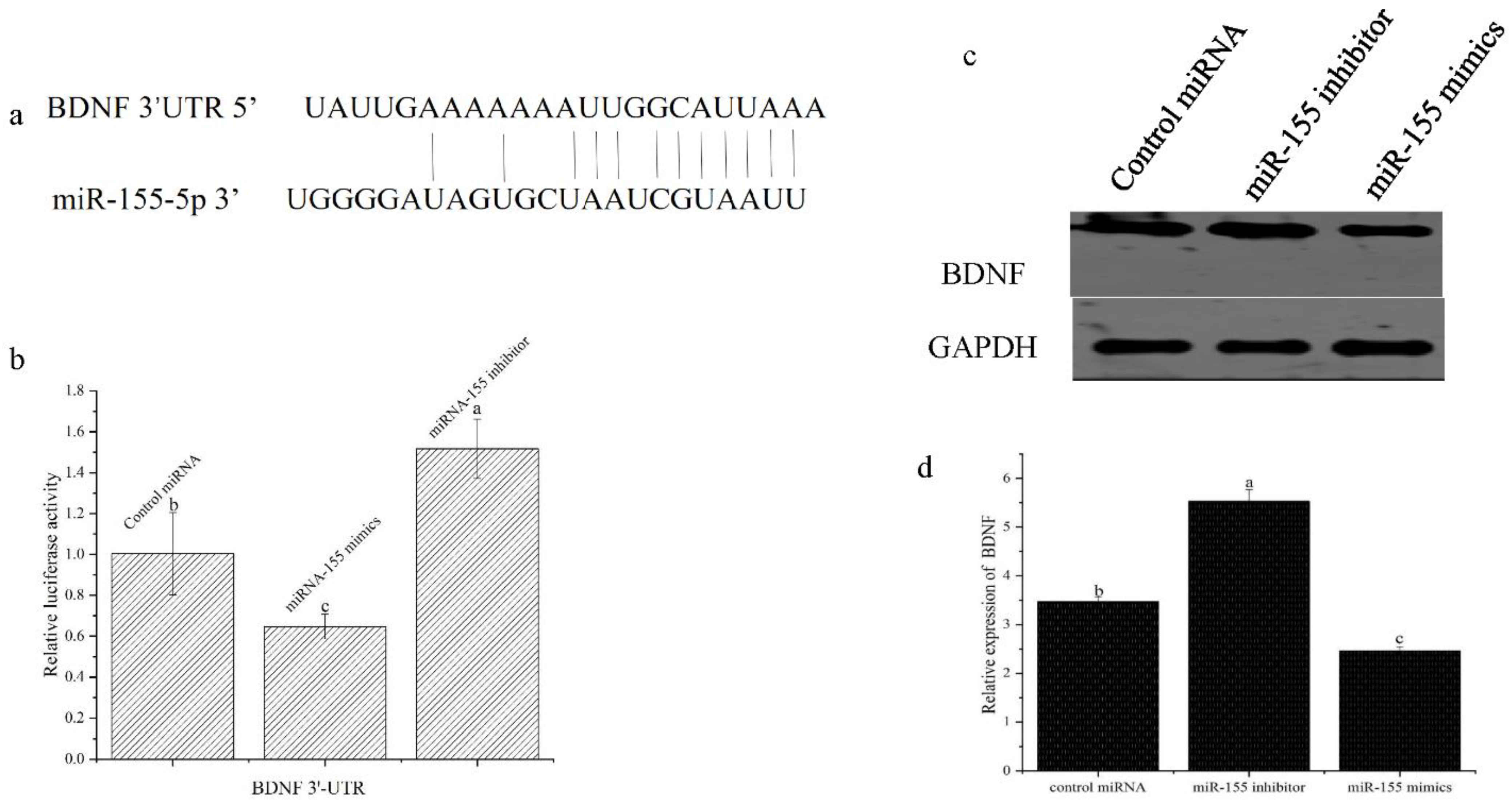
2.4. Luciferase Assay

The BDNF 3′-UTR region, identified as a putative miR-155 target region, was amplified via PCR and inserted into a pGL3 vector (Promega) downstream of a luciferase stop codon. Then, HEK-293 cells were transfected using Lipofectamine 3000 (Invitrogen, Shanghai, China) with 0.8 μg of either a control Renilla luciferase vector or the prepared firefly luciferase reporter vector containing the identified target site, together with pRL-TK (Promega, Beijing, China), and 100 nM of miR-155 mimic, inhibitor, or control miRNA constructs. At 24 h post-transfection, a dual-luciferase reporter assay system was used based on the provided directions to assess luciferase activity, with Renilla luciferase activity being used for normalization.

2.5. Cell Culture and Transfection

HEK-293 cells were purchased from Beyotime Biotechnology. Inc. (Shanghai, China) and cultured in DMEM containing 10% FBS, 10 mM L-glutamine, 0.1 mg/mL streptomycin, and 100 U/mL penicillin in a humidified 37 °C and 5% CO2 incubator. Cells in 6-well plates were transfected with miR-155 mimic, inhibitor, and control miRNA (20 μM) constructs using Lipofectamine 3000 (Invitrogen) based on the provided directions. At 8 h post-transfection, transfection media were exchanged for 2 mL of fresh media per well, and cells were incubated for a further 24 h prior to the downstream analysis.

2.6. qPCR

An RNA extraction kit (BioFlux, Beijing, China) was used to extract the total RNA from the gonads of the experimental Atlantic salmon, with the extracted RNA, ultimately, being eluted in 25 μL of RNase-free water. TransScript First-Strand cDNA Synthesis SuperMix (TransGen Biotech, Beijing, China) was used for the first-strand cDNA synthesis using 20 μL reactions containing 0.5 μL of an oligo dT primer (0.5 μg/μL), 1 µg of suspended RNA, and 2× TS reaction mix. The miR-155 was assayed with the miScript miRNA PCR Array (QIAGEN, China) according to the instructions.
A SYBR TransStart Green qPCR SuperMix (Transgen Biotech, Beijing, China) and an Eppendorf Mastercycler® ep realplex S real-time PCR instruments (Eppendorf, Hamburg, Germany) were used for all qPCR analyses. β-actin served as a reference gene, and the 2-delta Ct method was used to quantify relative gene expression. Primers used to analyze β-actin, FSH-R, and LH-R expression are provided in Table 1. Individual PCR reactions were conducted based on the provided directions in a total volume of 20 μL containing 0.4 μL each of a forward and a reverse primer, 10 μL of 2× TransScript Top Green qPCR Super Mix, 0.4 μL Passive Reference Dye I, and 7.8 μL of ddH2O.

2.7. Western Immunoblotting

A lysis buffer (50 mM Tris-Hcl pH 7.5, 5 mM EDTA, 150 mM NaCl, 0.6% NP-40) was used to extract cellular proteins on ice for 45 min, after which a Bio-Rad Protein Assay was used to measure protein concentrations. The protein samples were then separated via 10% SDS-PAGE, transferred to nitrocellulose membranes, and blots were blocked using 5% nonfat milk in PBST prior to incubation overnight with primary anti-BDNF or anti-GAPDH. The blots were then rinsed with PBST, followed by incubation with appropriate secondary antibodies (Servicebio, biological. Inc. Wuhan, China). An enhanced Western Bright ECL reagent was then used for the protein band detection.

2.8. Statistical Analysis

SPSS 22.0 was used for all statistical testing. Data were reported as means ± standard deviation ( x ¯ ± SD) and were compared with one-way ANOVAs and Tukey’s test. Analyses were repeated in triplicate, and p < 0.05 was the significance threshold.

3. Results

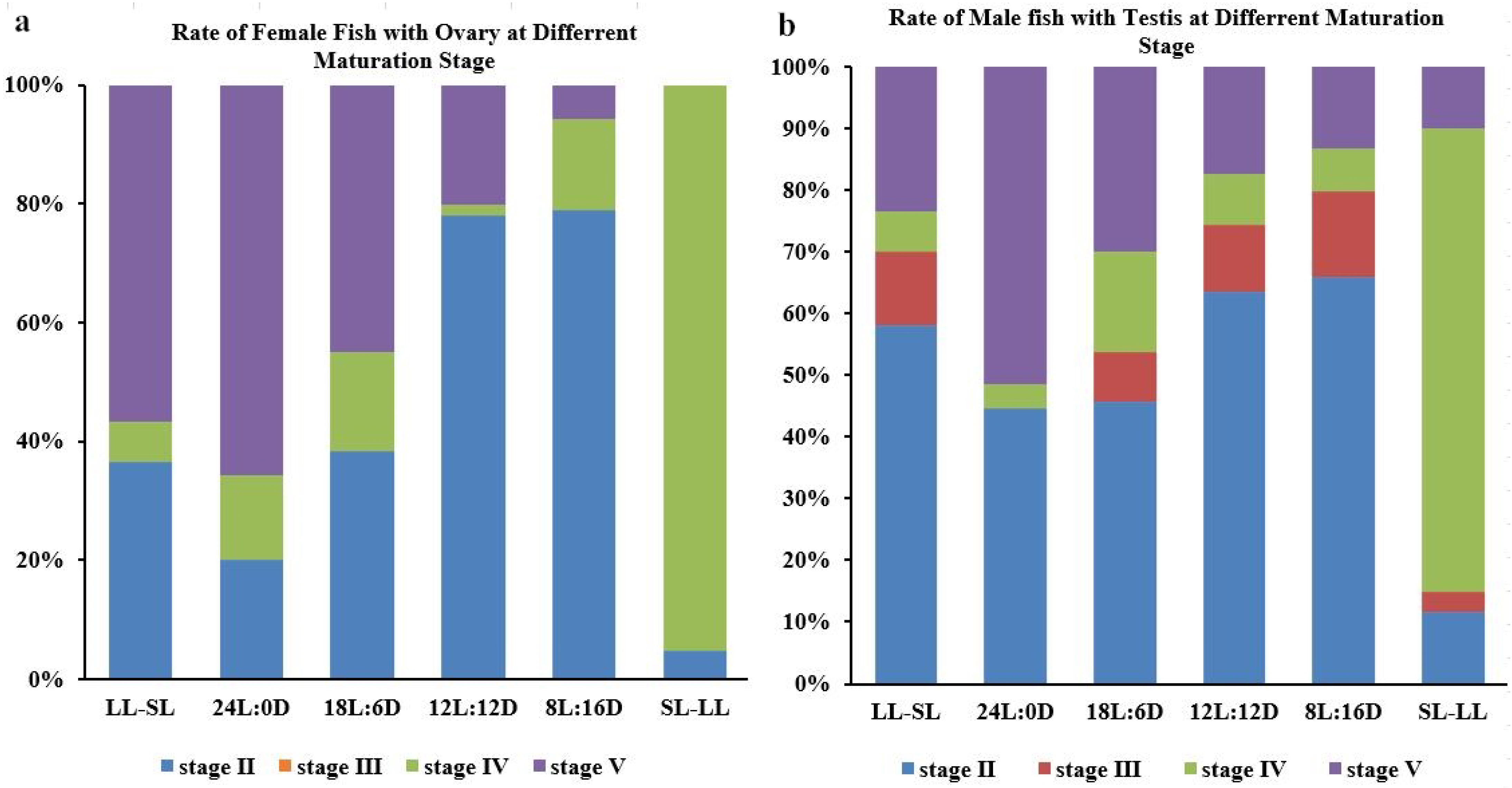
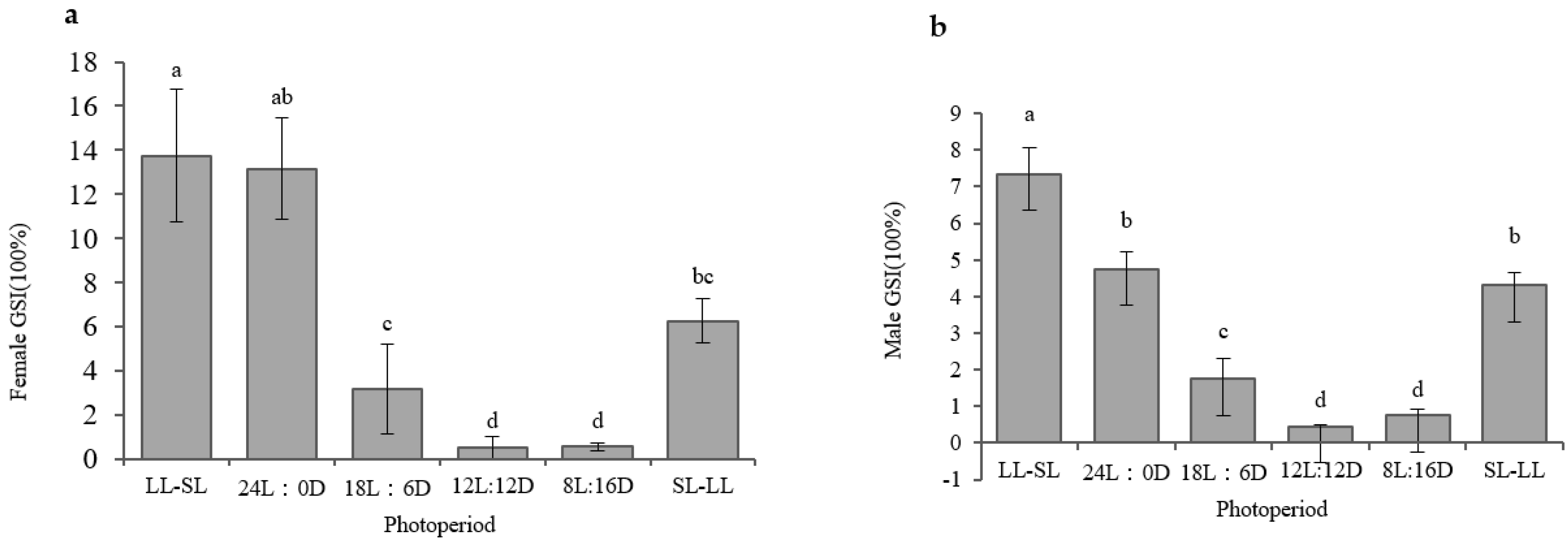
3.1. The Impact of the Photoperiod on Atlantic Salmon’s Gonadal Development in RAS Systems

Following a 5-month culture period, the highest proportion of premature (stage IV) and mature females (stage V) was evident in the SL–LL group (93%), followed by the 24L:0D (78.8%) and LL–SL (58.2%) groups. The highest proportion of premature (stage IV) and mature males (stage V) was observed in the SL–LL group (85.2%), followed by the 24L:0D (55.1%) and 18L:6D (46.2%) groups. In contrast, these levels were lowest in the 12L:12D and 8L:16D groups (Figure 1). The level of gonad stage is shown in Supplemental Figure S1 and Supplemental Figure S2.
The gonadosomatic index (GSI) values were similar across the photoperiods in males and females, with the highest GSI values being evident in the female and male salmon in the LL–SL group (13.75 ± 3.0% and 7.35 ± 0.71%, respectively), followed by the 24L:0D group (13.14 ± 2.30% and 4.75 ± 0.48%, respectively), and the SL–LL group (6.26 ± 1.00% vs. 4.35 ± 0.57%, respectively). The lowest GSI values were observed among female and male salmon in the 8L:16D (0.55 ± 0.30% and 0.75 ± 0.17%, respectively) and 12L:12D (0.53 ± 0.50% and 0.4 ± 0.06%, respectively) groups (Figure 2).

3.2. Expression Pattern of miR-155 in the Hypothalamus of Atlantic Salmon under Different Photoperiods

To determine whether the expression of miR-155 is influenced by photoperiod, we examined the expression of miR-155 in the hypothalamus of salmon reared under different photoperiods. We detected that the photoperiod affected the expression of miR-155 in the hypothalamus. In particular, we found that, at the beginning of the experiment, the levels of miR-155 transcripts were the lowest in the 24L:0D group, followed by the LL–SL group; these were the two treatments with the longest photoperiods. We did not detect significant differences between the 18L:6D and 12L:12D photoperiod treatments. The highest expression of miR-155 transcripts was shown in the 8L:16D photoperiod group, followed by the SL–LL photoperiod group. At the end of the experiment, the lowest levels of miR-155 transcripts were detected in the 24L:0D group, followed by the SL–LL group, whereas we did not observe any significant differences among the 18L:6D and 12L:12D photoperiod treatments. The highest expression of miR-155 transcripts was shown in the 8L:16D photoperiod treatment, followed by the LL-SL photoperiod treatment. These data suggested that the hypothalamic expression of miR-155 was regulated by the photoperiod (Figure 3).

3.3. miR-155 Suppresses Atlantic Salmon Photoperiodic Reproduction

To explore the ability of miR-155 to serve as a regulator of photoperiodic reproduction, miR-155 inhibitor or mimic constructs were next introduced into Atlantic salmon, after which reproduction-related hormone and gene expression in these experimental animals was analyzed. As shown in Figure 4, the miR-155 inhibitor administration resulted in a marked rise in the expression of reproduction-related genes, including GnRH, FSHr, and LHr. The hypothalamic levels of melatonin, a hormone exclusively secreted during the nighttime and which serves as an essential regulator that transmits photoperiod-related information to the neuroendocrine–gonadal axis, also increased following the miR-155 inhibitor treatment. Conversely, melatonin expression was markedly suppressed following the miR-155 mimic administration. These findings suggested that miR-155 can serve as a negative regulator of Atlantic salmon photoperiodic reproduction.

3.4. miR-155 Targets the BDNF mRNA in Atlantic Salmon

To better understand the mechanistic basis for the observed inhibitory effects of miR-155 documented above, candidate miR-155 target genes were next explored using PicTar and TargetScan. These putative targets included the brain-derived neurotrophic factor (BDNF), which serves as a key regulator in gonadotrophic axis functionality in vertebrates (Figure 5a). To directly test the ability of miR-155 to regulate BDNF expression, the identified BDNF 3′-UTR target sequence complementary to miR-155 was cloned into a luciferase reporter plasmid that was then transfected into HEK-293 cells. In this assay system, the miR-155 inhibitor transfection was sufficient to promote enhanced BDNF reporter activity (Figure 5b), with an increase in relative luciferase activity from 1.0 to 1.7. In contrast, the miR-155 mimic transfection reduced such luciferase activity to 0.6, consistent with the suppression of BDNF reporter activity. These results highlighted the ability of miR-155 to inhibit BDNF expression by binding this target 3′-UTR sequence. The introduction of the miR-155 inhibitor consistently resulted in a significant upregulation of the BDNF at the protein level, as measured via Western immunoblotting (Figure 5c), while the miR-155 mimic treatment had the opposite effect, downregulating BDNF expression relative to levels in the control group.
To verify the status of the BDNF as a functional miR-155 target gene in Atlantic salmon, these fish were treated with an siRNA specific for the BDNF. As shown in Figure 6, the BDNF knockdown resulted in a marked drop in hypothalamic GnRH and melatonin levels in these fish. These results suggested that miR-155 was, thus, able to negatively regulate the photoperiod reproduction in Atlantic salmon, at least in part via targeting the BDNF.

4. Discussion

Given their important functions as regulators of diverse physiological processes, miRNA dysregulation can play an important role in the onset and progression of various forms of neurodegenerative diseases [27]. Studies on miRNAs as regulators of seasonal responses in species other than flowering plants, however, have been limited to date [28]. Some studies determined that miRNAs may play important roles in regulating downstream responsivity to seasonal changes in photoperiods, given that photoperiod-related changes in miRNA expression have been reported in female sheep and white-crowned sparrows [29,30]. Details regarding the roles of miRNAs as regulators of seasonal reproductive behaviors, however, are lacking at present. Accordingly, this study was designed to examine the impact of photoperiod changes on the Atlantic salmon’s growth and gonadal development in RAS systems, with particular focus on how miR-155 regulates these photoperiodic responses during gonadal development.
A positive correlation between the photoperiod length and the growth and gonadal development of Atlantic salmon was detected in this study [31]. The highest specific growth rate (SGR) and body weight (Supplemental Figure S3) of salmon were observed in salmon exposed to the longest photoperiod (24L:0D), consistent with the known ability of a longer photoperiod in supporting growth in this species. The measured SGR values for all fish varied across the study period, declining suddenly during the second month in salmon grown under long photoperiods relative to the first month when fish exhibited stage II gonads. In contrast, the SGR of salmon reared under a short photoperiod remained stable. This may suggest that fish raised under a long photoperiod begin redirecting energetic resources to support reproductive growth and gonadal development [32]. During the third month, fish under all photoperiods exhibited rapid growth (SGR ≥ 0.9%/d), potentially due to appropriately low-density levels, with slower growth during the following months when a larger proportion of energy was directed towards reproductive development as observed previously [33].
Longer photoperiod exposure was associated with both the growth and maturation of Atlantic salmon, with more male and female fish exhibiting stage IV and V gonads being evident in the long photoperiod (24L:0D and 18L:6D) and SL–LL groups, while slower gonadal development was observed in the other groups. In prior reports, continuous light exposure was shown to effectively promote growth and interfere with salmonid gonadal maturation [34], in contrast to the present results, which suggested that longer photoperiods may prevent energetic resource distribution away from somatic growth and towards gonadal growth and reproduction.
The timing of photoperiod exposure can have a marked impact on how these photoperiods ultimately impact specific organisms [35]. A so-called “gating theory” has been proposed to explain the mechanisms through which photoperiods regulate reproduction, suggesting that individuals begin undergoing sexual maturation in response to a photoperiod-mediated switch once sufficient energetic requirements have been met [36]. Atlantic salmon gonadal development may be under the control of a similar mechanism. Some studies have shown that the extension of the illumination time during the winter or early spring can decrease the percentage of salmonids reaching sexual maturity, whereas exposure to longer photoperiods during the summer solstice could increase the overall proportion of salmon that reached puberty. In this paper, in order to investigate whether the photoperiod would have a cumulative effect on the development of fish, we set two changing photoperiod group, one group changed the photoperiod from LL to SL, and another group changed the photoperiod from SL to LL. From the results in this study, we think the effect of the photoperiod on fish gonad development is instantaneous. These two changing photoperiod groups had similar effects with the constant photoperiod groups. Furthermore, at the end of the experiment, although matured fish were mostly found in the long photoperiods (24L:0D and SL–LL), stage V was predominant in the 24:0D photoperiod, but stage IV was predominant in the SL–LL group. We think the development of fish in the 24L:0D photoperiod was faster than fish in the SL–LL group because, in the early stages, the long photoperiod could promote the growth of fish. Therefore, the gonad development of fish in the 24L:0D photoperiod might have occurred earlier than fish in the SL–LL group. These results led to the development of novel photoperiod regimens for use in RAS systems, with a long photoperiod (24L:0D) being used to suppress gonadal development in Atlantic salmon before puberty, after which this photoperiod was shortened to 18L:6D or 12L:12D following puberty to similarly delay gonadal development. This strategy could reduce the proportion of sexually matured Atlantic salmon from 40 down to 20% in RAS systems.
This study focused on testing the possibility that the photoperiod could regulate Atlantic salmon gonadal development through a mechanism at least partially mediated through miR-155. As miRNAs can regulate the expression of many target mRNAs in specific physiological contexts, they are ideal facilitators of the seasonal coordination of physiological and behavioral changes that occur in organisms. However, the roles of miRNAs as regulators of seasonal phenotypes outside of flowering plants have largely not been investigated [21]. As such, the impact of photoperiod changes on miR-155 expression was initially examined, revealing that longer photoperiods suppressed hypothalamic miR-155 levels in Atlantic salmon, while exposure to shorter photoperiods instead increased the hypothalamic levels of this miRNA. For salmon reared under changing photoperiod conditions, miR-155 expression was suppressed in the long photoperiod groups and enhanced in the short photoperiod groups. These findings demonstrated that the photoperiod could influence the hypothalamic miR-155 expression in Atlantic salmon. To directly confirm the functional role of this miRNA as a regulator of the gonadotrophic axis, the ICV injection of miR-155 mimic or inhibitor constructs was performed, revealing that inhibiting miR-155 significantly enhanced FSH and GnRH expression. Together, these results demonstrated that miR-155 is an important regulator of gonadotrophic signaling in Atlantic salmon.
To understand the molecular mechanisms whereby miR-155 exerted the observed effects, the PicTar and TargetScan databases were queried, ultimately, identifying the BDNF as a candidate miR-155 target gene. The BDNF is a critical neurotrophic signaling factor that supports dorsal ganglion neuron survival, neuroplasticity, neuronal growth, and synapse formation [37,38]. However, there have been only a limited number of studies examining the link between the BDNF and the gonadotrophic axis. Przybyl et al. determined that the BDNF was able to impact GnRH gene expression and alter the ability of pituitary cells to secrete LH and FSH [39]. Therefore, we first identified the interaction of miR-155 and the BDNF, and we found that Atlantic salmon’s miR-155-5p could bind to the 3′-untranslated region (3′-UTR) of the BDNF. This result demonstrated that miR-155 might regulate the expression of the BDNF in the hypothalamus of Atlantic salmon. In order to confirm this point, we then detected the expression of the BDNF 3′-UTR and the expression of the BDNF protein. The results showed that when we used an miR-155 inhibitor, both the luciferase activity of the BDNF 3′-UTR and protein expression of the BDNF increased, and the miR-155 mimics could decrease their activity and expression. All this evidence demonstrated that miR-155 could directly target the BDNF mRNA, suggesting that the suppression of BDNF expression may be a mechanism through which miR-155 can control the Atlantic salmon gonadotrophic axis, because small interfering RNA (siRNA) could specifically reduce the expression of targeted genes [40]. Therefore, in order to verify whether the BDNF in the Atlantic salmon’s hypothalamus could regulate the gonadotrophic axis, we used siRNA to reduce the expression of the BDNF. The results showed that when the expression of the BDNF was interfered with using siRNA, the expression of GnRH also reduced. The results stated that the BDNF could regulate the expression of GnRH. Atlantic salmon photoperiodic reproduction has previously been reported to be under the control of the hypothalamic kisspeptin/kissr pathway [15]. Therefore, we believe that the photoperiodic control of reproduction is very important for the survival and continuation of species, and multiple signaling pathways might be involved in the regulation of this physiological process, thus, deserving further study.

5. Conclusions

We, firstly, developed new photoperiod regimens for use in RAS systems, with a long photoperiod (24L:0D) being used to suppress gonadal development in Atlantic salmon before puberty, after which this photoperiod was shortened to 18L:6D or 12L:12D following puberty to similarly delay gonadal development. Then, we found that the photoperiod that regulated the gonads in Atlantic salmon might be mediated through miR-155 by targeting the BDNF in the Atlantic salmon’s hypothalamus.

Supplementary Materials

The following are available online at https://www.mdpi.com/article/10.3390/fishes8070345/s1, Figure S1: The external morphological characteristics and histological observation of Atlantic salmon ovary; (a) the characteristics of the stage III ovary; (b) the characteristics of the stage IV ovary; (c) the characteristics of the stage V ovary; (d) the histological observation of the stage III ovary; (e) the histological observation of the stage IV ovary; (f) the histological observation of the stage V ovary, Figure S2: The external morphological characteristics and histological observation of Atlantic salmon testis; (a) the characteristics of the stage II testis; (b) the characteristics of the stage III testis; (c) the characteristics of the stage IV testis; (d) the characteristics of the stage V testis; (e) the histological observation of the stage II testis; (f) the histological observation of the stage III testis; (g) the histological observation of the stage IV testis; (h) the histological observation of the stage V testis; SG: spermatogonia; SL: seminal lobule; PSC: primary spermatocytes; SSC: secondary spermatocytes; S: spermatozoa, Figure S3: The mean weight of fish under the different groups at the end of the experiment, Figure S4: (a) the expression of miR-155 when treated by different dose of miR-155 mimics; (b) the expression of miR-155 when treated by different dose of miR-155 inhibitor, Figure S5: The PCR of BDNF when fish were treated by different dose of siRNA, the primer used for Atlantic salmon BDNF were listed as: F: TGAGCAGGTGATTGAGGAGCT; R: TCGCACGTAGGACTGGGTTGT; PCR product size is 511 bp.

Author Contributions

Conceptualization, L.C. and Q.L.; methodology, T.G., Y.W. and P.J.; software, formal analysis, J.L.; investigation, Y.W. and J.L.; resources, Y.W.; data curation, J.L. and L.C.; writing—original draft preparation, T.G. and L.C.; writing—review and editing, L.C. and Q.L.; project administration, Q.L. and L.C.; funding acquisition, Q.L. and L.C. All authors have read and agreed to the published version of the manuscript.

Funding

This work was financially supported by the National Key Research and Development Program (2018YFD0901204), the China Agriculture Research System (CARS-47-G21), STS Project (KFZD-SW-106, ZSSD-019, 2017T3017, and KFJ-STS-QYZX-020), the Qingdao National Laboratory for Marine Science and Technology (2018SDKJ0502-2 and 2015ASKJ02), the National Natural Science Foundation of China (31672672,31402283), and the Agricultural Application Technology Innovation Project of Shandong Province in 2018/2019 (SD2019YY011).

Institutional Review Board Statement

The study was approved by the Institutional Animal Care and Use Committee of the Institute of Oceanography, the Chinese Academy of Sciences, and the ethical committee of the Qingdao Agricultural University (#QAU20215891).

Informed Consent Statement

Not applicable.

Data Availability Statement

The authors confirm that the data supporting the findings of this study are available within the article.

Conflicts of Interest

The authors declare no conflict of interest.

References

  1. Badruzzaman, M.; Bapary, M.A.J.; Takemura, A. Possible roles of photoperiod and melatonin in reproductive activity via changes in dopaminergic activity in the brain of a tropical damselfish, Chrysiptera cyanea. Gen. Comp. Endocrinol. 2013, 194, 240–247. [Google Scholar] [CrossRef]
  2. Carrillo, M.; Zanuy, S.; Felip, A.; Bayarri, M.J.; Moles, G.; Gomez, A. Hormonal and Environmental Control of Puberty in Perciform Fish The Case of Sea Bass. Ann. N. Y. Acad. Sci. 2009, 1163, 49–59. [Google Scholar] [CrossRef]
  3. Bromage, N.; Porter, M.; Randall, C. The environmental regulation of maturation in farmed finfish with special reference to the role of photoperiod and melatonin. Aquaculture 2001, 197, 63–98. [Google Scholar] [CrossRef]
  4. Nilsen, T.O.; Ebbesson, L.O.; Kiilerich, P.; Björnsson, B.T.; Madsen, S.S.; McCormick, S.D.; Stefansson, S.O. Endocrine systems in juvenile anadromous and landlocked Atlantic salmon (Salmo salar): Seasonal development and seawater acclimation. Gen. Comp. Endocrinol. 2008, 155, 762–772. [Google Scholar] [CrossRef] [PubMed]
  5. Björnsson, B.T.; Stefansson, S.O.; McCormick, S.D. Environmental endocrinology of salmon smoltification. Gen. Comp. Endocrinol. 2011, 170, 290–298. [Google Scholar] [CrossRef] [PubMed]
  6. Maitra, S.K.; Chattoraj, A.; Mukherjee, S.; Moniruzzaman, M. Melatonin: A potent candidate in the regulation of fish oocyte growth and maturation. Gen. Comp. Endocrinol. 2013, 181, 215–222. [Google Scholar] [CrossRef]
  7. Falcón, J.; Migaud, H.; Muñoz-Cueto, J.; Carrillo, M. Current knowledge on the melatonin system in teleost fish. Gen. Comp. Endocrinol. 2010, 165, 469–482. [Google Scholar] [CrossRef] [Green Version]
  8. Imsland, A.K.; Handeland, S.O.; Stefansson, S.O. Photoperiod and temperature effects on growth and maturation of pre- and post-smolt Atlantic salmon. Aquac. Int. 2014, 22, 1331–1345. [Google Scholar] [CrossRef]
  9. Martinez, E.P.; Balseiro, P.; Stefansson, S.O.; Kaneko, N.; Norberg, B.; Fleming, M.S.; Imsland, A.K.; Handeland, S.O. Interaction of temperature and feed ration on male postsmolt maturation of Atlantic salmon (Salmo salar L.). Aquaculture 2023, 562, 738877. [Google Scholar] [CrossRef]
  10. Handeland, S.O.; Berge, Å.; Björnsson, B.T.; Lie, Ø.; Stefansson, S.O. Seawater adaptation by out-of-season Atlantic salmon (Salmo salar L.) smolts at different temperatures. Aquaculture 2000, 181, 377–396. [Google Scholar] [CrossRef]
  11. McCormick, S.D.; O’Dea, M.F.; Moeckel, A.M.; Björnsson, B.T. Endocrine and physiological changes in Atlantic salmon smolts following hatchery release. Aquaculture 2003, 222, 45–57. [Google Scholar] [CrossRef]
  12. Kalsbeek, A.; Palm, I.F.; La Fleur, S.E.; Scheer, F.A.J.L.; Perreau-Lenz, S.; Ruiter, M.; Kreier, F.; Cailotto, C.; Buijs, R.M. SCN Outputs and the Hypothalamic Balance of Life. J. Biol. Rhythm. 2006, 21, 458–469. [Google Scholar] [CrossRef] [PubMed] [Green Version]
  13. Shahjahan, M.; Al-Emran, M.; Islam, S.M.; Baten, S.A.; Rashid, H.; Haque, M.M. Prolonged photoperiod inhibits growth and reproductive functions of rohu Labeo rohita. Aquac. Rep. 2020, 16, 100272. [Google Scholar] [CrossRef]
  14. Migaud, H.; Davie, A.; Taylor, J.F. Current knowledge on the photoneuroendocrine regulation of reproduction in temperate fish species. J. Fish Biol. 2010, 76, 27–68. [Google Scholar] [CrossRef]
  15. Chi, L.; Li, X.; Liu, Q.; Liu, Y. Photoperiod regulate gonad development via kisspeptin/kissr in hypothalamus and saccus vasculosus of Atlantic salmon (Salmo salar). PLoS ONE 2017, 12, e0169569. [Google Scholar] [CrossRef] [PubMed] [Green Version]
  16. Hasuwa, H.; Ueda, J.; Ikawa, M.; Okabe, M. MiR-200b and miR-429 Function in Mouse Ovulation and Are Essential for Female Fertility. Science 2013, 341, 71–73. [Google Scholar] [CrossRef]
  17. Gay, S.; Bugeon, J.; Bouchareb, A.; Henry, L.; Delahaye, C.; Legeai, F.; Montfort, J.; Le Cam, A.; Siegel, A.; Bobe, J.; et al. MiR-202 controls female fecundity by regulating medaka oogenesis. PLoS Genet. 2018, 14, e1007593. [Google Scholar] [CrossRef] [PubMed]
  18. Cardona, E.; Guyomar, C.; Desvignes, T.; Montfort, J.; Guendouz, S.; Postlethwait, J.H.; Skiba-Cassy, S.; Bobe, J. Circulating miRNA repertoire as a biomarker of metabolic and reproductive states in rainbow trout. BMC Biol. 2021, 19, 235. [Google Scholar] [CrossRef]
  19. Leng, R.-X.; Pan, H.-F.; Qin, W.-Z.; Chen, G.-M.; Ye, D.-Q. Role of microRNA-155 in autoimmunity. Cytokine Growth Factor Rev. 2011, 22, 141–147. [Google Scholar] [CrossRef]
  20. Seddiki, N.; Brezar, V.; Ruffin, N.; Lévy, Y.; Swaminathan, S. Role of miR-155 in the regulation of lymphocyte immune function and disease. Immunology 2014, 142, 32–38. [Google Scholar] [CrossRef]
  21. Surbhi; Schatz, K.C.; Kyne, R.F.; Nelson, R.J.; Paul, M.J. Photoperiod regulates hypothalamic miR-155 gene expression in female, but not male, Siberian hamsters (Phodopus sungorus). Behav. Neurosci. 2019, 133, 240–246. [Google Scholar] [CrossRef] [PubMed]
  22. Messina, A.; Langlet, F.; Chachlaki, K.; Roa, J.; Rasika, S.; Jouy, N.; Gallet, S.; Gaytan, F.; Parkash, J.; Tena-Sempere, M. A microRNA switch regulates the rise in hypothalamic GnRH production before puberty. Nat. Neurosci. 2016, 19, 835–844. [Google Scholar] [CrossRef]
  23. Thorstad, E.B.; Bliss, D.; Breau, C.; Damon-Randall, K.; Sundt-Hansen, L.E.; Hatfield, E.M.; Horsburgh, G.; Hansen, H.; Maoiléidigh, N.; Sheehan, T.; et al. Atlantic salmon in a rapidly changing environment—Facing the challenges of reduced marine survival and climate change. Aquat. Conserv. Mar. Freshw. Ecosyst. 2021, 31, 2654–2665. [Google Scholar] [CrossRef]
  24. Martins, C.; Eding, E.H.; Verdegem, M.C.; Heinsbroek, L.T.; Schneider, O.; Blancheton, J.-P.; d’Orbcastel, E.R.; Verreth, J. New developments in recirculating aquaculture systems in Europe: A perspective on environmental sustainability. Aquac. Eng. 2010, 43, 83–93. [Google Scholar] [CrossRef] [Green Version]
  25. Irachi, S.; Hall, D.J.; Fleming, M.S.; Maugars, G.; Björnsson, B.T.; Dufour, S.; Uchida, K.; McCormick, S.D. Photoperiodic regulation of pituitary thyroid-stimulating hormone and brain deiodinase in Atlantic salmon. Mol. Cell. Endocrinol. 2021, 519, 111056. [Google Scholar] [CrossRef] [PubMed]
  26. Ytrestøyl, T.; Hjelle, E.; Kolarevic, J.; Takle, H.; Rebl, A.; Afanasyev, S.; Krasnov, A.; Brunsvik, P.; Terjesen, B.F. Photoperiod in recirculation aquaculture systems and timing of seawater transfer affect seawater growth performance of Atlantic salmon (Salmo salar). J. World Aquac. Soc. 2023, 54, 73–95. [Google Scholar] [CrossRef]
  27. Zingale, V.D.; Gugliandolo, A.; Mazzon, E. MiR-155: An Important Regulator of Neuroinflammation. Int. J. Mol. Sci. 2021, 23, 90. [Google Scholar] [CrossRef]
  28. Mishra, A.; Bohra, A. Non-coding RNAs and plant male sterility: Current knowledge and future prospects. Plant Cell Rep. 2018, 37, 177–191. [Google Scholar] [CrossRef]
  29. Di, R.; He, J.; Song, S.; Tian, D.; Liu, Q.; Liang, X.; Ma, Q.; Sun, M.; Wang, J.; Zhao, W.; et al. Characterization and comparative profiling of ovarian microRNAs during ovine anestrus and the breeding season. BMC Genom. 2014, 15, 899. [Google Scholar] [CrossRef] [Green Version]
  30. Larson, T.A.; Lent, K.L.; Bammler, T.K.; MacDonald, J.W.; Wood, W.E.; Caras, M.L.; Thatra, N.M.; Budzillo, A.; Perkel, D.J.; Brenowitz, E.A. Network analysis of microRNA and mRNA seasonal dynamics in a highly plastic sensorimotor neural circuit. BMC Genom. 2015, 16, 905. [Google Scholar] [CrossRef] [Green Version]
  31. Nordgarden, U.; Oppedal, F.; Taranger, G.; Hemre, G.I.; Hansen, T. Seasonally changing metabolism in Atlantic salmon (Salmo salar L.) I–Growth and feed conversion ratio. Aquac. Nutr. 2003, 9, 287–293. [Google Scholar] [CrossRef]
  32. Taranger, G.L.; Carrillo, M.; Schulz, R.W.; Fontaine, P.; Zanuy, S.; Felip, A.; Weltzien, F.-A.; Dufour, S.; Karlsen, Ø.; Norberg, B. Control of puberty in farmed fish. Gen. Comp. Endocrinol. 2010, 165, 483–515. [Google Scholar] [CrossRef] [PubMed] [Green Version]
  33. Kråkenes, R.; Hansen, T.; Stefansson, S.O.; Taranger, G.L. Continuous light increases growth rate of Atlantic salmon (Salmo salar L.) postsmolts in sea cages. Aquaculture 1991, 95, 281–287. [Google Scholar] [CrossRef]
  34. Andersson, E.; Schulz, R.W.; Male, R.; Bogerd, J.; Patiña, D.; Benedet, S.; Norberg, B.; Taranger, G.L. Pituitary gonadotropin and ovarian gonadotropin receptor transcript levels: Seasonal and photoperiod-induced changes in the reproductive physiology of female Atlantic salmon (Salmo salar). Gen. Comp. Endocrinol. 2013, 191, 247–258. [Google Scholar] [CrossRef] [PubMed]
  35. Mørkøre, T.; Rørvik, K.-A. Seasonal variations in growth, feed utilisation and product quality of farmed Atlantic salmon (Salmo salar) transferred to seawater as 0+ smolts or 1+ smolts. Aquaculture 2001, 199, 145–157. [Google Scholar] [CrossRef]
  36. Davie, A.; Porter, M.J.; Bromage, N.R.; Migaud, H. The role of seasonally altering photoperiod in regulating physiology in Atlantic cod (Gadus morhua). Part I. Sexual maturation. Can. J. Fish. Aquat. Sci. 2007, 64, 84–97. [Google Scholar] [CrossRef]
  37. Peiris, T.S.; Machaalani, R.; Waters, K.A. Brain-derived neurotrophic factor mRNA and protein in the piglet brainstem and effects of Intermittent Hypercapnic Hypoxia. Brain Res. 2004, 1029, 11–23. [Google Scholar] [CrossRef]
  38. Lalo, U.; Bogdanov, A.; Moss, G.W.; Pankratov, Y. Astroglia-Derived BDNF and MSK-1 Mediate Experience- and Diet-Dependent Synaptic Plasticity. Brain Sci. 2020, 10, 462. [Google Scholar] [CrossRef]
  39. Przybył, B.J.; Szlis, M.; Wójcik-Gładysz, A. Brain-derived neurotrophic factor (BDNF) affects the activity of the gonadotrophic axis in sheep. Horm. Behav. 2021, 131, 104980. [Google Scholar] [CrossRef]
  40. Xia, H.; Mao, Q.; Paulson, H.L.; Davidson, B.L. siRNA-mediated gene silence in vitro and in vivo. Nat. Biotechnol. 2002, 20, 1006–1010. [Google Scholar] [CrossRef]
Figure 1. The proportions of Atlantic salmon with gonads at different stages of maturation under the indicated photoperiod treatments. (a) Female; (b) male.
Fishes 08 00345 g001
Figure 2. Gonadosomatic index (GSI) values for (a) female and (b) male Atlantic salmon exposed to the indicated photoperiod treatments. Data show means ± SD and were compared via one-way ANOVAs. Different letters indicate significant differences between groups (p < 0.05).
Fishes 08 00345 g002
Figure 3. Hypothalamic miR-155 expression in Atlantic salmon exposed to different photoperiod treatments. Expression was assessed (a) during the early phases of the study (most fish gonad developmental stage II/III at this phase) and (b) during the later phases of the study (most fish gonad developmental stage IV/V at this phase). Data show means ± SD and were compared via one-way ANOVAs. Different letters indicate significant differences between groups (p < 0.05).
Fishes 08 00345 g003
Figure 4. The effects of miR-155 on gonadal axis-related expression. Atlantic salmon were treated with miR-155 mimics or inhibitors, after which the expression of (a) melatonin, (b) GnRH, (c) FSHr, and (d) LHr was assessed. Data show means ± SD and were compared via one-way ANOVAs. Different letters indicate significant differences between groups (p < 0.05).
Fishes 08 00345 g004
Figure 5. miR-155 targets BDNF in Atlantic salmon. (a) Alignment of the miR-155 sequence with its putative target sequence in the BDNF 3′-UTR. (b) Following cotransfection for 48 h with miR-155 mimic, inhibitor, or control constructs, together with a reporter plasmid encoding the identified BDNF 3′-UTR segment and the pRL-TK control Renilla luciferase plasmid, relative luciferase activity in each group was analyzed. (c) Fish were ICV administered miR-155 mimic, inhibitor, or control miRNA constructs, and BDNF levels were assessed via Western immunoblotting, with GAPDH as a control. (d) Quantitative data of BDNF are shown in graph; data show means ± SD and were compared via one-way ANOVAs. Different letters indicate significant differences between groups (p < 0.05).
Fishes 08 00345 g005
Figure 6. BDNF is a miR-155 target gene in Atlantic salmon. (a) Salmon were ICV-administered siRNAs targeting BDNF, after which (a) GnRH and (b) melatonin expression levels were analyzed. Data show means ± SD and were compared via one-way ANOVAs. Different letters indicate significant differences between groups (p < 0.05).
Fishes 08 00345 g006
Table 1. The primers used in the experiment.
GenesSequences of PrimersProducts (bp)Annealing
Temperature
(°C)
GnRHF: GTGGTGGTGTTGGCGTTGGTA
R: GTGATGCTGAATGTCTGCTTG
28560 °C
LhRF: 5′-CGCCCATCTCGTTCTTCGCTATATCC-3′30658 °C
R:5′-GCAATGGCAGAGGGTCCATCATTTGTG-3′
fshRF:5′-GGGGTAAGCAGCTACAGCAAGGTGAG-3′26558 °C
R: 5′-CAGAGAGGGCGAAGAAGGAAATAGGC-3′
β-actinF: 5′-GACGCGACCTCACAGACTACCT-3′28258 °C
R: 5′-CGTGGATACCGCAAGACTCCATAC-3′
Note: F: forward primer; R: reverse primer.
Disclaimer/Publisher’s Note: The statements, opinions and data contained in all publications are solely those of the individual author(s) and contributor(s) and not of MDPI and/or the editor(s). MDPI and/or the editor(s) disclaim responsibility for any injury to people or property resulting from any ideas, methods, instructions or products referred to in the content.

