Communication

Knockdown of klotho Leads to Cell Movement Impairment during Zebrafish Gastrulation

1
Division of Nephrology, Department of Internal Medicine, Keelung Chang Gung Memorial Hospital, Keelung 204, Taiwan
2
College of Medicine, Chang Gung University, Taoyuan 333, Taiwan
3
Community Medicine Research Center, Keelung Chang Gung Memorial Hospital, Keelung 204, Taiwan
4
Graduate Institute of Clinical Medicine, College of Medicine, National Taiwan University, Taipei 100, Taiwan
5
Department of Chemistry, Tamkang University, No. 151, Ying-Chuan Road, Tamsui, New Taipei City 251, Taiwan
6
Department of Medical Research, Koo Foundation Sun Yat-Sen Cancer Center, Taipei 112, Taiwan
*
Authors to whom correspondence should be addressed.
Fishes 2023, 8(9), 440; https://doi.org/10.3390/fishes8090440
Submission received: 4 August 2023 / Revised: 16 August 2023 / Accepted: 21 August 2023 / Published: 29 August 2023

Abstract

:
(1) Background: Klotho is a multifunctional protein, but its biophysiological roles during fish early development are unclear. (2) Methods: We carried out anti-sense Klotho morpholino injection and whole-mount in situ hybridization experiments in this study. (3) Results: Our results showed that in the Klotho MO1-injected group, embryos displayed longitudinal shapes and swelling yolks compared to those of the un-injected groups. Their length/width ratio by 9 hpf for the uninfected group is 1 ± 0.1; but for 0.1 mM-, 0.2 mM-, and 0.5 mM-MO-injected are 1.1 ± 0.1, 1.2 ± 0.1, and 1.3 ± 0.1, respectively, suggesting that the klotho knockdown-induced length/width ratio changes are in a dose-dependent manner. Whole-mount in situ hybridization showed that several cell migration-related gene expressions, including goosecoid, floating head, bone morphogenetic protein 4, and even-skipped-liked 1 were affected in the Klotho MO-injected embryos. (4) Conclusions: We suggest that knockdown of klotho leads to cell movement impairment during zebrafish gastrulation.
Key Contribution: Zebrafish is an efficient model for studying cell migration. We found that klotho might play a role during zebrafish gastrulation.

1. Introduction

Klotho is a well-known anti-aging protein [1,2]. The Klotho gene encodes a 1012-amino-acid single transmembrane polypeptide (αKlotho), which is composed of an intracellular domain (11 amino acids), a short transmembrane domain (21 amino acids), and a long extracellular domain (980 amino acids; a 450-amino-acid KL1 domain and a 430-amino-acid KL2 domain) [2]. In mice, overexpression of Klotho increases the life span [3], whereas a low level of Klotho is associated with age-related disorders such as diabetes and cardiovascular disease [4,5]. In addition, many studies have demonstrated that Klotho is involved in various biophysiological events, including inflammation, cell apoptosis, cell movement, and cytokinesis [6,7,8,9]. These observations suggest that Klotho is a multifunctional protein.
In fishes, klotho genes have been identified in many fish species, such as cavefish, goldfish, yellowfin seabream, killifish, and zebrafish [10,11]. The zebrafish klotho gene encodes a 990 amino-acid polypeptide that shares more than 60% homology with human Klotho. Zebrafish klotho gene was identified, and its expressions were reported to be from 5 h postfertilization (hpf) embryos to adult fish, which were more broadly expressed than mammalian Klotho [10]. Zebrafish carried a non-sense mutation in the klotho gene (possessed a truncated form of Klotho, 1–305 amino acids), causing reduced motor mobility and a short life span [12]. A similar observation was also reported: the mutations in klotho gene affect age-associated vascular calcification and life span [13]. These results highlighted the importance that zebrafish and mice Klotho have conserved functions in regulating aging and aging-associated biological processes.
Cell movement is a fundamental step for embryonic development as well as tumor cell metastasis. In human cervical carcinoma cells, Klotho was demonstrated to be able to inhibit cell migration and invasion by regulating Wnt/ß-catenin target gene expression [14]. In addition, Klotho was reported to affect the EGF-induced pathway and cause cell migration inhibition in human renal carcinoma cells [15]. In non-tumor cells, Klotho was reported to be involved in the regulation of cell migration in the vascular smooth muscle cells and the pulmonary fibroblasts [7,16]. Although Klotho is a well-known aging-related gene, little is known regarding the association between Klotho and cell movement during early embryogenesis.
In animals, after fertilization, zygotes undergo a series of biological events, such as cell division, cell movement, and axis formation, that are well-known early development stages, including cleavage and gastrulation [17]. In particular, cell movement is a very important biological event during gastrulation. Zebrafish are an efficient model for studying cell movement due to their transparent embryos and rapid development [18,19]. In this study, we performed anti-sense morpholino (MO) knockdown and in situ hybridization experiments to investigate the associations between klotho and cell movement. This strategy provides an efficient means of studying the biological roles of klotho during early development.

2. Materials and Methods

2.1. Fish Care, Embryo Collection, and Whole-Mount In Situ Hybridization

The experimental protocols for fish care, embryo collection, and whole-mount in situ hybridization have been described previously [20,21], except that gsc (NM_131017.1) bmp4 (NM_131342.2), eve1 (X71845) and flh (L48017) were used as the probes [22,23,24]. The animal studies and all procedures were approved by the Use of Laboratory Animal Committee, Tamkang University.

2.2. Morpholino (MO) Preparation, Microinjection, Phenotypes Recording, and Images

Klotho MO1 (5′-GCAGAGGAATCCATGTCACTTTCAT-3′; Gene Tools, Philomath, OR, USA) and Klotho MO2 (5′-GTGCCGACGGCCCACATAAACTTAT-3′; Gene Tools) were established using the zebrafish klotho cDNA sequence (XM_685705) to block the translation start site (nucleotide positions 51–65) and coding region (nucleotide positions 202–226), respectively (Figure 1). A control MO, CTL-MO (5′-CCTCTTACCTCAGTTACAATTTATA-3′, random sequence), was used for injection control. P53 MO (5′-GCGCCATTGCTTTGCAAGAATTG-3′; Gene Tools) was established using the zebrafish p53 cDNA sequence (XM_005165101.4) to block the translation start site (nucleotide positions 15–37). The protocols for MO preparation, microinjection, and image capture were as previously described [8,25]. In brief, embryos by 1-cell stage were collected and divided into four groups: un-injected, CTL-MO-, Klotho MO1-, and Klotho-MO2-injected group [60 embryos (n = 60) per group for each injection]. The Klotho MO1 was injected into embryos at three different doses (i.e., 0.1 mM, 0.2 mM, and 0.5 mM; n = 60 per dose/embryos, repeated six times, N = 6), whilst CTL-MO and Klotho-MO2 were only injected into embryos at a single dose (i.e., 0.5 mM; n = 60 per dose/embryos, repeated six times, N = 6). For phenotype recording, embryos derived from all groups were incubated at 28.5 °C and their phenotypic changes were observed and recorded at 5 hpf and 9 hpf. In general, zebrafish embryos developed 50% epiboly (by 5 hpf) and 90% epiboly (by 9 hpf). Embryos were recorded as abnormal if they didn’t develop to either 50% epiboly (by 5 hpf) or 90% epiboly (by 9 hpf). All photos were taken under a microscope (Leica) equipped with a DIC lens.

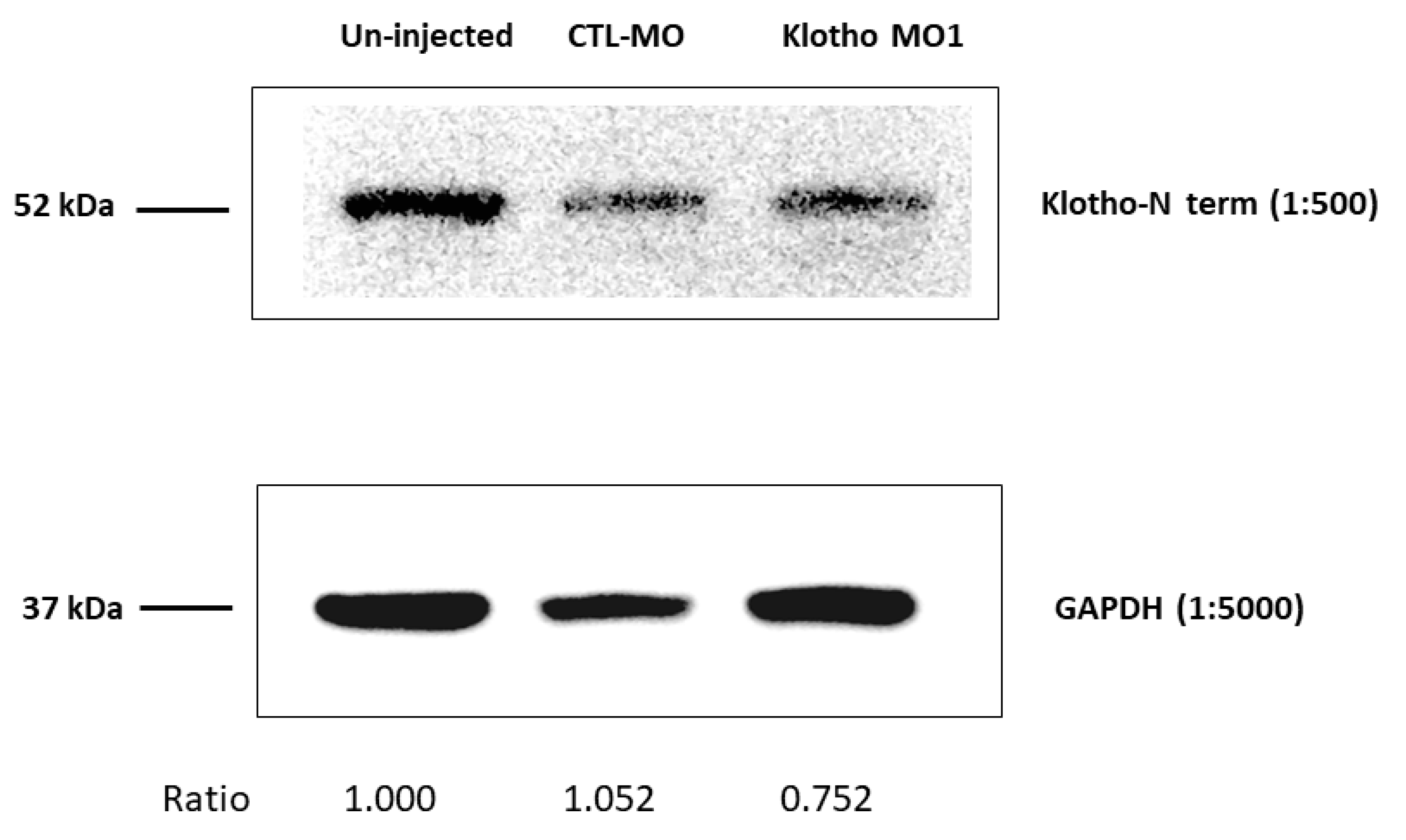
2.3. Embryonic Protein Extraction and Western Blotting

The embryonic protein lysates were extracted from the 5 hpf zebrafish embryos without morpholino injection (un-injected group), injected with CTL-MO, or injected with Klotho MO1 (around 60 embryos per group). The protocols for protein extraction and Western blotting were referred to as “The Zebrafish Book” as described previously [26]. Primary antibodies: Klotho N terminal region (Aviva, ARP36695_P050); GAPDH (14C10, Cell Signaling Technology, Danvers, MA, USA). Secondary antibodies: Anti-rabbit IgG, HRP-linked Antibody (Cell Signaling Technology). The bands were first visualized using the ECL method, and then their intensity was quantified by Image J software (Version 1.41, NIH free software).

2.4. Statistical Analysis

The data were expressed as averages ± SD and tested by Microsoft Excel TTEST. p < 0.05 was identified as statistically significant.

3. Results

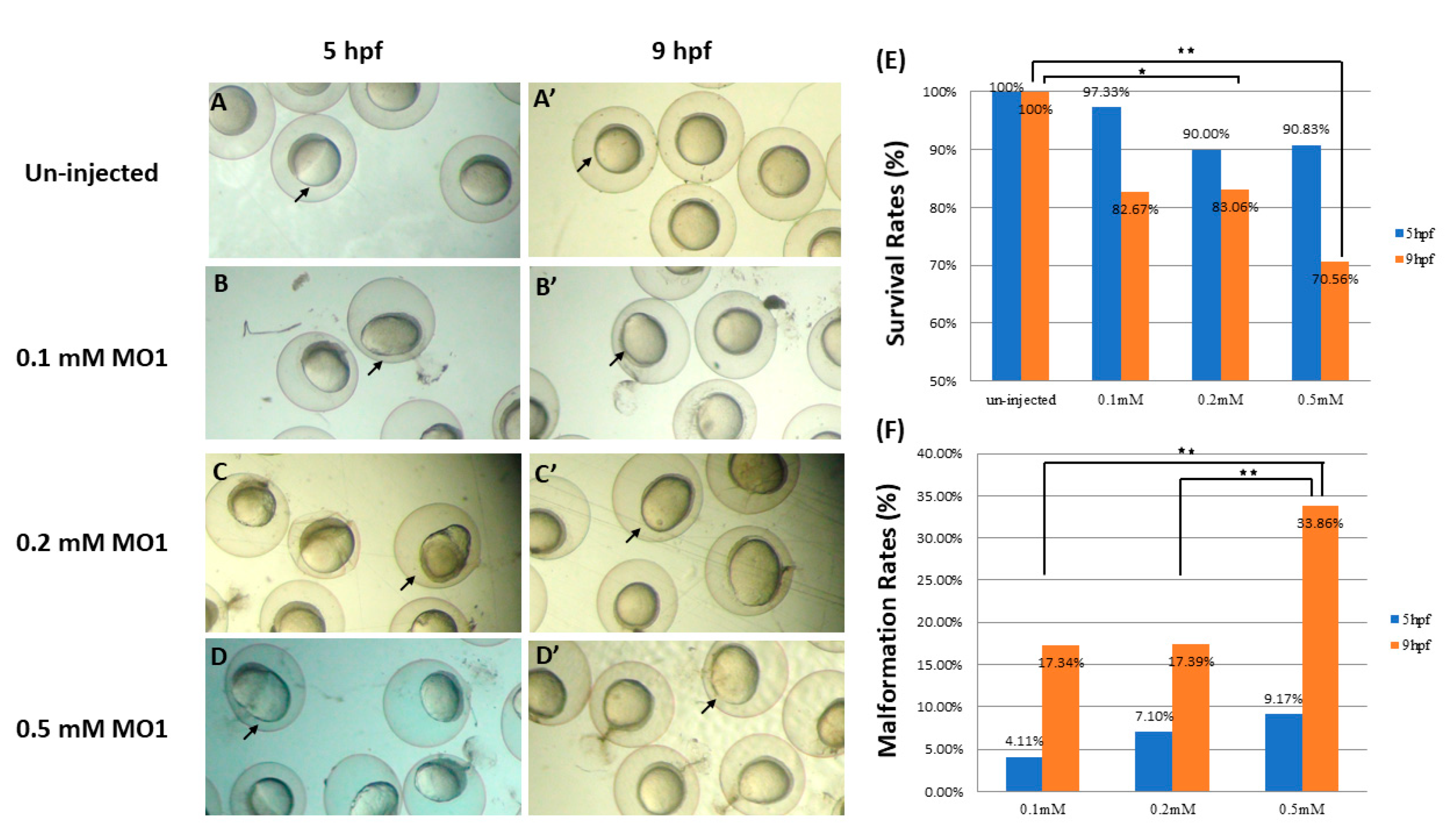
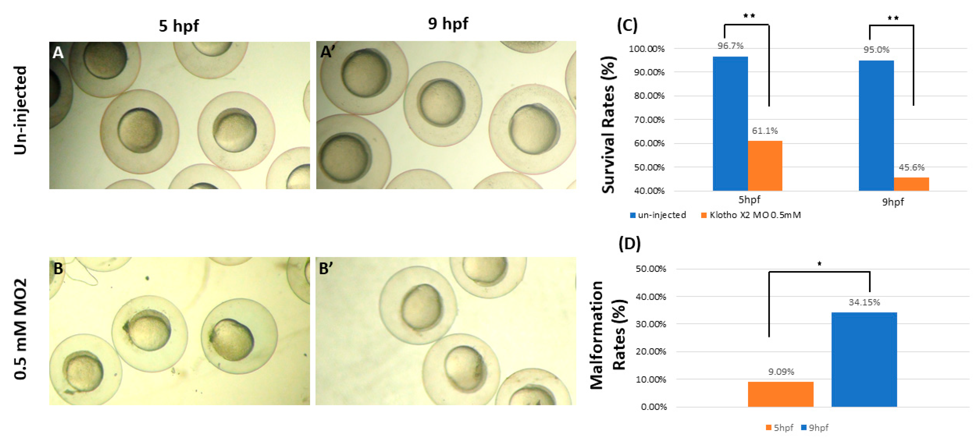
3.1. Knockdown of Zebrafish klotho Expression Led to Abnormal Embryonic Shapes

To determine the biological function of Klotho during zebrafish early development, we designed and injected two Klotho morpholinos (MO1 and MO2) to inactivate endogenous zebrafish klotho gene expression. Klotho MO1 was designed to target the translation start site (~1–8 a.a.), whereas Klotho MO2 was designed to target the N-terminal region (~68–73 a.a.) (Figure 1). Endogenous Klotho protein expressions were evaluated by Western blotting analysis, and results showed that in the Klotho MO1-injected group, the endogenous Klotho expressions were around 0.752 folds compared to those of the un-injected and CTL-MO-injected group (Figure 2). Furthermore, in the un-injected group, embryos showed normal migration patterns (50% epiboly by 5 hpf and 90% epiboly by 9 hpf, as shown in Figure 3A,A’, arrows, indicated). In the Klotho MO1-injected group, embryos displayed longitudinal shapes and swelling yolks compared to those of un-injected groups (Figure 3B,B’,C,C’,D,D’, arrows indicated). In addition, embryos that were co-injected with Klotho MO1 and P53 MO exhibited similar phenotypes, suggesting that those changes were not due to off-target effects. The survival rates in the Klotho MO1-injected groups had no significant differences among the different injection doses, 0.1, 0.2, and 0.5 mM (97.33% ± 1.33%~90.33% ± 2.31% by 5 hpf, 82.67% ± 6.02%~70.56 ± 10.48% by 9 hpf; Figure 3E, n = 60, N = 6). However, the malformation rates increased as the injection doses increased (17.34% ± 2.91%~33.86 ± 6.36% by 9 hpf, Figure 3F). A similar defective phenotypic type was observed in the 0.5 mM Klotho MO2-injected group (Figure 4). Thus, we suggest that the knockdown of endogenous klotho gene expressions led to longitudinal shapes and swelling yolks in the developing zebrafish embryos.

3.2. Knockdown of klotho Led to Longitudinal Shapes in a Dose-Dependent Manner

To further dissect whether the knockdown of klotho-induced longitudinal shapes is in a dose-dependent manner or not, we measured the length (L) and width (W) of each embryo from every experimental group (un-injected, 0.1, 0.2, and 0.5 mM, Figure 3A,A’,B,B’). As a result, the L/W ratios for un-injected (n = 38) is 1 ± 0; but for 0.1 mM (n = 57), 0.2 mM (n = 25), and 0.5 mM (n = 31), they are 1.2 ± 0.1, 1 ± 0.1, and 1.2 ± 0.1, respectively, indicating that L/W ratios had no significant differences by 5 hpf (Figure 5C). However, the L/W ratios by 9 hpf for un-injected (n = 67), 0.1 mM (n = 19), 0.2 mM (n = 19), and 0.5 mM (n = 48) are 1 ± 0.1, 1.1 ± 0.1, 1.2 ± 0.1, and 1.3 ± 0.1, respectively (Figure 5D). These observations suggest that longitudinal shapes induced by Klotho MO1 injection are dose-dependent.

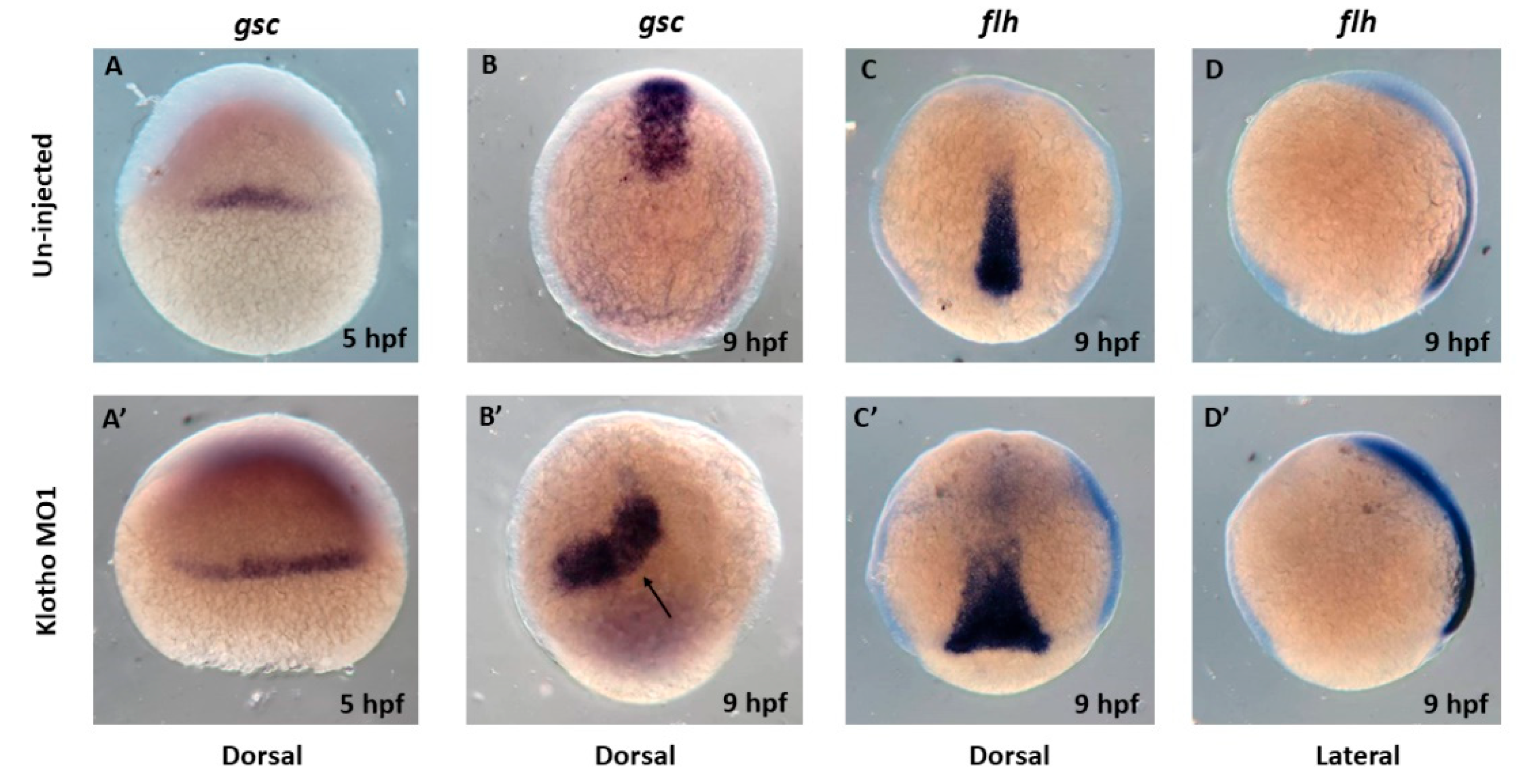
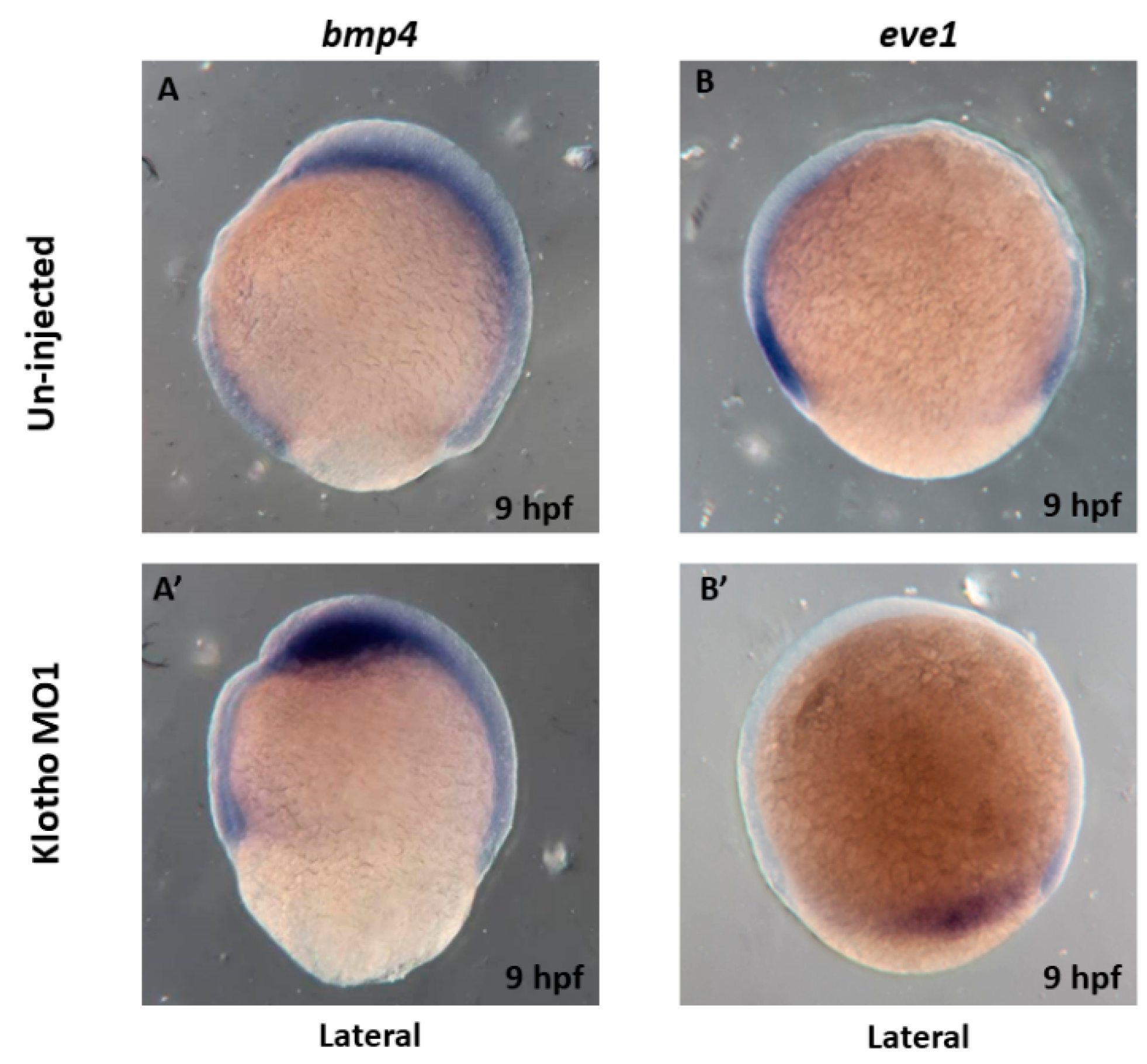
3.3. Longitudinal Shapes Embryos Might Be Due to Abnormal Cell Movement

To further investigate what caused longitudinal shapes in embryos in the Klotho MO-injected embryos, we performed whole-mount in situ hybridization experiments to examine several known cell migration gene markers. First of all, we examined the expressions of zebrafish goosecoid (gsc) in both the un-injected- and Klotho-MO-injected groups. At the early gastrulation stage (~5 hpf), gsc is expressed in the cell at the dorsal midline, and by 9 hpf, gsc is expressed in the cell anterior to the presumptive notochord [27]. Our results showed that embryos derived from the un-injected group displayed the same gsc expression patterns as previously described (Figure 6A,B). However, in Klotho MO1-injected embryos, the gsc expressed in the cell at the dorsal midline but in a broad range by 5 hpf (Figure 6A’). By 9 hpf, gsc expression domain was observed to have a heart-like shape, not restricted in the cell anterior to the presumptive notochord but in the bottom-right position closed to the midline (Figure 6B’). These observations indicate that cell movement is impaired in Klotho MO1-injected embryos. Next, we examined the expressions of the floating head (flh), a notochord marker (also known as a dorsal marker), and found that transcripts of flh were strongly detectable in the presumptive notochord region and extended toward the top (Figure 6C,D); but in the Klotho MO1-injected embryo, the flh-positive cells were accumulated in the anterior marginal zone in a more broadly defined range (Figure 6C’,D’, arrow indicates).
To further explore the molecular mechanism underlying the cell movement impairment in the Klotho MO1-injected embryos, we next examined the expressions of bone morphogenetic protein 4 (bmp4) and even-skipped-liked 1 (eve1) at 9 hpf. Signals for bmp4, a ventral posterior marker [28], were distributed evenly in the future head and the ventral regions in un-injected embryos (Figure 7A), but in the Klotho MO1-injected embryo, the bmp4 signals were weak in the ventral and seemed to be accumulated in the future head region (Figure 7A’). Finally, the signals for eve1, a ventral marker, were easily detected in the ventral region in un-injected embryos (Figure 7B), but the expression domains of eve1 in Klotho MO-injected embryos appeared in the anterior-dorsal parts of the marginal zone (Figure 7B’). Taken together, we suggested that Klotho played a role in cell movement in the zebrafish gastrulation stage.

4. Discussion

As previously reported, endogenous klotho mRNA expression could be detected as early as 5 hpf through adulthood. In adult tissue, klotho mRNA expression was detected in many organs, including the testis and ovary [10]. In this study, we also showed that endogenous Klotho protein expression could be observed as early as 5 hpf (Figure 3). These observations suggested that Klotho might play a role in early development. However, results revealed by Singh et al. [13] and Ogura et al. [12] do not report any defect in gastrulation in Klotho mutation zebrafish (possessed a truncated form of Klotho, 1–305 amino acids). This inconsistency might be due to hypomorphic effects or maternal inheritance of Klotho protein. It needs to be further investigated.
In zebrafish gastrula stages, epiblasts underwent epiboly, and notochord precursor cells’ convergence and extension are two obvious events for observing cell movements. Under normal conditions, epiboly will reach 50% by 5 hpf and 90% by 9 hpf. Notochord precursors first appeared in the anterior germ ring, migrated to the embryonic shield, and then underwent convergence and extended to the future head region. Once the process of cell movement is impaired, phenotypic changes can easily be observed. Our results showed that Klotho MO-injected embryos performed abnormal epiboly (Figure 3 and Figure 4) and migration defects in gsc- and flh-positive cells (Figure 6). This study provides new evidence that zebrafish Klotho protein could be associated with cell movement.
During zebrafish gastrulation, cell movement is regulated by several factors, including WNT, BMP, Nodal, and FGF signals [29]. In particular, the FGF signal has demonstrated that it can regulate gastrulation cell movements and morphology through a target, the neurotrophin receptor homolog [30]. Klotho was also reported to be an FGF co-receptor [31], and could be involved in the BMP signaling pathway [32]. Our results showed that bmp4 expression was affected in the Klotho MO-injected embryos (Figure 7A vs. Figure 7A’). These observations suggest that Klotho may play a role during gastrulation through these signal transduction pathways.
In the blastula stage, cells will undergo a series of mitosis but not movement. After midblastula transition (~3 hpf), cells begin to move and enter the gastrulation stage [33]. In this regard, if the mitosis process in the blastula is impaired, it might lead to the following gastrulation defects. Our previous study showed that klotho knockdown caused dysregulated cytokinesis [8]. Thus, we suggested that Klotho might be involved not only in cytokinesis but also in gastrulation cell movement.

5. Conclusions

In conclusion, this study provides a novel insight into studying the biological role of Klotho during early embryonic development. We found that inhibition of klotho expression by morpholino injection led to cell migration defects during the zebrafish gastrulation stage.

Author Contributions

H.-C.P. and C.-Y.S. conceived the experiments; K.-C.L., Y.-H.W., H.-T.H. and S.-C.C. performed the experiments and analyzed the data; Y.-H.C. wrote the paper. All authors have read and agreed to the published version of the manuscript.

Funding

The study is supported by the research grant of Chang Gung Memorial Hospital (CMRPG2K0093 and CORPG2M0131).

Institutional Review Board Statement

The animal studies and all procedures were approved by the Use of Laboratory Animal Committee, Tamkang University (TKU106003).

Informed Consent Statement

Not applicable.

Data Availability Statement

All data used to support the findings of this study are available from the corresponding author upon request.

Acknowledgments

The study is supported by the research grant of Chang Gung Memorial Hospital (H.-C.P., Grant No: CMRPG2K0093 and C.-Y.S., Grant No: CORPG2M0131). We are also grateful to the Zebrafish Core in Academia Sinica (ZCAS) for providing the AB strain of zebrafish.

Conflicts of Interest

The authors declare no conflict of interest.

References

  1. Kurosu, H.; Yamamoto, M.; Clark, J.D.; Pastor, J.V.; Nandi, A.; Gurnani, P.; McGuinness, O.P.; Chikuda, H.; Yamaguchi, M.; Kawaguchi, H.; et al. Suppression of aging in mice by the hormone Klotho. Science 2005, 309, 1829–1833. [Google Scholar] [CrossRef] [PubMed]
  2. Xu, Y.; Sun, Z. Molecular basis of klotho: From gene to function in aging. Endocr. Rev. 2015, 36, 174–193. [Google Scholar] [CrossRef]
  3. Kuro-o, M. Klotho and aging. Biochim. Biophy. Acta 2009, 1790, 1049–1058. [Google Scholar] [CrossRef]
  4. Donate-Correa, J.; Martín-Núñez, E.; Mora-Fernández, C.; Muros-de-Fuentes, M.; Pérez-Delgado, N.; Navarro-González, J.F. Klotho in cardiovascular disease: Current and future perspectives. World J. Biol. Chem. 2015, 6, 351–357. [Google Scholar] [CrossRef] [PubMed]
  5. Nie, F.; Wu, D.; Du, H.; Yang, X.; Yang, M.; Pang, X.; Xu, Y. Serum Klotho protein levels and their correlations with the progression of type 2 diabetes mellitus. J. Diabetes Compl. 2017, 31, 594–598. [Google Scholar] [CrossRef]
  6. Markiewicz, M.; Panneerselvam, K.; Marks, N. Role of Klotho in migration and proliferation of human dermal microvascular endothelial cells. Microvasc. Res. 2016, 107, 76–82. [Google Scholar] [CrossRef]
  7. Yu, S.; Chen, Y.; Chen, S.; Ye, N.; Li, Y.; Sun, Y. Klotho inhibits proliferation and migration of angiotensin II-induced vascular smooth muscle cells (VSMCs) by modulating NF-kB p65, Akt, and extracellular signal regulated kinase (ERK) signaling activities. Med. Sci. Monit. 2018, 24, 4851–4860. [Google Scholar] [CrossRef]
  8. Sun, C.Y.; Chou, C.Y.; Hsieh, Y.T.; Lo, K.C.; Liou, Y.L.; Chen, Y.H. Klotho exerts an emerging role in cytokinesis. Genes 2020, 11, 1048. [Google Scholar] [CrossRef] [PubMed]
  9. Typiak, M.; Piwkowska, A. Antiinflammatory actions of Klotho: Implications for therapy of diabetic nephropathy. Int. J. Mol. Sci. 2021, 22, 956. [Google Scholar] [CrossRef] [PubMed]
  10. Sugano, Y.; Lardelli, M. Identification and expression analysis of the zebrafish orthologue of Klotho. Dev. Genes Evol. 2011, 221, 179–186. [Google Scholar] [CrossRef] [PubMed]
  11. Zupkovitz, G.; Kabiljo, J.; Martin, D.; Laffer, S.; Schöfer, C.; Pusch, O. Phylogenetic analysis and expression profiling of the Klotho gene familyin the short-lived African killifish Nothobranchius furzeri. Dev. Genes Evol. 2018, 228, 255–265. [Google Scholar] [CrossRef] [PubMed]
  12. Ogura, Y.; Kaneko, R.; Ujibe, K.; Wakamatsu, Y.; Hirata, H. Loss of αklotho causes reduced motor ability and short life span in zebrafish. Sci. Rep. 2021, 11, 15090. [Google Scholar] [CrossRef] [PubMed]
  13. Singh, A.P.; Sosa, M.X.; Fang, J.; Shanmukhappa, S.K.; Hubaud, A.; Fawcett, C.H.; Molind, G.J.; Tsai, T.; Capodieci, P.; Wetzel, K.; et al. αKlotho regulates age-associated vascular calcification and lifespan in zebrafish. Cell Rep. 2019, 28, 2767–2776. [Google Scholar] [CrossRef] [PubMed]
  14. Chang, B.; Kim, J.; Jeong, D.; Jeon, S.; Jung, S.I.; Yang, Y.; Kim, K.I.; Lim, J.S.; Kim, C.; Lee, M.S. Klotho inhibits the capacity of cell migration and invasion in cervical cancer. Oncol. Rep. 2012, 28, 1022–1028. [Google Scholar] [CrossRef] [PubMed]
  15. Dehghani, M.; Brobey, R.; Wang, Y.; Souza, G.; Amato, R.J.; Rosenblatt, K.P. Klotho inhibits EGF-induced cell migration in Caki-1 cells through inactivation of EGFR and p38 MAPK signaling pathways. Oncotargets 2018, 9, 26737–26750. [Google Scholar] [CrossRef] [PubMed]
  16. Huang, Q.; Chen, Y.; Shen, S.; Wang, Y.; Liu, L.; Wu, S.; Xu, W.; Zhao, W.; Lin, M.; Wu, J. Klotho antagonizes pulmonary fibrosis through suppressing pulmonary fibroblasts activation, migration, and extracellular matrix production: A therapeutic implication for idiopathic pulmonary fibrosis. Aging 2020, 12, 5812–5831. [Google Scholar] [CrossRef] [PubMed]
  17. Gilbert, S.F. Developmental Biology, 6th ed.; Sinauer Associates, Inc. Press: Sunderland, MA, USA, 2000. [Google Scholar]
  18. Rohde, L.A.; Heisenberg, C.-P. Zebrafish gastrulation: Cell movements, signals and mechanisms. Int. Rev. Cytol. 2007, 261, 159–192. [Google Scholar]
  19. Marlow, F. Setting up for gastrulation in zebrafish. Curr. Top. Dev. Biol. 2020, 136, 33–83. [Google Scholar] [PubMed]
  20. Wu, H.J.; Fong, T.H.; Chen, S.L.; Wei, J.C.; Wang, I.J.; Wen, C.C.; Chang, C.Y.; Chen, X.G.; Chen, W.Y.; Chen, H.M.; et al. Perturbation of cytosolic calcium by 2-aminoethoxydiphenyl borate and caffeine affects zebrafish myofibril alignment. J. Appl. Toxicol. 2015, 35, 287–294. [Google Scholar] [CrossRef]
  21. Tsai, J.N.; Sun, C.Y.; Ding, Y.J.; Wang, Y.H.; Lo, K.C.; Weng, C.C.; Lin, J.W.; Chang, C.F.; Hsu, L.S.; Chen, H.M.; et al. Embryonic exposure to 4-methylimidazole leads to zebrafish myofibril misalignment. Environ. Toxicol. 2018, 33, 1321–1328. [Google Scholar] [CrossRef]
  22. Toyama, R.; O’Connell, M.L.; Wright, C.V.; Kuehn, M.R.; Dawid, I.B. Nodal induces ectopic goosecoid and lim1 expression and axis duplication in zebrafish. Development 1995, 121, 383–391. [Google Scholar] [CrossRef] [PubMed]
  23. Hammerschmidt, M.; Serbedzija, G.N.; McMahon, A.P. Genetic analysis of dorsoventral pattern formation in the zebrafish: Requirement of a BMP-like ventralizing activity and its dorsal repressor. Genes Dev. 1996, 10, 2452–2461. [Google Scholar] [CrossRef] [PubMed]
  24. Griffin, K.; Patient, R.; Holder, N. Analysis of FGF function in normal and no tail zebrafish embryos reveals separate mechanisms for formation of the trunk and the tail. Development 1995, 121, 2983–2994. [Google Scholar] [CrossRef]
  25. Lee, G.H.; Chang, M.Y.; Hsu, C.H.; Chen, Y.H. Essential roles of basic helix-loop-helix transcription factors, Capsulin and Musculin, during craniofacial myogenesis of zebrafish. Cell. Mol. Life Sci. 2011, 68, 4065–4078. [Google Scholar] [CrossRef] [PubMed]
  26. Westerfield, M. The Zebrafish Book: A Guide for the Laboratory Use of Zebrafish (Danio Rerio), 5th ed.; University of Oregon Press: Eugene, OR, USA, 2007. [Google Scholar]
  27. Schulte-Merker, S.; Hammerschmidt, M.; Beuchle, D.; Cho, K.W.; De Robertis, E.M.; Nüsslein-Volhard, C. Expression of zebrafish goosecoid and no tail gene products in wild-type and mutant no tail embryos. Development 1994, 120, 843–852. [Google Scholar] [CrossRef]
  28. Stickney, H.L.; Imai, Y.; Draper, E.; Moens, C.; Talbot, W.S. Zebrafish bmp4 functions during late gastrulation to specify ventroposterior cell fates. Dev. Biol. 2007, 310, 71–84. [Google Scholar] [CrossRef] [PubMed]
  29. Morgani, S.M.; Hadjantonakis, A.-K. Signaling regulation during gastrulation: Insights from mouse embryos and in vitro system. Curr. Top. Dev. Biol. 2020, 137, 391–431. [Google Scholar]
  30. Chung, H.A.; Hyodo-Miura, J.; Nagamune, T.; Ueno, N. FGF signal regulates gastrulation cell movements and morphology through its target NRH. Dev. Biol. 2005, 282, 95–110. [Google Scholar] [CrossRef] [PubMed]
  31. Urakawa, I.; Yamazaki, Y.; Shimada, T.; Iijima, K.; Hasegawa, H.; Okawa, K.; Fujita, T.; Fukumoto, S.; Yamashita, T. Klotho converts canonical FGF receptor into a specific receptor for FGF23. Nature 2006, 444, 770–774. [Google Scholar] [CrossRef]
  32. Lin, Y.; Sun, Z. Klotho-deficiency-induced arterial calcification involves osteoblastic transition of VSMCs and activation of BMP signaling. J. Cell. Physiol. 2022, 237, 720–729. [Google Scholar] [CrossRef]
  33. Kan, D.A.; Kimmel, C. The zebrafish midblastula transition. Development 1993, 119, 447–456. [Google Scholar] [CrossRef] [PubMed]
Figure 1. Location of Klotho MO1 and MO2. Zebrafish klotho sequence was obtained from NCBI website according to an accession number XM_685705. The sequence of Klotho MO1 was based on the nucleotide positions 51–65 (for amino acids position, ~1–8 a.a.). The red bold ATG represents the translation start site of klotho gene. The sequence of Klotho MO2 was based on the nucleotide positions 202–256 (for amino acids position, ~68–73 a.a.).
Fishes 08 00440 g001
Figure 2. Endogenous Klotho protein expressions were detected in zebrafish embryos. Proteins were extracted from un-injected, CTL-MO-injected, or Klotho MO1-injected-embryos, and their endogenous Klotho protein expressions were detected by Western blotting analysis. Upper panel: a rabbit polyclonal antibody against the Klotho N-terminal region was used to detect the endogenous zebrafish Klotho protein expression among each group. A 52 kDa band was observed as indicated. Bottom panel: a rabbit monoclonal antibody against GAPDH was used as the loading control. A 37 kDa band was observed as indicated.
Fishes 08 00440 g002
Figure 3. Morphology, survival rates and malformation rates analysis in zebrafish embryos. Pictures of zebrafish embryos without injecting (A,A’) or being injected with 0.1 mM (B,B’), 0.2 mM (C,C’), or 0.5 mM (D,D’) of Klotho MO1. The survival rates and malformation rates analysis for each group were shown in panels (E,F). Photos were taken by 5 hpf (AD) or 9 hpf (A’D’). (* p < 0.05; ** p < 0.01). Arrows in (A,A’): normal cell migration; arrows in (B,B’,C’,C’,D,D’): malformed embryos.
Fishes 08 00440 g003
Figure 4. Morphology, survival rates and malformation rates analysis in Klotho MO2-injected embryos. Pictures of zebrafish embryos without injecting (A,A’) or being injected with 0.5 mM (B,B’) of Klotho MO2. The survival rates and malformation rates analysis for each group were shown in panels (C,D). Photos were taken by 5 hpf (A,B) or 9 hpf (A’,B’). (* p < 0.05; ** p < 0.01).
Fishes 08 00440 g004
Figure 5. Measurement of the length/width (L/W) ratio in the zebrafish embryos. Pictures of zebrafish embryos without injecting (A,B) or being injected with 0.5 mM of Klotho MO1 (A’,B’). Statistical analysis of the L/W ratio in each group was shown at (C) 5 hpf, and (D) 9 hpf. (* p < 0.05).
Fishes 08 00440 g005
Figure 6. Zebrafish embryos derived from (AD) the un-injected group or (A’D’) the Klotho MO-injected group were stained by either the anti-sense gsc (A,B,A’,B’) or flh (C,D,C’,D’) riboprobe through whole mount in situ hybridization experiment. (A,A’) By 5 hpf, and (BD,B’D’) by 9 hpf. Arrow: abnormal expression pattern.
Fishes 08 00440 g006
Figure 7. Expressions of bone morphogenetic protein 4 (bmp4) and even-skipped-liked 1 (eve1) were affected in the Klotho Mo-injected embryos. Zebrafish embryos derived from (A,B) the un-injected group or (A’,B’) the Klotho MO-injected group were stained by either the anti-sense bmp4 (A,A’) or eve1 (B,B’) riboprobe through a whole-mount in situ hybridization experiment by 9 hpf.
Fishes 08 00440 g007
Disclaimer/Publisher’s Note: The statements, opinions and data contained in all publications are solely those of the individual author(s) and contributor(s) and not of MDPI and/or the editor(s). MDPI and/or the editor(s) disclaim responsibility for any injury to people or property resulting from any ideas, methods, instructions or products referred to in the content.

