Article

Potential Probiotic Bacillus Strains with Antioxidant and Antimutagenic Activity Increased Weight Gain and Altered hsp70, cxc, tnfα, il1β, and lysC Gene Expression in Clarias gariepinus

by
Radomir Viktorovich Skripnichenko
1,2,*,
Daria Sergeevna Chelombitskaya
2,
Evgeniya Valer’evna Prazdnova
1,2,*,
Maxim Pavlovich Kulikov
1,2,
Alexey Mikhailovich Neurov
1,2,
Anna Andreevna Zaikina
2,
Vadim Alekseevich Grigoryev
1,3,
Marina Nikolaevna Sorokina
1,3,
Vladimir Anatolievich Chistyakov
1,
Michael Leonidas Chikindas
1,4,5 and
Dmitriy Vladimirovich Rudoy
1
1
Center for Agrobiotechnology, Don State Technical University, Gagarina Sq. 1, Rostov-on-Don 344002, Russia
2
Academy of Biology and Biotechnology, Southern Federal University, Stachki Ave., 194/1, Rostov-on-Don 344090, Russia
3
Federal Research Center, Southern Scientific Center of the Russian Academy of Sciences, Chekhov Ave. 4, Rostov-on-Don 344006, Russia
4
Health Promoting Naturals Laboratory, School of Environmental and Biological Sciences, Rutgers State University, 65 Dudley Road, New Brunswick, NJ 08901, USA
5
Department of General Hygiene, I.M. Sechenov First Moscow State Medical University, Trubetskaya Str. 8, Bldg 2, Moscow 119991, Russia
*
Authors to whom correspondence should be addressed.
Fishes 2024, 9(12), 476; https://doi.org/10.3390/fishes9120476
Submission received: 23 October 2024 / Revised: 15 November 2024 / Accepted: 21 November 2024 / Published: 25 November 2024
(This article belongs to the Special Issue The Effects of Feed on the Growth Immunity and Metabolism of Fishes)

Abstract

:
The potential probiotic properties of three Bacillus strains were studied. A probiotic supplement for the African catfish Clarias gariepinus was produced via the solid-state fermentation protocol and incorporated into the fish feed for a period of seven weeks. Since the 36th day of the experiment, all experimental groups had a statistically significant increase in their weight gain than the control group. The maximum weight gain observed in fish fed the probiotic-supplemented feed was 29.16% higher than that of the control group, and the maximum feed conversion rate improvement was 24%. Cell-free extracts from these strains showed antioxidant (11.55–27.40%) and DNA-protective (45.33–61.83%) activity in a series of in vitro biosensor tests. Further investigation into the antimutagenic activity of the strains revealed that two of them reduced the level of induced mutagenesis in an Escherichia coli model (by 33.58% and 54.35%, respectively). We also assessed the impact of probiotic strains on the expression of several key genes in the host (C. gariepinus), including hsp70, cxc, tnfα, il1β, and lysC. More than a 10-fold increase in expression rates was observed for hsp70 in gonads and liver; for cxc in muscles and gonads; for tnfα in brain, gills, and liver; for il1β in the brain, gills, gonads, and liver; and for lysC in gills, gonads, liver, and muscles. This study provides evidence that probiotics exhibiting antioxidant and antimutagenic properties can provide significant benefits in vivo within aquaculture systems. The molecular effects of these probiotics appear to be complex and tissue-specific, with both upregulation and downregulation of immune system genes observed. Nevertheless, at the organismal level, the impact was unequivocally positive in terms of aquaculture objectives, manifested as enhanced body weight gain in the fish. Consequently, these Bacillus strains warrant serious consideration as potential probiotics for this species.
Key Contribution: The probiotic potential of three Bacillus strains exhibiting antioxidant and antimutagenic activity was assessed in Clarias gariepinus. Bacillus velezensis R5 demonstrated the most significant enhancement of weight gain (29.16% increase) compared to the other two strains. This treatment resulted in alterations in gene expression within the brain, gills, gonads, liver, and muscle tissues of the fish, suggesting a potential mechanism for the observed growth promotion.

1. Introduction

Probiotics are widely used in aquaculture to enhance the health of fish, particularly C. gariepinus (reviewed in [1,2,3]). Developing novel probiotic formulations has shifted the focus from identifying new sources to selecting the best criteria for in vitro screening.
Antioxidant and antimutagenic (DNA-protective) activity, demonstrably linked to probiotic efficacy in in vivo trials [4], represents a promising criterion. Such activities have been associated with positive systemic effects in avian and other agricultural species, including improved reproductive metrics and reduced reproductive senescence [5,6]. External antioxidants have been shown to improve immune system function, in particular by intercepting excess free radicals generated by the cells of the immune system itself. [7]. While the precise mechanisms of this relationship remain incompletely understood, research in this field is accumulating over time.
This study investigated whether this effect translates to aquaculture fish. To assess potential systemic probiotic effects, we examined changes in the expression of key genes indicative of fish health status, using analogous models (see below).
Understanding the mechanisms of action of probiotics requires consideration of their influence on the host defense systems. Studies have demonstrated that probiotics can upregulate genes associated with inflammatory responses and post-translational modifications in fish, highlighting the profound impact of the gut microbiome on host gene expression related to immunity and metabolism [8]. Further investigation of these effects may elucidate immunomodulation mechanisms [9], probiotic influence on host signaling pathways, the gut–brain axis, and other phenomena [10].
Such “probiotics-host” interactions in fish are still under-researched, although some studies have explored the effects of probiotics on gene expression, focusing on interleukins [11,12]. A recent study by Lee et al. [13] reported no effect of Pseudomonas probiotics on gene expression, yet other groups of probiotics may exert different beneficial effects [14]. Therefore, further investigation of novel probiotics for aquaculture at various levels, including epigenetics, seems worthwhile.
C. gariepinus is a suitable model organism for probiotic studies. Its ready acceptance of artificial diets [15] facilitates controlled feeding experiments, enabling standardized diets and consistent probiotic delivery across experimental groups. The combination of rapid growth, enhanced disease resistance, reproductive efficiency, and adaptability to artificial diets allows for diverse experimental designs. Its importance in Russian aquaculture [16] and wider use in other studies [17] further justify its selection for testing novel probiotics.
C. gariepinus also holds significant commercial importance in aquaculture, particularly in Africa and parts of Asia. Its rapid growth, enabling a relatively swift return on investment in aquaculture, allows it to reach substantial sizes, with reported maximum lengths of 1.7 m and weights of up to 59 kg [18]. High market demand, attributable to its palatable flesh and high nutritional value (approximately 15.65% protein content), drives its popularity both locally and internationally. C. gariepinus thrives in diverse environmental conditions, including saline and brackish water unsuitable for conventional agriculture. This adaptability supports aquaculture development in regions with limited agricultural potential [19].
The species is often used in integrated aquaculture systems, such as rice-fish farming, where it contributes to nutrient recycling, enhancing rice yields while providing additional protein sources [20]. The catfish industry provides significant employment opportunities and helps mitigate food insecurity by providing affordable protein sources in developing countries [21].
To assess the systemic effects of potential probiotics, we evaluated their impact on the expression of genes linked to stress response and immunity, specifically genes hsp70, cxc, tnfα, il1β, and lysC.
HSP70, a heat shock protein involved in cellular homeostasis against stressors in fish, serves as an early stress biomarker in C. gariepinus [22]. Chemokines (cxc) attract immune cells to infection or inflammation sites, alongside lysozyme. IL-1β and TNF-α are pro-inflammatory cytokines crucial in the innate immune response of fish [23,24].
The objective of this study was to investigate the effects of three new Bacillus strains, isolated from the microbiota of healthy fish and exhibiting antioxidant activity, on C. gariepinus. The key outcomes assessed were body weight gain, feed conversion efficiency, and gene expression related to immunity and stress.

2. Materials and Methods

2.1. Preparation of Cell-Free Supernatants (CFSs) of Strains

The strains were grown on 37 °C LB medium for 48 h. Cell-free preparations were obtained via 20 min centrifugation at 14,000× g using MiniSpin (Eppendorf, Hamburg, Germany), followed by filtration. Serial tenfold dilutions were prepared, with final preparation concentrations on the plates of 0.001, 0.01, and 0.1 of the original culture supernatant.

2.2. Biosensor Test on Antioxidant and DNA-Protective Activity

Cell-free supernatants of the strains were assessed for antioxidant activity against hydrogen peroxide and superoxide anion radical, as well as for DNA protective activity, using a panel of LUX biosensors based on E. coli. Biosensor strains MG 1655 (pSoxS-lux), MG1655 (pKatG-lux), MG1655 (pRecA-lux), and MG1655 (pColD-lux), harboring plasmids expressing the luxCDABE operon of Photorhabdus luminescens under control of the respective stress-inducible E. coli promoters (PsoxS, PkatG, PrecA), were used. This operon encodes the bacterial luciferase and its regulators. The resulting bioluminescence, serving as a reporter function, was quantified using a luminometer. Biosensors with PkatG and PsoxS promoters detect oxidants that form hydroperoxides and superoxide anion radicals, respectively, in the cell. The pRecA biosensor detects DNA-damaging agents [25].

2.3. Inducers and Protectors

1,4-Dioxide 2,3-quinoxaline dimethanol (dioxidine) (Biosintez, Penza, Russia) at a concentration of 2.25 × 10⁻⁵ M was used as a DNA damaging agent; hydrogen peroxide at 10−3 M and paraquat (1,1′-dimethyl-4,4′-dipyridylium dichloride) at the same concentration were used as prooxidants.
Tocopherol acetate (Lumi, Saint Petersburg, Russia) at a concentration of 10−2 M served as a positive control for antioxidant and DNA-protective activity.

2.4. Growth Conditions

E. coli cell cultures were grown in an LB medium supplemented with ampicillin (100 μg/mL).
Bacterial cultures were grown in a liquid medium at 37 °C to the mid-logarithmic phase. Overnight cultures were diluted with fresh medium to an optical density (OD600) of 0.01–0.1 McFarland units (3 × 10⁶–3 × 10⁷ cells/mL), as determined using a DEN-1B densitometer (Biosan, Riga, Latvia). Cultures were then incubated for 2 h to reach the early logarithmic phase.

2.5. Testing of CFS and Bioluminescence Measurement

Ninety microlitres of the culture were transferred to each well of a 96-well plate. There were ten microlitres of the test preparation (or deionized water for control wells). Luminescence was measured using an LM-01T microplate luminometer (Immunotech, Prague, Czech Republic). The plate was placed in a luminometer and incubated at 30 °C. The bioluminescence intensity was measured every 10–15 min for 2 h. All experiments were performed in triplicate.
The SOS response/oxidative stress/DNA damage induction factor (Is) was calculated using the following formula:
Is = Le/Lk − 1,
where Lk—luminescence intensity of the control sample (c. u.); Le—luminescence intensity of the experimental sample (c. u.).
The protective activity index (A, %) was calculated by the following formula:
A = (1 − Ia/Ip) × 100%,
where Ia—the SOS response induction factor by the studied exposure in the presence of an inhibitor; Ip—SOS response induction factor by ciprofloxacin.
All experiments were performed in triplicate.

2.6. Culture Fluid Antimutagenic Activity Study

To assess the antimutagenic activity of metabolites from potential probiotic strains, 150 μL of the culture liquid of the studied strains was collected and centrifuged for 20 min at 13,400× g. The resulting supernatant was used for testing. Dioxidine (hydroxymethylquinoxylindioxide) was added to 900 μL of medium, acting as a mutagen according to a previously described method [26].
An overnight culture of E. coli MG1655 was grown in liquid LB medium at 37 °C for 18–20 h. The following options have been explored:
1. Control: 800 µL LB + 200 µL water;
2. Mutagen: 800 µL LB + 100 µL dioxidine solution + 100 µL water;
3. Antimutagen: 800 µL LB + 100 µL culture liquid + 100 µL water;
4. Antimugen + mutagen: 800 µL LB + 100 µL dioxidine solution + 100 µL culture liquid.
One hundred microlitres of culture were plated on LB agar plates with and without rifampicin at a concentration of 100 μg/mL according to the standard method. Colonies were counted after 48 h’ of incubation.

2.7. Preparation and Use of Potential Probiotic Formulations

The formulations of the potential probiotic strains were prepared via solid-phase fermentation as follows. The pure cultures of B. subtilis R1, B. subtilis R4, and B. velezensis R5 were used as inocula. Presoaked, autoclaved, and cooled to 60 °C soybeans were inoculated with the starter culture and thoroughly mixed, then incubated for 24 h at 45°. The fermented beans were ground in a grinder, and the resulting mass was dried and then milled in a coffee grinder. The technology is described in more detail in our previous works [6,27]. The preparations were then added to the feed formulation described below.

2.8. Fish Containment Condition

Juvenile C. gariepinus were used to evaluate the effects of the potential probiotic strains B. subtilis R1, B. subtilis R4, and B. velezensis R5. Fifty fish were randomly assigned to each of three replicate 100-litre tanks. There was no distinguishment between females and males since all fish and their gonads were immature. The average weight of a juvenile at the beginning of the trial was 10 g. Fish were fed the following (Table 1): wheat, pork meat meal, fish meal, chicken meat and bone meal, pork hemoglobin, corn gluten, gaprin, feeding yeast, flax meal, pea protein, rapeseed meal, soybean protein concentrate, toasted soybean meal, premix 4% (juvenile), refined rapeseed oil, and fish grease.
The feeding rate was set to 5% body weight. Experimental groups received the described basal diet supplemented with one of the potential probiotic preparations: B. subtilis R1 (Group 1), B. subtilis R4 (Group 2), or B. velezensis R5 (Group 3). The final concentration of potential probiotic bacilli in the feed in all three groups was the same and amounted to 105 CFU/g. The control group instead of the probiotic received the same feed supplemented with 1% of the soybean prepared as described in Section 2.7, excluding the inoculation step. The water flow rate during the cultivation was set to 1–3 cm/s, according to the fish mass of 50–1000 mg.

2.9. Evaluation of the Effects on Gene Expression Associated with Immunity and Stress

To assess the effect of the studied strains on the expression of genes associated with stress and immunity (hsp70, cxc, tnfα, il1β, and lysC) in the tissues of different C. gariepinus organs (muscles, brain, liver, gonads, and gills), quantitative real-time PCR was employed. The total RNA was isolated from tissue samples using the RNA Fixer solution (Evrogen, Moscow, Russia) for storage and the ExtraRNA kit (Evrogen). Reverse transcription was performed using the MMLV-Evrogen kit. The resulting cDNA was subjected to qRT-PCR analysis using qPCRmix-HS SYBR (Evrogen) in an ANK-32-M (Sintol, Moscow, Russia) qRT-PCR amplifier. Target gene expression was quantified using the 2−ΔΔCt method.
All target genes were normalized to the reference β-actin gene (actβ). Primers were partially designed using NCBI Blast and previous findings [28,29,30]. The nucleotide sequences are presented in Table 2.

2.10. Estimation of the Growth Parameters of C. gariepinus

To assess the effect of the potential probiotics on the weight gain of C. gariepinus, the fish were weighed on the 1st, 12th, 24th, 36th, 48th and 53rd (end of the experiment). Body weight gain (BWG) was calculated using the following formula:
BWG = FW − IW,
where BWG—total weight gain (g); FW—final weight (g); IW—initial weight (g).
To assess the growth rate of the fish, the average daily gain (ADG) was calculated using the following formula:
ADG = BWG/T,
where ADG—average daily gain (mg); BWG—total weight gain (g); and T—number of days on feed.
The feed conversion ratio (FCR) was calculated as follows:
FCR = FG/BWG,
where FG—total feed given (g); BWG—total weight gain (g).

2.11. Statistical Analysis

The statistical significance was evaluated using a t-test for normally distributed data and a Wilcoxon test for non-normally distributed data. The data normality check was performed using the Shapiro test. All calculations were performed in Rstudio (R 4.2.1).
In biosensor tests, the standard deviation of the induction factor S was calculated by the following formula:
S = I × sqrt ((sLe/Le)2 + (sLk/Lk)2),
where the subscripts e and k refer to experience and control, respectively.

3. Results

3.1. Evaluation of Antioxidant and DNA Protective Activity

Cell-free preparations were tested in a series of ten-fold dilutions. The maximum superoxide anion scavenging activity was observed at a 10% dilution of the original supernatant. No statistically significant effects were observed at other dilutions (p < 0.05). In contrast, DNA-protective activity was most pronounced at dilutions of 1–0.1% of the initial concentration, whereas higher concentrations exhibited bactericidal activity against E. coli and led to the death of biosensor cells (Figure 1).
Strains of Bacillus exhibited some antioxidant activity, with its indices ranging from 11.55% to 27.40%. The DNA protective activity of potential probiotic strains, determined using the E. coli MG1655 pRecA-lux biosensor, varied significantly among potential probiotic strains, although none exhibited an effect comparable to that of tocopherol (70.44%). The B. subtilis R4 strain displayed the most potent DNA-protective activity (61.83%), followed by B. subtilis R1 (57.87%) and B. velezensis R5 (45.33%). All values were significantly different from the control (p < 0.05), but no significant difference was observed between the DNA protective effects of B. subtilis R1 and B. subtilis R4.

3.2. Study of the Antimutagenic Activity of CFS

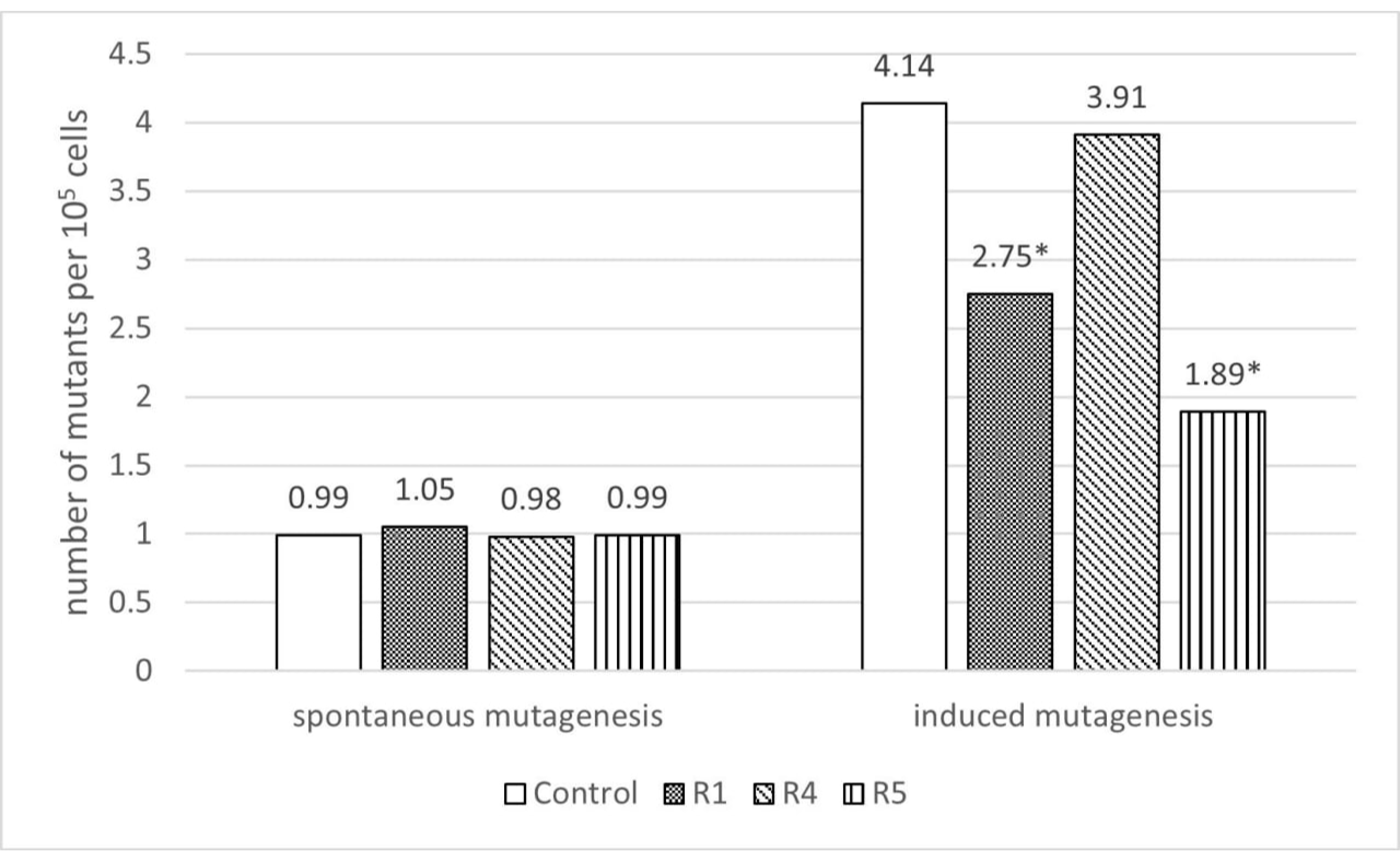
The results of antimutagenic activity testing are shown in Figure 2.
The frequency of spontaneous mutagenesis in E. coli MG1655 was 0.99 × 10⁻⁵, and the frequency of induced mutagenesis was 4.14 × 10⁻⁵. The addition of cell-free preparations of B. subtilis R1 and B. velezensis R5 strains did not affect the level of spontaneous mutagenesis but reduced induced mutagenesis (by 33.58% and 54.35%, respectively). B. subtilis R4 did not exhibit antimutagenic properties. Overall, these data correlate with the biosensor test, where the cell-free preparation of B. velezensis R5 had the most significant (p < 0.05) antigenotoxic properties.

3.3. Effects on Gene Expression

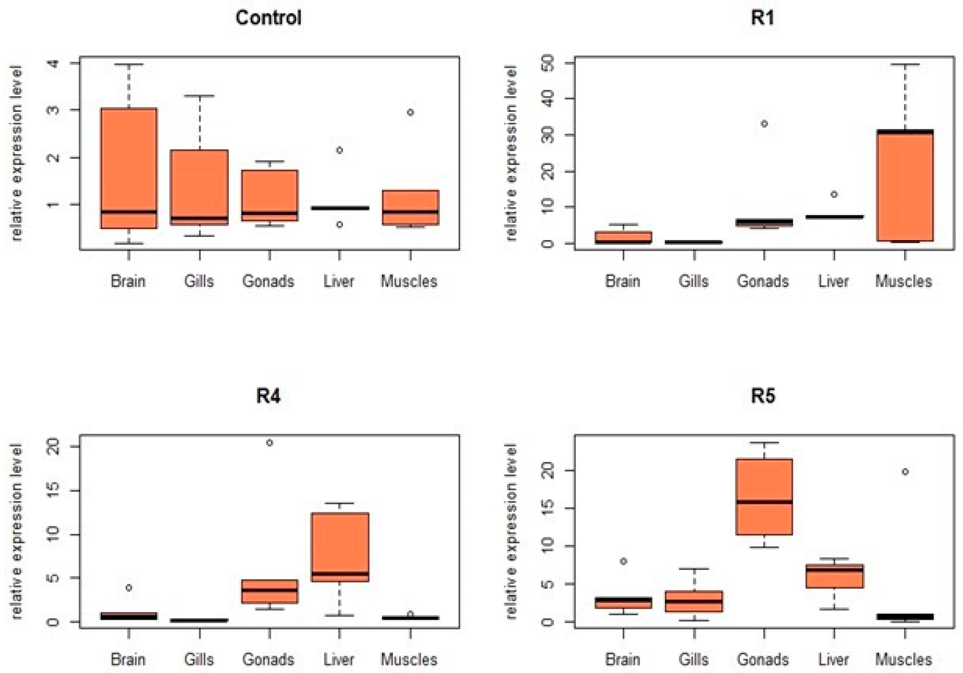
Hsp70 expression increased in all three experimental groups, but the tissue-specific impact of each potential probiotic strain differed. B. subtilis R1 increased the expression level of hsp70 in gills by 5.0 times, in gonads by 11.5 times, and in the liver by 16.2 times, but caused a slight decrease in brain and muscle. Both B. subtilis R4 and B. velezensis R5 increased the expression of hsp70 in gills, gonads, liver, and muscles by 3.6, 7.8, 8.1, 11.4, and 1.9, 3.6, 12.2, 7.6 times, respectively, with a modest reduction in its expression in the brain. In summary, all three strains increased the expression of hsp70 in all tissues except for the brain (and muscles in the case of B. subtilis R1), with increases in excess of 10-fold for the muscles, liver, and gonads (Figure 3).
B. subtilis R1 increased the cxc gene expression in muscle, hepatocytes, and gonads (2.6-, 8.5-, and 10.9-fold, respectively). Conversely, cxc expression in the brain and gills remained comparable to the control levels. B. velezensis R5 increased expression in all tissues, with the most pronounced effect in gonads (16.6-fold above control). B. subtilis R4 had the least impact on cxc expression. Only the brain, gonads, and liver exhibited increased expression (1.2-, 6.5- and 7.3-fold, respectively), whereas gills and muscle showed slight decreases (0.2- and 0.5-fold, respectively) (Figure 4).
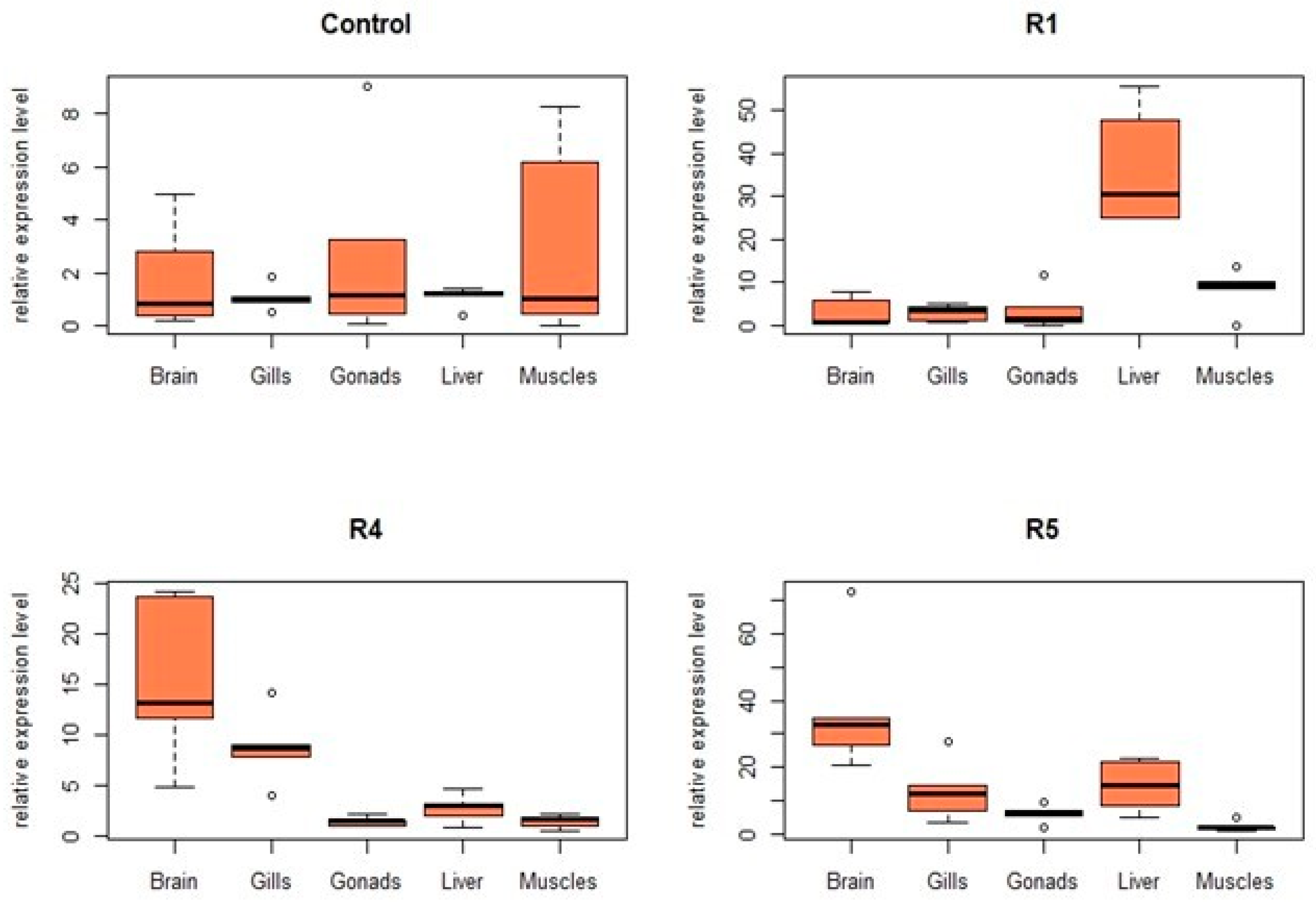
B. subtilis R1 significantly increased tnfα expression across all tissues, with the most pronounced effect observed in the liver (36.8-fold). B. subtilis R4 positively impacted the tnfα expression in the brain (15.4-fold), gills (8.7-fold), and liver (2.7-fold), with minor increases observed in both gonads and muscles (1.4-fold each). B. velezensis R5 remarkably enhanced the tnfα transcription in the brain (37.4-fold), gills (13-fold), and liver (14.5 times); a lesser positive effect was observed in the gonads (6.2-fold and muscles (2.4 times). Overall, all three strains demonstrated varying degrees of gene expression increase across all five tissues in our experiment (Figure 5).
Both B. subtilis R1 and B. velezensis R5 increased the il1β expression in all tissues, approximately 10-fold in the case of the brain, gonads, gills, and liver (Figure 6). B. subtilis R4, however, increased il1β expression in the brain (3-fold), gonads (1.6-fold), and gills (50-fold), with the latter effect being among the most pronounced observed. The other two tissues exhibited a slight decrease in il1β expression (Figure 6).
B. subtilis R1 significantly increased the lysC expression in the gonads and liver (32.9-fold and 36.6-fold, respectively), and to a lesser extent in the gills (8.2-fold). Muscle lysC expression was largely unaffected by B. subtilis R1, while a slight decrease was observed in brain tissue. B. subtilis R4 caused the most pronounced increase in lysC expression across all tissues, in gills and gonads, with 54-fold and 56.8-fold increases in gills and gonads, respectively, while brain expression remained largely unchanged. B. velezensis R5 also markedly increased lysC expression in four of five tissues (gills, gonads, liver, and muscles, by 33, 46.4, 27.2, and 44 times, respectively), and to a lesser extent in the brain (6.3-fold). Thus, lysC expression increased more than any other gene examined in this study (Figure 7).

3.4. Estimating the Increase in Weight Gain of C. gariepinus

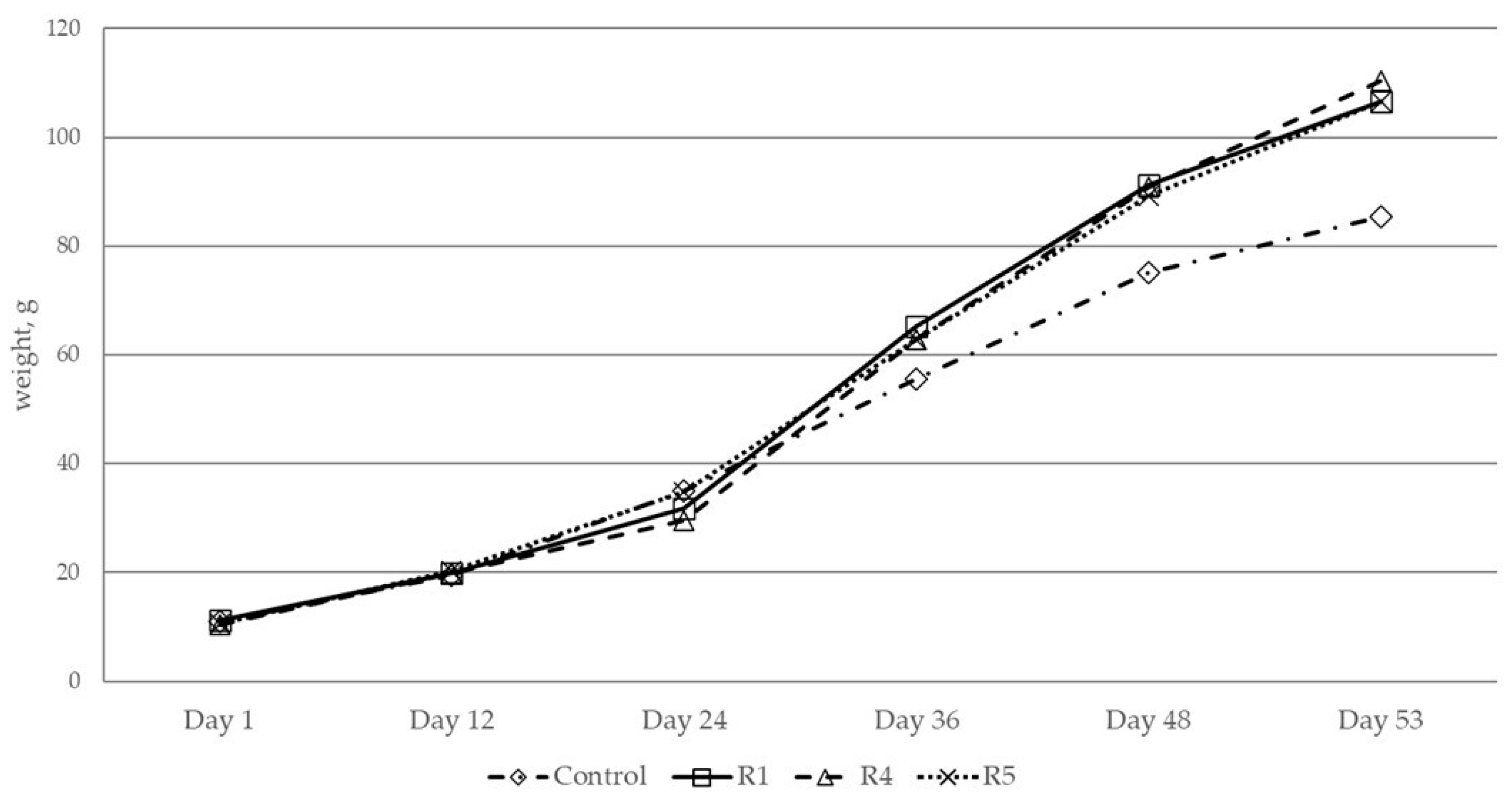
The results of C. gariepinus weighing (Figure 8 and Figure 9) show that the use of all three potential probiotics as a supplement to the main feed for C. gariepinus had a positive effect on weight gain. The weight gain curve and final values for the 53rd day of the experiment are given.
In Group 1, treated with B. subtilis R1 (initial weight 11.18 ± 1.43 g; final weight 106.63 ± 3.06 g), and Group 2, treated with B. subtilis R4 (initial weight 11.58 ± 1.13 g final weight 106.44 ± 3.07 g), the final weight was significantly greater than the control group (initial weight 11.06 ± 0.32 g; final weight 85.42 ± 2.96 g) by 24.83% and 24.61%, respectively (p < 0.05).
The largest increase in C. gariepinus weight was observed in group 3, treated with the B. velezensis R5 potential probiotic (initial weight 10.42 ± 0.32 g; final weight 110.33 ± 2.96 g). Compared to the control group (initial weight 11.06 ± 0.32 g; final weight 85.42 ± 2.96 g), the mass of C. gariepinus increased by 29.16% (p-value < 0.05).
Feed conversion ratios (Table 3) indicate that all three probiotic-supplemented diets were more efficient than the control, with a maximum improvement of 24% for B. subtilis R4.
Overall, the effects of the selected strains we identified can be summarized in Table 4.

4. Discussion

Bacillus strains have repeatedly demonstrated their health-promoting potential in aquaculture [31]. Their effectiveness is attributed to various mechanisms, including bacteriocin production, suppression of virulence gene expression, competition for adhesion sites with pathogens, stimulation of immunity and organic acid production [32], direct antimicrobial action [33], and quorum-sensing inhibition in pathogens [30].
B. velezensis, among other Bacillus probiotics, is considered a promising candidate due to its numerous functions in aquaculture [28]. Supplementation with B. velezensis has been shown to enhance the growth of Rhynchocypris lagowskii [34], stimulate immunity, exhibit antimicrobial and anti-stress effects in Oreochromis niloticus [35], and improve growth, hepatic metabolism, and gut microbiome in Pelteobagrus fulvidraco × Pelteobagrus vachelli [36]. In Oncorhynchus mykiss, the probiotic B. velezensis positively impacted growth, immunity, serum enzyme activity, gut microbiota, and resistance to Aeromonas salmonicida infection [37]. Bacillus amyloliquefaciens R8 improved hepatic glucose levels and lipid metabolism, reduced oxidative stress, and strengthened host immunity in Danio rerio, with increased expression of innate immunity genes also supporting its probiotic potential [38]. Similarly, Paralichthys olivaceus supplemented with Bacillus clausii exhibited enhanced growth and feed efficiency compared to the controls [39].
In C. gariepinus, supplementation with Bacillus cereus improved body mass, weight gain, growth rate, lysosomal activity, and serum antioxidant levels [40].
These findings align with our results, demonstrating weight gain in C. gariepinus supplemented with potential probiotics, with a maximum 29.16% increase observed in the group receiving B. velezensis R5.
Probiotic Bacillus strains, particularly B. subtilis, are known to enhance the viability of beneficial gut microbiota, such as Lactobacillus and Bifidobacterium species. This effect may be linked to the production of catalase and subtilisin by B. subtilis [41]. Given the antimutagenic properties of some probiotic Bacillus strains, they may protect other beneficial bacteria from harmful factors and enhance the overall antimutagenic potential of the host microbiota in conjunction with commensal microorganisms [4].
Importantly, the DNA-protective effects observed for B. subtilis R1 and B. velezensis R5 correlate with their antimutagenic activity (reduced chemically induced mutagenesis in E. coli). In contrast, only in vitro DNA protection was observed for B. subtilis R4. These differences may reflect the production of distinct metabolites by each strain. Future work should compare the metabolic profiles of these strains to identify specific antimutagenic compounds.
Interpreting changes in stress- and immunity-related gene expression requires consideration of the following facts. In numerous papers on fish models, the increased expression of such genes can be interpreted as either a negative effect of stressors (e.g., heavy metals) or a positive effect—activation of the host’s defense mechanisms (e.g., in response to immunostimulants like probiotics); often, practically identical effects are interpreted as opposite [42,43,44,45]. This apparent duality in interpretation highlights the need for a nuanced approach to evaluating these responses. Specifically, the magnitude of the response should be considered: moderate expression of pro-inflammatory cytokines, for example, may contribute to immune homeostasis and infection resistance [20], whereas exceeding a critical threshold could lead to negative outcomes such as chronic inflammation. A standardized scale for quantifying these effects in fish models is currently lacking.
Furthermore, when evaluating the effects of probiotics in animal feed, improvements in physiological parameters such as weight gain and feed conversion efficiency are strong indicators of a positive impact. In our study, all experimental groups exhibited significantly greater weight gain than the control group (24.83%, 24.60%, and 29.16%), suggesting commercial value for these probiotic interventions. However, the long-term consequences for fish health and the potential for extrapolation to other organisms require further investigation, as does the need for histopathological analysis to determine the pro- or anti-inflammatory nature of the probiotic effects.
Below, we discuss the effects on individual genes in the context of existing data.

4.1. Influence on the Stress and Immunity Genes

4.1.1. Influence on hsp70

The heat shock protein 70 (Hsp70) gene (hsp70) is often used as a biomarker of cellular stress. The HSP protein family plays a crucial role in cellular protection by preventing protein aggregation, assisting in protein folding, stabilizing and refolding damaged proteins, as well as directing incorrectly folded proteins to degradation pathways [46].
In fish models, the upregulation of hsp70 expression is considered a stress marker, with downregulation indicating a return to homeostasis. Nasrullah et al. [22] demonstrated hsp70 induction in C. gariepinus following bacterial infection, transportation, heat shock, and high nitrite exposure. Conversely, the introduction of mixed Bacillus species has been shown to enhance early innate immune reactions and reduce Hsp70 levels [47]. In several studies, the downregulation of hsp70 is also considered a positive effect of probiotics [30,48].
However, some studies on probiotic effects interpret hsp70 upregulation as an increased stress resilience in fish. For instance, tnfα, il1β, and hsp70 expressions have been observed to increase in fish fed with probiotics, with researchers concluding that probiotics enhance defense against hypoxia in O. niloticus [49]. Similarly, dietary supplementation with immunostimulants produced by probiotic bacteria resulted in an upregulation of hsp70 expression [50]. The upregulation of immunity-related genes, including those encoding hepatic HSP70, IL-1β, and TNF-α, splenic TNF-α and IL-1β, and intestinal C-lysozyme and TNF-α, was observed in O. niloticus at the 1.5–2-fold level [51].
We can conclude that research on the impact of probiotics on hsp70 expression in C. gariepinus shows both upregulation and downregulation, depending on the specific probiotic strain and experiment conditions [30].
In our research, we observed a decrease in hsp70 expression in the brain with all probiotics and an increase in the gills, gonads, and liver. Strains B. subtilis R4 and B. velezensis R5 also increased its level in the muscles. The most significant increase in activity was observed in the liver. This tissue-specific response, exhibiting both upregulation and downregulation, aligns with existing data.

4.1.2. Influence on the Immunity Genes

Currently, the precise mechanism by which probiotic bacteria modulate the fish immune system remains unclear. However, some hypotheses suggest a role for β-glucans as signaling molecules, recognized by immune cell receptors [40]. The interaction of β-glucans with receptors on macrophages and dendritic cells appears to trigger the production of various cytokines, which in turn activate B- or T-lymphocytes, generating a systemic immune response [52,53]. Oral probiotic administration is associated with increased immune parameters and upregulation of immunity-related genes, including those encoding IgM, TCR-ß, MHC-IIα, C3, TNF-α, and IL-1ß [52,53].
Studies using D. rerio models treated with various probiotics have also demonstrated significant alterations in immune gene expression following pathogen exposure [19].
Moderately increased transcription of immune-related genes, such as pro-inflammatory cytokines, may contribute to immune homeostasis and enhance infection resistance [20,54,55]. Potential mechanisms include the action of microbially associated molecular patterns (MAMPs), such as peptidoglycan, lipopolysaccharides, flagella, and nucleic acids, on Toll-like receptors [20,56,57]. However, these MAMPs can be produced by any bacteria, and this mechanism does not explain the specific effects of particular probiotic strains.

Influence on cxc

Chemokines are small secretory and transmembrane cytokines stimulating the production of leukocytes [58]. They also induce directed chemotaxis of macrophages and neutrophils in pathological and homeostatic conditions [59,60].
The CXC chemokine gene (cxc) is often used as an indicator of immune status in C. gariepinus. Increased cxc expression is generally considered a positive effect. Hamid et al. [30] found that probiotic treatment in C. gariepinus × Clarias macrocephalus hybrids resulted in a 1.9- to 2.2-fold increase in liver cxc expression and a 3- to 5-fold increase in head kidney cxc expression. Similar studies using Bacillus spp. in Oreochromis spp. showed increases in liver (1.5-fold), spleen (2.5-fold), and kidney (3.5-fold) cxc expression, suggesting a potential positive impact on immunity and infection resistance [61].
In our study, the potential probiotics B. subtilis R1 and B. subtilis R4 exhibited varied effects on cxc expression in different tissues, excluding the brain. In contrast, B. velezensis R5 demonstrated a 3- to 16-fold increase in cxc expression across all tissues, including the brain. This suggests a correlation between the intensity of the probiotic effect and cxc expression, with B. velezensis R5 being the most effective of the three strains tested.

Influence on tnfa

TNF-α is a key cytokine in fish, modulating inflammation, immunity, and development [62,63]. It also participates in adaptive immune response in teleosts by initiating apoptosis [53]. This gene is usually upregulated in response to infection [62]. However, probiotics can both decrease and increase tnfa expression.
Some studies report a decrease in tnfα levels in D. rerio and C. gariepinus following probiotic supplementation, both in infected and uninfected groups [12,64].
Conversely, other studies have shown that a positive effect of probiotic feeding (60 days) on weight gain was accompanied by increases in il1β (4.1-fold) and tnfα (2.3-fold) expression [65]. In O. niloticus × Oreochromis aureus, B. subtilis C-3102 increased tnfα expression by 18-fold [66]. The probiotic effect on tnfα expression likely exhibits tissue specificity. For instance, Gioacchini et al. [67] found that Lacticaseibacillus (formerly Lactobacillus) rhamnosus in D. rerio decreased tnfα expression in the liver (1.3-fold) but increased it in the intestine (9-fold).
Our study revealed an increase in the expression of this gene by all strains. B. subtilis R1 slightly enhanced its expression in all tissues, but a significant effect was observed only in the liver. B. velezensis R5 affected all tissues except muscles, with a notable increase in its expression in the brain. B. subtilis R4 influenced only the brain, gills, and liver, with the most significant effect observed in the brain. Given the observed effects on brain tnfα expression, further investigation into the potential behavioral impacts of these probiotics is warranted.

Influence on il1β

The interleukin-1β gene (il1β) is crucial for activating innate immunity in fish, driving the secretion of anti-inflammatory cytokines. Increased il1β expression typically correlates with positive probiotic effects, activating T and B lymphocyte proliferation, accelerating immune responses to pathogens, stimulating phagocytosis, and suppressing inflammation [32,68].
Recent studies have shown that probiotic supplementation increases cytokine gene expression, including il1β, il6, and il10, in fish [10]. For example, feeding D. rerio with probiotic Lbs. rhamnosus resulted in increased il1β expression in the liver (by 1.2-fold) and intestines (by 8.5-fold) [67] and Enterococcus faecium increased il1β expression in the spleen (1.4-fold) and in the kidneys (3.5-fold) in Oncorhynchus mykiss, with L. rhamnosus showing further increases in spleen (2.5-fold) and kidney (2-fold) expression [69]. A 16-fold increase in il1β expression was observed in another study using postbiotic feed supplements [50].
Conversely, decreased il1β expression can be a consequence of immunosuppressive xenobiotics. For instance, lead nitrate and fipronil exposure in C. gariepinus decreased il1β expression by 1.5- to 4-fold, an effect mitigated by probiotic metabolite β-1,3-glucan supplementation [23].
In our study, B. subtilis R1 and B. velezensis R5 increased il1β expression in all tissues except muscle, while B. subtilis R4 acted more tissue-specifically, primarily affecting the muscle and gills. In gills, B. subtilis R4 induced a substantial increase (up to 50-fold, potentially indicating a pro-inflammatory effect). The maximum induction values for B. subtilis R1 and B. velezensis R5 were lower (no more than 13-fold), comparable to previously reported results.
Based on our published data, il1β expression appears to be a robust genetic marker of fish condition. Unlike some genes with ambiguous interpretations, increased il1β expression is consistently associated with a positive immunostimulatory effect. However, it is crucial to acknowledge that exceeding a specific threshold may lead to a pro-inflammatory and ultimately detrimental response, although the precise threshold remains to be determined for any fish model.

Influence on lysC

Lysozyme is one of the important components of the humoral innate immunity in fish, exhibiting glycosidase activity on bacterial cell walls, causing their destruction, stimulating phagocytic activity of macrophages and neutrophils, and possessing anti-inflammatory and chitinolytic properties [70]. Increased lysozyme expression or activity is generally considered a positive factor in most studies. However, like other immune markers, lysozyme expression also increases in response to stressors such as bacterial infection [71].
In aquaculture, lysozyme activity in serum is frequently measured over gene expression, as a routinely assessed biochemical parameter. Increases of several-fold are commonly observed [64,72,73], with Bacillus species among the probiotics inducing this effect [74,75]. Hamid et al. [30] demonstrated a significant increase in lysozyme activity in C. gariepinus × C. macrocephalus hybrids after the addition of probiotics (E. hirae). Ashouri et al. [76] reported a 7-fold upregulation of various lysozyme genes in the intestine of Lates calcarifer following probiotic introduction.
In our study, B. velezensis R5 increased lysozyme expression in all tissues. B. subtilis R1 increased expression in gills, gonads, and liver, but decreased it in the brain. B. subtilis R4 increased expression in all tissues except the brain, with the most pronounced effects (50-fold or greater) observed for this strain.
Thus, the tissue-specific effect on gene expression is consistent across this part of the model. Lysozyme gene expression may therefore serve as a marker of immunostimulation.

4.2. Interpretation of Systemic Effects

It can be noted that the strain B. velezensis R5, which exhibited the greatest positive effect on weight gain, also showed the most widespread and pronounced impact on gene expression across various tissues. However, its effects were less dramatic than those of B. subtilis R4, which, despite affecting a more limited number of tissues, induced a substantial (50-fold or greater) increase in the expression of immunity-related genes.
It can be hypothesized that while all three potential probiotics promoted similar growth outcomes, B. subtilis R4 exhibits a more aggressive action, and its stimulation of fish growth may be mediated by a stress-like reaction of the organism.
Analysis of gene expression changes indicates tissue-specific probiotic effects, which in some cases may mimic the impact of stress factors by modulating the expression of stress-related genes. For example, probiotic treatment in D. rerio has been shown to increase innate immune markers in intestinal tissue while decreasing stress and apoptosis markers in the liver [67]. These effects are likely mediated by the probiotic influence on cytokine production (e.g., IL-1β and TNF-α) and immune cell activation [77]. Another probiotic regulatory action may involve the production or modulation of neurotransmitters, such as dopamine, noradrenaline, serotonin, GABA, acetylcholine, and histamine [77]. A study focusing on oxidative stress biomarkers in rainbow trout has also revealed different antioxidant responses to external factors in muscle, gills, liver, and brain tissues [78]. It seems logical that probiotics, which also have an antioxidant effect, will have different effects on the balance of prooxidants and antioxidants in different parts of the fish.
However, the overall positive impact on fish weight gain indicates that the three probiotic strains investigated may offer significant benefits for the aquaculture industry.
We suggest the use of a similar analysis pipeline involving in vitro and in vivo screening in order to distinguish probiotics based on their preferred mode of action. It is clear that each probiotic strain operates through multiple pathways to establish symbiosis with the host. Some may exert their effects indirectly by modulating the microbiota, while others, such as Bacillus velezensis R5, may have a more direct impact on the host itself.
Looking ahead, our research will focus on elucidating the genetic and metabolic potentials of these strains, as well as identifying the specific metabolites responsible for the observed benefits. While understanding the functionality of probiotics in healthy animals is crucial for their potential use as health-promoting or prophylactic agents, our future objectives will also encompass the evaluation of selected probiotics in animals subjected to various stressors and challenges.

5. Conclusions

This study demonstrates the significant impact of Bacillus supplementation on morphometric parameters in C. gariepinus, with B. velezensis R5 exhibiting the most pronounced effect. R5’s superior antimutagenic properties in a chemically-induced E. coli mutagenesis model suggest that antimutagenicity may be a more reliable predictor of probiotic efficacy than DNA-protective capacity alone.
Tissue-specific stimulated gene expression, including pro-inflammatory cytokines, suggests a complex interplay between the host immune system and probiotic strains. Notably, strain R4 induced a significantly higher upregulation of certain genes, reaching up to 50-fold, thus potentially indicating a response approaching a threshold for adverse effects. In contrast, R5 exerted a broader influence on gene expression, modulating a diverse array of genes involved in immunity and stress response.
Based on these observations, we propose that the rate of increase in il1β and lysC gene expression may serve as a valuable marker of positive immunostimulatory activity during the selection of potential probiotic strains in the C. gariepinus model.

Author Contributions

Conceptualization, E.V.P.; methodology, E.V.P., M.P.K., V.A.G., M.N.S. and V.A.C.; software, M.P.K.; validation, D.S.C., M.P.K., A.M.N., A.A.Z., V.A.G. and M.N.S.; formal analysis, R.V.S., D.S.C., M.P.K., E.V.P., A.M.N., A.A.Z., V.A.G. and M.N.S.; investigation, D.S.C., M.P.K., A.M.N., A.A.Z., V.A.G. and M.N.S.; resources, E.V.P., V.A.C. and D.V.R.; data curation, D.S.C., E.V.P. and M.P.K.; writing—original draft preparation, R.V.S.; writing—review and editing, M.L.C.; visualization, M.P.K.; supervision, project administration, and funding acquisition, D.V.R. All authors have read and agreed to the published version of the manuscript.

Funding

This research was funded by the Russian Science Foundation, grant number 23-76-30006.

Institutional Review Board Statement

The current study followed a standard working methodology approved by the Ethics Committee of the Don State Technical University, Rostov-on-Don, Russia (Protocol No. 67-43-2) on 15 February 2023.

Data Availability Statement

The raw data supporting the conclusions of this article will be made available by the authors upon request.

Acknowledgments

Authors thank Elena Ponomareva for the support and provided materials.

Conflicts of Interest

The authors declare no conflicts of interest.

References

  1. Eyo, V.O.; Ekane, A.P. Dietary Probiotic Supplementation; Effects on Fecundity and Relationship with Biometric Parameters of Clarias gariepinus. J. Microbes Res. 2023, 2, 2836-2187. [Google Scholar] [CrossRef] [PubMed]
  2. Todorov, S.D.; Lima, J.M.S.; Bucheli, J.E.V.; Popov, I.V.; Tiwari, S.K.; Chikindas, M.L. Probiotics for Aquaculture: Hope, Truth, and Reality. Probiotics Antimicrob. Proteins 2024, 16, 2007–2020. [Google Scholar] [CrossRef] [PubMed]
  3. Torres-Maravilla, E.; Parra, M.; Maisey, K.; Vargas, R.A.; Cabezas-Cruz, A.; Conzalez, A.; Tello, M.; Bermúdez-Humarán, L.G. Importance of Probiotics in Fish Aquaculture: Towards the Identification and Design of Novel Probiotics. Microorganisms 2024, 12, 626. [Google Scholar] [CrossRef] [PubMed]
  4. Prazdnova, E.V.; Mazanko, M.S.; Chistyakov, V.A.; Bogdanova, A.A.; Refeld, A.G.; Kharchenko, E.Y.; Chikindas, M.L. Antimutagenic Activity as a Criterion of Potential Probiotic Properties. Probiotics Antimicrob. Proteins 2022, 14, 1094–1109. [Google Scholar] [CrossRef]
  5. Makarenko, M.S.; Chistyakov, V.A.; Usatov, A.V.; Mazanko, M.S.; Prazdnova, E.V.; Bren, A.B.; Gorlov, I.F.; Komarova, Z.B.; Chikindas, M.L. The Impact of Bacillus subtilis KATMIRA1933 Supplementation on Telomere Length and Mitochondrial DNA Damage of Laying Hens. Probiotics Antimicrob. Proteins 2019, 11, 588–593. [Google Scholar] [CrossRef]
  6. Mazanko, M.S.; Gorlov, I.F.; Prazdnova, E.V.; Makarenko, M.S.; Usatov, A.V.; Bren, A.B.; Chistyakov, V.A.; Tutelyan, A.V.; Komarova, Z.B.; Mosolova, N.I.; et al. Bacillus Probiotic Supplementations Improve Laying Performance, Egg Quality, Hatching of Laying Hens, and Sperm Quality of Roosters. Probiotics Antimicrob. Proteins 2018, 10, 367–373. [Google Scholar] [CrossRef]
  7. Shareef, R.H.; Sharba, Z.F.; Hameed, E.N. The Positive Role of Antioxidants on Body Immunity: An Overview. Med. J. Babylon 2021, 18, 169. [Google Scholar] [CrossRef]
  8. Sadeghi, J.; Chaganti, S.R.; Heath, D.D. Regulation of host gene expression by gastrointestinal tract microbiota in Chinook Salmon (Oncorhynchus tshawytscha). Mol. Ecol. 2023, 32, 4427–4446. [Google Scholar] [CrossRef]
  9. Gorreja, F. Gene Expression Changes as Predictors of the Immune-Modulatory Effects of Probiotics: Towards a Better Understanding of Strain-Disease Specific Interactions. NFS J. 2019, 14–15, 1–5. [Google Scholar] [CrossRef]
  10. Liu, M.; Chen, J.; Dharmasiddhi, I.P.W.; Chen, S.; Liu, Y.; Liu, H. Review of the Potential of Probiotics in Disease Treatment: Mechanisms, Engineering, and Applications. Processes 2024, 12, 316. [Google Scholar] [CrossRef]
  11. Choi, W.; Moniruzzaman, M.; Bae, J.; Hamidoghli, A.; Lee, S.; Choi, Y.H.; Min, T.; Bai, S.C. Evaluation of Dietary Probiotic Bacteria and Processed Yeast (GroPro-Aqua) as the Alternative of Antibiotics in Juvenile Olive Flounder Paralichthys olivaceus. Antibiotics 2022, 11, 129. [Google Scholar] [CrossRef] [PubMed]
  12. Ehsannia, S.; Ahari, H.; Kakoolaki, S.; Anvar, S.A.; Yousefi, S. Effects of Probiotics on Zebrafish Model Infected with Aeromonas hydrophila: Spatial Distribution, Antimicrobial, and Histopathological Investigation. BMC Microbiol. 2022, 22, 167. [Google Scholar] [CrossRef] [PubMed]
  13. Lee, S.-J.; Noh, D.-I.; Lee, Y.-S.; Hasan, M.T.; Hur, S.W.; Lee, S.; Jeong, S.-M.; Lee, J.M.; Lee, E.-W.; Kim, K.-W.; et al. Effects of Host-Associated Low-Temperature Probiotics in Olive Flounder (Paralichthys olivaceus) aquaculture. Sci. Rep. 2024, 14, 2134. [Google Scholar] [CrossRef] [PubMed]
  14. Zhao, Y.; Zeng, Y.; Zeng, D.; Wang, H.; Zhou, M.; Sun, N.; Xin, J.; Khalique, A.; Rajput, D.S.; Pan, K.; et al. Probiotics and MicroRNA: Their Roles in the Host–Microbe Interactions. Front. Microbiol. 2021, 11, 604462. [Google Scholar] [CrossRef] [PubMed]
  15. Fakhri, M.; Ekawati, A.; Arifin, N.; Yuniarti, A.; Hariati, A. Effect of Probiotics on Survival Rate and Growth Performance of Clarias gariepinus. Nat. Environ. Pollut. Technol. 2019, 18, 313–316. [Google Scholar]
  16. Romanova, E.M.; Lyubomirova, V.N.; Romanov, V.V.; Mukhitova, M.E.; Shlenkina, T.M. Seasonal Studies of Caviar Production and the Growth Rate of the African Catfish (Clarias gariepinus, Burchell, 1822). Egypt. J. Aquat. Res. 2018, 44, 315–319. [Google Scholar] [CrossRef]
  17. Mutalib, A.; Alhassan, E.; Larbi Ayisi, C. Evaluating the Impact of Varied Probiotic Levels (Bacillus Subtilis 200) on Feed Utilization, Growth Performance, and Proximate Composition in African Catfish (Clarias gariepinus). J. Energy Nat. Resour. Manag. 2024, 9, 52–63. [Google Scholar] [CrossRef]
  18. Strauch, S.M.; Wenzel, L.C.; Bischoff, A.; Dellwig, O.; Klein, J.; Schüch, A.; Wasenitz, B.; Palm, H.W. Commercial African catfish (Clarias gariepinus) recirculating aquaculture systems: Assessment of element and energy pathways with special focus on the phosphorus cycle. Sustainability 2018, 10, 1805. [Google Scholar] [CrossRef]
  19. El-Shebly, A. Evaluation of Growth Performance, Production and Nutritive Value of the African Catfish, Clarias gariepinus. Cultured In Earthen Ponds. Egypt. J. Aquat. Biol. Fish. 2006, 10, 55–67. [Google Scholar] [CrossRef]
  20. Skelton, P. A Complete Guide to the Freshwater Fishes of Southern Africa; Halfway House: Southern Book Publishers Ltd.: Cape Town, South Africa, 2015. [Google Scholar]
  21. Dienye, H.E.; Olopade, O.A.; Obi, C.O. Socio-Economic and Cost Benefits of Catfish (Clarias gariepinus) Marketing in Obio-Akpor Local Government Area, Rivers State, Nigeria. J. Limnol. Freshw. Fish. Res. 2021, 7, 40–48. [Google Scholar] [CrossRef]
  22. Nasrullah, H.; Layinatuzzain, L.; Soelistiyowati, D.T.; Widanarni, W.; Alimuddin, A. Heat-Shock Protein 70: Sequence and Expression Analysis in African Catfish Clarias gariepinus after Bacterial Infection and Stress Exposures. Genet. Aquat. Org. 2022, 6, GA509. [Google Scholar] [CrossRef]
  23. Elmowalid, G.A.; Ghonimi, W.A.M.; Abd Allah, H.M.; Abdallah, H.; El-Murr, A.; Abdelwahab, A.M. β-1,3-Glucan Improved the Health and Immunity of Juvenile African Catfish (Clarias gariepinus) and Neutralized the Histological Changes Caused by Lead and Fipronil Pollutants. BMC Vet. Res. 2023, 19, 45. [Google Scholar] [CrossRef] [PubMed]
  24. Popoola, O.M.; Oyelade, A.M.; Torhukerıjho, S.T. Expression of Immune-Related Gene from African Mud Catfish Clarias gariepinus Reared in Bioflocs Systems after Aeromonas hydrophilia Infection. Biotech Stud. 2022, 31, 17–27. [Google Scholar] [CrossRef]
  25. Zavilgelsky, G.B.; Kotova, V.Y.; Manukhov, I.V. Action of 1,1-Dimethylhydrazine on Bacterial Cells is Determined by Hydrogen Peroxide. Mutat. Res./Genet. Toxicol. Environ. Mutagen. 2007, 634, 172–176. [Google Scholar] [CrossRef] [PubMed]
  26. Mazanko, M.S.; Chistyakov, V.A.; Prazdnova, E.V.; Pokudina, I.O.; Churilov, M.N.; Chmyhalo, V.K.; Batyushin, M.M. Dioxidine Induces Bacterial Resistance to Antibiotics. Mol. Genet. Microbiol. Virol. 2016, 31, 227–232. [Google Scholar] [CrossRef]
  27. Chistyakov, V.; Melnikov, V.; Chikindas, M.L.; Khutsishvili, M.; Chagelishvili, A.; Bren, A.; Kostina, N.; Cavera, V.; Elisashvili, V. Poultry-Beneficial Solid-State Bacillus amyloliquefaciens B-1895 Fermented Soybean Formulation. Biosci. Microbiota Food Health 2015, 34, 25–28. [Google Scholar] [CrossRef]
  28. Swaleh, S.B.; Banday, U.Z.; Asadi, M.-A.; Usmani, N. Biochemical Profile and Gene Expression of Clarias gariepinus as a Signature of Heavy Metal Stress. Environ. Pollut. 2020, 264, 114693. [Google Scholar] [CrossRef]
  29. Kari, Z.A.; Kabir, M.A.; Dawood, M.A.O.; Razab, M.K.A.A.; Ariff, N.S.N.A.; Sarkar, T.; Pati, S.; Edinur, H.A.; Mat, K.; Ismail, T.A.; et al. Effect of Fish Meal Substitution with Fermented Soy Pulp on Growth Performance, Digestive Enzyme, Amino Acid Profile, and Immune-Related Gene Expression of African Catfish (Clarias gariepinus). Aquaculture 2022, 546, 737418. [Google Scholar] [CrossRef]
  30. Hamid, N.H.; Daud, H.M.; Kayansamruaj, P.; Hassim, H.A.; Mohd Yusoff, M.S.; Abu Bakar, S.N.; Srisapoome, P. Short- and Long-Term Probiotic Effects of Enterococcus hirae Isolated from Fermented Vegetable Wastes on the Growth, Immune Responses, and Disease Resistance of Hybrid Catfish (Clarias gariepinus × Clarias macrocephalus). Fish Shellfish Immunol. 2021, 114, 1–19. [Google Scholar] [CrossRef]
  31. Huang, Q.; Li, Y.L.; Xu, X.; Huang, Y.; Cui, Z.W.; Yu, D.Y.; Li, W.F. Modulatory effects of Bacillus subtilis BS02 on viability and immune responses of RAW 264.7 murine macrophages. J. Anim. Vet. Adv. 2012, 11, 1934–1938. [Google Scholar]
  32. Kuebutornye, F.K.A.; Abarike, E.D.; Lu, Y.; Hlordzi, V.; Sakyi, M.E.; Afriyie, G.; Wang, Z.; Li, Y.; Xie, C.X. Mechanisms and the Role of Probiotic Bacillus in Mitigating Fish Pathogens in Aquaculture. Fish Physiol. Biochem. 2020, 46, 819–841. [Google Scholar] [CrossRef] [PubMed]
  33. Vazirzadeh, A.; Roosta, H.; Masoumi, H.; Farhadi, A.; Jeffs, A. Long-Term Effects of Three Probiotics, Singular or Combined, on Serum Innate Immune Parameters and Expressions of Cytokine Genes in Rainbow Trout during Grow-Out. Fish Shellfish Immunol. 2020, 98, 748–757. [Google Scholar] [CrossRef] [PubMed]
  34. Nakharuthai, C.; Boonanuntanasarn, S.; Kaewda, J.; Manassila, P. Isolation of Potential Probiotic Bacillus spp. from the Intestine of Nile Tilapia to Construct Recombinant Probiotic Expressing CC Chemokine and Its Effectiveness on Innate Immune Responses in Nile Tilapia. Animals 2023, 13, 986. [Google Scholar] [CrossRef] [PubMed]
  35. Gantner, B.N.; Simmons, R.M.; Canavera, S.J.; Akira, S.; Underhill, D.M. Collaborative Induction of Inflammatory Responses by Dectin-1 and Toll-like Receptor 2. J. Exp. Med. 2003, 197, 1107–1117. [Google Scholar] [CrossRef]
  36. Ji, Z.; Lu, X.; Xue, M.; Fan, Y.; Tian, J.; Dong, L.; Zhu, C.; Wen, H.; Jiang, M. The Probiotic Effects of Host-Associated Bacillus velezensis in Diets for Hybrid Yellow Catfish (Pelteobagrus fulvidraco ♀ × Pelteobagrus vachelli ♂). Anim. Nutr. 2023, 15, 114–125. [Google Scholar] [CrossRef]
  37. Zhao, C.; Men, X.; Dang, Y.; Zhou, Y.; Ren, Y. Probiotics mediate intestinal microbiome and microbiota-derived metabolites regulating the growth and immunity of rainbow trout (Oncorhynchus mykiss). Microbiol. Spectr. 2023, 11, e03980-22. [Google Scholar] [CrossRef]
  38. Lin, Y.-S.; Saputra, F.; Chen, Y.-C.; Hu, S.-Y. Dietary Administration of Bacillus amyloliquefaciens R8 Reduces Hepatic Oxidative Stress and Enhances Nutrient Metabolism and Immunity against Aeromonas hydrophila and Streptococcus agalactiae in Zebrafish (Danio rerio). Fish Shellfish Immunol. 2019, 86, 410–419. [Google Scholar] [CrossRef]
  39. Cámara-Ruiz, M.; Balebona, M.C.; Moriñigo, M.Á.; Esteban, M.Á. Probiotic Shewanella putrefaciens (SpPdp11) as a Fish Health Modulator: A Review. Microorganisms 2020, 8, 1990. [Google Scholar] [CrossRef]
  40. Reda, R.M.; El-Hady, M.A.; Selim, K.M.; El-Sayed, H.M. Comparative Study of Three Predominant Gut Bacillus Strains and a Commercial B. amyloliquefaciens as Probiotics on the Performance of Clarias gariepinus. Fish Shellfish Immunol. 2018, 80, 416–425. [Google Scholar] [CrossRef]
  41. Salinas, I.; Cuesta, A.; Esteban, M.Á.; Meseguer, J. Dietary Administration of Lactobacillus delbrüeckii and Bacillus subtilis, Single or Combined, on Gilthead Seabream Cellular Innate Immune Responses. Fish Shellfish Immunol. 2005, 19, 67–77. [Google Scholar] [CrossRef]
  42. Ahmed, H.; Bakry, K.A.; Abdeen, A.; El bagny, H.E.K.; Abdo, M.; Imbrea, F.; Fericean, L.; Elshemy, M.A.; Ibrahim, S.F.; Shukry, M.; et al. The involvement of antioxidant, stress, and immune-related genes in the responsive mechanisms of common carp (Cyprinus carpio) to hypersalinity exposure. Front. Mar. Sci. 2023, 10, 1195016. [Google Scholar] [CrossRef]
  43. Beemelmanns, A.; Zanuzzo, F.S.; Sandrelli, R.M.; Rise, M.L.; Gamperl, A.K. The Atlantic salmon’s stress- and immune-related transcriptional responses to moderate hypoxia, an incremental temperature increase, and these challenges combined. G3 2021, 11, jkab102. [Google Scholar] [CrossRef] [PubMed]
  44. Kumar, N.; Thorat, S.T.; Gunaware, M.A.; Kumar, P.; Reddy, K.S. Unraveling gene regulation mechanisms in fish: Insights into multistress responses and mitigation through iron nanoparticles. Front. Immunol. 2024, 15, 1410150. [Google Scholar] [CrossRef]
  45. Guo, H.; Dixon, B. Understanding acute stress-mediated immunity in teleost fish. Fish Shellfish Immunol. Rep. 2021, 12, 100010. [Google Scholar] [CrossRef]
  46. Jeyachandran, S.; Chellapandian, H.; Park, K.; Kwak, I.-S. A Review on the Involvement of Heat Shock Proteins (Extrinsic Chaperones) in Response to Stress Conditions in Aquatic Organisms. Antioxidants 2023, 12, 1444. [Google Scholar] [CrossRef]
  47. Eissa, E.A.; El-Sayed, T.I.; Attia, A.A.; Rashed, M.E.; Refaat, H.M. Proinflammatory cytokines in plasma of patients with typhoid fever and resistance to therapy. Egypt. J. Microbiol. 2018, 53, 141–149. [Google Scholar] [CrossRef]
  48. Shukry, M.; Abd El-Kader, M.F.; Hendam, B.M.; Dawood, M.A.O.; Farrag, F.A.; Aboelenin, S.M.; Soliman, M.M.; Abdel-Latif, H.M.R. Dietary Aspergillus oryzae Modulates Serum Biochemical Indices, Immune Responses, Oxidative Stress, and Transcription of HSP70 and Cytokine Genes in Nile Tilapia Exposed to Salinity Stress. Animals 2021, 11, 1621. [Google Scholar] [CrossRef]
  49. Dawood, M.A.O.; Eweedah, N.M.; Moustafa, E.M.; Farahat, E.M. Probiotic Effects of Aspergillus oryzae on the Oxidative Status, Heat Shock Protein, and Immune Related Gene Expression of Nile Tilapia (Oreochromis niloticus) under Hypoxia Challenge. Aquaculture 2020, 520, 734669. [Google Scholar] [CrossRef]
  50. Arciuch-Rutkowska, M.; Nowosad, J.; Gil, Ł.; Czarnik, U.; Kucharczyk, D. Synergistic Effect of Dietary Supplementation with Sodium Butyrate, β-Glucan and Vitamins on Growth Performance, Cortisol Level, Intestinal Microbiome and Expression of Immune-Related Genes in Juvenile African Catfish (Clarias gariepinus). Int. J. Mol. Sci. 2024, 25, 4619. [Google Scholar] [CrossRef]
  51. Omar, A.A.; Gado, M.S.; Kandel, H.E.; Farrag, F.A.; Shukry, M. Probiotic Efficacy in Aquaculture: The Role of Technospore® (Bacillus coagulans) in Improving Nile Tilapia (Oreochromis niloticus) Performance and Disease Resistance: A Study on Gut Health, Immunological Response, and Gene Expression. Probiotics Antimicrob. Proteins 2024, 1, 1–18. [Google Scholar] [CrossRef]
  52. Rodrigues, M.V.; Zanuzzo, F.S.; Koch, J.F.A.; de Oliveira, C.A.F.; Sima, P.; Vetvicka, V. Development of Fish Immunity and the Role of β-Glucan in Immune Responses. Molecules 2020, 25, 5378. [Google Scholar] [CrossRef] [PubMed]
  53. Li, K.; Qiu, H.; Yan, J.; Shen, X.; Wei, X.; Duan, M.; Yang, J. The Involvement of TNF-α and TNF-β as Proinflammatory Cytokines in Lymphocyte-Mediated Adaptive Immunity of Nile Tilapia by Initiating Apoptosis. Dev. Comp. Immunol. 2021, 115, 103884. [Google Scholar] [CrossRef] [PubMed]
  54. Adams, C.A. The Probiotic Paradox: Live and Dead Cells Are Biological Response Modifiers. Nutr. Res. Rev. 2010, 23, 37–46. [Google Scholar] [CrossRef] [PubMed]
  55. Hasan, M.T.; Jang, W.J.; Lee, J.M.; Hur, S.W.; Lim, S.G.; Kim, K.W.; Han, H.S.; Kong, I.S. Effects of Immunostimulants, Prebiotics, Probiotics, Synbiotics, and Potentially Immunoreactive Feed Additives on Olive Flounder (Paralichthys olivaceus): A Review. Rev. Fish. Sci. Aquac. 2019, 27, 417–437. [Google Scholar] [CrossRef]
  56. Akhter, N.; Wu, B.; Memon, A.M.; Mohsin, M. Probiotics and Prebiotics Associated with Aquaculture: A Review. Fish Shellfish Immunol. 2015, 45, 733–741. [Google Scholar] [CrossRef]
  57. Montalban-Arques, A.; De Schryver, P.; Bossier, P.; Gorkiewicz, G.; Mulero, V.; Gatlin, D.M.; Galindo-Villegas, J. Selective Manipulation of the Gut Microbiota Improves Immune Status in Vertebrates. Front. Immunol. 2015, 6, 512. [Google Scholar] [CrossRef]
  58. Sommer, F.; Torraca, V.; Meijer, A.H. Chemokine Receptors and Phagocyte Biology in Zebrafish. Front. Immunol. 2020, 11, 325. [Google Scholar] [CrossRef]
  59. DeVries, M.E.; Kelvin, A.A.; Xu, L.; Ran, L.; Robinson, J.; Kelvin, D.J. Defining the Origins and Evolution of the Chemokine/Chemokine Receptor System1. J. Immunol. 2006, 176, 401–415. [Google Scholar] [CrossRef]
  60. Sun, X.; Fang, Z.; Yu, H.; Zhao, H.; Wang, Y.; Zhou, F.; Zhao, L.; Sun, J.; Tian, Y. Effects of Enterococcus faecium (R8a) on Nonspecific Immune Gene Expression, Immunity and Intestinal Flora of Giant Tiger Shrimp (Penaeus monodon). Sci. Rep. 2024, 14, 1823. [Google Scholar] [CrossRef]
  61. Waiyamitra, P.; Zoral, M.A.; Saengtienchai, A.; Luengnaruemitchai, A.; Decamp, O.; Gorgoglione, B.; Surachetpong, W. Probiotics modulate tilapia resistance and immune response against tilapia lake virus infection. Pathogens 2020, 9, 919. [Google Scholar] [CrossRef]
  62. Li, Y.; Xiao, T.; Zou, J. Fish TNF and TNF Receptors. Sci. China Life Sci. 2021, 64, 196–220. [Google Scholar] [CrossRef] [PubMed]
  63. Zou, J.; Secombes, C.J. The function of fish cytokines. Biology 2016, 5, 23. [Google Scholar] [CrossRef] [PubMed]
  64. Aini, N.; Putri, D.S.Y.R.; Achhlam, D.H.; Fatimah, F.; Andriyono, S.; Hariani, D.; Do, H.D.K.; Wahyuningsih, S.P.A. Supplementation of Bacillus subtilis and Lactobacillus casei to increase growth performance and immune system of catfish (Clarias gariepinus) due to Aeromonas hydrophila infection. Vet. World 2024, 17, 602–611. [Google Scholar] [CrossRef] [PubMed]
  65. Hooshyar, Y.; Kenari, A.A.; Hamed, P.; Gandomi, H. Effects of Lactobacillus rhamnosus ATCC 7469 on different parameters related to health status of rainbow trout (Oncorhynchus mykiss) and the protection against Yersinia ruckeri. Probiotics Antimicrob. Proteins 2020, 12, 1370–1384. [Google Scholar] [CrossRef]
  66. He, Y.Q.; Zhu, J.H.; Cui, Z.; He, J.; Jia, W.H. The association between the polymorphisms of TNF-α and non-Hodgkin lymphoma: A meta-analysis. Tumor Biol. 2014, 35, 12509–12517. [Google Scholar] [CrossRef]
  67. Gioacchini, G.; Giorgini, E.; Olivotto, I.; Maradonna, F.; Merrifield, D.L.; Carnevali, O. The Influence of Probiotics on Zebrafish Danio Rerio Innate Immunity and Hepatic Stress. Zebrafish 2014, 11, 98–106. [Google Scholar] [CrossRef]
  68. Nandi, A.; Banerjee, G.; Dan, S.K.; Ghosh, K.; Ray, A.K. Evaluation of in vivo probiotic efficiency of Bacillus amyloliquefaciens in Labeo rohita challenged by pathogenic strain of Aeromonas hydrophila MTCC 1739. Probiotics Antimicrob. Proteins 2017, 10, 391–398. [Google Scholar] [CrossRef]
  69. Panigrahi, A.; Kiron, V.; Satoh, S.; Hirono, I.; Kobayashi, T.; Sugita, H.; Puangkaew, J.; Aoki, T. Immune modulation and expression of cytokine genes in rainbow trout Oncorhynchus mykiss upon probiotic feeding. Dev. Comp. Immunol. 2007, 31, 372–382. [Google Scholar] [CrossRef]
  70. Li, L.; Cardoso, J.C.R.; Félix, R.C.; Mateus, A.P.; Canário, A.V.M.; Power, D.M. Fish lysozyme gene family evolution and divergent function in early development. Dev. Comp. Immunol. 2021, 114, 103772. [Google Scholar] [CrossRef]
  71. Chen, H.; Chen, X.; Song, T.Y.; Ge, J.Q. Expression Profiles of Zebrafish (Danio rerio) Lysozymes and Preparation of c-Type Lysozyme with High Bacteriolytic Activity against Vibrio vulnificus. Antibiotics 2022, 11, 1803. [Google Scholar] [CrossRef]
  72. Zhu, L.; Kong, Y.; Chang, X.; Feng, J.; Wang, X.; Hou, L.; Zhaou, X.; Pei, C.; Kong, X. Effects of two fish-derived probiotics on growth performance, innate immune response, intestinal health, and disease resistance of Procambarus clarkii. Aquaculture 2023, 562, 738765. [Google Scholar] [CrossRef]
  73. El-Kady, A.A.; Magouz, F.I.; Mahmoud, S.A.; Abdel-Rahim, M.M. The Effects of Some Commercial Probiotics as Water Additive on Water Quality, Fish Performance, Blood Biochemical Parameters, Expression of Growth and Immune-Related Genes, and Histology of Nile Tilapia (Oreochromis Niloticus). Aquaculture 2022, 546, 737249. [Google Scholar] [CrossRef]
  74. Giri, S.S.; Sen, S.S.; Chi, C.; Kim, H.J.; Yun, S.; Park, S.C.; Sukumaran, V. Effect of cellular products of potential probiotic bacteria on the immune response of Labeo rohita and susceptibility to Aeromonas hydrophila infection. Fish Shellfish Immunol. 2015, 46, 716–722. [Google Scholar] [CrossRef] [PubMed]
  75. Han, B.; Long, W.; He, J.; Liu, Y.; Si, Y.; Tian, L. Effects of Dietary Bacillus Licheniformis on Growth Performance, Immunological Parameters, Intestinal Morphology and Resistance of Juvenile Nile Tilapia (Oreochromis Niloticus) to Challenge Infections. Fish Shellfish Immunol. 2015, 46, 225–231. [Google Scholar] [CrossRef]
  76. Ashouri, G.; Mahboobi Soofiani, N.; Hoseinifar, S.H.; Jalali, S.A.H.; Morshedi, V.; Valinassab, T.; Bagheri, D.; Doan, H.V.; Mozanzadeh, M.T.; Carnevali, O. Influence of dietary sodium alginate and Pediococcus acidilactici on liver antioxidant status, intestinal lysozyme gene expression, histomorphology, microbiota, and digestive enzymes activity, in Asian sea bass (Lates calcarifer) juveniles. Aquaculture 2020, 518, 734638. [Google Scholar] [CrossRef]
  77. Mohammadi, G.; Hafezieh, M.; Karimi, A.A.; Azra, M.N.; Doan, H.V.; Tapingkae, W.; Abdelrahman, H.A.; Dawood, M.A.O. The synergistic effects of plant polysaccharide and Pediococcus acidilactici as a synbiotic additive on growth, antioxidant status, immune response, and resistance of Nile tilapia (Oreochromis niloticus) against Aeromonas hydrophila. Fish Shellfish Immunol. 2022, 120, 304–313. [Google Scholar] [CrossRef]
  78. Tkachenko, H.; Kurhaluk, N.; Grudniewska, J.; Andriichuk, A. Tissue-specific responses of oxidative stress biomarkers and antioxidant defenses in rainbow trout Oncorhynchus mykiss during a vaccination against furunculosis. Fish Physiol. Biochem. 2014, 40, 1289–1300. [Google Scholar] [CrossRef]
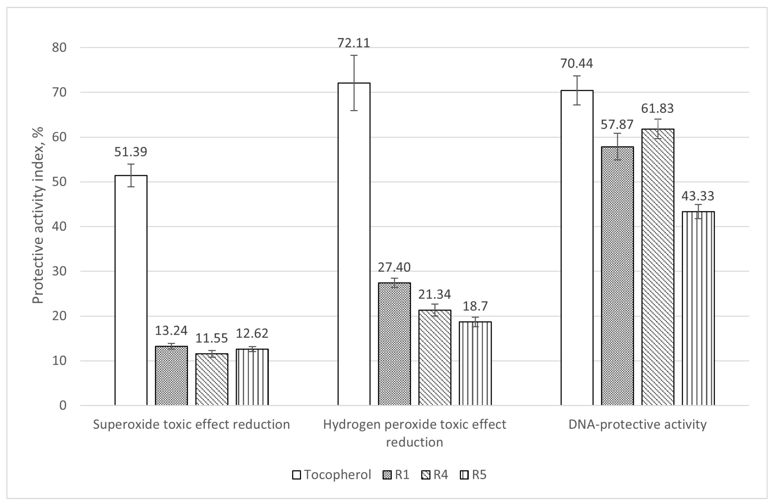
Figure 1. Antioxidant and DNA-protective activity of potential probiotic strains. All effects were statistically significant (p < 0.05). Tocopherol was used as a standard antioxidant.
Fishes 09 00476 g001
Figure 2. Frequencies of spontaneous and dioxidine-induced mutagenesis in E. coli MG1655 under the action of preparations of the studied strains. * These experimental data have a statistically significant difference from the control (p < 0.05).
Fishes 09 00476 g002
Figure 3. Relative mRNA expression of the hsp70 gene in different tissues of C. gariepinus (p-values for the brain, gill, gonad, liver, and muscle tissues were 0.007937, 0.07276, 0.09663, 0.01015, and 0.01889 (for B. subtilis R1); 0.5476, 0.06088, 0.125, 0.07507, and 0.02579 (for B. subtilis R4); and 0.15, 0.08589, 0.1718, 0.02247, and 1.1157 (for B. velezensis R5), respectively). Thick lines inside the boxes indicate the medians; circles outside the boxes indicate the outliers.
Fishes 09 00476 g003
Figure 4. Relative mRNA expression of the cxc gene in different tissues of C. gariepinus (p-values for the brain, gill, gonad, liver, and muscle tissues were 0.9802, 0.08762, 0.1558, 0.003346 (for B. subtilis R1); 0.6268, 0.09122, 0.207, 0.09524, and 0.1676 (for B. subtilis R4); and 0.2877, 0.2723, 0.004556, 0.01587, and 0.466 (for B. velezensis R5), respectively). Thick lines inside the boxes indicate the medians; circles outside the boxes indicate the outliers.
Fishes 09 00476 g004
Figure 5. Relative expression of mRNA of the tnfa gene in different tissues of C. gariepinus (p-values for the brain, gill, gonad, liver, and muscle tissues were 0.5445, 0.1272, 0.774, 0.007937, and 0.1091 (for B. subtilis R1); 0.01978, 0.008842, 0.4466, 0.09524, and 0.3561 (for B. subtilis R4); and 0.01753, 0.04634, 0.1412, 0.007937, 0.6802 (for B. velezensis R5), respectively). Thick lines inside the boxes indicate the medians; circles outside the boxes indicate the outliers.
Fishes 09 00476 g005
Figure 6. Relative expression of mRNA of the il1β gene in different tissues of C. gariepinus (p-values for the brain, gill, gonad, liver, and muscle tissues were 0.1005, 0.005556, 0.03175, 0.07213, and 0.3931 (for B. subtilis R1); 0.07737, 0.007937, 0.8413, 0.02697, and 0.07423 (for B. subtilis R4); and 0.1121, 0.1508, 0.03175, 0.2175, and 0.615 (for B. velezensis R5), respectively). Thick lines inside the boxes indicate the medians; circles outside the boxes indicate the outliers.
Fishes 09 00476 g006
Figure 7. Relative expression of mRNA of the lysC gene in different tissues of C. gariepinus (p-values for the brain, gill, gonad, liver, and muscle tissues was 0.1149, 0.003685, 0.02024, 0.007937, 0.8413 (for B. subtilis R1); 0.8046, 0.0006603, 0.03012, 0.09524, and 0.01587 (for B. subtilis R4); and 0.01873, 0.01638, 0.001216, 0.007937, and 0.007937 (for B. velezensis R5), respectively). Thick lines inside the boxes indicate the medians; circles outside the boxes indicate the outliers.
Fishes 09 00476 g007
Figure 8. Weight gain dynamic throughout the experiment. Since Day 36, all groups have a statistically significant difference from the control (p < 0.05).
Fishes 09 00476 g008
Figure 9. Effect of potential probiotic Bacillus strains on weight gain. All groups have a statistically significant difference from the control (p < 0.05).
Fishes 09 00476 g009
Table 1. Feed composition.
ComponentMass Content, %
Probiotic preparation (experimental groups)1
Non-inoculated soybean preparation (control group)1
Protein, total53
Fat, total12
Fiber, total1
Ash, total33
Table 2. Oligonucleotide sequences for qPCR-RT.
GeneForward Primer Sequence, 5′-3′Reverse Primes Sequence, 5′-3′
actβGTTGGGCACAAGGCATCCTA GGACTCCATACCCAGGAAAGATGG
hsp70GTTTCAGGCAAGCACGTGAGGTTCCCTGAGGCTGTTCGAT
cxcAGATCACCGGGAACTGTGAC GTCCTCACTTCAGCTTGCCT
tnfαTCTCAGGTCAATACAACCCGCGAGGCCTTTGCGGAAAATCTTG
il1βTGCAGTGAATCCAAGAGCTACAGCCCACCTTTCAGAGTGAATGCCAGC
lysCTGCTAAACAGTATGATCGGTGTGA TATCTGGAAAATGCCGTAGTCTGT
Table 3. Feed conversion ratios (FCR) in different experiment variations.
GroupFCR
Control1.34
R11.04
R4 1.02
R51.08
Table 4. Summarized probiotic properties of the studied Bacillus strains.
Strain/EffectDNA Protection, %Antimu-Tagenic, %Weight Gain Increase, %GeneExpression Rates Increase (Compared to Control)
BrainGillsGonadsLiverMuscle
R157.8733.5824.83hsp70-+4.0+10.5+15.2-
cxc+0.8-+9.9+7.5+21.6
tnfα+1.9+1.8+2.6+35.8+7.3
il1β+9.4+8.3+13.9+10.8+1.0
lysC-+7.2+31.9+35.6+0.4
R461.83024.60hsp70-+2.6+6.8+7.1+10.4
cxc0.2-+5.5+6.3-
tnfα+14.4+7.7+0.4+1.7+0.4
il1β+2.0+49.7+0.6--
lysC+1.7+52.9+55.8+23.9+3.6
R545.3354.3529.16hsp70-+0.9+2.6+11.2+6.6
cxc+2.4+2.0+15.6+4.8+3.4
tnfα+36.4+12.0+5.2+13.5+1.4
il1β+10.0+13.6+7.7+9.2+0.7
lysC+5.3+31.9+45.4+26.2+43.0
Semibold values stand for the cases where the expression increase was more than 13 times (see discussion). A dash stands for the decreased expression.
Disclaimer/Publisher’s Note: The statements, opinions and data contained in all publications are solely those of the individual author(s) and contributor(s) and not of MDPI and/or the editor(s). MDPI and/or the editor(s) disclaim responsibility for any injury to people or property resulting from any ideas, methods, instructions or products referred to in the content.