Article

Protective Effects of Roselle Aqueous Extracts against UV-Induced Damage in Zebrafish Fins

1
Department of Chemistry, Tamkang University, Tamsui, New Taipei City 25137, Taiwan
2
Department of Mathematics, Tamkang University, Tamsui, New Taipei City 25137, Taiwan
*
Authors to whom correspondence should be addressed.
These authors contributed equally to this work.
Fishes 2024, 9(6), 199; https://doi.org/10.3390/fishes9060199
Submission received: 28 April 2024 / Revised: 17 May 2024 / Accepted: 24 May 2024 / Published: 26 May 2024

Abstract

:
(1) Background: Roselle (Hibiscus sabdariffa) is a flowering plant reported to have anti-obesity, antioxidant, antibacterial, and anti-inflammatory effects. This study aims to evaluate the UV-absorbing and antioxidant activities of roselle aqueous extracts (RAE) and test the protective effects of RAE against UV radiation in zebrafish embryos. (2) Methods: DPPH assay and UV-spectrum methods were applied to evaluate the antioxidant and UV-absorbing activities, respectively. The protective effects of RAE were evaluated using fin morphology recording, Kaplan–Meier analysis, and Cox proportional hazards regression. Real-time PCR experiments were also applied to detect both the UV- and RAE-induced gene expressions. (3) Results: Our results show that (i) RAE had UV-absorbing abilities and significantly reduced ROS production in vitro; (ii) the mean times of malformed fins in the UV + RAE (36 and 48 ppm) groups were 3.56 and 4.44 days, respectively, and were prolonged compared to those in the UV-only group (3.36 days); (iii) zebrafish in the UV + RAE (36 and 48 ppm) groups were 0.963 and 0.496 (p < 0.001) times more likely to develop to malformed fins, respectively, than those in the UV-only group; and (iv) the RAE treatment led to the 0.19- to 0.62-fold downregulation of the p53, p21, mdm2, and bcl2 gene expressions, compared to the UV-only group. (4) Conclusions: The UV-protective effects of RAE might derive from both the in vitro UV-absorbing activity and in vivo regulation of the p53, p21, mdm2, and bcl2 gene expressions.
Key Contribution: This study highlights the important roles of roselle for UV protection in a zebrafish model.

1. Introduction

Roselle (Hibiscus sabdariffa) is a flowering plant native to Africa. Its flowers and calyces contain many bioactive compounds, such as anthocyanins, citric acid, hibiscus acid, flavonoids, gossypetin, hibiscetine, and quercetin, and these active ingredients have anti-obesity, antioxidant, antibacterial, or anti-inflammatory effects [1,2]. Roselle is renowned for its high anthocyanin content. Anthocyanins can inhibit transcription factors through various pathways, achieving anti-inflammatory effects. Through regulating the expression of cancer-related genes and proteins, roselle can suppress cancer cell growth [3]. It also has outstanding anti-obesity effects, effectively inhibiting the accumulation and formation of lipids, thereby preventing cardiovascular diseases and hyperlipidemia.
Additionally, anthocyanins can suppress UV-induced oxidative stress and apoptosis, thereby preventing oxidative damage to skin tissues [4]. The flavonoid compound quercetin similarly achieves protection against UV damage by scavenging reactive oxygen species (ROS) and inducing the expression of the P53 protein, accelerating cancer cell apoptosis [5].
The ability of roselle to eliminate free radicals has been confirmed in both in vivo and in vitro experiments. In a mouse model, the extract of roselle was confirmed to exhibit antioxidant, antihypertensive, and immune-stimulating activities at non-toxic doses [6]. In particular, its antioxidant and antihypertensive properties can reduce the severity of multi-organ damage [7]. The extract has shown significant cellular gene-protective efficacy against H2O2-induced DNA damage [8]. It was inferred that roselle may have the ability to restore the intracellular antioxidant system, protecting mouse hematopoietic stem cells (HSC) from oxidative damage induced by ROS [8]. The ability of roselle to remove ROS has been reflected in animal experiments and confirmed by a reduction in luminol chemiluminescence intensity. It has been demonstrated that roselle can eliminate free radicals generated through radiation [9].
This study aims to investigate the antioxidant ability and UV radiation protection efficiency of roselle aqueous extracts (RAE) in a zebrafish model in vivo. Embryonic zebrafish fins are very sensitive to UV radiation, which could be used as an indicator to evaluate UV-induced damage and the protective effects of RAE.

2. Materials and Methods

2.1. Preparation of Roselle Aqueous Extract (RAE) and UV Absorbance Assay

Dried roselle was bought from the local market (Taitung Area Farmers’ Cooperative Supermarket, Taitung, Taiwan). For roselle aqueous extract (RAE) preparation, around 10 g of dried roselle was mixed with sterile distilled water (200 mL) and was heated to 100 °C (5 min), to harvest their aqueous extracts. At room temperature, the supernatant was filtered through filter paper. The volume of RAE was measured and stored at 4 °C before use. Regarding the calculation of the RAE concentration, the air-dried unsolvable parts of the roselle weighed 6.2 g; thus, the stock concentration of RAE in this study was around 24,000 ppm (mg/L). Different concentrations of RAE were used for the UV absorbance assay, and their UV absorbance from 400 to 700 nm was measured using a UV-3100 Spectrophotometer (Apple Valley, MN, USA).

2.2. In Vitro Antioxidant Assay

We used 1,1-diphenyl-2-picryl hydrazyl (DPPH, DPPH Antioxidant Assay Kit) (Dojindo Co., Rockville, MD, USA) assay to evaluate the free-radical scavenging property (antioxidant activity) of RAE. The detailed protocols for this assay are described in the manufacturers’ instructions. In brief, DPPH was dissolved in 10 mL of 99.5% ethanol to make a stock solution. Then, 200 μL of the solution containing DPPH in 100 µL of ethanol was used as the control. In another test tube, 100 μL DPPH was combined with 100 µL of RAE (60–1500 ppm) as test samples. Subsequently, the tubes were kept in darkness at 25 °C for 30 min, and the absorbance was detected at 517 nm. By using the following formula, the percentage of antioxidant activity was calculated:
Percentage (%) of antioxidant activity (free radical scavenging activity) = [(Acs − As) ÷ Acs] × 100
where Acs—control reaction absorbance; As—testing specimen absorbance.

2.3. Experimental Animals and Images

Wild-type (AB strain) zebrafish were obtained and maintained as previously described [10]. The Use of Laboratory Animal Committee, Tamkang University approved the animal studies and all procedures. All embryos were observed under a DM 2500 microscope (Leica, Wetzlar, Germany).

2.4. Survival Rate Analysis, UV Radiation, RAE Treatment, and Fin Morphology Recording

Each experimental group (20 embryos/each) was treated with two different exposure protocols (Methods I and II) and for different concentrations (0, 36, 48, 60, 300, and 600 ppm) of RAE to calculate their survival rates. All experiments were repeated three times. For the UV exposure and RAE treatment, the detailed protocols are described in the previously published paper [11]. In brief, each group (embryos by 72 hpf, 50 embryos/each group) was exposed to either water (UV only) or to different concentrations of RAE (36, 48, and 60 ppm) in parallel to receive 100 mJ/cm2 of UVB (302 nm, UV cross-linker, XL-1000, Spectroline, Melville, NY, USA) three times. For fin morphology recording, the pelvic fins of the experimental groups were compared to the pelvic fins of non-experimental healthy zebrafish and subjectively classified as normal (>80%), reduced (20–80%), or absent (<20%) [11,12,13].

2.5. Real-Time PCR

The RNA extraction, cDNA synthesis, and real-time PCR analysis experimental procedures were described previously [11,12]. In this study, five primer sets were synthesized to detect the expressions of β-actin (internal control; 5′-CGAGCAGGAGATGGGAACC-3′, and 5′-CAACGGAAACGCTCATTGC-3′), p53 (5′-GGCTCTTGCTGGGACATCAT-3′, and 5′-TGGATGGCTGAGGCTGTTCT-3′), p21 (5′-CAGCTTCAGGTGTTCCTCAGC-3′, and 5′-CGAGTGAACGTAGGATCCGC-3′), mdm2 (5′-GTGAACCAGATCGAGGACCC-3′, and 5′-GTCAGGGAAAAGCTGTCCGA-3′), and bcl2 (5′-CCTTCAATAAAGCAGTGGAGGAA-3′, and 5′-CGGGCTATCAGGCATTCAGA-3′).

2.6. Statistical Analysis

We used the R packages “RcmdrPlugin.survival” and “RcmdrPlugin.EZR” (R version 3.5.0) to do statistical analysis. “Malformation” is defined as “fin absent or death.” We treated “progress to malformation” as the event, and regarded embryos that did not achieve “malformation” before the end of the fifth day as censored data. The Kaplan–Meier and the Cox proportional hazards methods were used for all assays [11,12,13].

3. Results

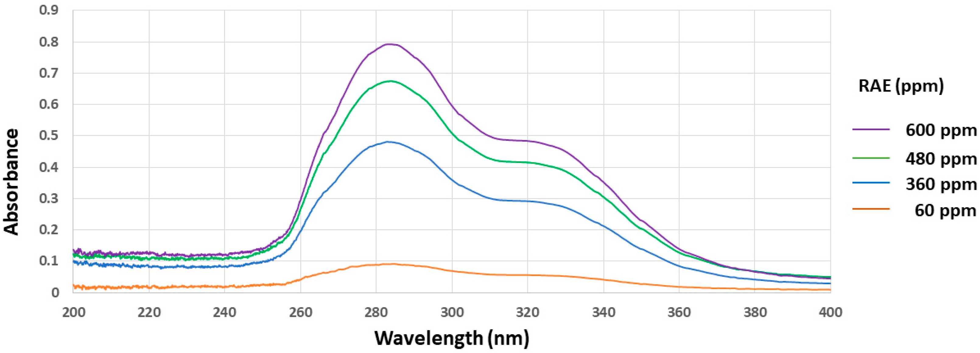
3.1. In Vitro Free-Radical Scavenging Activities and UV Absorbance of Roselle Aqueous Extract (RAE)

Previous studies have shown that UV exposure leads to free radical production. As an efficient UV-protective substance, roselle might possess free radical scavenging activity and/or UV-absorbing activity. In this study, we first detected the free radical scavenging activity of RAE using a DPPH assay. As shown in Figure 1, the high concentration (600 ppm) of RAE possessed a 26.5% inhibition ratio, whereas the low concentration (<60 ppm) of RAE still had a 1.6% inhibition ratio, following a dose-dependent manner. For the UV absorbance in the RAE experiment, different concentrations of RAE (0, 60, 360, 480, and 600 ppm) were used to measure the absorbance (200–400 nm). Figure 2 showed the RAE had UV absorbance, especially in the 260–350 nm wavelength range. The absorbance peak of RAE appeared in the wavelength of 283 nm (in the range of UVB). Additionally, the UV absorbance of RAE followed a concentration-dependent manner. These results indicate that RAE possessed both UV-absorbance and free radical scavenging (antioxidant) activity.

3.2. Survival Rate Analysis of RAE

Zebrafish embryos were divided into 20 embryos per group. Then, each experiment was treated with different concentrations of RAE (0, 36, 48, 60, 300, and 600 ppm) through Methods I or II (Figure 3), and their survival rates were calculated. The results show that, when using exposure protocol Method I (0–96 hpf), the survival rates were 65–85% in the low concentrations of the RAE-exposure groups (36, 48, and 60 ppm), but decreased to 0–55% in the 300 and 600 ppm groups (Figure 3). These observations suggest that the survival rates and RAE concentrations followed a dose- and duration-dependent manner. Additionally, when using exposure protocol Method II (24–96 h), the survival rates were high (60–100%) in almost all the RAE-exposure groups (Figure 3). Based on these observations, we selected low concentrations of the RAE-exposure groups (36, 48, and 60 ppm) for the subsequent UV-induced fin damage experiments.

3.3. Effects of RAE on Fin Repair

Zebrafish fins are sensitive to UV radiation, which makes them an efficient indicator for monitoring UV-induced damage [13]. In this study, pelvic fins after UVB exposure were examined to evaluate the preventive effect of RAE (Figure 4). First, we used the Kaplan–Meier method to describe time-to-malformation phenomena. The non-malformed rate curve for each group is presented in Figure 4, and the mean times (with the standard error) and median times (with 95% confidence limits) of “progress to malformation” are listed in Table 1. The results reveal that the 48 ppm group (n = 48) had the longest average time (4.44 days) and a median time (5 days) of “progress to malformation” (Table 1), and for the estimated rates of the non-malformed embryos, for example, after exposure to UV for 4 days, the UV-only group had 17.71%. In contrast, the rate was around 6.25% for the 36 ppm group, 58.33% for the 48 ppm group, and 2.08% for the 60 ppm group (Figure 4).
We also assessed the effect of RAE on the time to malformation via the Cox proportional hazards regression method. Table 2 showed that the relative probabilities of “progress to malformation” (with corresponding 95% confidence limits) for the 36 ppm, 48 ppm, and 60 ppm groups, compared to the control (UV-only) group, were 0.963 (0.675~1.370), 0.496 (0.345~0.715), and 1.360 (0.953~1.930), respectively. This indicates that the zebrafish embryos in the 48 ppm group were 0.496 times more likely to achieve malformation than those in the control (UV-only) group, with significant evidence (p-value < 0.001).

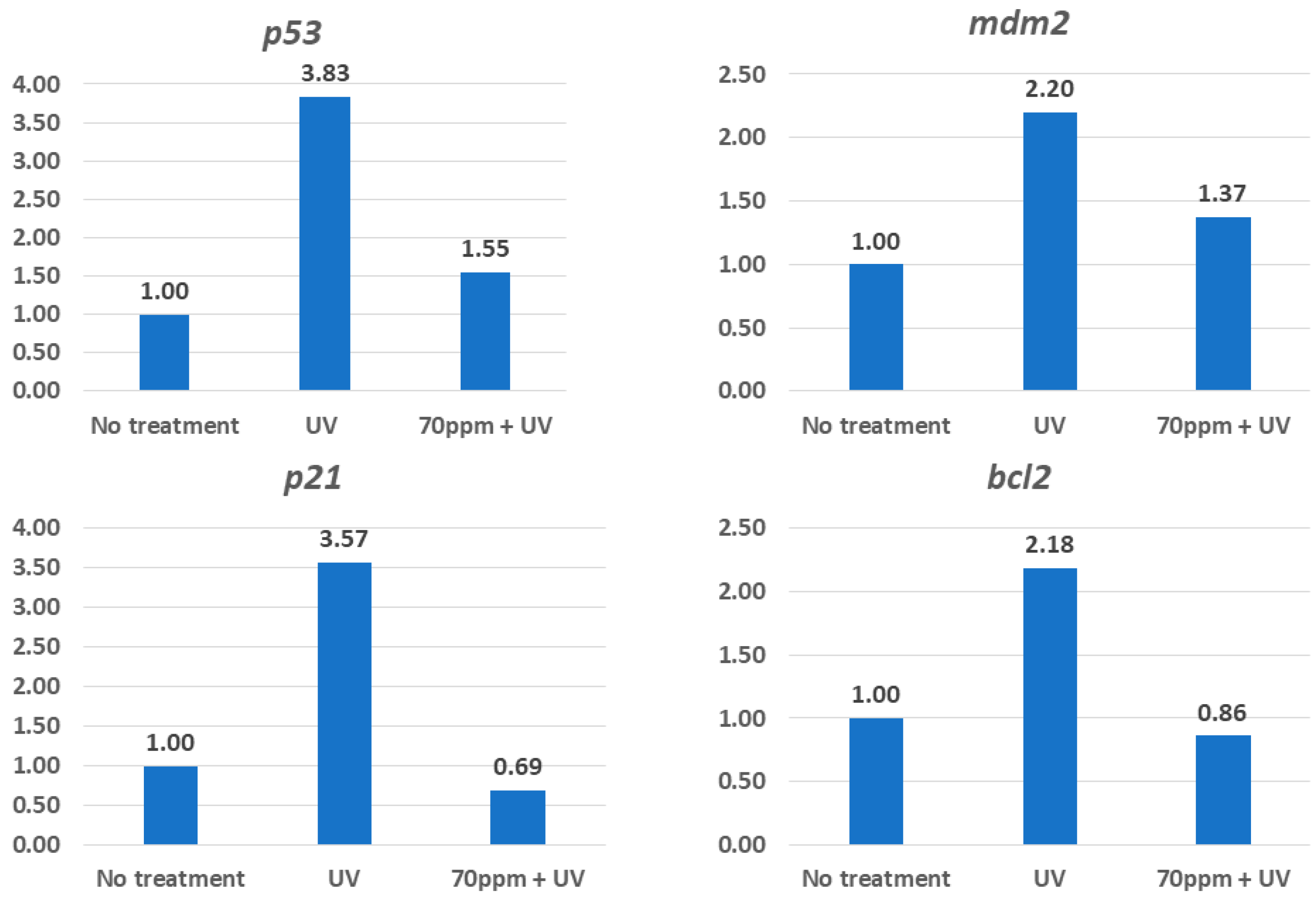
3.4. Possible Mechanisms That Underlie Protection from UV by RAE

It has been reported that P53 signaling is one of the most evident pathways after UV exposure, including the expressions of the p53, p21, mdm2, and bcl2 genes. As shown in Figure 5, the expression levels of p53, p21, mdm2, and bcl2 in the embryos derived from the UV groups increased by 3.83-, 3.57-, 2.20-, and 2.18-fold, respectively, in comparison to those of the embryos derived from the no-treatment group. However, the expression levels of p53, p21, mdm2, and bcl2 from the UV + RAE (48 ppm) embryos decreased by 1.55-, 0.69-, 1.37-, and 0.86-fold, respectively, in comparison to those of the embryos derived from the no-treatment group (Figure 5). This indicates that the groups treated with RAE had around 0.19- to 0.62-fold more protection than those of the embryos derived from the UV group. This suggests that RAE exposure might attenuate the UV-induced P53-related pathway.

4. Discussion

It has been reported that UV-induced damage might be due to the production of free radicals, leading to DNA damage [14,15]. Our previously published paper demonstrated that UV exposure led to cell apoptosis and caused damage in zebrafish fins [11,13]. In this study, we observed that fin damage in zebrafish embryos caused by UV could be attenuated by roselle aqueous extract (RAE) exposure. The UV protection effects of RAE might come from three aspects: (i) the absorption of extracellular UV, reducing its energy; (ii) acting as an antioxidant to scavenge free radicals; or (iii) the enhancement of zebrafish embryos’ repair system through the regulation of p53-related genes.
As shown in Figure 2, our results indicate that RAE absorbed UV, especially in the 260–350 nm wavelength range. Previous reports have shown that roselle extracts contain many known chemicals, including anthocyanins (~22%), citric acids (~12–22%), chlorogenic acids (~18%), quercetin (<0.5%), and rutin (<0.5%) [16,17]. They all reported that they possess UV-absorbing activities but in different wavelength ranges. It has been reported that the UV-absorbing wavelength ranges for anthocyanins, citric acids, chlorogenic acids, quercetin, and rutin are 260–280 nm, 200 nm, 200–400 nm, 240–500 nm, and 200–400 nm, respectively [18,19,20,21,22]. Figure 2 shows that the maximum UV-absorbing wavelength for RAE occurred at 283 nm. Anthocyanins and citric acids are hydrophilic. Compared to the UV absorbances of RAE and the above-mentioned known chemicals, we suggest that the UV-absorbing abilities of RAE might derive from its anthocyanins and citric acids.
This study demonstrated that RAE had free radical-scavenging activity in vitro (Figure 1). RAE is rich in anthocyanins, citric acids, chlorogenic acids, quercetin, and rutin, which suggests that it possesses free radical-scavenging (anti-oxidant) activity. For example, anthocyanins, chlorogenic, quercetin, and rutin have been found to have protective effects against UV-irradiated DNA damage and ROS-scavenging activities in cultured human cancer cells as well as in fibroblasts [4,5,23,24,25,26]. Based on these observations, we suggest that the free radical-scavenging activity of RAE might come from its anthocyanins, citric acids, chlorogenic, quercetin, and rutin. Considering the water solubility, anthocyanins and citric acids might contribute more than chlorogenic, quercetin, and rutin in the RAE.
The results in Figure 5 show that RAE treatment regulated UV-induced gene expressions, such as p53-related genes. Previous reports have demonstrated that cells treated with anthocyanins, chlorogenic, and quercetin-regulated p53, mdm2, bcl2, and p21 expressions [27,28,29,30]. These observations strongly suggest that the activity of RAE-attenuated UV-induced P53-related gene expressions might come from these chemicals. In conclusion, our data demonstrate that RAE (a natural product) is efficient in protecting cells from being damaged by UV radiation. Therefore, RAE might have potential applications in the cosmetics industry.

5. Conclusions

In conclusion, we suggest that the UV-protective effects of RAE might derive from both its in vitro UV-absorbing activity and in vivo regulation of the p53, p21, mdm2, and bcl2 gene expressions.

Author Contributions

Conceptualization, B.-C.W. and Y.-H.C.; methodology, I.-T.L., C.-Y.H., W.-L.S. and T.M.T.; software, C.-C.W.; formal analysis, I.-T.L. and C.-Y.H.; writing—original draft preparation, I.-T.L.; writing—review and editing, B.-C.W.; supervision, project administration, and funding acquisition, Y.-H.C. All authors have read and agreed to the published version of the manuscript.

Funding

This research was funded by the National Science and Technology Council, Taiwan, grant number 104-2313-B-032-002 (YHC).

Institutional Review Board Statement

The animal study protocol was approved by the Use of Laboratory Animal Committee at Tamkang University, Tamsui, New Taipei City, Taiwan (approval no. 103001).

Informed Consent Statement

Not applicable.

Data Availability Statement

All data generated or analyzed during this study are included in this published article.

Acknowledgments

The authors thank the Zebrafish Core in Academia Sinica (ZCAS) for providing the AB strain zebrafish.

Conflicts of Interest

The authors declare no conflicts of interest.

References

  1. Zhen, J.; Villani, T.S.; Guo, Y.; Qi, Y.; Chin, K.; Pan, M.H.; Ho, C.T.; Simon, J.E.; Wu, Q. Phytochemistry, antioxidant capacity, total phenolic content and anti-inflammatory activity of Hibiscus sabdariffa leaves. Food Chem. 2016, 190, 673–680. [Google Scholar] [CrossRef]
  2. Ojulari, O.V.; Lee, S.G.; Nam, J.O. Beneficial effects of natural bioactive compounds from Hibiscus sabdariffa L. on obesity. Molecules 2019, 24, 210. [Google Scholar] [CrossRef] [PubMed]
  3. Lin, B.W.; Gong, C.C.; Song, H.F.; Cui, Y.Y. Effects of anthocyanins on the prevention and treatment of cancer. Br. J. Pharmacol. 2017, 174, 1226–1243. [Google Scholar] [CrossRef] [PubMed]
  4. Li, W.; Tan, L.; Zou, Y.; Tan, X.; Huang, J.; Chen, W.; Tang, Q. The effects of ultraviolet A/B treatments on anthocyanin accumulation and gene expression in dark-purple tea cultivar ‘Ziyan’ (Camellia sinensis). Molecules 2020, 25, 354. [Google Scholar] [CrossRef]
  5. Zhu, X.; Li, N.; Wang, Y.; Ding, L.; Chen, H.; Yu, Y.; Shi, X. Protective effects of quercetin on UVB irradiation-induced cytotoxicity through ROS clearance in keratinocyte cells. Oncol. Rep. 2017, 37, 209–218. [Google Scholar] [CrossRef]
  6. Fakeye, T. Toxicity and immunomodulatory activity of fractions of Hibiscus sabdariffa Linn (family Malvaceae) in animal models. Afr. J. Tradit. Complement. Altern. Med. 2008, 5, 394–398. [Google Scholar] [CrossRef] [PubMed]
  7. Seujange, Y.; Leelahavanichkul, A.; Yisarakun, W.; Khawsuk, W.; Meepool, A.; Phamonleatmongkol, P.; Saechau, W.; Onlamul, W.; Tantiwarattanatikul, P.; Oonsook, W.; et al. Hibiscus sabdariffa linnaeus aqueous extracts attenuate the progression of renal injury in 5/6 nephrectomy rats. Ren. Fail. 2013, 35, 118–125. [Google Scholar] [CrossRef]
  8. Abdul Hamid, Z.; Lin, W.H.; Abdalla, B.J.; Bee Yuen, O.; Latif, E.S.; Mohamed, J.; Rajab, N.F.; Paik Wah, C.; Wak Harto, M.K.; Budin, S.B. The role of Hibiscus sabdariffa L. (Roselle) in maintenance of ex vivo murine bone marrow-derived hematopoietic stem cells. Sci. World J. 2014, 2014, 258192. [Google Scholar] [CrossRef]
  9. Murillo Pulgarín, J.A.; García Bermejo, L.F.; Carrasquero Durán, A. A fast and simple FIA-chemiluminescence method for the evaluation of Roselle flowers as scavenger of the free radicals generated by UV irradiated antibiotics. J. Pharm. Biomed. Anal. 2019, 164, 630–635. [Google Scholar] [CrossRef]
  10. Chen, Y.H.; Chang, C.Y.; Chang, C.F.; Chen, P.C.; Lee, Y.T.; Chern, C.Y.; Tsai, J.N. Pro-angiogenic effects of chalcone derivatives in zebrafish embryos in vivo. Molecules 2015, 20, 12512–12524. [Google Scholar] [CrossRef]
  11. Cheng, C.C.; Chou, C.Y.; Chang, Y.C.; Wang, H.W.; Wen, C.C.; Chen, Y.H. Protective role of comfrey leave extracts on UV-induced zebrafish fin damage. J. Toxicol. Pathol. 2014, 27, 115–121. [Google Scholar] [CrossRef] [PubMed]
  12. Chen, Y.H.; Chen, Y.J.; Chou, C.Y.; Wen, C.C.; Cheng, C.C. UV-protective activities of pineapple leaf extract in zebrafish embryos. Res. Chem. Intermed. 2019, 45, 65–75. [Google Scholar] [CrossRef]
  13. Wang, Y.H.; Wen, C.C.; Yang, Z.S.; Cheng, C.C.; Tsai, J.N.; Ku, C.C.; Wu, H.J.; Chen, Y.H. Development of a whole-organism model to screen new compounds for sun protection. Mar. Biotechnol. 2009, 11, 419–429. [Google Scholar] [CrossRef]
  14. Valko, M.; Leibfritz, D.; Moncol, J.; Cronin, M.T.; Mazur, M.; Telser, J. Free radicals and antioxidants in normal physiological functions and human disease. Int. J. Biochem. Cell Biol. 2007, 39, 44–84. [Google Scholar] [CrossRef]
  15. Mullendlers, L.H.F. Solar UV damage to cellular DNA: From mechanisms to biological effects. Photochem. Photobiol. Sci. 2018, 17, 1842–1852. [Google Scholar] [CrossRef]
  16. Yang, D.; Ding, X.Y.; Xu, H.X.; Guo, Y.X.; Zhang, Q.F. Chemical profile of roselle extract and its inhibitory activities on three digestive enzymes in vitro and in vivo. Int. J. Biol. Macromol. 2023, 253, 126902. [Google Scholar] [CrossRef] [PubMed]
  17. Izquierdo-Vega, J.A.; Arteaga-Badillo, D.A.; Sánchez-Gutiérrez, M.; Morales-González, J.A.; Vargas-Mendoza, N.; Gómez-Aldapa, C.A.; Castro-Rosas, J.; Delgado-Olivares, L.; Madrigal-Bujaidar, E.; Madrigal-Santillán, E. Organic acids from roselle (Hibiscus sabdariffa L.)-A brief review of its pharmacological effects. Biomedicines 2020, 8, 100. [Google Scholar] [CrossRef] [PubMed]
  18. Krukowski, S.; Karasiewicz, M.; Kolodziejski, W. Convenient UV-spectrophotometric determination of citrates in aqueous solutions with applications in the pharmaceutical analysis of oral electrolyte formulations. J. Food Drug Anal. 2017, 25, 717–722. [Google Scholar] [CrossRef]
  19. Wen, J.; Kang, L.; Liu, H.; Xiao, Y.; Zhang, X.; Chen, Y. A validated UV-HPLC method for determination of chlorogenic acid in Lepidogrammitis drymoglossoides (Baker) Ching, Polypodiaceae. Pharmacogn. Res. 2012, 4, 148–153. [Google Scholar]
  20. Buchweitz, M.; Kroon, P.A.; Rich, G.T.; Wilde, P.J. Quercetin solubilisation in bile salts: A comparison with sodium dodecyl sulphate. Food Chem. 2016, 211, 356–364. [Google Scholar] [CrossRef]
  21. Saha, S.; Singh, J.; Paul, A.; Sarkar, R.; Khan, Z.; Banerjee, K. Anthocyanin profiling using UV-Vis spectroscopy and liquid chromatography mass spectrometry. J. AOAC Int. 2020, 103, 23–39. [Google Scholar] [CrossRef]
  22. Pinto, D.; Lameirão, F.; Delerue-Matos, C.; Rodrigues, F.; Costa, P. Characterization and stability of a formulation containing antioxidants-enriched castanea sativa shells extract. Cosmetics 2021, 8, 49. [Google Scholar] [CrossRef]
  23. Ryan, E.M.; Duryee, M.J.; Hollins, A.; Dover, S.K.; Pirruccello, S.; Sayles, H.; Real, K.D.; Hunter, C.D.; Thiele, G.M.; Mikuls, T.R. Antioxidant properties of citric acid interfere with the uricase-based measurement of circulating uric acid. J. Pharm. Biomed. Anal. 2019, 164, 460–466. [Google Scholar] [CrossRef]
  24. Liang, N.; Kitts, D.D. Role of chlorogenic acids in controlling oxidative and inflammatory stress conditions. Nutrients 2015, 8, 16. [Google Scholar] [CrossRef] [PubMed]
  25. Gęgotek, A.; Rybałtowska-Kawałko, P.; Skrzydlewska, E. Rutin as a mediator of lipid metabolism and cellular signaling pathways interactions in fibroblasts altered by UVA and UVB radiation. Oxidative Med. Cell. Longev. 2017, 2017, 4721352. [Google Scholar] [CrossRef]
  26. Tena, N.; Martín, J.; Asuero, A.G. State of the art of anthocyanins: Antioxidant activity, sources, bioavailability, and therapeutic effect in human health. Antioxidants 2020, 9, 451. [Google Scholar] [CrossRef] [PubMed]
  27. Shih, P.H.; Yeh, C.T.; Yen, G.C. Effects of anthocyanidin on the inhibition of proliferation and induction of apoptosis in human gastric adenocarcinoma cells. Food Chem. Toxicol. 2005, 43, 1557–1566. [Google Scholar] [CrossRef]
  28. Lo, C.W.; Huang, H.P.; Lin, H.M.; Chien, C.T.; Wang, C.J. Effect of Hibiscus anthocyanins-rich extract induces apoptosis of proliferating smooth muscle cell via activation of P38 MAPK and p53 pathway. Mol. Nutr. Food Res. 2007, 51, 1452–1460. [Google Scholar] [CrossRef] [PubMed]
  29. Anwar, S.; Fratantonio, D.; Ferrari, D.; Saija, A.; Cimino, F.; Speciale, A. Berry anthocyanins reduce proliferation of human colorectal carcinoma cells by inducing caspase-3 activation and p21 upregulation. Mol. Med. Rep. 2016, 14, 1397–1403. [Google Scholar] [CrossRef]
  30. Clemente-Soto, A.F.; Salas-Vidal, E.; Milan-Pacheco, C.; Sánchez-Carranza, J.N.; Peralta-Zaragoza, O.; González-Maya, L. Quercetin induces G2 phase arrest and apoptosis with the activation of p53 in an E6 expression-independent manner in HPV-positive human cervical cancer-derived cells. Mol. Med. Rep. 2019, 19, 2097–2106. [Google Scholar] [CrossRef]
Figure 1. In vitro antioxidant assay of RAE. The free-radical scavenging property (antioxidant) of RAE was detected using 1,1-diphenyl-2-picryl hydrazyl (DPPH, DPPH Antioxidant Assay Kit). The percentage of antioxidant activity (free radical scavenging activity) was calculated using the following formula: percentage (%) of antioxidant activity = [(Acs − As) ÷ Acs] × 100. Acs: control reaction absorbance; As testing specimen absorbance. x-axis: concentrations of RAE; y-axis: percentage (%) of antioxidant activity (inhibition ratio). The regression line indicates that the inhibition radio increased as the concentrations of RAE increased.
Fishes 09 00199 g001
Figure 2. The UV absorbance activity of RAE. RAE with 60 (orange line), 360 (blue line), 480 (green line), and 600 (purple line) ppm was used to measure the absorbance between 200 and 400 nm, respectively.
Fishes 09 00199 g002
Figure 3. Exposure methods for survival rate analysis were used in this study. (A) Schematic illustration of exposure protocols. (B) Zebrafish (AB strain) embryos were treated with different concentrations (0, 36, 48, 60, 300, and 600 ppm) of RAE from 0 to 96 hpf (Method I) or from 24 to 96 hpf (Method II). Twenty embryos were used for each group. All experiments were repeated three times. hpf: Hours postfertilization.
Fishes 09 00199 g003
Figure 4. UV radiation and RAE treatment exposure methods, fin morphology recording, and statistical analysis were used in this study. Zebrafish embryos that developed at 72 h postfertilization (hpf) were collected (30 embryos per experimental group), exposed to UV, and treated in water (no RAE, 0 ppm), or in RAE (36, 48, and 60 ppm). Embryos displayed normal fins before UV exposure but exhibited reduced- or absent-fin phenotypes after exposure to UV (left panels). Non-malformation rate curves for the four groups (UV only, UV + 36 ppm, UV + 48 ppm, and UV + 60 ppm), estimated using the Kaplan–Meier method (right panel).
Fishes 09 00199 g004
Figure 5. RAE affects the expressions of p53, p21, mdm2, and bcl2. Relative quantification of mRNA expression using the comparative CT method (CT: cycles of real-time PCR; relative fold to control group = 2−ΔΔCT). The RAE + UV group is significantly different from the corresponding UV-treated group.
Fishes 09 00199 g005
Table 1. Summarized results of Kaplan–Meier analysis for each group.
Experimental Groupn (Number of Embryos)Mean Time of MalformationSE of Mean Time
UV only963.360.103
UV + 36 ppm483.560.110
UV + 48 ppm484.440.110
UV + 60 ppm483.080.128
Table 2. Cox proportional hazards regression for assessing the effects of each treatment on the time to malformation.
Experimental GroupRelative Probabilityp-Value
UV + 36 ppm0.9630.834
UV + 48 ppm0.496<0.001
UV + 60 ppm1.3600.091
Disclaimer/Publisher’s Note: The statements, opinions and data contained in all publications are solely those of the individual author(s) and contributor(s) and not of MDPI and/or the editor(s). MDPI and/or the editor(s) disclaim responsibility for any injury to people or property resulting from any ideas, methods, instructions or products referred to in the content.

