Review

Harnessing Hue: Advances and Applications of Fish Skin Pigmentation Genetics in Aquaculture

by
Jialong Liu
1,2,
Miaomiao Yin
1,2,
Zhi Ye
1,2,*,
Jingjie Hu
1,2,* and
Zhenmin Bao
1,2
1
Key Laboratory of Tropical Aquatic Germplasm of Hainan Province, Sanya Oceanographic Institution, Ocean University of China, Sanya 572024, China
2
MOE Key Laboratory of Marine Genetics and Breeding, College of Marine Life Sciences, Ocean University of China, Qingdao 266100, China
*
Authors to whom correspondence should be addressed.
Fishes 2024, 9(6), 220; https://doi.org/10.3390/fishes9060220
Submission received: 7 May 2024 / Revised: 5 June 2024 / Accepted: 6 June 2024 / Published: 10 June 2024
(This article belongs to the Special Issue Genetic Breeding and Developmental Biology of Aquaculture Animals)

Abstract

:
Fish exhibit a broad spectrum of colors and patterns facilitated by specialized cells known as chromatophores. The vibrant coloration of fish, controlled by complex genetic and environmental interactions, serves critical roles in ecological functions such as mating, predation, and camouflage. This diversity not only makes fish an invaluable model for exploring the molecular mechanisms of pigmentation but also significantly impacts their economic value within the aquaculture industry, where color traits can drive marketability and breeding choices. This review delves into the sophisticated biological processes governing fish pigmentation and discusses their applications in enhancing aquaculture practices. By exploring the intersection of genetic regulation, environmental influences, and advanced breeding techniques, this review highlights both the scientific understanding and practical applications of fish coloration, providing a bridge between basic biological research and its application in commercial aquaculture.
Key Contribution: This paper provides a comprehensive review of the genetic and molecular mechanisms underlying fish pigmentation, particularly highlighting recent advances in pigment cell differentiation, gene regulation, and their applications in aquaculture, thus offering a valuable framework for future research and genetic engineering strategies in the field.

1. Introduction

Fish are distinguished by a vibrant and diverse palette of body colors, which vary significantly across species, populations, and even individuals. These color variations are not merely aesthetic but serve crucial biological functions such as communication and reproduction, as well as camouflage and predation, which are essential for the survival of many fish species [1,2,3]. In the context of sexual dimorphism, coloration is particularly pivotal during mating. For instance, female peacock cichlids (Cichla) show a preference for mates displaying bright orange-red hues [4]. Furthermore, pigmentation is vital for the growth and development of fish, providing protection against DNA damage from excessive ultraviolet radiation, which can lead to skin lesions, developmental abnormalities, or even unexpected mortality [5,6,7].
Beyond its biological significance, coloration in fish is a key economic trait in aquaculture, influencing market value profoundly. Ornamental species like goldfish (Carassius auratus) are prized for their vivid colors and unique shapes [8,9,10], while species like red tilapia (Oreochromis spp.) are valuable due not only to their vibrant color but also to their taste and nutrition [11]. The market value of the leopard coral grouper (Plectropomus leopardus), for instance, increases with the intensity of its red body coloration [12,13], underlining the economic importance of pigment traits. Consequently, the controlled and effective enhancement of color traits through genetic selection remains a focal point in aquaculture.
The underlying determinants of fish skin color involve intricate interactions among various pigment cells which originate from the developmental processes of neural crest cells (NCCs). Extensive research has elucidated that while many pigment cells share a common intermediate destiny, it is the differential regulation of genes and signaling pathways that dictates their fate [14,15,16,17,18]. Additionally, the synthesis and accumulation of pigments within these cells are critical for color variability, influenced by the ratio of pigments like pteridines and carotenoids [19].
Furthermore, the determination of coloration is influenced by an interplay of genetic, environmental, dietary, and physiological factors, with genetics playing a particularly critical role [20]. Over recent decades, advancements in genomics, transcriptomics, metabolomics, and proteomics have shed light on the regulatory networks that underpin color formation and have identified key genes regulating color variations [21,22,23,24,25,26]. Alongside these advancements, techniques such as hybrid breeding, genomic selection, and gene editing have been successfully applied to breed fish with desired color traits [9,27,28,29,30,31].
This review aims to provide a comprehensive overview of the cellular and structural bases of fish coloration, the factors influencing color variation, and the recent progress in color-related fish breeding. By delving into the mechanisms for the differentiation of pigment cells and the molecular processes regulating pigment accumulation, and discussing the influence of various factors such as nutrition, light, and hormones on color variation, this article bridges fundamental biological research and its practical applications in commercial aquaculture.

2. Cellular and Molecular Basis of Pigmentation

Fish exhibit a wide array of both colors and patterns on the skin, from the stark horizonal blue and yellow stripes of zebrafish (Danio rerio) to the bright red body of the leopard coral grouper adorned with blue iridescent spots. The diversity of coloration traits in fish is determined by the types of pigment cells present, their pigment accumulation and distribution across the scales, fins, and skin [32]. Therefore, a fundamental understanding of the types of pigment cells, their differentiation mechanisms, and the molecular processes driving pigment accumulation is essential for the molecular breeding of color traits in aquaculture. This fundamental understanding will become the cornerstone of research into the genetic manipulation of coloration in fish.

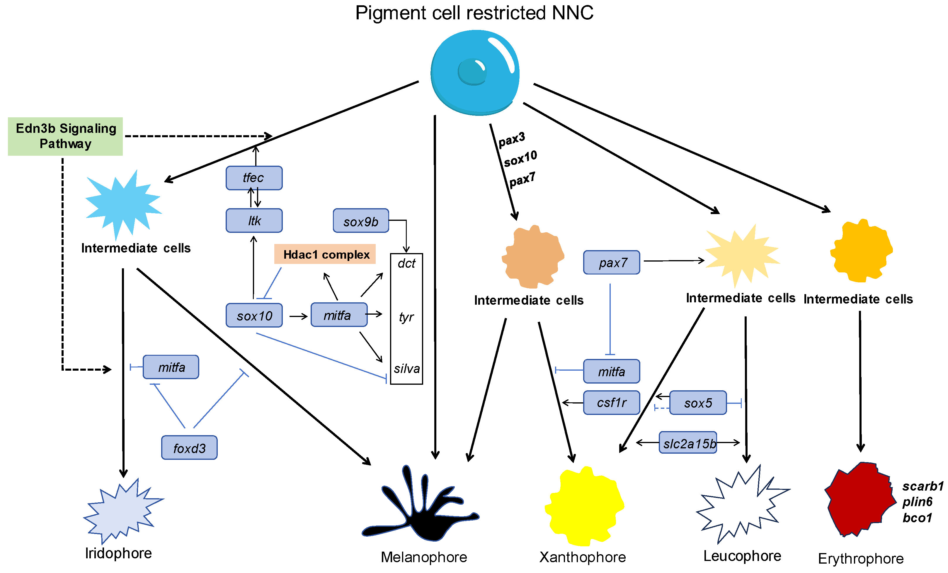
2.1. Pigment Cells and Their Development in Fish

Six primary types of pigment cells have been identified, including melanophore, erythrophore, xanthophore, iridophore, leucophore, and the rarely reported cyanophore. Fish also exhibit specialized pigment cells known as dichromatic chromatophores, which uniquely combine the characteristics of two distinct types of pigment cells [33,34], adding complexity to their coloration. Based on their mechanisms of coloration, pigment cells are further classified into pigment cells and structural cells.
Melanophores are pigment cells characterized by their large size and radially branched morphology with dendritic extensions. These cells typically range from 100 to 300 μm in diameter, are larger than other types of cells, and contain melanin granules. The distribution of these granules affects the color intensity of the fish’s surface—dispersing to darken the surface and aggregating to lighten it. Melanophores primarily house two types of melanin: eumelanin and pheomelanin, with granules generally measuring between 0.3 and 0.7 μm [35,36].
Xanthophores and erythrophores are closely related pigment cells that share a common progenitor in zebrafish [37]. Xanthophores, usually measuring between 50 and 100 μm in diameter, contain pteridine pigments that impart a yellow hue, whereas erythrophores are filled with carotenoids responsible for red coloration. Interestingly, pteridine pigments and carotenoids can coexist within the same cell, influencing the color outcome based on their relative concentrations [38,39]. It is noteworthy that animals cannot synthesize carotenoids themselves and must obtain them from food. Through a series of processes including absorption, transportation, oxidation, and deposition within the body, carotenoids eventually manifest in the coloration of the body surface [40,41].
Iridophores are structural cells characterized by a polygonal or oval shape and contain reflective substances such as guanine, hypoxanthine, and adenine that create iridescence through light reflection. Interference and scattering phenomena occur when the incident light goes through the reflection plates, resulting in the display of different colors when layered with other pigment cells. The size of iridophores is similar to that of xanthophores, but their dendrites are thicker [42,43].
Leucophores, also categorized as structural cells, utilize uric acid-formed reflectors to produce white or silver-white colors, distinguishing them from the iridescent colors of iridophores. The size of leucophores is similar to that of xanthophores and erythrophores. Research on leucophores is relatively abundant on the medaka (Oryzias latipes) [44,45].
Pigment cells in teleost fish originate from NCCs during embryonic development. NCCs are a special population of embryonic stem cells in vertebrates with transient, highly migratory, and multipotent characteristics [46]. After their delamination from the dorsal neural tube, NCCs migrate extensively throughout the embryo, settling in diverse locations and differentiating into various cell types, including neurons, glial cells, chondrocytes, bone cells, and pigment cells critical for skin coloration [47,48].
The mechanisms governing the fate determination of NCCs have not been fully resolved yet. Early studies proposed the direct fate restriction (DFR) model which suggests that NCCs differentiate directly into specific cell types at defined locations under the influence of targeted signals such as bone morphogenetic protein (BMP), neuregulin (NRG), and Wnt. These signals drive the differentiation of NCCs into sensory neuron cells, smooth muscle cells, and melanophores, respectively [49,50,51]. However, emerging research has indicated a notable heterogeneity in the expression of key marker genes among early migrating and pre-migratory NCCs across different conditions. This observation has led to the proposal of the progressive fate restriction (PFR) model, which suggests a more dynamic differentiation process involving a series of intermediate progenitor cells [52,53,54,55,56]. These progenitors differentiate into specific cell types as they migrate to appropriate sites within the embryo. For instance, bipotent sympathetic neuroglial progenitor cells which have potential for sympathetic neuron and neuroglial cell fates, migrate towards the aorta to form sympathetic ganglia [57,58]. Moreover, the differentiation trajectories of NCCs in other vertebrates like mice (Mus musculus) and chickens (Gallus gallus) support the PFR model, showing potential common progenitors for melanophores and glial cells [59,60]. The advent of single-cell sequencing technologies has further refined our understanding of these processes, revealing the transcriptional heterogeneity of NCCs in multiple model species [16,17,18] and the expression of pigment cell-specific genes in pre-migratory NCCs in zebrafish [18]. These supplementary research findings have led to a broader acceptance of the PFR model.
The zebrafish is distinguished by its possession of the most common types of pigment cells found in fish—melanophores, xanthophores, iridophores, and leucophores. Coupled with attributes such as embryo transparency, rapid development, and an ease of genetic manipulation, the zebrafish has proven to be an exemplary model for studying the origins, differentiation pathways, and patterning mechanisms of pigment cells [61,62,63]. Research has demonstrated that these pigment cells originate from NCCs, and during their differentiation, zebrafish exhibit bipotent pigment progenitor cells, aligning with the PFR model of NCC differentiation [14,15]. In-depth studies have elucidated the cellular mechanisms underlying the fate determination of these cells, particularly how NCCs differentiate into specific pigment cells [64,65,66].
Melanophores, the most extensively studied pigment cell type across animal species, are heavily influenced by a network of regulatory factors. The transcription factor Sox10 is pivotal in the fate specification of neural crest-derived cells. Mutations in sox10 result in the apoptosis of NCCs and the subsequent failure to develop melanophores, xanthophores, and iridophores [67]. Initially, the upregulation of sox10 during embryonic development triggers the activation of mitfa, a critical gene directing the melanophore lineage [68]. Interestingly, as development progresses, sox10 is rapidly downregulated, which inversely affects the expression of pigment synthesis genes such as dct, tyr, and silva, mediated by mitfa [69]. This dynamic expression is potentially regulated by feedback mechanisms involving the Hdac1 histone deacetylase. Furthermore, studies have indicated that sox9b can independently drive melanophore differentiation apart from mitfa regulation, stabilizing the presence of melanophores in the organism [69]. The Wnt signaling pathway also plays a crucial role throughout the process of melanophore differentiation, exerting a continuous influence up to 72 hpf [70,71]. Initially, this pathway synergizes with sox10 to activate the expression of mitfa, thereby facilitating the early stages of melanophore differentiation. As development progresses, a positive feedback loop emerges between the Wnt signaling pathway and mitfa, which plays a pivotal role in the morphological evolution of melanophores. This dynamic regulatory interaction underscores the complexity of the cellular signaling involved in pigment cell development.
Recent advancements have significantly deepened our understanding of the mechanisms for iridophores’ differentiation, particularly their close linkage with melanophore differentiation. Central to this process is transcription factor tfec, a member of the MiT transcription factor family, similar to mitfa. Tfec serves as the master regulator guiding multipotent progenitors to differentiate into iridophores [72]. It is co-expressed with sox10 and leukocyte tyrosine kinase (ltk), which encodes an insulin-like receptor tyrosine kinase, in early NCCs. This expression synergy promotes the differentiation of these cells into specific iridophore precursors [66,73]. Sox10 plays a pivotal role in sustaining tfec expression within these precursors, while tfec and ltk reciprocally maintain each other’s expression through a positive feedback loop [66]. Moreover, iridophore precursors also express mitfa, and intriguingly, in mitfa mutant zebrafish where melanophores are absent, an increase in iridophore numbers is observed. This indicates the bipotent capability of iridophore precursors to differentiate into either melanophores or iridophores [66,72]. Additionally, the forkhead box transcription factor encoded by foxd3 is expressed in pre-migratory NCCs and is crucial for iridophore differentiation [74]. Studies have shown that in zebrafish, foxd3 inhibits mitfa expression by binding to its promoter, which not only promotes iridophore development but also prevents these precursors from differentiating into melanophores [75,76,77]. The Edn3b signaling pathway also plays a crucial role in both iridophore differentiation and pigment accumulation. The knockdown of edn3b results in a significant reduction in iridophore numbers by 48hpf [78], and the knockout of its receptor, ednrba, in adult zebrafish leads to decreased iridophore numbers and defects in stripe patterns [79]. Furthermore, other transcription factors like gbx2, alx4a, and mpv17, which encode various regulatory proteins, contribute to the specialization of iridophores, although the specifics of their downstream regulatory mechanisms remain to be fully elucidated [80,81,82].
The differentiation of xanthophores involves a complex network of multiple genes, including pax3, pax7, csf1r, sox5, and sox10. Studies using the targeted knockdown of pax3 in specific NCC populations have demonstrated its critical role, as its absence results in the depletion of xanthophores and enteric neurons, highlighting pax3′s essential function in xanthophore development [83]. Additionally, pax7 proves to be pivotal in establishing the xanthophore lineage; zebrafish embryos with double mutations in pax7a and pax7b exhibit a significant reduction in xanthophore precursor cells and a complete absence of mature xanthophores [65]. Moreover, pax7 mutants display an increase in melanophores, which migrate more extensively and contribute to a darker phenotype in embryos and larvae. This suggests that pax7 influences pigment cell differentiation by regulating bipotent xantho-melanophore precursor cells. Further analysis reveals that the regulatory interactions between sox10, pax3, and mitfa are evolutionarily conserved and work synergistically to enhance mitfa expression, which plays a critical role in directing the fate of these bipotent cells. Intriguingly, pax7 can modulate mitfa to preferentially differentiate these cells into xanthophores rather than melanophores, thus influencing the fate selection of xantho-melanophore cells in zebrafish. This mechanism of fate determination is analogous to the regulation observed between melanophores and iridophores, where the expression of mitfa by transcription factors is a deciding factor [84]. In addition, studies have found that csf1r (also known as fms), which encodes the type III receptor tyrosine kinase, is indispensable for the maturation of xanthophore precursors into fully developed xanthophores and also impacts the distribution of melanophores [85]. Furthermore, sox5 plays an important role in overall pigment cell differentiation by acting antagonistically against sox10 and loss of sox5 can rescue the loss of three types of pigment cells including xanthophore in the sox10 mutant in zebrafish. However, in the medaka, which has abundant numbers of leucophores, sox5 interacts with sox10 in the xanthophore/leucophore progenitors to promote the xanthophore fate [86].
Research on erythrophores has predominantly concentrated on the aspects of pigment synthesis, accumulation, and degradation, with relatively few studies addressing the early differentiation mechanisms of these cells. This gap in research is partly due to the absence of erythrophores in zebrafish, a commonly used model organism. To bridge this knowledge gap, Huang et al. leveraged the presence of erythrophores in Danio albolineatus, a close relative of zebrafish, to explore the differentiation dynamics between erythrophores and xanthophores [37]. Their study discovered that in the fins of D. albolineatus, both erythrophores and xanthophores originate from shared orange progenitor cells and make distinct fate decisions based on the cell distribution within the tissue. Remarkably, under specific conditions, differentiated erythrophores have the capability to transform into xanthophores, underscoring a significant developmental flexibility and interconnection between these pigment cells. This observation is corroborated by the findings of Sušnik et al. [87]. Further research in Spotted scat (Scatophagus argus) provided insights into the genetic regulation of erythrophore function. It was observed that genes involved in carotenoid metabolism (scarb1, plin6, and bco1), tetrahydrobiopterin synthesis (gch2), and pigment granule differentiation (slc2a15b and csf1ra) exhibit a marked increase in expression from 24 to 36 hpf. This upregulation reflects the critical developmental phases of erythrophores, elucidating the temporal gene expression patterns during their early embryonic development [88].
The medaka has become a valuable model organism for studying pigment cell differentiation mechanisms due to its possession of key cell types, including melanophores, iridophores, xanthophores, and leucophores [89]. Although leucophores used to be thought to be more related to iridophores as they share the same mechanism of purine-dependent light reflection, research has elucidated that the differentiation pathways of leucophores in medaka closely mirror those of xanthophores. Specifically, transcription factor Pax7a is crucial for differentiating both leucophores and xanthophores [90]. It was also shown that pax7 can regulate the formation of xantho-leucophores or melanophores through the modulation of mitfa [84]; furthermore, the gene slc2a15b has been identified as promoting the development of xantho-leucophores into leucophores [90]. Expanding on these insights, studies have shown that sox5 serves as a regulatory switch in the fate determination of these bipotent progenitor cells. The expression of sox5 in medaka directs progenitor cells towards a xanthophore lineage, whereas a reduction in sox5 function shifts the balance towards leucophore differentiation [91]. This dynamic role of sox5 has been further validated by research conducted by Nagao et al., which demonstrates that while sox5 supports xanthophore lineage differentiation in medaka, it acts conversely in zebrafish, highlighting a fascinating aspect of species-specific regulatory mechanisms in pigment cell development [86]. The mechanisms for the differentiation of these pigment cells are illustrated in Figure 1.

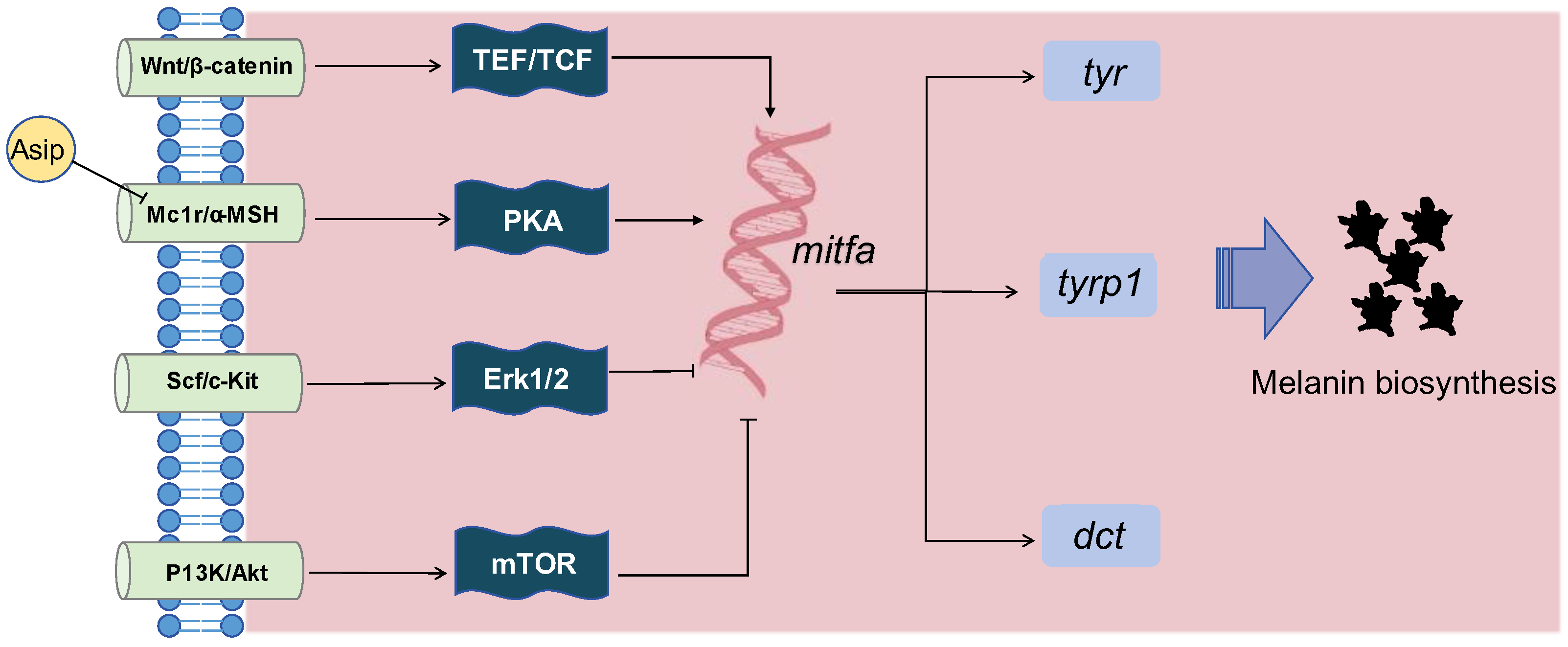
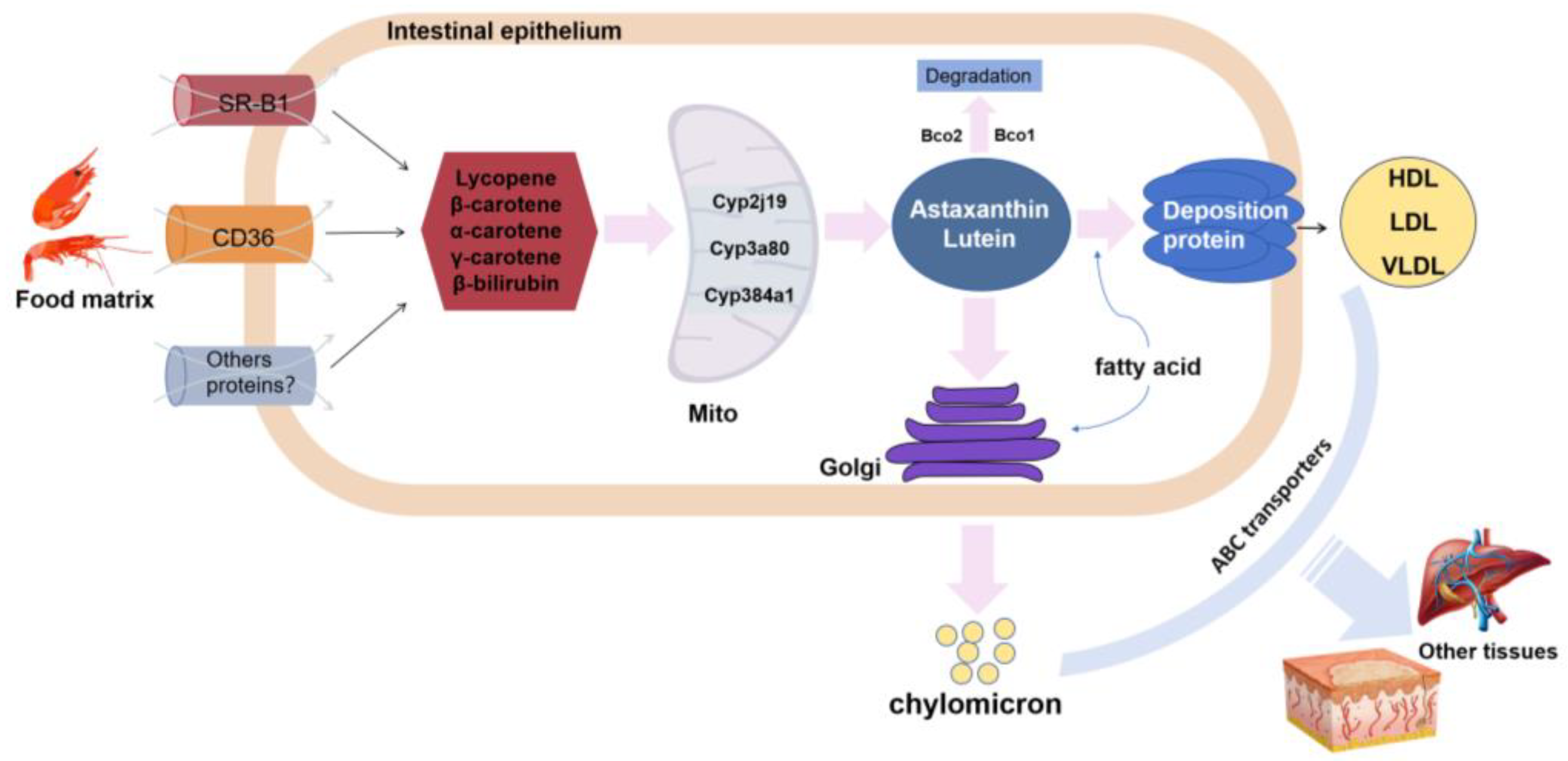
2.2. Accumulation of Pigments and Body Color Differences

The differentiation of NCCs into various pigment cells and the subsequent accumulation of pigments are pivotal processes that dictate the diverse coloration observed in fish. The process of pigment accumulation is regulated by specific genes and influenced by a myriad of factors including the genetic makeup, intercellular interactions, and environmental conditions.
In melanophores, melanin synthesis is a critical determinant of coloration, governed by the tyrosine enzyme family. This family includes tyrosinase (Tyr), tyrosinase-related protein 1 (Tyrp1), and dopachrome tautomerase (Dct), which are essential for the conversion of tyrosine into melanin [92]. Tyr, as the rate-limiting enzyme, plays a decisive role in initiating melanin biosynthesis, while Tyrp1 and Dct further catalyze the conversion of intermediate dopaquinone into actual melanin [93]. These enzymatic activities are intricately regulated by Mitfa, which influences melanin production by binding to the promoters of tyr, tyrp1, and dct [94]. Several signaling pathways are implicated in the regulation of melanin synthesis (Figure 2). The Wnt/β-catenin signaling pathway modulates the gene expression involved in melanogenesis through the regulation of mitfa [95,96]. Exposure to ultraviolet radiation triggers the Mc1r/α-MSH pathway, which then drives the expression of mitfa to enhance melanin synthesis to provide photoprotection [97,98]. Conversely, Asip’s interaction with Mc1r can suppress Mitfa expression, thereby reducing melanin synthesis [99]. Additionally, the Scf/c-Kit signaling pathway negatively regulates melanin production by activating Erk1/2 phosphorylation [100,101]. Similar to this, the P13K/Akt pathway can also influence melanin production by regulating the expression of mitfa [102,103]. In this process, mTOR cooperates with this signal to decrease mitfa expression, thereby inhibiting melanin synthesis [104].
In xanthophores, pigments responsible for coloration include pteridines and carotenoids. Thus, the biosynthesis of pteridine substances forms the foundation for pigment accumulation in xanthophores. In species like zebrafish and medaka, the biosynthesis of pteridines primarily involves key enzymes, such as GTP cyclohydrolase (Gch), 6-pyruvoyltetrahydropterin synthase (Ptps), and sepiapterin reductase (Spr). The gch gene encodes the first crucial rate-limiting enzyme in this pathway, and its high expression leads to an increased pteridine metabolism and pigment content [105]. Mutations in gch2 result in grayish xanthophores in zebrafish larvae, which gradually return to normal during development due to compensatory effects from gch1 [106]. ptps encodes the second crucial enzyme in pteridine synthesis, and its loss results in a lethal albino phenotype in silkworms [107]. spr catalyzes the conversion of sepiapterin to tetrahydrobiopterin (BH4), and its expression increases with the proliferation of xanthophores [105].
Erythrophores primarily contain carotenoids which animals cannot synthesize and must obtain from food. Thus, the metabolism of carotenoids is closely related to pigment accumulation in erythrophores. The process involves several biological stages such as absorption, oxidation, transportation, deposition, and degradation (Figure 3). Carotenoid absorption relies on transport molecules, including scavenger receptor class B type 1 (SR-B1) [108], cluster of differentiation 36 (CD36) [109], and ATP-binding cassette transporters [110]. After being absorbed, carotenoids undergo oxidation via ketolases before being assimilated by the organism. For instance, cyprinid fish oxidize zeaxanthin into astaxanthin, creating more vivid body colors. Some marine fish produce bright yellow substances through tunacrysin, a byproduct of astaxanthin oxidation [111]. Notably, six ketolases, namely Cyp2j19, Cyp3a80, Cyp384a1, Crtw, Crto, and Crts, have been identified. A mutation in the cyp2ae2 gene significantly reduces the astaxanthin content in the fin region containing erythrophores of Danio albolineatus, while the relative amount of zeaxanthin increases, indicating that cyp2ae2 encodes a carotenoid ketolase affecting oxidation [37]. In addition, carotenoids are typically packaged into chylomicrons and some lipoproteins for transport to other tissues for utilization by the body [112,113]. Carotenoid-binding proteins, such as crustacyanin, the asteriarubin protein, the Linckiacyanin protein, and the shrimp ovary green protein mediate the transportation and deposition of carotenoids in animals [114,115]. Finally, carotenoid cleavage dioxygenases such as Bco1 and Bco2 degrade carotenoids by acting on different chemical sites [116,117].
Iridophores exhibit a unique feature: their pigment composition lacks internal pigments but uses reflective plates made of guanine crystals to display various colors [43]. Therefore, guanine synthesis directly affects iridophore coloration. This pathway includes inosine monophosphate (IMP) synthesis, guanosine monophosphate (GMP), and finally guanine [118]. Enzymes encoded by the gart and paics genes are crucial for IMP synthesis. Mutations in these genes lead to a significant deficiency in the guanine required for iridophore pigmentation in zebrafish embryos [119]. The pnp4a gene encodes purine nucleoside phosphorylase which converts guanosine to guanine. Mutations in pnp4a in medaka significantly reduce the guanine deposition in iridophores throughout their lifecycle, affecting iridophore coloring [120]. While guanine synthesis is a key determinant of iridophore coloration, other factors such as the angle of incident light, the cytoplasmic refractive index, and the spacing of reflective plates also play significant roles. Together, these factors interact to create the observed variations in iridophore coloration [43]. Leucophores, structurally similar to iridophores, also possess reflective plates. Recently, research in medaka has found that the primary high-refractive-index substance is uric acid [121]. Uric acid scatters light, creating a white appearance. Thus, uric acid synthesis may influence leucophore coloration and subsequently contribute to body color variation.
In summary, NCCs differentiate into various pigment cell types via diverse molecular mechanisms. Pigment cells accumulate pigments under complex gene networks and signaling pathways. Interactions between neighboring cells, signal regulation in the cellular environment, and external factors ultimately lead to body color differences within the same species. This regulatory network and differentiation process accounts for the diversity of fish coloration, providing an essential focus for understanding biodiversity and evolutionary adaptation.

3. Factors Influencing Pigmentation

The manifestation of color changes in fish primarily stems from two fundamental aspects: morphology and physiology. These facets are intricately regulated by a multitude of factors, encompassing environmental variables, nutritional statuses, and intrinsic biological determinants. The interplay of diverse conditions, such as disparate lighting regimes and nutritional compositions, can yield a spectrum of body colors among fish populations. Dynamic alterations in body coloration, triggered by external stimuli, are mediated by the intricate orchestration of physiological processes, including the secretion of hormones. Furthermore, certain species undergo metamorphic transitions during their developmental stages, engendering profound shifts in body pigmentation and consequent individual variations in coloration. Consequently, within stable genetic frameworks, a myriad of factors exerts discernible influences on the coloration of fish. These color dynamics epitomize an adaptive mechanism, facilitating an enhanced resilience to fluctuating environmental conditions and fostering superior acclimatization within their habitat.

3.1. Nutritional Influences

Zooplankton stand as a vital component of the natural diet for fish, furnishing them with a rich array of nutrients, including docosahexaenoic acid (DHA) and eicosapentaenoic acid (EPA). The consumption of zooplankton by fish precipitates the deposition of skin pigments, which has been observed in species such as the Turbot (Scophthalmus maximus), the Dover sole (Solea solea), and the Atlantic halibut (Hippoglossus hippoglossus) [122,123,124]. An insufficiency of DHA can detrimentally impact melanin synthesis. Skin tissues undergo heightened sensitivity during the pre- and post-metamorphic phases of fish development, with pigment progenitor cells transitioning into fully mature pigment cells. Throughout this critical period, the nutritional milieu emerges as a pivotal determinant influencing skin pigmentation dynamics. Noteworthy instances include observations in Senegalese sole (Solea senegalensis) fry, where supplementation with arachidonic acid (ARA) during early developmental stages manifests in a bleaching phenotype, hampering the accumulation of skin pigments [125]. Thus, fatty acids are underscored as indispensable contributors to the intricacies of fish coloration.
Numerous economically significant fish species, such as the leopard coral grouper and the red tilapia, command premium prices in the market when exhibiting hues leaning towards red. This desirable coloration is primarily attributed to the deposition of ample carotenoids on the body surface [111]. Among these, astaxanthin stands out, yet it is noteworthy that animals lack the ability to synthesize it endogenously [126]. Instead, astaxanthin is assimilated and metabolized by the intestines post-consumption, predominantly sourced from dietary intake. Once absorbed, it accumulates in the skin tissues, imparting vivid red, orange, and yellow hues to the body [127,128]. However, color degradation can occur in artificial aquaculture settings, often mitigated by dietary supplementation strategies incorporating astaxanthin or astaxanthin-rich sources such as microalgae into the fish’s diet [129].

3.2. Environmental Factors

Illumination plays a pivotal role in regulating the circadian rhythms of fish and ensuring the synchronization of their biological clocks [130]. In natural habitats, light conditions that align with the visual acuity of fish facilitate efficient foraging, thereby enhancing their overall survival rates [131]. Moreover, distinct light spectra exhibit varying degrees of transmission, and the selection of specific lighting conditions in fish rearing settings can significantly influence body pigmentation and even impact survival outcomes. Notably, investigations have revealed that exposure to white light inhibits the embryonic development of zebrafish and the deposition of xanthophores, underscoring the significance of mitigating light pollution [3]. In aquaculture practices, adjustable LED lights are commonly employed as artificial light sources to emulate natural lighting conditions, thereby fostering optimal conditions for breeding and eliciting desired body coloration traits. For instance, in the case of the spotted snakehead fish (Channa punctata), it has been observed that monochromatic light influences the distribution of melanophores. Blue light exhibited a favorable effect on the fish’s coloration, whereas continuous exposure to white and black light compromised the aesthetic appearance of C. punctata [132].
In addition to external lighting conditions, the selection of an appropriate background color for fish rearing holds significant importance. Research has underscored the profound impact of background color on various aspects of fish biology, particularly in terms of pigmentation [10,133]. Fish adapt to their background hues, leading to changes in their body coloration. This adaptive response has been thoroughly examined in rainbow trout, where individuals raised in tanks of different colors exhibited distinct color variations, with trout in the white tank displaying the most vibrant coloration, while those in the black tank showed the darkest hues [134]. This suggests that lighter background hues may enhance body color brightness. Similar observations have been corroborated in the leopard coral grouper, where employing white as the background color was found to be particularly advantageous in maintaining vibrant red body pigmentation [135]. However, contrasting results were observed in goldfish, wherein a white background was found to compromise skin coloration while simultaneously promoting fish growth, indicating a complex interaction between background color and physiological responses [136].
These external factors—lighting and background color—are perceived as stimuli that can lead to variations in hormone secretion, reflecting the organism’s internal response to environmental changes. For instance, research on rainbow trout revealed that rearing them against different background colors resulted in varying levels of secretion of melanin-concentrating hormone 1 (Mch1) and pro-opiomelanocortin (Pmoc), with higher secretion observed in environments with white and black backgrounds, respectively [134]. Similarly, catfish reared in tanks of different colors showed elevated cortisol levels for those kept in black tanks compared to those in white, yellow, and green tanks [137]. These findings underscore the need for further research to explore how background color affects body coloration through changes in hormone secretion, enhancing our understanding of the physiological mechanisms underlying these environmental effects on fish pigmentation.
In addition to the aforementioned factors, various other environmental parameters exert notable influences on the body coloration of fish within aquaculture settings. For instance, factors such as temperature and salinity have been identified as key determinants shaping fish coloration dynamics. Notably, maintaining a water temperature of 28 ± 2 °C and incorporating astaxanthin or microalgae into the diet have been shown to significantly enhance the vibrancy of goldfish body coloration [138]. Similarly, when rainbow trout are reared under conditions of 30‰ salinity, they exhibit elevated total carotenoid contents compared to those raised in fresh water [139]. Furthermore, stocking density emerges as a pivotal factor influencing body color variations among fish populations. High stocking densities can induce pressure mechanisms, leading to observable changes in skin coloration. For instance, Atlantic mackerel (Scomber scombrus) subjected to elevated rearing densities may transition from green to blue hues [140]. Additionally, physiological responses to acute stress in teleost can evoke a rapid whitening of body coloration, a phenomenon attributed to the prompt release of adrenaline into the bloodstream [13]. These multifaceted interactions underscore the intricate interplay between environmental factors and fish pigmentation within aquaculture systems.

3.3. Hormonal Influences

Alterations in fish body coloration are not solely contingent upon external influences such as nutrition and lighting but are also intricately linked to hormonal regulation. Notably, stress-induced responses in fish prompt the secretion of two key hormones: melanin-concentrating hormone (Mch) and melanocyte-stimulating hormone (Msh), instigating rapid color changes as a coping mechanism in response to external stimuli [141]. Moreover, as research endeavors delve deeper into color-related studies, mounting evidence underscores the pivotal roles played by hormones such as thyroid hormone (Th), somatolactin (Sl), and prolactin in modulating the differentiation and development of pigment cells. These insights illuminate the multifaceted interplay between hormonal regulation and fish pigmentation dynamics, underscoring the complexity of the underlying physiological processes.
In addition to exerting influences on the physiological dynamics of fish body coloration, the hormones Mch and Msh play pivotal roles in modulating the differentiation and synthesis of melanophores. Notably, research elucidates that Mch curtails the heightened activity of melanophores in trout, while Msh stimulates melanin synthesis and serves as a pivotal differentiation factor for melanophores [36,142]. Meanwhile, Sl, a member of the growth hormone superfamily, contributes to a myriad of physiological processes encompassing reproduction, growth, and immune responses [143,144]. Within Cichlasoma dimerus, Sl has been observed to foster melanophore proliferation, whereas in medaka, it selectively orchestrates the morphogenesis of specific pigment cells [145,146]. Further investigations unveil a close physiological and morphological interdependence between Mch and Sl in effecting body color changes, collectively influencing pigment cell differentiation [147]. Additionally, prolactin has been noted in Nile tilapia (Oreochromis niloticus) to disperse erythrophores and xanthophores, thereby facilitating the development of corresponding skin hues [148,149].
Th serves as a pivotal regulator in various physiological facets, encompassing abnormal development, growth, reproduction, and metabolism in animals. This review centers on elucidating its influence specifically on the differentiation mechanism of pigment cells. McMenamin et al. elucidated the dependency of pigment cell differentiation on Th through the zebrafish pigment differentiation model [150]. Furthermore, leveraging advancements in single-cell sequencing technology, Saunders et al. conducted an intricate analysis of this mechanism. Their findings revealed that Th fosters the differentiation of pigment progenitor cells into melanophores while concurrently constraining the total melanophore population generated. Moreover, in xanthophores, Th facilitates the accumulation of carotenoids, thereby imbuing color to the cells [151]. Additionally, research has underscored the significant role of Th signaling in the visual adaptation of vertebrates, potentially linking changes in illumination to alterations in body coloration mediated by fish vision [152]. Consequently, the question arises: do illumination factors induce changes in body color through Th signaling?
The intricacies of body coloration, whether in its formation or alteration, entail a complex interplay of processes involving pigment cell differentiation, pigment synthesis, and accumulation. These intricate processes are tightly regulated by the organism’s own gene expression networks. Factors such as nutrition, light exposure, background color, hormonal influences, among others, exert their effects on body coloration primarily through modulating the expression of genes linked to pigmentation. Consequently, elucidating the genetic mechanisms underpinning body color traits in economically significant species stands as a paramount objective in aquaculture breeding programs. The overarching goal is to delineate the key genes governing body color formation, thereby facilitating the selective breeding of aquaculture species harboring desirable color traits. Ultimately, these genetic insights are leveraged within aquaculture practices to optimize productivity, as aquaculture species exhibiting coveted body color characteristics are selectively bred to meet market demands.

4. Advances in Breeding Technologies for Color Traits

To enhance the skin color and economic viability of cultured and ornamental fish species, researchers continue to delve into the genetic regulatory mechanisms underlying pigmentation, and by harnessing cutting-edge breeding technologies such as gene editing, selective breeding, and hybrid breeding, researchers are able to pinpoint and manipulate the genetic determinants of pigmentation, propelling the development of targeted breeding strategies in aquaculture.

4.1. Gene Editing

Gene editing, particularly through the CRISPR/Cas9 system, has revolutionized the precision with which we can alter the genetic makeup of organisms. This technology allows for the targeted modification of DNA to include point mutations, gene knockouts, and large-scale genomic rearrangements, thereby enabling researchers to directly assess gene function and trait manifestation [153,154,155].
Focused gene exploration by gene editing has been pivotal in expanding our understanding and manipulation of pigmentation pathways into aquaculture fish species. The study by Xu et al. has shown that knocking out the tyrosinase (tyr) genes in Oujiang color common carp leads to the absence of melanin, creating golden mutants in the initial (F0) generation [156]. The disruption of mc1r in Oujiang color common carp resulted in a significant reduction of skin melanin levels, which underscored the pivotal role of mc1r in skin melanin spot formation [157]. Moreover, utilizing CRISPR-Cas9 targeting tyr genes in Atlantic salmon, crucian carp, and zebrafish, as well as the asip gene in Oujiang color carp, researchers have probed various aspects of melanin regulation in these species [158,159,160,161].
Additionally, strategic disruptions in pigment-related genes using CRISPR-Cas9 have led to groundbreaking advances in the breeding of fish with desirable color traits. For instance, targeted knockouts in melanin-related genes such as pmela and pmelb have resulted in Nile tilapia variants displaying a pale yellow coloration and reduced melanophore density, pivotal in the development of the golden Nile tilapia variant [162]. Similarly, the creation of homozygous mutants in the hps4 gene, critical for melanin synthesis, has produced silver-white Nile tilapia variants [163]. Furthermore, precise gene editing of csf1ra has yielded Nile tilapia with gray and gray-black tails, illustrating the refined control achievable over pigment cell development [164]. Remarkably, the knockout of a single gene, agrp2, has enabled the reconstitution of stripe patterns in a non-striped cichlid, showcasing the potential of gene editing to manipulate specific patterns in fish [165]. This discovery opens new avenues for breeding ornamental fish with customized patterns through targeted genetic manipulation.
Gene editing not only offers a pathway to deepen our understanding of genetic factors underlying fish pigmentation but also serves as a crucial technology for sustainable fishery development. Its ability to achieve precise trait incorporation rapidly and without introducing foreign DNA addresses both biosafety concerns and accelerates breeding cycles. Ongoing advancements in delivery techniques, such as microinjection, electroporation, and nanoparticle delivery, are expected to further enhance the efficiency and scope of gene editing in aquaculture. CRISPR-Cas9′s role in modern aquaculture exemplifies a shift towards more controlled and sustainable breeding practices, promising significant advancements in fish farming efficiency and genetic diversity preservation.

4.2. Selective Breeding and Genome-Wide Association Studies (GWAS)

Selective breeding is instrumental in enhancing and manipulating fish coloration, a process critical for both commercial and conservation purposes [20,166]. By strategically pairing individuals that exhibit desirable color traits, breeders can produce offspring with intensified hues. This selective process typically focuses on individuals displaying the most vibrant and distinct colors, facilitating the hereditary transmission of these traits. Achieving effective selection, however, necessitates a comprehensive understanding of the genetic foundations that link genotypes to phenotypes. While this is relatively straightforward in instances where a single or a few genes govern the color trait, the complexity increases substantially when the trait is influenced by multiple genes or when the specific gene or cis-regulatory elements remain unidentified. Fortunately, advancements in next-generation sequencing technologies, coupled with decreasing costs, have empowered researchers and breeders to identify molecular markers linked to specific color traits. These markers can then be employed in marker-assisted selection or genomic selection strategies to precisely enhance color traits in fish populations.
Genome-Wide Association Studies (GWAS) utilize statistical methods based on linkage disequilibrium to identify correlations between genetic variants—particularly single nucleotide polymorphisms (SNPs)—and phenotypic traits across a broad spectrum of aquatic organisms [167]. This approach has been pivotal in researching growth, disease resistance, stress tolerance, and pigmentation traits in fish [168,169,170,171]. GWAS not only aid in gene discovery but also significantly advance molecular marker-assisted breeding, enhancing both the precision and the efficiency of these processes [172].
Recent applications of GWAS have profoundly impacted the study of fish pigmentation, elucidating the complex genetic architecture that governs color traits. For example, Xu et al. (2014) employed whole-genome sequencing to analyze the genomes of various strains of common carp (Cyprinus carpio) and identified genetic loci associated with scale patterns and color variations, highlighting the wnt/β-catenin signaling pathway as a crucial element in pigmentation processes [173]. GWAS were used to identify 158 genes within 50kb of 57 SNPs linked to black color traits [174] and 10 SNPs with 35 associated genes which are correlated to the red color traits in the leopard coral grouper [171]. In Huanghe carp, GWAS analysis revealed 18 significant SNP loci on chromosome 11, subsequently localizing to five candidate genes (mitf, oca2, ap1m1, apope, and lprp8) associated with pigment deposition [175].
Additionally, GWAS continue to be instrumental in other species such as the Yesso scallop (Patinopecten yessoensis), the giant grouper (Epinephelus lanceolatus), and the large yellow croaker (Larimichthys crocea), where it has helped identify genes linked to traits like shell or skin color and growth, significantly enhancing aquaculture practices [176,177,178]. These findings underscore the value of GWAS in uncovering the genetic foundations of economically important traits and in fostering the sustainable development of aquaculture.
As genomic technologies and methodologies continue to advance, the scope and impact of GWAS in fish research are expected to expand, offering more profound insights into genetic mechanisms and enabling more targeted breeding strategies. This ongoing evolution promises to refine our understanding of fish genetics and to support the conservation and enhancement of fish populations worldwide.

4.3. Hybrid Breeding

Hybrid breeding, a method involving the crossbreeding of individuals from different populations, varieties, or subspecies, leverages genetic diversity to produce offspring with desirable traits. This method enhances phenotypic characteristics such as growth rate, disease resistance, stress tolerance, and notably, diverse color patterns, thereby increasing the adaptability, productivity, and economic value of the offspring [179].
Research has shown that complex color patterns in fish can emerge from the hybridization of simpler motifs, a phenomenon supported by mathematical models. Miyazawa et al. (2010) provided empirical support for this model by documenting the emergence of intricate, curved, worm-like and labyrinthine patterns in hybrids of White-spotted charr (Salvelinus leucomaenis) and Masu salmon (Oncorhynchus masou), where bright and dark spots, respectively, combined to produce new patterns consistent with simulation predictions [180]. Subsequent studies by Miyazawa (2020) further elucidated the mechanistic underpinnings of these camouflaged labyrinthine patterns, finding strong associations with simple spot motifs across a broad analysis of 18,114 fish species, which underscored the robustness of the pattern blending hypothesis [181].
While traditionally utilized in the breeding of ornamental fish, targeted hybridization for color traits has also shown potential in modifying skin coloration in food fish, catering to niche market preferences. For example, hybrid groupers (Epinephelus fuscoguttatus × Epinephelus lanceolatus) have demonstrated notable variations in skin color, including normal, white, and yellow hues, potentially enhancing market appeal [182]. Additionally, hybrid breeding offers a strategic approach to amalgamating favorable color traits with other advantageous characteristics like growth rates from different species or strains [182,183]. This breeding technique also plays a crucial role in elucidating the genetic mechanisms underlying pigmentation. For instance, research by Nwachi et al. on the African catfish (Clarias gariepinus) leveraged hybrid breeding to probe the genetic determinants of albinism, revealing gender-specific patterns in the inheritance of body color traits [184].
In summary, hybrid breeding emerges as a formidable strategy in aquaculture, enabling the development of fish with tailored traits that not only enhance visual appeal and commercial value but also foster a deeper understanding of genetic and phenotypic evolution. As biotechnological advancements progress, and our understanding of fish pattern formation deepens, the precision and effectiveness of hybrid breeding for color trait enhancement are anticipated to advance, further solidifying its role in promoting sustainable aquaculture practices.

5. Conclusions and Prospects

This review has illustrated the intricate interplay of genetic, environmental, and nutritional factors that govern the pigmentation in fish. Understanding these complex mechanisms is crucial not only for the scientific breeding of aesthetically and economically valuable aquaculture species but also for contributing to biodiversity conservation and ecological studies.
While substantial advancements have been made, research on fish pigmentation continues to confront significant controversies and limitations. For instance, the traditional PFR model has been a longstanding framework for understanding pigment cell differentiation. However, recent insights from Subkhankulova et al. propose an alternative Cyclical Fate Restriction (CFR) model, which suggests that pigment cells arise directly from highly multipotent progenitor cells that express various fate-biased markers cyclically [185,186]. Upon receiving specific signals, these cells commit to distinct fates, enriching our understanding of the cellular pathways involved in pigment cell differentiation and highlighting the dynamic nature of genetic regulation in this field.
Despite having identified numerous key genes responsible for the differentiation of NCCs into specific pigment cell types, our understanding remains limited regarding the specific mechanisms through which these genes are upregulated or downregulated along different developmental pathways. Evolutionary research in cichlids and Danio species has suggested that cis-regulatory modifications other than those of the coding region often underlie variations in color patterns among fish [187]. Moving forward, it is imperative that future studies focus on identifying and conducting a comprehensive analysis of the regulatory factors and their corresponding cis-regulatory elements. Such research will significantly deepen our understanding of the molecular mechanisms that control the formation of pigment cells in fish.
The zebrafish stands out as an exemplary model organism for investigating the development of fish pigment cells, owing to its possession of the three primary types of pigment cells found in fish, along with leucophores—a fourth type only recently identified in this species [188]. Despite this, there remains a significant gap in research concerning specialized pigment cells such as erythrophores, leucophores, and the less frequently studied cyanophores. Although the pigment accumulation in erythrophores is well-documented, the differentiation mechanism of erythrophores in fish remains relatively unexplored. Research on leucophores has primarily focused on medaka, with limited numbers of studies on other species. Moreover, specialized pigment cells such as the light-absorbing dichromatic chromatophores, which contain both erythrosomes and cyanosomes in the same cell, have been identified in the mandarin fish Synchiropus splendidus [33], and dichromatic chromatophores with reddish pigments and reflective platelets have been observed in the reddish-violet skin regions of Pseudochromis diadema [34]. These findings suggest that further types of pigment cells are likely to be discovered across the diverse range of fish species. Moreover, cross-species comparative analyses of pigment cells are crucial for a comprehensive understanding of the interspecies variations in these cells. Our current knowledge is largely based on in-depth analyses of a limited number of model species. To advance our understanding and application of this knowledge, particularly in aquaculture, it is essential to broaden these studies to include a wider array of fish species.
Moreover, the influence of environmental factors like light exposure, dietary composition, and hormonal fluctuations on body coloration is well-documented but often studied only at the phenomenological level. There is a need for deeper molecular investigations to elucidate how these factors interact with genetic pathways to cause visible changes in pigmentation. Additionally, while color traits are economically significant in aquaculture, they have received less attention compared to growth and stress resilience. Addressing this research gap could lead to more refined breeding strategies that enhance both the aesthetic and commercial values of aquaculture species.
In summary, the field of fish pigmentation genetics offers vast potential for enhancing our understanding of biological diversity and for applying these insights to improve aquaculture practices. As research methodologies advance and new genetic tools become available, the scope for developing innovative breeding strategies that emphasize pigment traits will expand, promising significant contributions to sustainable aquaculture and conservation biology.

Author Contributions

Conceptualization, Z.Y. and J.L.; writing—original draft preparation, J.L. and M.Y.; writing—review and editing, Z.Y., J.L., J.H. and Z.B.; visualization, J.L.; supervision, Z.Y.; funding acquisition, Z.Y. and J.H. All authors have read and agreed to the published version of the manuscript.

Funding

This work was supported by the National Key R&D Program of China (grant number: 2022YFD2400500), Shandong Provincial Special Funds for Taishan Scholars (grant number: tsqn202306104), the Hainan Province ‘South China Sea New Star’ Science and Technology Innovation Talent Platform Project (grant number: NHXXRCXM202365), and the Hainan Provincial Joint Project of Sanya Yazhou Bay Science and Technology City Grant (grant number: 2021JJLH0090).

Institutional Review Board Statement

Not applicable.

Data Availability Statement

Not applicable.

Conflicts of Interest

The authors declare no conflicts of interest.

References

  1. Hubbard, J.K.; Uy, J.A.C.; Hauber, M.E.; Hoekstra, H.E.; Safran, R.J. Vertebrate pigmentation: From underlying genes to adaptive function. Trends Genet. 2010, 26, 231–239. [Google Scholar] [CrossRef] [PubMed]
  2. Rodgers, G.M.; Kelley, J.L.; Morrell, L.J. Colour change and assortment in the western rainbowfish. Anim. Behav. 2010, 79, 1025–1030. [Google Scholar] [CrossRef]
  3. Üstündağ, Ü.V.; Çalıskan-Ak, E.; Ateş, P.S.; Ünal, İ.; Eğilmezer, G.; Yiğitbaşı, T.; Ata Alturfan, A.; Emekli-Alturfan, E. White LED light exposure inhibits the development and xanthophore pigmentation of zebrafish embryo. Sci. Rep. 2019, 9, 10810. [Google Scholar] [CrossRef] [PubMed]
  4. Willis, S.C.; Nunes, M.S.; Montaña, C.G.; Farias, I.P.; Lovejoy, N.R. Systematics, biogeography, and evolution of the neotropical peacock basses Cichla (Perciformes: Cichlidae). Mol. Phylogenetics Evol. 2007, 44, 291–307. [Google Scholar] [CrossRef] [PubMed]
  5. Icoglu Aksakal, F.; Ciltas, A. The impact of ultraviolet B (UV-B) radiation in combination with different temperatures in the early life stage of zebrafish (Danio rerio). Photochem. Photobiol. Sci. 2018, 17, 35–41. [Google Scholar] [CrossRef] [PubMed]
  6. Vásquez, P.; Llanos-Rivera, A.; Castro, L.; Fernandez, C. UV radiation effects on the embryos of anchoveta (Engraulis ringens) and common sardine (Strangomera bentincki) off central Chile. Mar. Freshw. Res. 2015, 67, 195–209. [Google Scholar] [CrossRef]
  7. Mueller, K.P.; Neuhauss, S.C. Sunscreen for fish: Co-option of UV light protection for camouflage. PLoS ONE 2014, 9, e87372. [Google Scholar] [CrossRef] [PubMed]
  8. Paripatananont, T.; Tangtrongpairoj, J.; Sailasuta, A.; Chansue, N. Effect of astaxanthin on the pigmentation of goldfish Carassius auratus. J. World Aquac. Soc. 1999, 30, 454–460. [Google Scholar] [CrossRef]
  9. Li, H.; Wang, X.; Zhang, R.; Liu, L.; Zhu, H. Generation of golden goldfish Carassius auratus via tyrosinase gene targeting by CRISPR/Cas9. Aquaculture 2024, 583, 740594. [Google Scholar] [CrossRef]
  10. Eslamloo, K.; Akhavan, S.R.; Eslamifar, A.; Henry, M.A. Effects of background colour on growth performance, skin pigmentation, physiological condition and innate immune responses of goldfish, Carassius auratus. Aquac. Res. 2015, 46, 202–215. [Google Scholar] [CrossRef]
  11. Fang, W.; Huang, J.; Li, S.; Lu, J. Identification of pigment genes (melanin, carotenoid and pteridine) associated with skin color variant in red tilapia using transcriptome analysis. Aquaculture 2022, 547, 737429. [Google Scholar] [CrossRef]
  12. Hao, R.; Zhu, X.; Tian, C.; Zhu, C.; Li, G. Analysis of body color formation of leopard coral grouper Plectropomus leopardus. Front. Mar. Sci. 2022, 9, 964774. [Google Scholar] [CrossRef]
  13. Zhao, N.; Jiang, K.; Ge, X.; Huang, J.; Wu, C.; Chen, S.X. Neurotransmitter norepinephrine regulates chromatosomes aggregation and the formation of blotches in coral trout Plectropomus leopardus. Fish Physiol. Biochem. 2024, 50, 705–719. [Google Scholar] [CrossRef] [PubMed]
  14. Howard, A.G., IV; Baker, P.A.; Ibarra-García-Padilla, R.; Moore, J.A.; Rivas, L.J.; Tallman, J.J.; Singleton, E.W.; Westheimer, J.L.; Corteguera, J.A.; Uribe, R.A. An atlas of neural crest lineages along the posterior developing zebrafish at single-cell resolution. eLife 2021, 10, e60005. [Google Scholar] [CrossRef] [PubMed]
  15. Liu, C.; Li, R.; Li, Y.; Lin, X.M.; Zhao, K.C.; Liu, Q.; Wang, S.W.; Yang, X.Q.; Shi, X.Y.; Ma, Y.T.; et al. Spatiotemporal mapping of gene expression landscapes and developmental trajectories during zebrafish embryogenesis. Dev. Cell 2022, 57, 1284–1298. [Google Scholar] [CrossRef]
  16. Pajanoja, C.; Hsin, J.; Olinger, B.; Schiffmacher, A.; Yazejian, R.; Abrams, S.; Dapkunas, A.; Zainul, Z.; Doyle, A.D.; Martin, D. Maintenance of pluripotency-like signature in the entire ectoderm leads to neural crest stem cell potential. Nat. Commun. 2023, 14, 5941. [Google Scholar] [CrossRef]
  17. Soldatov, R.; Kaucka, M.; Kastriti, M.E.; Petersen, J.; Chontorotzea, T.; Englmaier, L.; Akkuratova, N.; Yang, Y.; Häring, M.; Dyachuk, V. Spatiotemporal structure of cell fate decisions in murine neural crest. Science 2019, 364, eaas9536. [Google Scholar] [CrossRef] [PubMed]
  18. Lencer, E.; Prekeris, R.; Artinger, K.B. Single-cell RNA analysis identifies pre-migratory neural crest cells expressing markers of differentiated derivatives. eLife 2021, 10, e66078. [Google Scholar] [CrossRef] [PubMed]
  19. Andrade, P.; Carneiro, M. Pterin-based pigmentation in animals. Biol. Lett. 2021, 17, 20210221. [Google Scholar] [CrossRef]
  20. Luo, M.K.; Lu, G.Q.; Yin, H.R.; Wang, L.M.; Atuganile, M.; Dong, Z.J. Fish pigmentation and coloration: Molecular mechanisms and aquaculture perspectives. Rev. Aquac. 2021, 13, 2395–2412. [Google Scholar] [CrossRef]
  21. Yue, G.H.; Wang, L. Current status of genome sequencing and its applications in aquaculture. Aquaculture 2017, 468, 337–347. [Google Scholar] [CrossRef]
  22. Zhu, X.; Hao, R.; Tian, C.; Zhang, J.; Zhu, C.; Li, G. Integrative transcriptomics and metabolomics analysis of body color formation in the leopard coral grouper (Plectropomus leopardus). Front. Mar. Sci. 2021, 8, 726102. [Google Scholar] [CrossRef]
  23. Wu, S.; Huang, J.; Li, Y.; Zhao, L.; Liu, Z. Analysis of yellow mutant rainbow trout transcriptomes at different developmental stages reveals dynamic regulation of skin pigmentation genes. Sci. Rep. 2022, 12, 256. [Google Scholar] [CrossRef] [PubMed]
  24. Yang, B.-T.; Wen, B.; Ji, Y.; Wang, Q.; Zhang, H.-R.; Zhang, Y.; Gao, J.-Z.; Chen, Z.-Z. Comparative metabolomics analysis of pigmentary and structural coloration in discus fish (Symphysodon haraldi). J. Proteom. 2021, 233, 104085. [Google Scholar] [CrossRef] [PubMed]
  25. Li, B.; Chen, L.; Yan, M.; Jiang, Z.; Xue, Y.; Xu, P. Integrative transcriptomics and metabolomics analysis of body color formation in the common carp. Aquaculture 2024, 579, 740143. [Google Scholar]
  26. Wen, X.; Yang, M.; Zhou, K.; Huang, J.; Fan, X.; Zhang, W.; Luo, J. Transcriptomic and proteomic analyses reveal the common and unique pathway (s) underlying different skin colors of leopard coral grouper (Plectropomus leopardus). J. Proteom. 2022, 266, 104671. [Google Scholar] [CrossRef] [PubMed]
  27. Nakamura, Y.; Mori, K.; Saitoh, K.; Oshima, K.; Mekuchi, M.; Sugaya, T.; Shigenobu, Y.; Ojima, N.; Muta, S.; Fujiwara, A.; et al. Evolutionary changes of multiple visual pigment genes in the complete genome of Pacific bluefin tuna. Proc. Natl. Acad. Sci. USA 2013, 110, 11061–11066. [Google Scholar] [CrossRef] [PubMed]
  28. Ota, S.; Hisano, Y.; Ikawa, Y.; Kawahara, A. Multiple genome modifications by the CRISPR/Cas9 system in zebrafish. Genes Cells 2014, 19, 555–564. [Google Scholar] [CrossRef] [PubMed]
  29. Dunham, R.A.; Elaswad, A. Catfish Biology and Farming. Annu. Rev. Anim. Biosci. 2018, 6, 305–325. [Google Scholar] [CrossRef]
  30. Balamurugan, J.; Ajith Kumar, T.T.; Kathiresan, K.; Meenakumari, B. Determination of growth, colour and other traits in F1 hybrid of Amphiprion percula (male) × A. ocellaris (female). Aquac. Res. 2017, 48, 2989–3003. [Google Scholar] [CrossRef]
  31. Song, H.; Dong, T.; Wang, W.; Yan, X.; Jiang, B.; Xu, S.; Hu, H. Whole-genome resequencing of Russian sturgeon (Acipenser gueldenstaedtii) reveals selection signatures associated with caviar color. Aquaculture 2024, 582, 740545. [Google Scholar] [CrossRef]
  32. Inaba, M.; Yamanaka, H.; Kondo, S. Pigment Pattern Formation by Contact-Dependent Depolarization. Science 2012, 335, 677. [Google Scholar] [CrossRef] [PubMed]
  33. Goda, M.; Fujiyoshi, Y.; Sugimoto, M.; Fujii, R. Novel dichromatic chromatophores in the integument of the mandarin fish Synchiropus splendidus. Biol. Bull. 2013, 224, 14–17. [Google Scholar] [CrossRef] [PubMed]
  34. Goda, M.; Ohata, M.; Ikoma, H.; Fujiyoshi, Y.; Sugimoto, M.; Fujii, R. Integumental reddish-violet coloration owing to novel dichromatic chromatophores in the teleost fish, Pseudochromis diadema. Pigment Cell Melanoma Res. 2011, 24, 614–617. [Google Scholar] [CrossRef] [PubMed]
  35. Hoekstra, H.E. Genetics, development and evolution of adaptive pigmentation in vertebrates. Heredity 2006, 97, 222–234. [Google Scholar] [CrossRef] [PubMed]
  36. Sugimoto, M. Morphological color changes in fish: Regulation of pigment cell density and morphology. Microsc. Res. Tech. 2002, 58, 496–503. [Google Scholar] [CrossRef] [PubMed]
  37. Huang, D.L.; Lewis, V.M.; Foster, T.N.; Toomey, M.B.; Corbo, J.C.; Parichy, D.M. Development and genetics of red coloration in the zebrafish relative Danio albolineatus. eLife 2021, 10, e70253. [Google Scholar] [CrossRef] [PubMed]
  38. Leclercq, E.; Taylor, J.F.; Migaud, H. Morphological skin colour changes in teleosts. Fish Fish. 2010, 11, 159–193. [Google Scholar] [CrossRef]
  39. Kimler, V.A.; Taylor, J.D. Morphological studies on the mechanisms of pigmentary organelle transport in fish xanthophores and melanophores. Microsc. Res. Tech. 2002, 58, 470–480. [Google Scholar] [CrossRef]
  40. Pham, M.A.; Byun, H.G.; Kim, K.D.; Lee, S.M. Effects of dietary carotenoid source and level on growth, skin pigmentation, antioxidant activity and chemical composition of juvenile olive flounder Paralichthys olivaceus. Aquaculture 2014, 431, 65–72. [Google Scholar] [CrossRef]
  41. Djurdjevič, I.; Kreft, M.E.; Sušnik Bajec, S. Comparison of pigment cell ultrastructure and organisation in the dermis of marble trout and brown trout, and first description of erythrophore ultrastructure in salmonids. J. Anat. 2015, 227, 583–595. [Google Scholar] [CrossRef] [PubMed]
  42. Ryozo, F.; Noriko, O. Control of chromatophore movements in teleost fishes. Zool. Sci. 1986, 3, 13–47. [Google Scholar]
  43. Lythgoe, J.; Shand, J. The structural basis for iridescent colour changes in dermal and corneal iridophores in fish. J. Exp. Biol. 1989, 141, 313–325. [Google Scholar] [CrossRef]
  44. Lynn Lamoreux, M.; Kelsh, R.N.; Wakamatsu, Y.; Ozato, K. Pigment pattern formation in the medaka embryo. Pigment Cell Res. 2005, 18, 64–73. [Google Scholar] [CrossRef] [PubMed]
  45. Menter, D.G.; Obika, M.; Tchen, T.; Taylor, J.D. Leucophores and iridophores of Fundulus heteroclitus: Biophysical and ultrastructural properties. J. Morphol. 1979, 160, 103–119. [Google Scholar] [CrossRef] [PubMed]
  46. Graham, A.; Begbie, J.; McGonnell, I. Significance of the cranial neural crest. Dev. Dyn. 2004, 229, 5–13. [Google Scholar] [CrossRef] [PubMed]
  47. Hutchins, E.J.; Kunttas, E.; Piacentino, M.L.; Howard, A.G.A.; Bronner, M.E.; Uribe, R.A. Migration and diversification of the vagal neural crest. Dev. Biol. 2018, 444, S98–S109. [Google Scholar] [CrossRef] [PubMed]
  48. Williams, A.L.; Bohnsack, B.L. Neural crest derivatives in ocular development: Discerning the eye of the storm. Birth Defects Res. Part C-Embryo Today-Rev. 2015, 105, 87–95. [Google Scholar] [CrossRef] [PubMed]
  49. Fraser, S.E.; Bronner-Fraser, M. Migrating neural crest cells in the trunk of the avian embryo are multipotent. Development 1991, 112, 913–920. [Google Scholar] [CrossRef]
  50. Stemple, D.L.; Anderson, D.J. Isolation of a stem cell for neurons and glia from the mammalian neural crest. Cell 1992, 71, 973–985. [Google Scholar] [CrossRef]
  51. Hari, L.; Miescher, I.; Shakhova, O.; Suter, U.; Chin, L.; Taketo, M.; Richardson, W.D.; Kessaris, N.; Sommer, L. Temporal control of neural crest lineage generation by Wnt/β-catenin signaling. Development 2012, 139, 2107–2117. [Google Scholar] [CrossRef] [PubMed]
  52. Le Douarin, N.M. Cell line segregation during peripheral nervous system ontogeny. Science 1986, 231, 1515–1522. [Google Scholar] [CrossRef] [PubMed]
  53. Baroffio, A.; Dupin, E.; Le Douarin, N.M. Clone-forming ability and differentiation potential of migratory neural crest cells. Proc. Natl. Acad. Sci. USA 1988, 85, 5325–5329. [Google Scholar] [CrossRef] [PubMed]
  54. Baroffio, A.; Dupin, E.; Douarin, N.M.L. Common precursors for neural and mesectodermal derivatives in the cephalic neural crest. Development 1991, 112, 301–305. [Google Scholar] [CrossRef] [PubMed]
  55. Weston, J. Regulation of neural crest cell migration and differentiation. In Cell Interactions and Development: Molecular Mechanisms; John Wiley and Sons, Inc.: Hoboken, NJ, USA, 1983; pp. 153–184. [Google Scholar]
  56. Weston, J.A. 6 sequential segregation and fate of developmentally restricted intermediate cell populations in the neural crest lineage. Curr. Top. Dev. Biol. 1991, 25, 133–153. [Google Scholar] [PubMed]
  57. Krispin, S.; Nitzan, E.; Kassem, Y.; Kalcheim, C. Evidence for a dynamic spatiotemporal fate map and early fate restrictions of premigratory avian neural crest. Development 2010, 137, 585–595. [Google Scholar] [CrossRef] [PubMed]
  58. Ruhrberg, C.; Schwarz, Q. In the beginning: Generating neural crest cell diversity. Cell Adhes. Migr. 2010, 4, 622–630. [Google Scholar] [CrossRef] [PubMed]
  59. Reedy, M.V.; Faraco, C.D.; Erickson, C.A. The delayed entry of thoracic neural crest cells into the dorsolateral path is a consequence of the late emigration of melanogenic neural crest cells from the neural tube. Dev. Biol. 1998, 200, 234–246. [Google Scholar] [CrossRef] [PubMed]
  60. Nitzan, E.; Krispin, S.; Pfaltzgraff, E.R.; Klar, A.; Labosky, P.A.; Kalcheim, C. A dynamic code of dorsal neural tube genes regulates the segregation between neurogenic and melanogenic neural crest cells. Development 2013, 140, 2269–2279. [Google Scholar] [CrossRef]
  61. Quigley, I.K.; Parichy, D.M. Pigment pattern formation in zebrafish: A model for developmental genetics and the evolution of form. Microsc. Res. Tech. 2002, 58, 442–455. [Google Scholar] [CrossRef]
  62. Lister, J.A. Development of pigment cells in the zebrafish embryo. Microsc. Res. Tech. 2002, 58, 435–441. [Google Scholar] [CrossRef] [PubMed]
  63. Singh, A.P.; Nüsslein-Volhard, C. Zebrafish stripes as a model for vertebrate colour pattern formation. Curr. Biol. 2015, 25, R81–R92. [Google Scholar] [CrossRef] [PubMed]
  64. Kenny, C.; Dilshat, R.; Seberg, H.E.; Van Otterloo, E.; Bonde, G.; Helverson, A.; Franke, C.M.; Steingrímsson, E.; Cornell, R.A. TFAP2 paralogs facilitate chromatin access for MITF at pigmentation and cell proliferation genes. PLoS Genet. 2022, 18, e1010207. [Google Scholar] [CrossRef] [PubMed]
  65. Nord, H.; Dennhag, N.; Muck, J.; von Hofsten, J. Pax7 is required for establishment of the xanthophore lineage in zebrafish embryos. Mol. Biol. Cell 2016, 27, 1853–1862. [Google Scholar] [CrossRef] [PubMed]
  66. Petratou, K.; Subkhankulova, T.; Lister, J.A.; Rocco, A.; Schwetlick, H.; Kelsh, R.N. A systems biology approach uncovers the core gene regulatory network governing iridophore fate choice from the neural crest. PLoS Genet. 2018, 14, e1007402. [Google Scholar] [CrossRef] [PubMed]
  67. Elworthy, S.; Lister, J.A.; Carney, T.J.; Raible, D.W.; Kelsh, R.N. Transcriptional regulation of mitfa accounts for the sox10 requirement in zebrafish melanophore development. Development 2003, 130, 2809–2818. [Google Scholar] [CrossRef]
  68. Opdecamp, K.; Nakayama, A.; Nguyen, M.-T.T.; Hodgkinson, C.A.; Pavan, W.J.; Arnheiter, H. Melanocyte development in vivo and in neural crest cell cultures: Crucial dependence on the Mitf basic-helix-loop-helix-zipper transcription factor. Development 1997, 124, 2377–2386. [Google Scholar] [CrossRef] [PubMed]
  69. Greenhill, E.R.; Rocco, A.; Vibert, L.; Nikaido, M.; Kelsh, R.N. An iterative genetic and dynamical modelling approach identifies novel features of the gene regulatory network underlying melanocyte development. PLoS Genet. 2011, 7, e1002265. [Google Scholar] [CrossRef] [PubMed]
  70. Vibert, L.; Aquino, G.; Gehring, I.; Subkankulova, T.; Schilling, T.F.; Rocco, A.; Kelsh, R.N. An ongoing role for Wnt signaling in differentiating melanocytes in vivo. Pigment Cell Melanoma Res. 2017, 30, 219–232. [Google Scholar] [CrossRef]
  71. Dorsky, R.I.; Moon, R.T.; Raible, D.W. Control of neural crest cell fate by the Wnt signalling pathway. Nature 1998, 396, 370–373. [Google Scholar] [CrossRef]
  72. Petratou, K.; Spencer, S.A.; Kelsh, R.N.; Lister, J.A. The MITF paralog tfec is required in neural crest development for fate specification of the iridophore lineage from a multipotent pigment cell progenitor. PLoS ONE 2021, 16, e0244794. [Google Scholar] [CrossRef]
  73. Ben-Neriah, Y.; Bauskin, A.R. Leukocytes express a novel gene encoding a putative transmembrane protein-kinase devoid of an extracellular domain. Nature 1988, 333, 672–676. [Google Scholar] [CrossRef] [PubMed]
  74. Lister, J.A.; Cooper, C.; Nguyen, K.; Modrell, M.; Grant, K.; Raible, D.W. Zebrafish Foxd3 is required for development of a subset of neural crest derivatives. Dev. Biol. 2006, 290, 92–104. [Google Scholar] [CrossRef]
  75. Ignatius, M.S.; Moose, H.E.; El-Hodiri, H.M.; Henion, P.D. colgate/hdac1 Repression of foxd3 expression is required to permit mitfa-dependent melanogenesis. Dev. Biol. 2008, 313, 568–583. [Google Scholar] [CrossRef]
  76. Curran, K.; Raible, D.W.; Lister, J.A. Foxd3 controls melanophore specification in the zebrafish neural crest by regulation of Mitf. Dev. Biol. 2009, 332, 408–417. [Google Scholar] [CrossRef] [PubMed]
  77. Curran, K.; Lister, J.A.; Kunkel, G.R.; Prendergast, A.; Parichy, D.M.; Raible, D.W. Interplay between Foxd3 and Mitf regulates cell fate plasticity in the zebrafish neural crest. Dev. Biol. 2010, 344, 107–118. [Google Scholar] [CrossRef] [PubMed]
  78. Krauss, J.; Frohnhöfer, H.G.; Walderich, B.; Maischein, H.M.; Weiler, C.; Irion, U.; Nüsslein-Volhard, C. Endothelin signalling in iridophore development and stripe pattern formation of zebrafish. Biol. Open 2014, 3, 503–509. [Google Scholar] [CrossRef]
  79. Parichy, D.M.; Mellgren, E.M.; Rawls, J.F.; Lopes, S.S.; Kelsh, R.N.; Johnson, S.L. Mutational analysis of endothelin receptor b1 (rose) during neural crest and pigment pattern development in the zebrafish Danio rerio. Dev. Biol. 2000, 227, 294–306. [Google Scholar] [CrossRef]
  80. Hozumi, S.; Shirai, M.; Wang, J.X.; Aoki, S.; Kikuchi, Y. The N-terminal domain of gastrulation brain homeobox 2 (Gbx2) is required for iridophore specification in zebrafish. Biochem. Biophys. Res. Commun. 2018, 502, 104–109. [Google Scholar] [CrossRef]
  81. Jang, H.S.; Chen, Y.; Ge, J.; Wilkening, A.N.; Hou, Y.; Lee, H.J.; Choi, Y.R.; Lowdon, R.F.; Xing, X.; Li, D. Epigenetic dynamics shaping melanophore and iridophore cell fate in zebrafish. Genome Biol. 2021, 22, 282. [Google Scholar] [CrossRef]
  82. D’Agati, G.; Beltre, R.; Sessa, A.; Burger, A.; Zhou, Y.; Mosimann, C.; White, R.M. A defect in the mitochondrial protein Mpv17 underlies the transparent casper zebrafish. Dev. Biol. 2017, 430, 11–17. [Google Scholar] [CrossRef] [PubMed]
  83. Minchin, J.E.; Hughes, S.M. Sequential actions of Pax3 and Pax7 drive xanthophore development in zebrafish neural crest. Dev. Biol. 2008, 317, 508–522. [Google Scholar] [CrossRef] [PubMed]
  84. Miyadai, M.; Takada, H.; Shiraishi, A.; Kimura, T.; Watakabe, I.; Kobayashi, H.; Nagao, Y.; Naruse, K.; Higashijima, S.-i.; Shimizu, T. A gene regulatory network combining Pax3/7, Sox10 and Mitf generates diverse pigment cell types in medaka and zebrafish. Development 2023, 150, dev202114. [Google Scholar] [CrossRef]
  85. Parichy, D.M.; Ransom, D.G.; Paw, B.; Zon, L.I.; Johnson, S.L. An orthologue of the kit-related gene is required for development of neural crest-derived xanthophores and a subpopulation of adult melanocytes in the zebrafish, Danio rerio. Development 2000, 127, 3031–3044. [Google Scholar] [CrossRef] [PubMed]
  86. Nagao, Y.; Takada, H.; Miyadai, M.; Adachi, T.; Seki, R.; Kamei, Y.; Hara, I.; Taniguchi, Y.; Naruse, K.; Hibi, M.; et al. Distinct interactions of Sox5 and Sox10 in fate specification of pigment cells in medaka and zebrafish. PLoS Genet. 2018, 14, e1007260. [Google Scholar] [CrossRef] [PubMed]
  87. Bajec, S.S.; Djurdjevic, I.; Andújar, C.L.; Kreft, M.E. Genetic and correlative light and electron microscopy evidence for the unique differentiation pathway of erythrophores in brown trout skin. Sci. Rep. 2022, 12, 1015. [Google Scholar] [CrossRef] [PubMed]
  88. Liao, Y.G.; Shi, H.J.; Han, T.; Jiang, D.N.; Lu, B.Y.; Shi, G.; Zhu, C.H.; Li, G.L. Pigment Identification and Gene Expression Analysis during Erythrophore Development in Spotted Scat (Scatophagus argus) Larvae. Int. J. Mol. Sci. 2023, 24, 15356. [Google Scholar] [CrossRef] [PubMed]
  89. Wakamátsu, Y.; Pristyazhnyuk, S.; Kinoshita, M.; Tanaka, M.; Ozato, K. The see-through medaka: A fish model that is transparent throughout life. Proc. Natl. Acad. Sci. USA 2001, 98, 10046–10050. [Google Scholar] [CrossRef]
  90. Kimura, T.; Nagao, Y.; Hashimoto, H.; Yamamoto-Shiraishi, Y.; Yamamoto, S.; Yabe, T.; Takada, S.; Kinoshita, M.; Kuroiwa, A.; Naruse, K. Leucophores are similar to xanthophores in their specification and differentiation processes in medaka. Proc. Natl. Acad. Sci. USA 2014, 111, 7343–7348. [Google Scholar] [CrossRef]
  91. Nagao, Y.; Suzuki, T.; Shimizu, A.; Kimura, T.; Seki, R.; Adachi, T.; Inoue, C.; Omae, Y.; Kamei, Y.; Hara, I.; et al. Sox5 Functions as a Fate Switch in Medaka Pigment Cell Development. PLoS Genet. 2014, 10, e1004246. [Google Scholar] [CrossRef]
  92. Hearing, V.J.; Tsukamoto, K. Enzymatic control of pigmentation in mammals. FASEB J. 1991, 5, 2902–2909. [Google Scholar] [CrossRef] [PubMed]
  93. Chang, T.S. Natural Melanogenesis Inhibitors Acting Through the Down-Regulation of Tyrosinase Activity. Materials 2012, 5, 1661–1685. [Google Scholar] [CrossRef]
  94. Bauer, G.L.; Praetorius, C.; Bergsteinsdóttir, K.; Hallsson, J.H.; Gísladóttir, B.K.; Schepsky, A.; Swing, D.A.; O’Sullivan, T.N.; Arnheiter, H.; Bismuth, K.; et al. The Role of MITF Phosphorylation Sites During Coat Color and Eye Development in Mice Analyzed by Bacterial Artificial Chromosome Transgene Rescue. Genetics 2009, 183, 581–594. [Google Scholar] [CrossRef] [PubMed]
  95. Sutton, G.; Kelsh, R.N.; Scholpp, S. The Role of Wnt/β-Catenin Signalling in Neural Crest Development in Zebrafish. Front. Cell Dev. Biol. 2021, 9, 782445. [Google Scholar] [CrossRef] [PubMed]
  96. Dorsky, R.I.; Raible, D.W.; Moon, R.T. Direct regulation of nacre, a zebrafish MITF homolog required for pigment cell formation, by the Wnt pathway. Genes Dev. 2000, 14, 158–162. [Google Scholar] [CrossRef] [PubMed]
  97. Liu, X.; Li, H.; Cong, X.; Huo, D.; Cong, L.; Wu, G. α-Msh-Pe38kdel kills melanoma cells Via modulating Erk1/2/Mitf/Tyr signaling in an Mc1r-dependent manner. OncoTargets Ther. 2020, 13, 12457–12469. [Google Scholar] [CrossRef] [PubMed]
  98. Herraiz, C.; Martínez-Vicente, I.; Maresca, V. The α-melanocyte-stimulating hormone/melanocortin-1 receptor interaction: A driver of pleiotropic effects beyond pigmentation. Pigment Cell Melanoma Res. 2021, 34, 748–761. [Google Scholar] [CrossRef]
  99. Wang, T.-Z.; Wu, Q.; Zhang, N.; Wang, D.-J.; Xu, Z.; Luo, W.; Du, Z.-J. Advances in research on melanin synthesis and signaling pathway in fish. China Biotechnol. 2020, 40, 84–93. [Google Scholar]
  100. Huang, H.C.; Chang, S.J.; Wu, C.Y.; Ke, H.J.; Chang, T.M. [6]-Shogaol Inhibits α-MSH-Induced Melanogenesis through the Acceleration of ERK and PI3K/Akt-Mediated MITF Degradation. BioMed Res. Int. 2014, 2014, 842569. [Google Scholar] [CrossRef]
  101. Wu, L.-C.; Lin, Y.-Y.; Yang, S.-Y.; Weng, Y.-T.; Tsai, Y.-T. Antimelanogenic effect of c-phycocyanin through modulation of tyrosinase expression by upregulation of ERK and downregulation of p38 MAPK signaling pathways. J. Biomed. Sci. 2011, 18, 74. [Google Scholar] [CrossRef]
  102. Choi, H.; Yoon, J.-H.; Youn, K.; Jun, M. Decursin prevents melanogenesis by suppressing MITF expression through the regulation of PKA/CREB, MAPKs, and PI3K/Akt/GSK-3β cascades. Biomed. Pharmacother. 2022, 147, 112651. [Google Scholar] [CrossRef] [PubMed]
  103. Phung, B.; Sun, J.; Schepsky, A.; Steingrimsson, E.; Rönnstrand, L. C-KIT signaling depends on microphthalmia-associated transcription factor for effects on cell proliferation. PLoS ONE 2011, 6, e24064. [Google Scholar] [CrossRef] [PubMed]
  104. Del Ama, L.F.; Jones, M.; Walker, P.; Chapman, A.; Braun, J.A.; Mohr, J.; Hurlstone, A.F. Reprofiling using a zebrafish melanoma model reveals drugs cooperating with targeted therapeutics. Oncotarget 2016, 7, 40348. [Google Scholar] [CrossRef] [PubMed]
  105. Ziegler, I.; McDonaldo, T.; Hesslinger, C.; Pelletier, I.; Boyle, P. Development of the pteridine pathway in the zebrafish, Danio rerio. J. Biol. Chem. 2000, 275, 18926–18932. [Google Scholar] [CrossRef] [PubMed]
  106. Lister, J.A. Larval but not adult xanthophore pigmentation in zebrafish requires GTP cyclohydrolase 2 (gch2) function. Pigment Cell Melanoma Res. 2019, 32, 724–727. [Google Scholar] [CrossRef] [PubMed]
  107. Tong, X.; Liang, P.; Wu, S.; Li, Y.; Qiao, L.; Hu, H.; Xiang, Z.; Lu, C.; Dai, F. Disruption of PTPS gene causing pale body color and lethal phenotype in the silkworm, Bombyx mori. Int. J. Mol. Sci. 2018, 19, 1024. [Google Scholar] [CrossRef] [PubMed]
  108. Shyam, R.; Vachali, P.; Gorusupudi, A.; Nelson, K.; Bernstein, P.S. All three human scavenger receptor class B proteins can bind and transport all three macular xanthophyll carotenoids. Arch. Biochem. Biophys. 2017, 634, 21–28. [Google Scholar] [CrossRef]
  109. Sakudoh, T.; Kuwazaki, S.; Iizuka, T.; Narukawa, J.; Yamamoto, K.; Uchino, K.; Sezutsu, H.; Banno, Y.; Tsuchida, K. CD36 homolog divergence is responsible for the selectivity of carotenoid species migration to the silk gland of the silkworm Bombyx mori. J. Lipid Res. 2013, 54, 482–495. [Google Scholar] [CrossRef]
  110. Huang, Y.; Zhang, L.; Wang, G.; Huang, S. De novo assembly transcriptome analysis reveals the genes associated with body color formation in the freshwater ornamental shrimps Neocaridina denticulate sinensis. Gene 2022, 806, 145929. [Google Scholar] [CrossRef]
  111. Maoka, T. Carotenoids in Marine Animals. Mar. Drugs 2011, 9, 278–293. [Google Scholar] [CrossRef]
  112. Parker, R.S. Absorption, metabolism, and transport of carotenoids. FASEB J. 1996, 10, 542–551. [Google Scholar] [CrossRef] [PubMed]
  113. Canene-Adams, K.; Erdman, J.W., Jr. Absorption, transport, distribution in tissues and bioavailability. In Carotenoids: Volume 5: Nutrition and Health; Springer: Basel, Switzerland, 2009; pp. 115–148. [Google Scholar]
  114. Keen, J.N.; Caceres, I.; Eliopoulos, E.E.; Zagalsky, P.F.; Findlay, J.B. Complete sequence and model for the C1 subunit of the carotenoprotein crustacyanin, and model for the dimer, β-crustacyanin, formed from the C1 and A2 subunits with astaxanthin. Eur. J. Biochem. 1991, 202, 31–40. [Google Scholar] [CrossRef] [PubMed]
  115. Zagalsky, P.; Haxo, F.; Hertzberg, S.; Liaaen-Jensen, S. Studies on a blue carotenoprotein, linckiacyanin, isolated from the starfish Linckia laevigata (Echinodermata: Asteroidea). Comp. Biochem. Physiol. Part B Comp. Biochem. 1989, 93, 339–353. [Google Scholar] [CrossRef]
  116. dela Seña, C.; Narayanasamy, S.; Riedl, K.M.; Curley, R.W.; Schwartz, S.J.; Harrison, E.H. Substrate specificity of purified recombinant human β-carotene 15, 15′-oxygenase (BCO1). J. Biol. Chem. 2013, 288, 37094–37103. [Google Scholar] [CrossRef] [PubMed]
  117. dela Seña, C.; Sun, J.; Narayanasamy, S.; Riedl, K.M.; Yuan, Y.; Curley, R.W.; Schwartz, S.J.; Harrison, E.H. Substrate specificity of purified recombinant chicken β-carotene 9′, 10′-oxygenase (BCO2). J. Biol. Chem. 2016, 291, 14609–14619. [Google Scholar] [CrossRef] [PubMed]
  118. Pareek, V.; Tian, H.; Winograd, N.; Benkovic, S.J. Metabolomics and mass spectrometry imaging reveal channeled de novo purine synthesis in cells. Science 2020, 368, 283–290. [Google Scholar] [CrossRef] [PubMed]
  119. Ng, A.; Uribe, R.A.; Yieh, L.; Nuckels, R.; Gross, J.M. Zebrafish mutations in gart and paics identify crucial roles for de novo purine synthesis in vertebrate pigmentation and ocular development. Development 2009, 136, 2601–2611. [Google Scholar] [CrossRef] [PubMed]
  120. Kimura, T.; Takehana, Y.; Naruse, K. pnp4a Is the Causal Gene of the Medaka Iridophore Mutant guanineless. G3 Genes Genomes Genet. 2017, 7, 1357–1363. [Google Scholar] [CrossRef]
  121. Goda, M.; Miyagi, A.; Kitamoto, T.; Kondo, M.; Hashimoto, H. Uric acid is a major chemical constituent for the whitish coloration in the medaka leucophores. Pigment Cell Melanoma Res. 2023, 36, 416–422. [Google Scholar] [CrossRef]
  122. Estevez, A.; Kanazawa, A. Effect of (n-3) PUFA and vitamin A Artemia enrichment on pigmentation success of turbot, Scophthalmus maximus. Aquac. Nutr. 1995, 1, 159–168. [Google Scholar] [CrossRef]
  123. Heath, P.L.; Moore, C.G. Rearing Dover sole larvae on Tisbe and Artemia diets. Aquac. Int. 1997, 5, 29–39. [Google Scholar] [CrossRef]
  124. Hamre, K.; Moren, M.; Solbakken, J.; Opstad, I.; Pittman, K. The impact of nutrition on metamorphosis in Atlantic halibut (Hippoglossus hippoglossus L.). Aquaculture 2005, 250, 555–565. [Google Scholar] [CrossRef]
  125. Darias, M.J.; Andree, K.B.; Boglino, A.; Rotllant, J.; Cerdá-Reverter, J.M.; Estévez, A.; Gisbert, E. Morphological and Molecular Characterization of Dietary-Induced Pseudo-Albinism during Post-Embryonic Development of Solea senegalensis (Kaup, 1858). PLoS ONE 2013, 8, e68844. [Google Scholar] [CrossRef]
  126. Maoka, T.; Sato, W.; Nagai, H.; Takahashi, T. Carotenoids of red, brown, and black specimens of plectropomus leopardus, the coral trout (Suziara in Japanese). J. Oleo Sci. 2017, 66, 579–584. [Google Scholar] [CrossRef] [PubMed]
  127. Pan, C.H.; Chien, Y.H. Effects of dietary supplementation of alga Haematococcus pluvialis (Flotow), synthetic astaxanthin and β-carotene on survival, growth, and pigment distribution of red devil, Cichlasoma citrinellum (Günther). Aquac. Res. 2009, 40, 871–879. [Google Scholar] [CrossRef]
  128. Tejera, N.; Cejas, J.R.; Rodríguez, C.; Bjerkeng, B.; Jerez, S.; Bolaños, A.; Lorenzo, A. Pigmentation, carotenoids, lipid peroxides and lipid composition of skin of red porgy (Pagrus pagrus) fed diets supplemented with different astaxanthin sources. Aquaculture 2007, 270, 218–230. [Google Scholar] [CrossRef]
  129. Kalinowski, C.T.; Robaina, L.E.; Fernández-Palacios, H.; Schuchardt, D.; Izquierdo, M.S. Effect of different carotenoid sources and their dietary levels on red porgy (Pagrus pagrus) growth and skin colour. Aquaculture 2005, 244, 223–231. [Google Scholar] [CrossRef]
  130. Bedrosian, T.A.; Nelson, R.J. Timing of light exposure affects mood and brain circuits. Transl. Psychiatry 2017, 7, e1017. [Google Scholar] [CrossRef] [PubMed]
  131. Lee, J.S.F.; Britt, L.L.; Cook, M.A.; Wade, T.H.; Berejikian, B.A.; Goetz, F.W. Effect of light intensity and feed density on feeding behaviour, growth and survival of larval sablefish Anoplopoma fimbria. Aquac. Res. 2017, 48, 4438–4448. [Google Scholar] [CrossRef]
  132. Ali, B.; Mishra, A. Effects of monochromatic lights on the melanophores arrangement in the spotted snakehead fish Channa punctata (Bloch, 1793). J. Fish Biol. 2023, 102, 1415–1424. [Google Scholar] [CrossRef]
  133. Liu, Q.; Yan, H.W.; Hu, P.F.; Liu, W.L.; Shen, X.F.; Cui, X.; Wu, Y.M.; Yuan, Z.; Zhang, L.; Zhang, Y.X.; et al. Growth and survival of Takifugu rubripes larvae cultured under different light conditions. Fish Physiol. Biochem. 2019, 45, 1533–1549. [Google Scholar] [CrossRef] [PubMed]
  134. Kasagi, S.; Miura, M.; Okazaki, T.; Mizusawa, K.; Takahashi, A. Effects of tank color brightness on the body color, somatic growth, and endocrine systems of rainbow trout Oncorhynchus mykiss. Gen. Comp. Endocrinol. 2020, 298, 113581. [Google Scholar] [CrossRef] [PubMed]
  135. Song, F.B.; Shi, L.P.; Yao, F.C.; Gu, Y.; Zheng, D.; Zhang, W.W.; Liang, Y.S.; Zhang, K.X.; Yang, M.; Wang, L.; et al. The Effect of Background Color on Skin Color Variation of Juvenile Plectropomus leopardus. Animals 2022, 12, 3349. [Google Scholar] [CrossRef] [PubMed]
  136. Yang, T.; Kasagi, S.; Takahashi, A.; Mizusawa, K. Effects of background color and feeding status on the expression of genes associated with body color regulation in the goldfish Carassius auratus. Gen. Comp. Endocrinol. 2021, 312, 113860. [Google Scholar] [CrossRef] [PubMed]
  137. Costa, D.C.; Mattioli, C.C.; Silva, W.S.; Takata, R.; Leme, F.O.P.; Oliveira, A.L.; Luz, R.K. The effect of environmental colour on the growth, metabolism, physiology and skin pigmentation of the carnivorous freshwater catfish Lophiosilurus alexandri. J. Fish Biol. 2017, 90, 922–935. [Google Scholar] [CrossRef] [PubMed]
  138. Gouveia, L.; Rema, P. Effect of microalgal biomass concentration and temperature on ornamental goldfish (Carassius auratus) skin pigmentation. Aquac. Nutr. 2005, 11, 19–23. [Google Scholar] [CrossRef]
  139. No, H.K.; Storebakken, T. Pigmentation of rainbow trout with astaxanthin and canthaxanthin in freshwater and saltwater. Aquaculture 1992, 101, 123–134. [Google Scholar] [CrossRef]
  140. Tveit, G.M.; Anders, N.; Bondo, M.S.; Mathiassen, J.R.; Breen, M. Atlantic mackerel (Scomber scombrus) change skin colour in response to crowding stress. J. Fish Biol. 2022, 100, 738–747. [Google Scholar] [CrossRef] [PubMed]
  141. Bertolesi, G.E.; Zhang, J.Z.; McFarlane, S. Plasticity for colour adaptation in vertebrates explained by the evolution of the genes pomc, pmch and pmchl. Pigment Cell Melanoma Res. 2019, 32, 510–527. [Google Scholar] [CrossRef]
  142. Baker, B.I.; Bird, D.J.; Buckingham, J.C. Effects of chronic administration of melanin-concentrating hormone on corticotrophin, melanotrophin, and pigmentation in the trout. Gen. Comp. Endocrinol. 1986, 63, 62–69. [Google Scholar] [CrossRef]
  143. Benedet, S.; Björnsson, B.T.; Taranger, G.L.; Andersson, E. Cloning of somatolactin alpha, beta forms and the somatolactin receptor in Atlantic salmon: Seasonal expression profile in pituitary and ovary of maturing female broodstock. Reprod. Biol. Endocrinol. 2008, 6, 42. [Google Scholar] [CrossRef] [PubMed]
  144. Kakisawa, S.; Kaneko, T.; Hasegawa, S.; Hirano, T. Effects of feeding, fasting, background adaptation, acute stress, and exhaustive exercise on the plasma somatolactin concentrations in rainbow trout. Gen. Comp. Endocrinol. 1995, 98, 137–146. [Google Scholar] [CrossRef] [PubMed]
  145. Cánepa, M.M.; Zhu, Y.; Fossati, M.; Stiller, J.W.; Vissio, P.G. Cloning, phylogenetic analysis and expression of somatolactin and its receptor in Cichlasoma dimerus: Their role in long-term background color acclimation. Gen. Comp. Endocrinol. 2012, 176, 52–61. [Google Scholar] [CrossRef] [PubMed]
  146. Fukamachi, S.; Sugimoto, M.; Mitani, H.; Shima, A. Somatolactin selectively regulates proliferation and morphogenesis of neural-crest derived pigment cells in medaka. Proc. Natl. Acad. Sci. USA 2004, 101, 10661–10666. [Google Scholar] [CrossRef] [PubMed]
  147. Bertolesi, G.E.; McFarlane, S. Melanin-concentrating hormone like and somatolactin. A teleost-specific hypothalamic-hypophyseal axis system linking physiological and morphological pigmentation. Pigment Cell Melanoma Res. 2021, 34, 564–574. [Google Scholar] [CrossRef] [PubMed]
  148. Oshima, N.; Goto, M. Prolactin signaling in erythrophores and xanthophores of teleost fish. Pigment Cell Res. 2000, 13, 35–40. [Google Scholar] [CrossRef]
  149. Oshima, N.; Makino, M.; Iwamuro, S.; Bern, H.A. Pigment dispersion by prolactin in cultured xanthophores and erythrophores of some fish species. J. Exp. Zool. 1996, 275, 45–52. [Google Scholar] [CrossRef]
  150. McMenamin, S.K.; Bain, E.J.; McCann, A.E.; Patterson, L.B.; Eom, D.S.; Waller, Z.P.; Hamill, J.C.; Kuhlman, J.A.; Eisen, J.S.; Parichy, D.M. Thyroid hormone-dependent adult pigment cell lineage and pattern in zebrafish. Science 2014, 345, 1358–1361. [Google Scholar] [CrossRef] [PubMed]
  151. Saunders, L.M.; Mishra, A.K.; Aman, A.J.; Lewis, V.M.; Toomey, M.B.; Packer, J.S.; Qiu, X.J.; McFaline-Figueroa, J.L.; Corbo, J.C.; Trapnell, C.; et al. Thyroid hormone regulates distinct paths to maturation in pigment cell lineages. eLife 2019, 8, e45181. [Google Scholar] [CrossRef]
  152. Volkov, L.I.; Kim-Han, J.S.; Saunders, L.M.; Poria, D.; Hughes, A.E.; Kefalov, V.J.; Parichy, D.M.; Corbo, J.C. Thyroid hormone receptors mediate two distinct mechanisms of long-wavelength vision. Proc. Natl. Acad. Sci. USA 2020, 117, 15262–15269. [Google Scholar] [CrossRef]
  153. Lu, J.; Fang, W.; Huang, J.; Li, S. The application of genome editing technology in fish. Mar. Life Sci. Technol. 2021, 3, 326–346. [Google Scholar] [CrossRef] [PubMed]
  154. Gutási, A.; Hammer, S.E.; El-Matbouli, M.; Saleh, M. Recent Applications of Gene Editing in Fish Species and Aquatic Medicine. Animals 2023, 13, 1250. [Google Scholar] [CrossRef] [PubMed]
  155. Robinson, N.A.; Østbye, T.K.K.; Kettunen, A.H.; Coates, A.; Barrett, L.T.; Robledo, D.; Dempster, T. A guide to assess the use of gene editing in aquaculture. Rev. Aquac. 2024, 16, 775–784. [Google Scholar] [CrossRef]
  156. Xu, X.; Chen, H.; Mandal, B.K.; Si, Z.; Wang, J.; Wang, C. Duplicated Tyr disruption using CRISPR/Cas9 reveals melanophore formation in Oujiang color common carp (Cyprinus carpio var. color). Reprod. Breed. 2022, 2, 37–45. [Google Scholar] [CrossRef]
  157. Mandal, B.K.; Chen, H.; Si, Z.; Hou, X.; Yang, H.; Xu, X.; Wang, J.; Wang, C. Shrunk and scattered black spots turn out due to MC1R knockout in a white-black Oujiang color common carp (Cyprinus carpio var. color). Aquaculture 2020, 518, 734822. [Google Scholar] [CrossRef]
  158. Jao, L.-E.; Wente, S.R.; Chen, W. Efficient multiplex biallelic zebrafish genome editing using a CRISPR nuclease system. Proc. Natl. Acad. Sci. USA 2013, 110, 13904–13909. [Google Scholar] [CrossRef] [PubMed]
  159. Edvardsen, R.B.; Leininger, S.; Kleppe, L.; Skaftnesmo, K.O.; Wargelius, A. Targeted mutagenesis in Atlantic salmon (Salmo salar L.) using the CRISPR/Cas9 system induces complete knockout individuals in the F0 generation. PLoS ONE 2014, 9, e108622. [Google Scholar] [CrossRef] [PubMed]
  160. Liu, Q.; Qi, Y.; Liang, Q.; Song, J.; Liu, J.; Li, W.; Shu, Y.; Tao, M.; Zhang, C.; Qin, Q. Targeted disruption of tyrosinase causes melanin reduction in Carassius auratus cuvieri and its hybrid progeny. Sci. China Life Sci. 2019, 62, 1194–1202. [Google Scholar] [CrossRef] [PubMed]
  161. Chen, H.; Wang, J.; Du, J.; Si, Z.; Yang, H.; Xu, X.; Wang, C. ASIP disruption via CRISPR/Cas9 system induces black patches dispersion in Oujiang color common carp. Aquaculture 2019, 498, 230–235. [Google Scholar] [CrossRef]
  162. Wang, C.; Xu, J.; Kocher, T.D.; Li, M.; Wang, D. CRISPR knockouts of pmela and pmelb engineered a golden tilapia by regulating relative pigment cell abundance. J. Hered. 2022, 113, 398–413. [Google Scholar] [CrossRef]
  163. Wang, C.; Kocher, T.D.; Lu, B.; Xu, J.; Wang, D. Knockout of hermansky-pudlak syndrome 4 (hps4) leads to silver-white tilapia lacking melanosomes. Aquaculture 2022, 559, 738420. [Google Scholar] [CrossRef]
  164. Lu, B.; Wang, C.; Liang, G.; Xu, M.; Kocher, T.D.; Sun, L.; Wang, D. Generation of ornamental Nile tilapia with distinct gray and black body color pattern by csf1ra mutation. Aquac. Rep. 2022, 23, 101077. [Google Scholar] [CrossRef]
  165. Kratochwil, C.F.; Liang, Y.; Gerwin, J.; Woltering, J.M.; Urban, S.; Henning, F.; Machado-Schiaffino, G.; Hulsey, C.D.; Meyer, A. Agouti-related peptide 2 facilitates convergent evolution of stripe patterns across cichlid fish radiations. Science 2018, 362, 457–460. [Google Scholar] [CrossRef] [PubMed]
  166. Hershberger, W.K. Selective breeding in aquaculture. Food Rev. Int. 1990, 6, 359–372. [Google Scholar] [CrossRef]
  167. Risch, N.; Merikangas, K. The future of genetic studies of complex human diseases. Science 1996, 273, 1516–1517. [Google Scholar] [CrossRef] [PubMed]
  168. Doan, Q.K.; Vandeputte, M.; Chatain, B.; Haffray, P.; Vergnet, A.; Breuil, G.; Allal, F. Genetic variation of resistance to Viral Nervous Necrosis and genetic correlations with production traits in wild populations of the European sea bass (Dicentrarchus labrax). Aquaculture 2017, 478, 1–8. [Google Scholar] [CrossRef]
  169. Liu, G.J.; Han, Z.F.; Jiang, D.; Li, W.B.; Zhang, W.J.; Ye, K.; Gu, L.L.; Dong, L.S.; Fang, M.; Wang, Z.Y. Genome-wide association study identifies loci for traits related to swim bladder in yellow drum (Nibea albiflora). Aquaculture 2020, 526, 735327. [Google Scholar] [CrossRef]
  170. Wu, Y.D.; Zhou, Z.X.; Pan, Y.; Zhao, J.; Bai, H.Q.; Chen, B.H.; Zhang, X.Y.; Pu, F.; Chen, J.; Xu, P. GWAS identified candidate variants and genes associated with acute heat tolerance of large yellow croaker. Aquaculture 2021, 540, 736696. [Google Scholar] [CrossRef]
  171. Wen, X.; Tang, H.; Zhou, M.; Yang, M.; Huang, J.; Liu, J.; Zhou, K.; Fan, X.; Zhang, W.; Luo, J. Genome-wide association study of red skin color in leopard coral grouper (Plectropomus leopardus) based on genome resequencing. Aquaculture 2023, 563, 739014. [Google Scholar] [CrossRef]
  172. Valette, T.; Leitwein, M.; Lascaux, J.M.; Desmarais, E.; Berrebi, P.; Guinand, B. Redundancy analysis, genome-wide association studies and the pigmentation of brown trout (Salmo trutta L.). J. Fish Biol. 2023, 102, 96–118. [Google Scholar] [CrossRef]
  173. Xu, P.; Zhang, X.F.; Wang, X.M.; Li, J.T.; Liu, G.M.; Kuang, Y.Y.; Xu, J.; Zheng, X.H.; Ren, L.F.; Wang, G.L.; et al. Genome sequence and genetic diversity of the common carp, Cyprinus carpio. Nat. Genet. 2014, 46, 1212–1219. [Google Scholar] [CrossRef] [PubMed]
  174. Tang, H.Z.; Liu, J.C.; Wang, Z.R.; Zhang, L.J.; Yang, M.; Huang, J.; Wen, X.; Luo, J. Genome-wide association study (GWAS) analysis of black color trait in the leopard coral grouper (Plectropomus leopardus) using whole genome resequencing. Comp. Biochem. Physiol. D-Genom. Proteom. 2023, 48, 101138. [Google Scholar] [CrossRef] [PubMed]
  175. Jiang, Y.L.; Li, B.J.; Yu, M.H.; Chang, S.H.; Li, S.Q.; Xu, J.; Feng, J.X.; Zhang, Q.; Zhang, H.Y.; Xu, P. Genome-wide association study and gene editing reveals the causal gene responsible for abnormal red skin color in Yellow River carp. Aquaculture 2022, 560, 738530. [Google Scholar] [CrossRef]
  176. Zhao, L.; Li, Y.P.; Li, Y.J.; Yu, J.C.; Liao, H.; Wang, S.Y.; Lv, J.; Liang, J.; Huang, X.T.; Bao, Z.M. A Genome-Wide Association Study Identifies the Genomic Region Associated with Shell Color in Yesso Scallop, Patinopecten yessoensis. Mar. Biotechnol. 2017, 19, 301–309. [Google Scholar] [CrossRef] [PubMed]
  177. Wu, L.N.; Yang, Y.; Li, B.J.; Huang, W.H.; Wang, X.; Liu, X.C.; Meng, Z.N.; Xia, J.H. First Genome-wide Association Analysis for Growth Traits in the Largest Coral Reef-Dwelling Bony Fishes, the Giant Grouper (Epinephelus lanceolatus). Mar. Biotechnol. 2019, 21, 707–717. [Google Scholar] [CrossRef] [PubMed]
  178. Zhou, Z.X.; Han, K.H.; Wu, Y.D.; Bai, H.Q.; Ke, Q.Z.; Pu, F.; Wang, Y.L.; Xu, P. Genome-Wide Association Study of Growth and Body-Shape-Related Traits in Large Yellow Croaker (Larimichthys crocea) Using ddRAD Sequencing. Mar. Biotechnol. 2019, 21, 655–670. [Google Scholar] [CrossRef] [PubMed]
  179. Lavanchy, G.; Schwander, T. Hybridogenesis. Curr. Biol. 2019, 29, R9–R11. [Google Scholar] [CrossRef] [PubMed]
  180. Miyazawa, S.; Okamoto, M.; Kondo, S. Blending of animal colour patterns by hybridization. Nat. Commun. 2010, 1, 66. [Google Scholar] [CrossRef]
  181. Miyazawa, S. Pattern blending enriches the diversity of animal colorations. Sci. Adv. 2020, 6, eabb9107. [Google Scholar] [CrossRef]
  182. Zhou, K.X.; Zhang, K.X.; Fan, X.; Zhang, W.W.; Liang, Y.S.; Wen, X.; Luo, J. The skin-color is associated with its physiological state: A case study on a colorful variety, hybrid grouper (Epinephelus fuscoguttatus × Epinephelus lanceolatus). Aquaculture 2022, 549, 737719. [Google Scholar] [CrossRef]
  183. Mohamad, S.N.; Noordin, W.N.M.; Ismail, N.F.; Hamzah, A. Red hybrid tilapia (Oreochromis spp.) broodstock development programme in Malaysia: Status, challenges and prospects for future development. Asian Fish. Sci 2021, 34, 73–81. [Google Scholar] [CrossRef]
  184. Nwachi, O.F.; Irabor, A.E.; Umehai, M.C.; Omonigho, T.; Sanubi, J.O. Pattern of color inheritance in African catfish (Clarias gariepinus): An expression of a Mendelian law. Fish Physiol. Biochem. 2023. [Google Scholar] [CrossRef]
  185. Subkhankulova, T.; Camargo Sosa, K.; Uroshlev, L.A.; Nikaido, M.; Shriever, N.; Kasianov, A.S.; Yang, X.; Rodrigues, F.S.; Carney, T.J.; Bavister, G. Zebrafish pigment cells develop directly from persistent highly multipotent progenitors. Nat. Commun. 2023, 14, 1258. [Google Scholar] [CrossRef] [PubMed]
  186. Dawes, J.H.; Kelsh, R.N. Cell fate decisions in the neural crest, from pigment cell to neural development. Int. J. Mol. Sci. 2021, 22, 13531. [Google Scholar] [CrossRef] [PubMed]
  187. Irion, U.; Nüsslein-Volhard, C. The identification of genes involved in the evolution of color patterns in fish. Curr. Opin. Genet. Dev. 2019, 57, 31–38. [Google Scholar] [CrossRef]
  188. Lewis, V.M.; Saunders, L.M.; Larson, T.A.; Bain, E.J.; Sturiale, S.L.; Gur, D.; Chowdhury, S.; Flynn, J.D.; Allen, M.C.; Deheyn, D.D. Fate plasticity and reprogramming in genetically distinct populations of Danio leucophores. Proc. Natl. Acad. Sci. USA 2019, 116, 11806–11811. [Google Scholar] [CrossRef]
Figure 1. A schematic diagram illustrating the differentiation mechanisms of pigment cells in fish. The diagram highlights the major regulatory genes that promote (arrows) or inhibit (“T” shaped marks) specific pigment cell fates. The sox5 gene plays a suppressive role in overall pigment cell differentiation, including xanthophores in zebrafish, by antagonizing sox10. However, in medaka, where leucophores are abundant, it promotes xanthophore differentiation over leucophores. Intermediate cells represent a stage with dual differentiation potential. Only genes with a known role in pigment cell differentiation are depicted. Hdac1 complex: histone deacetylase 1 complex.
Fishes 09 00220 g001
Figure 2. Signaling pathways influencing melanin synthesis in fish pigment cells. Illustrated are the Wnt/β-catenin signaling pathway, the Mc1r/α-MSH signaling pathway, the Scf/c-Kit signaling pathway, and the PI3K/Akt signaling pathway, each contributing to the regulation of melanin synthesis.
Fishes 09 00220 g002
Figure 3. The carotenoid metabolism in intestinal tissues of fish. Key organelles and lipoproteins are annotated, including the mitochondria (Mito), Golgi apparatus, high-density lipoprotein (HDL), low-density lipoprotein (LDL), and the very low-density lipoprotein (VLDL).
Fishes 09 00220 g003
Disclaimer/Publisher’s Note: The statements, opinions and data contained in all publications are solely those of the individual author(s) and contributor(s) and not of MDPI and/or the editor(s). MDPI and/or the editor(s) disclaim responsibility for any injury to people or property resulting from any ideas, methods, instructions or products referred to in the content.

