Article

Characterization of Ovarian Lipid Composition in the Largemouth Bronze Gudgeon (Coreius guichenoti) at Different Development Stages

1
Hubei Key Laboratory of Three Gorges Project for Conservation of Fishes, Chinese Sturgeon Research Institute, China Three Gorges Corporation, Yichang 443100, China
2
Key Laboratory of Freshwater Animal Breeding, Ministry of Agriculture, Fishery College, Huazhong Agricultural University, Wuhan 430070, China
*
Author to whom correspondence should be addressed.
These authors contributed equally to this work.
Fishes 2024, 9(7), 291; https://doi.org/10.3390/fishes9070291
Submission received: 1 May 2024 / Revised: 3 July 2024 / Accepted: 6 July 2024 / Published: 22 July 2024
(This article belongs to the Section Physiology and Biochemistry)

Abstract

:
The largemouth bronze gudgeon has experienced a sharp drop in its natural population and has been listed as a protected species in China. The frequent occurrence of ovarian development obstruction from stage III to IV has restricted artificial propagation. Due to lipids being a crucial factor for ovarian development, this study aimed to characterize the ovarian lipid profile at different development stages in largemouth bronze gudgeons. Using UPLC-MS/MS, 1353 lipids belonging to 46 subclasses were identified in ovaries from largemouth bronze gudgeons. The results showed that glycerolipids and glycerophospholipids were the dominant lipids during ovarian development. Lysophosphatidyl choline (LPC), phosphatidyl choline (PC), and phosphatidylserine (PS), as the crucial phospholipids for ovarian development, were significantly reduced from stage III to IV. This may be the main cause of ovarian development obstruction for largemouth bronze gudgeons. Meanwhile, the enrichment analysis revealed that lipid metabolites are present at different ovarian development stages. Glycerophospholipid, linoleic acid, and linolenic acid metabolism were significantly enriched at stage IV. This study shows the complete picture of the ovarian lipid composition profile, and also discovers that phospholipids may be the limiting factor for ovarian development; these findings offer a theoretical basis for the artificial propagation and release of the largemouth bronze gudgeon.
Key Contribution: The ovarian development of Coreius guichet is affected by environmental change. Decreased phospholipid content and active phospholipid metabolism were found at stage IV of ovarian development. Such changes are noteworthy in artificial propagation and release.

1. Introduction

Coreius guichenoti, also known as the largemouth bronze gudgeon, is a unique freshwater fish that can be found in the middle and upper reaches of the Yangtze River in China [1]. In recent years, the survival of this fish has been threatened by the construction of hydroelectric power stations [2], which block rivers and reduce the spawning grounds of the largemouth bronze gudgeon, leading to a sharp decline in their natural population [3,4]. Therefore, the largemouth bronze gudgeon has been listed as a protected species in China. At present, artificial propagation and release are effective means to protect and restore the population of the largemouth bronze gudgeon. Artificial propagation comprises the steps of broodstock cultivation, artificial breeding, and juvenile rearing. The juveniles are subsequently released to restore their population. Relevant research studies focusing on genetics [5], disease prevention, and intestinal bacteria [6,7] have been carried out to expand the scale of artificial propagation and release. In the current artificial breeding practices for largemouth bronze gudgeons, artificial compound feed is used. A study showed that a reasonable feeding frequency could reduce hepatic lipid accumulation, oxidative stress, and inflammatory response [8]. However, despite previous research efforts, it remains unclear as to why the development of the ovaries of the largemouth bronze gudgeon is blocked at the late stage. The development of fish ovaries is affected by environmental factors such as temperature and water flow [9,10]. Although some research studies have evaluated the impact of certain environmental factors on ovarian development [11], the relevant mechanisms have not been revealed.
Generally, ovarian developmental stages have been classified into five stages [12]. During the five stages, oocytes experience primary growth, the lipid stage, vitellogenesis, and then become mature. The primary growth stage is always followed by the differentiation of the vitelline envelope. Secondary growth commences with the accumulation of lipid droplets (lipid stage) and is then followed by vitellogenesis during the vitellogenic stage [13]. Recent studies have reported that ovarian development is correlated with feed lipid levels [14]. A study on spotted scat also revealed that fish oil could promote estradiol (E2) secretion and vitellogenin (VTG) mRNA expression [15]. As such, it can be seen that lipid metabolism is crucial for ovarian development [16]. During oocyte generation, lipids act as energy suppliers and membrane components [17]. In addition, lipids and vitellin are components of yolk granules. They usually accumulate in oocytes, working as nutrient suppliers for embryonic development [18,19]. Therefore, changes in lipid composition can directly affect the ovarian development process. Recently, the application of omics has facilitated the study of lipids, making it possible to track lipid metabolism during ovarian development.
Lipidomics is the study of lipids in cells or tissues and has been widely used in research. In a recent study, untargeted lipidomics was used to analyze lipid molecule profiling and conversion pathways during the fermentation of mandarin fish [20]. Furthermore, lipidomics can be used to unravel lipid metabolism pathways in specific fish [21] or to identify deviations in lipoid homeostasis when fish are in different environments [22]. In our study, lipidomics was used to investigate the composition of and changes in lipids during different stages of ovarian development. This study aimed to explore the reason why the development of the ovaries of largemouth bronze gudgeons is blocked in the late stage. Our results showed that phospholipids change greatly during ovarian development. Supplementation of phospholipids may be important for artificial propagation. This research provides a theoretical basis for the artificial propagation and release of the largemouth bronze gudgeon, promoting the restoration of its population.

2. Materials and Methods

2.1. Experimental Fish Treatment

The largemouth bronze gudgeons were obtained from the Jinsha River in Yunnan Province, China. Eight fish were selected for the experiment. There were three fish at stage II, three at stage III, and two at stage IV. To keep the experiment rigorous and scientific, all fish were healthy and normal in appearance, with no damage or disease. Experimental fish were dissected to observe the ovarian morphology. Ovarian tissues were sampled for H&E staining and lipidomics analysis.
All experiments were conducted according to the institutional ethical guidelines of Huazhong Agricultural University (HZAU) on the care and use of experimental animals. Animal research in this study gained approval from the Ethical Committee of Huazhong Agriculture University (identification code: Fish-2023-12-20).

2.2. Experiment Materials and Methods

2.2.1. H&E Staining Tests

Hematoxylin and eosin (H&E) staining tests were conducted according to the description in our publication [23]. In detail, samples were dehydrated in graded ethanol concentrations and embedded in paraffin wax (6–8 μm thick). Then, they were stained with hematoxylin and eosin (H&E). After staining, sagittal sections were mounted with neutral resin. The staining procedure was as follows. After xylene dewaxing, sagittal sections were rehydrated in graded ethanol (from 100% to 50%), and then stained with hematoxylin. Next, sagittal sections were rinsed and rehydrated in graded ethanol (from 50% to 95%), and then stained with hematoxylin. Finally, sagittal sections were cleared with xylene and mounted with neutral resin.

2.2.2. Sample Preparation

First, 20 mg of sample was taken, and then homogenized with a 1 mL mixture (including methanol MTBE and internal standard mixture) and steel ball. Next, the sample was whirled for 2 min and sonicated for 5 min. Then, 200 μL water was added and the sample was whirled for 1 min. Then, the mixture was centrifuged for 10 min (12,000 r/min, 4 °C). After that, 200 μL supernatant was extracted for concentration. Finally, the sample was reconstituted with 200 μL reconstitution solution for LC-MS/MS analysis.

2.2.3. Lipidomics

The data acquisition instrument system mainly includes ultra performance liquid chromatography, (UPLC) (ExionLC™ AD, Sciex, Shanghai, China) and tandem mass spectrometry (MS/MS) (QTRAP® 6500+, Sciex, Shanghai, China).
Liquid phase conditions: (1) Columns: Thermo Accucore™C30 (2.6 μm, 2.1 mm × 100 mm i.d.). (2) Mobile phase: A, acetonitrile/water (60/40, v/v) (including 0.1% formylic acid, 10 mmol/L ammonium formate). B, acetonitrile/ isopropanol (10/90, v/v) (including 0.1% formylic acid, 10 mmol/L ammonium formate). (3) Gradient program: A/B 80:20 (v/v) at 0 min, 70:30 (v/v) at 2 min, 40:60 (v/v) at 4 min, 15:85 (v/v) at 9 min, 10:90 (v/v) at 14 min, 5:95 (v/v) at 15.5 min, 5:95 (v/v) at 17.3 min, 80:20 (v/v) at 17.5 min, 80:20 (v/v) at 20 min. (4) Speed 0.35 mL/min, temperature 45 °C, injection volume 2 μL.
Mass spectrometry conditions: The electrospray ionization (ESI) source operation parameters were as follows. The source temperature was 500 °C. Ion spray voltage was 5500 V (positive), −4500 V (negative). The ion source gas 1 (GS1) was set at 45 psi, gas 2 (GS2) at 55 psi, and curtain gas (CUR) at 35 psi. DP and CE for individual MRM transitions were conducted with further DP and CE optimization. A specific set of MRM transitions was monitored for each period according to the metabolites eluted within this period.

2.2.4. KEGG Annotation and Enrichment Analysis

Identified metabolites were annotated using the KEGG compound database (http://www.kegg.jp/kegg/compound/, accessed on 16 September 2023). The annotated metabolites were then mapped to the KEGG Pathway database (http://www.kegg.jp/kegg/pathway.html, accessed on 16 September 2023). Pathways with significant metabolites were analyzed by metabolite set enrichment analysis (MSEA). Their significance was determined by the p-value of a hypergeometric test.

2.3. Statistical Analysis

Figures were made using Graph Pad Prism 8.0. Data were analyzed by orthogonal partial least squares discriminant analysis (OPLS-DA). Significantly different lipid molecules were considered when variable importance in projection (VIP) > 1 and p < 0.05.

3. Results

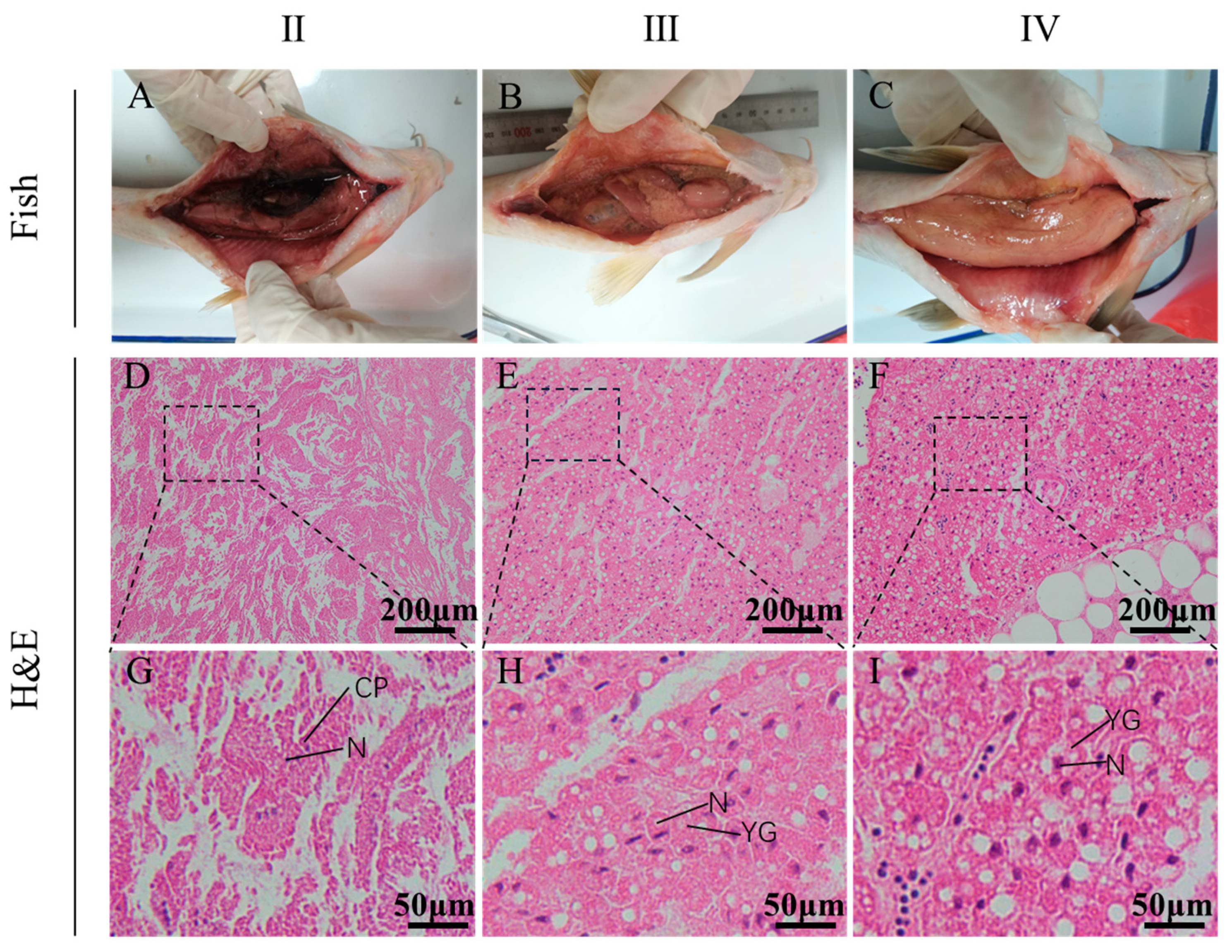
3.1. The Characteristic of Ovarian Morphology and Histology from Stage II to Stage IV

Ovarian morphology and histology from stage II to stage IV are shown in Figure 1. Compared to other stages, ovaries at stage IV were larger in size and redder in color with the increased number of blood vessels (Figure 1A–C). As shown in Figure 1G–I, the oocytes increased both in quantity and size from stage II to stage IV.
Meanwhile, the yolk granules emerged at stage III and significantly accumulated at stage IV, showing a distinct staged change.

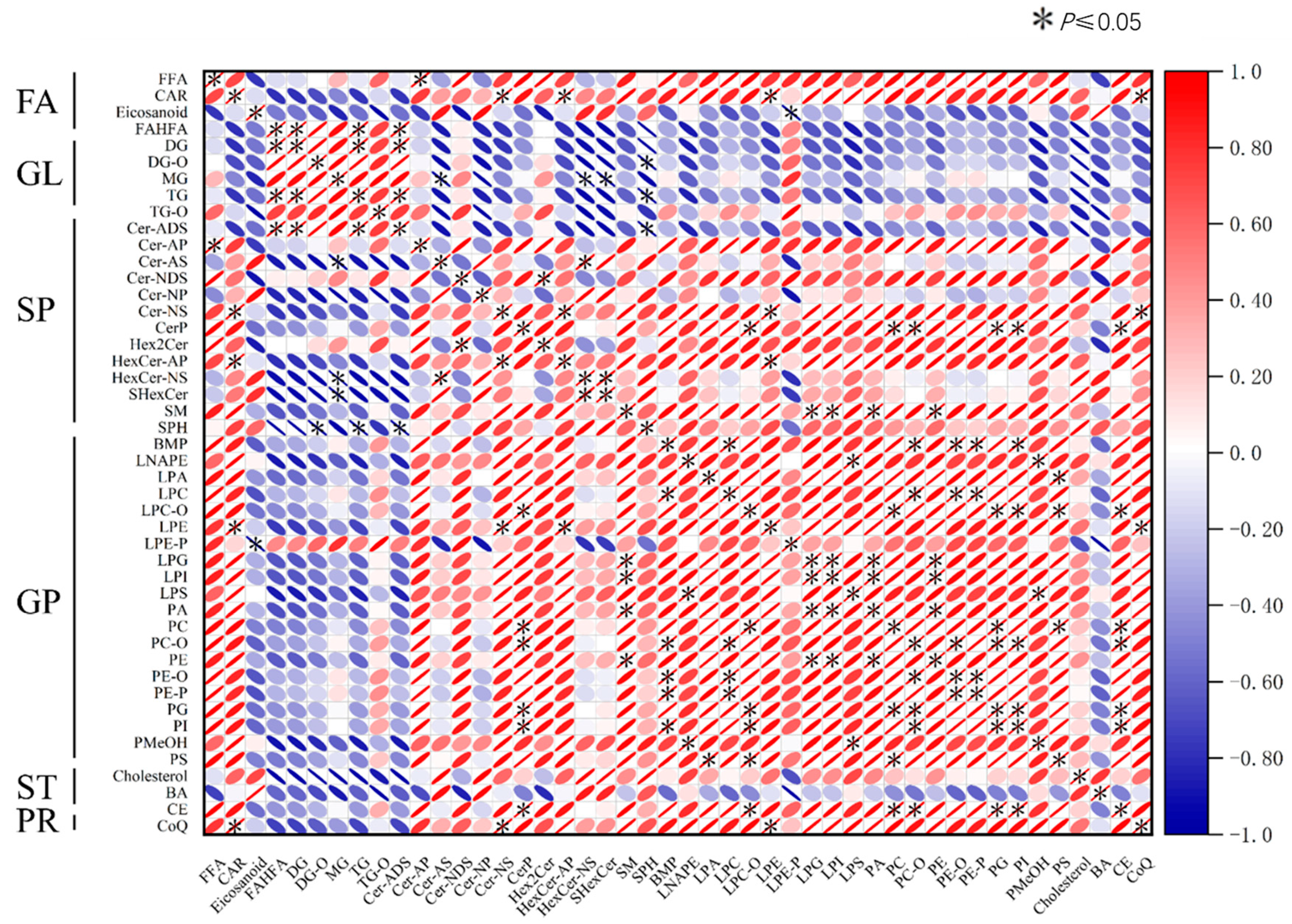
3.2. The Profile of Ovarian Lipid Composition and Correlation Analysis

Given that lipid metabolism is crucial for ovarian development, the profile of ovarian lipid composition in largemouth bronze gudgeons was investigated. A total of 1353 lipids, belonging to 46 subclasses, were identified (Figure 2). These lipids were classified into six categories, namely, fatty acyl (FA), glycerolipid (GL), glycerophospholipid (GP), sphingolipid (SP), sterol lipid (ST), and prenol lipids (PRs). Glycerophospholipid is the most abundant among them. However, saccharolipid (SL) and polyketide (PK) were not detected. Then, the correlation among these lipids was analyzed (Figure 2). The results showed that glycerophospholipid was negatively correlated with glycerolipid, but positively correlated with sphingolipid.

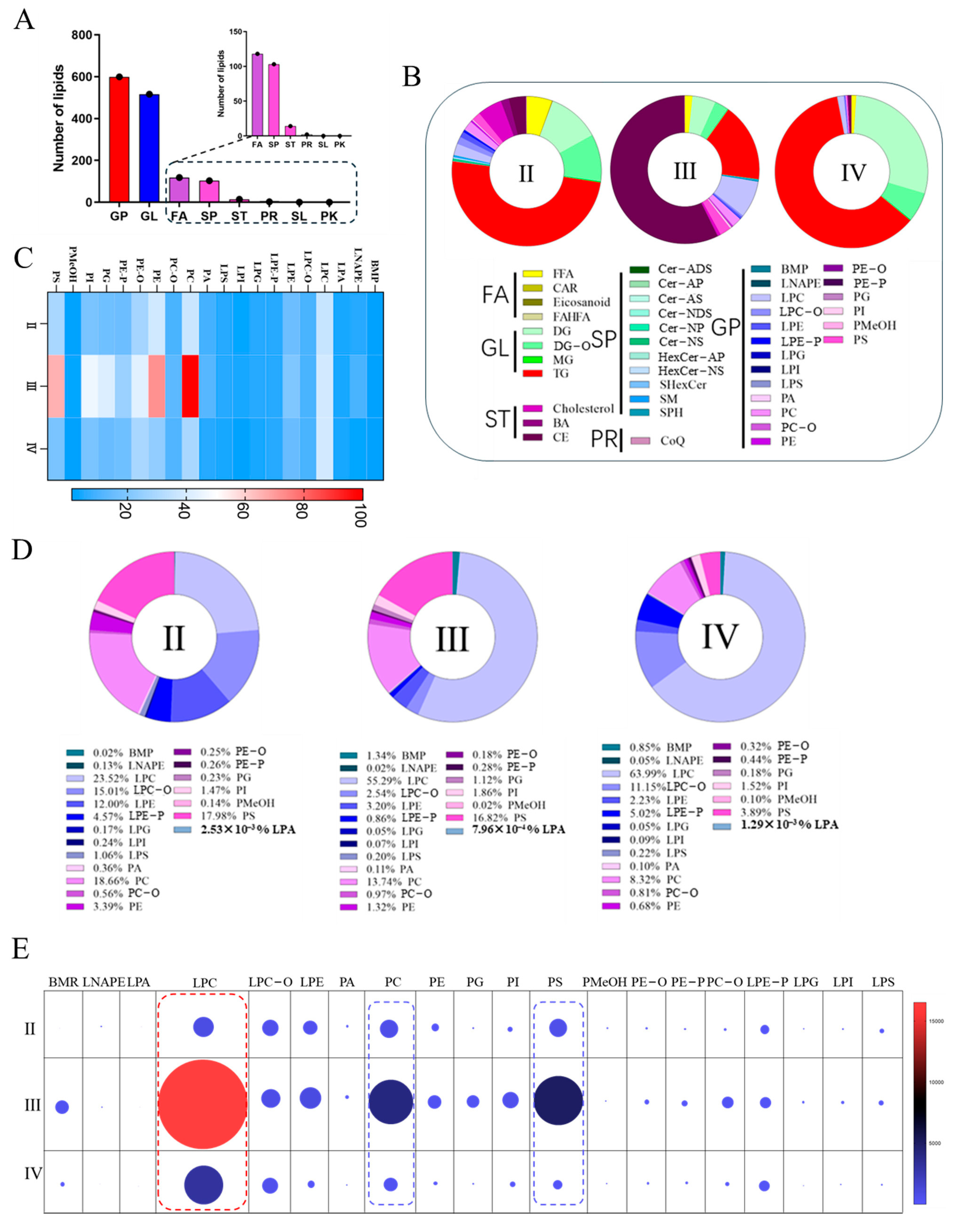
3.3. The Category and Content Analysis of Ovarian Lipid at Different Stages

The lipid abundance during ovarian development was analyzed. As shown in Figure 3A, for category analysis, the most abundant lipid is glycerophospholipid, and the next one is glycerolipid, suggesting they are the dominant lipids for ovarian development. As for the content analysis of total lipids, glycerolipids take the largest proportion; cholesterol lipids (CEs) increased significantly at stage III (Figure 3B). Glycerophospholipid significantly increased at stage III but decreased at stage IV (Figure 3C). Since great changes in glycerophospholipid were observed, its content and proportion were analyzed. The proportion of lysophosphatidyl choline (LPC), phosphatidylcholine (PC), and phosphatidylinositol (PS) decreased significantly at stage IV (Figure 3D). Corresponding to their changes in proportion, their lipid content also decreased significantly at stage IV (Figure 3E). In conclusion, the glycerophospholipids (mainly LPC, PC, PS) changed significantly in their content. They are differential lipids during Coreius guichenoti ovarian development. The results suggest a potential reason for the delay of ovarian development from stage III to IV.

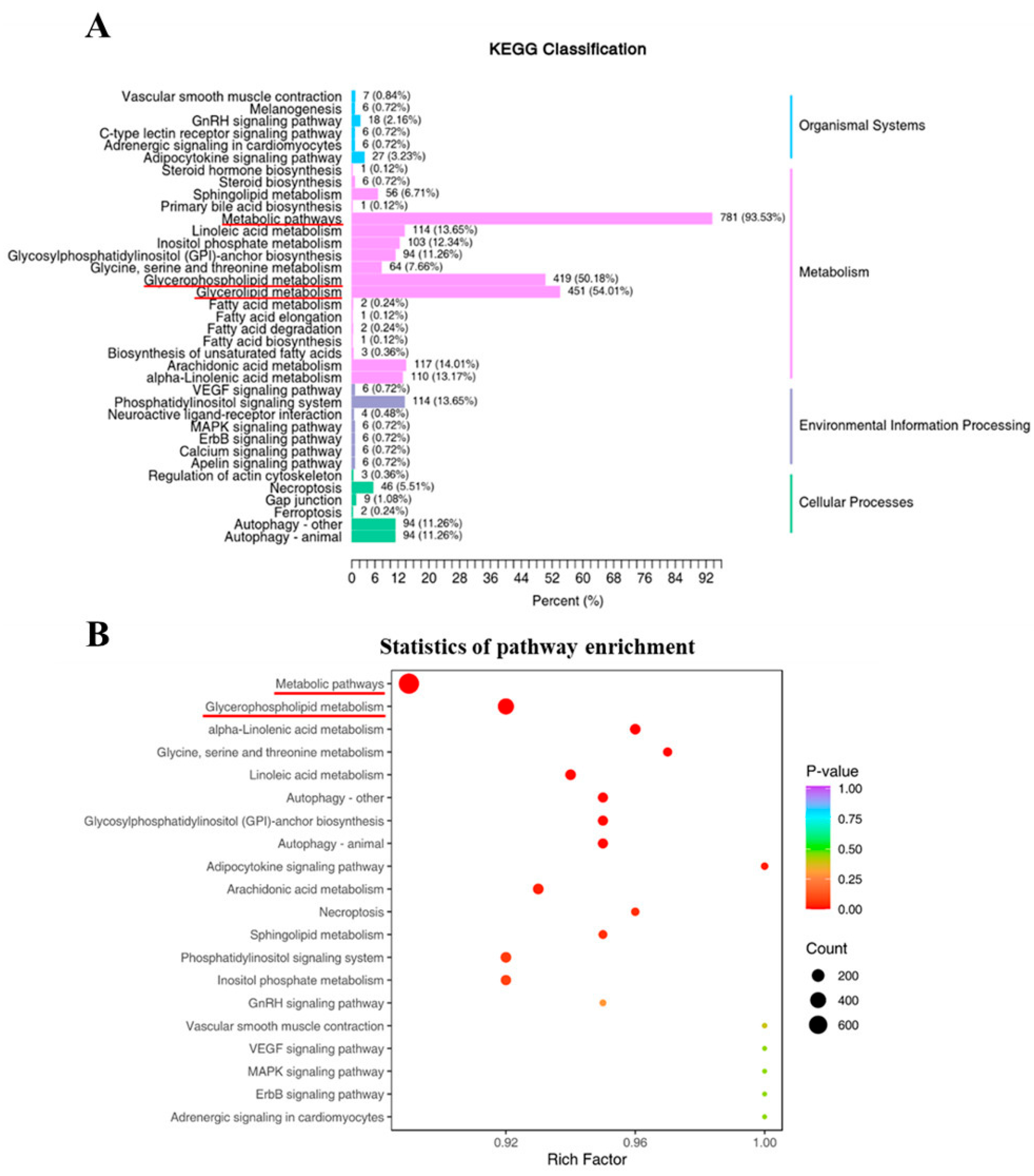
3.4. Lipid-Metabolism-Related Pathways and Their Differential Metabolic Lipids

Using the Kyoto Encyclopedia of Genes and Genomes (KEGG), all differential metabolic lipids were categorized into four categories: “organismal systems”, “metabolism”, “environmental information processing”, and “cellular processes”. Most of the differential metabolic lipids were mainly enriched in “metabolism”. In detail, 781 lipids were enriched in “metabolic pathway”, 419 were enriched in “glycerophospholipid metabolism”, and 451 were enriched in “glycerolipid metabolism”. In addition, a few lipids were enriched in “linoleic acid metabolism”, “phosphoinositide metabolism”, and “glycosylphosphatidylinositol (GPI)-anchor biosynthesis” (Figure 4A). The top 20 enriched pathways are listed in Figure 4B. The significantly upregulated pathways at stage IV were “metabolic pathways”, “glycerophospholipid metabolism”, and “α-linolenic acid metabolism”, suggesting the active glycerophospholipid metabolism at stage IV.

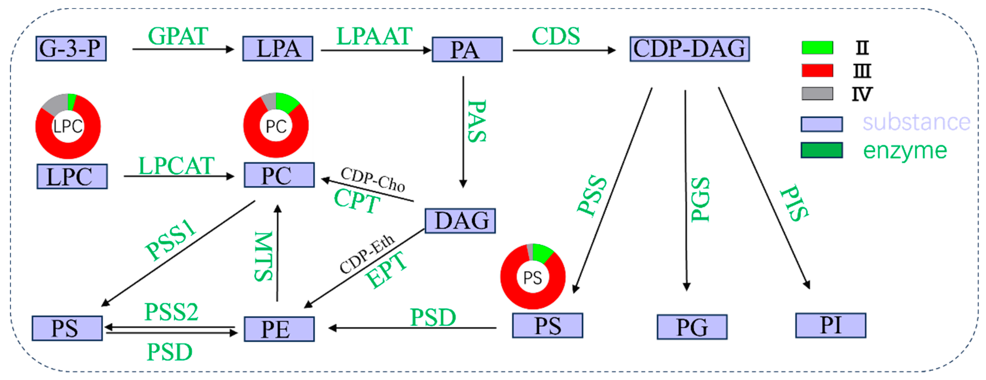
3.5. Key Phospholipid-Metabolism-Related Pathways during Ovarian Development

To clearly understand the glycerophospholipid metabolism during ovarian development, the main phospholipid synthesis pathways are concluded in Figure 5. There are two main pathways for phospholipid synthesis. Firstly, both pathways undergo the transition from glycerol triphosphate (G-3-P) to phosphatidic acid (PA), which need glycerol-3-phosphate acyltransferase (GPAT) and lysophosphatidic acid acyltransferase (LPAAT). After that, one pathway synthesizes phospholipids with diglycerides (DAG), and the other synthesizes phospholipids with cytidine diphosphate-diglycerides (CDP-DAG). There are also some mutual transformations during the phospholipid synthesis process. It is clear to see that differential metabolic lipids (LPC, PC, and PS) are closely related to the enzymes like lysophosphatidylcholine acyltransferase (LPCAT), choline phosphotransferase (CPT), methyltransferases (MTS), and phosphatidylserine synthetase (PSS). The decrease in LPC, PC, and PS at stage IV can be attribute to the low synthesis activity from stage III to IV.

4. Discussion

The construction of hydropower stations has encroached on the spawning ground of the largemouth bronze gudgeon, which is one of the main reasons for the sharp decline in their population [24]. Some measures, like artificial propagation and release, have been undertaken to restore their population. However, such an approach is limited by the delay of ovarian development. Therefore, it is essential to explore the mechanism of ovarian development, as well as the role that lipids play during ovarian development. In this study, lipidomics based on UPLC-MS/MS was used to explore the changes in lipid species, composition, and metabolism during Coreius guichenoti ovarian development. The results show that the lipid content changed significantly during ovarian development. We found that Glycerolipid accounts for the largest proportion among all lipids, and the glycerophospholipid content decreased significantly from stage III to stage IV. Such results show that glycerolipid and glycerophospholipid influenced the accumulation of yolk granules. Changes in lipid metabolism indicate the effect of glycerophospholipids on ovarian development. The present study uncovered the lipid changes during ovarian development through lipidomics and KEGG annotation.
For bony fishes, the ovarian development process includes oocyte growth, hormone secretion, vitellogenesis, and lipid deposition. The egg is the product of oocyte growth and differentiation. Generally, the primordial germ cells (PGCs) firstly develop into oocytes, and then release eggs. This progress is accompanied by many complex changes in the ovary [25]. Previous research has revealed that the synthesis and secretion of sex hormones can affect oocyte development. For example, estrogen and androgen can regulate oogenesis [26,27]. Estradiol (E2) stimulates the expression of the vitellogenin (VTG) mRNA [28] and VTG synthesis [29] in the liver. VTG enters the oocyte through endocytosis mediated by the relevant receptors. Then it is converted into yolk granules and deposited in the oocyte, maintaining embryonic development [30,31]. These biological processes are influenced by internal and external factors, which further affect the ovarian development. Using light and electron microscopy sectioning techniques, previous researchers revealed the morphological changes in primary oocytes when they developed into mature eggs [32].
In this study, dissection and the H&E staining test showed that there were no significant changes in ovarian morphology and histology. The oocyte gradually enlarged in size, and the nucleus became darker from stage II to stage IV. The yolk granules appeared at stage III, showing a normal ovarian development. However, the yolk granules were not densely deposited at stage IV, indicating that ovarian development was delayed due to changes in the living environment. Consistent with our results, previous studies have also shown that changes in hydrological conditions can affect the growth and spawning of the largemouth bronze gudgeon [11]. In addition, considering that the yolk granules are composed of lipids and proteins [33], the mechanism regarding ovarian development can be further explored through lipid composition analysis.
Various lipids have distinct functions in promoting fish growth and ovarian development. Generally, lipids can be used as components of cell membranes, energy suppliers, or materials for hormone synthesis [17,34]. The development of omics has laid a foundation for further research on lipid metabolism. In this study, lipidomics was performed on ovaries of largemouth bronze gudgeons. The results showed that fatty acyls, glycerolipids, glycerophospholipids, sphingolipids, sterol lipids, and prenol lipids were all involved in the ovarian development.
Recently, some researchers have reported the role of glycerolipids and glycerophospholipid during ovarian development. Glycerolipids are neutral lipids, serving as energy supplementation for gonadal development. They usually accumulate in the ovaries as lipid droplets, supporting oocyte and embryonic development by maintaining tissue morphology and providing energy [35]. Glycerophospholipids are involved in yolk synthesis in oocytes. They are the key component of the yolk [19]. Together with other substances, such as lipids, carbohydrates, and vitamins, glycerophospholipids ensure the normal development of the embryo [18]. In our study, we observed that most glycerolipids were positively correlated with glycerophospholipids during ovarian development from stage II to stage IV. This result indicates the function of these lipids in Coreius guichenoti ovarian development. However, phospholipids content decreased significantly at stage IV, which may be an important reason for the delay of ovarian development.
As discussed previously, lipids play an important role in tissue composition and function [19,35,36]. In this study, glycerophospholipids and glycerolipids were found significantly more than other lipids. This result indicates that glycerophospholipids and glycerolipids were the dominant lipids affecting the ovarian development. Glycerolipids account for the largest proportion of all lipids from stage II to IV. They usually accumulate in the ovaries in the form of lipid droplets, providing energy for ovarian development [35]. At stage III, we found that the proportion of cholesterol (CE) was more than 50 percent. Cholesterol is the precursor to all steroid hormones [37], including glucocorticoids and mineral corticosteroids, sex hormones, and vitamin D. These hormones are involved in the regulation of carbohydrates metabolism and reproductive homeostasis. High levels of cholesterol indicate that ovaries were undergoing extensive hormone synthesis at stage III to promote ovarian development. Meanwhile, glycerophospholipids, such as LPC, PC, and PS, decreased significantly after the increase at stage III. However, their proportions did not follow the same trend. Low proportion of glycerophospholipids was found during ovarian development. We speculate that this may be due to the interconversion of phospholipids during ovarian development. Relevant studies have reported that phospholipids can be converted into other phospholipids [38]. For example, PS can be converted to PC by phospholipid synthase [39], and PE can be methylated to PC [38]. We noted that the number of phospholipids, especially LPC, PC, and PS, decreased at stage IV after increasing at stage III. In conclusion, the decrease in glycerophospholipids hindered the ovarian development at stage IV. Thus, ovaries of largemouth bronze gudgeons failed to naturally develop to stage V for spawning.
Lipids, as one of the basic nutrients in fish, are involved in the composition of cells and regulation of metabolism, maintaining the lipid balance in the body [40]. In our study, “glycerophospholipid metabolism” was the most significant lipid metabolism pathway at stage IV. Other differential metabolites were mainly enriched in “glyceride metabolism”, “linoleic acid metabolism”, “α-linolenic acid metabolism”, and “linoleic acid metabolism”. This indicates that the lipid metabolism, especially glycerophospholipid metabolism, occurs frequently at stage IV. This result also proves that the decrease in glycerophospholipids hindered the ovarian development of largemouth bronze gudgeons.
Phospholipids play a key role in oocyte development. Vitellogenin (VTG), a vital protein for teleost oocyte development, consists of 20% lipids and 80% proteins. The lipids in VTG are mainly phospholipids [33]. Phospholipids participate in the synthesis of vitellin by promoting the transport of lipids from the liver to the ovary. Thus, phospholipids can promote the vitellogenesis and embryonic development [41]. Previous studies have reported the process of phospholipid synthesis [38,39,42]. Phospholipids are synthesized by corresponding enzymes or converted from other phospholipids. For example, PC and PE can be converted to PS by PSS [42], and PS can be converted to PC by phospholipid synthase [39]. The coordinated transformation of various phospholipids jointly promotes the development of oocytes. Phospholipids, especially PC and PE, are usually stored in yolks to promote yolk deposition [43]. PC can be broken down to provide energy for embryonic development [44]. In a catfish study, PC was found to be the main phospholipid in vitellin [45]. Consistent with our findings, previous studies have found that LPC [46] is the major component of cell membranes and lipoproteins. LPC also participates in protein synthesis. Other phospholipids play a coordinate role during ovarian development. The construction of hydropower stations has altered the spawning environment of the largemouth bronze gudgeon, which affects the lipid metabolism during ovarian development. As a result, the development of the largemouth bronze gudgeon is threatened, and artificial propagation is less effective.

5. Conclusions

The present study investigated the morphology of ovary and oocyte, as well as their lipid composition and metabolism at different ovarian development stages. The results showed that the Coreius guichenoti ovarian development was affected by environmental changes. Phospholipids like PC and LPC, which play a key role in ovarian development, decreased significantly from stage III to stage IV, showing a hindered ovarian development. The results of KEGG and enrichment analysis showed that there was frequent lipid metabolism occurring at stage IV. Among them, the glycerophospholipid metabolism pathway significantly enriched, indicating frequent phospholipid metabolism during ovarian development. In this study, the ovarian development was studied at the cellular and molecular levels. The results provided a theoretical basis for the artificial propagation and release of the largemouth bronze gudgeon.

Author Contributions

J.Z. and Y.S. designed the experiment. N.H. and Y.X. conducted the experiment and data analysis with the help of L.W. and X.L. Y.S. drafted the manuscript. J.Z. revised the manuscript. All authors have read and agreed to the published version of the manuscript.

Funding

This work was funded by the Hubei Key Laboratory of Three Gorges Project for Conservation of Fishes (grant No. 2022057-ZHX).

Institutional Review Board Statement

All experiments were conducted according to the institutional ethical guidelines of Huazhong Agricultural University (HZAU) on the care and use of experimental animals. Animal research in this study gained approval from the Ethical Committee of Huazhong Agriculture University (identification code: Fish-2023-03-11).

Data Availability Statement

Data are contained within the article. The data that support the findings of this study are available from the corresponding author upon reasonable request.

Conflicts of Interest

The authors declare no conflicts of interest.

References

  1. Xu, D.; Li, P.; Zhang, Y.; Peng, Z. Comparative study of the complete mitochondrial genomes of the bronze gudgeon (Coreius heterodon) and largemouth bronze gudgeon (Coreius guichenoti). Mitochondr. DNA 2013, 24, 189–190. [Google Scholar] [CrossRef] [PubMed]
  2. Liao, X.; Yu, X.; Chang, J.; Tong, J. Polymorphic microsatellites in largemouth bronze gudgeon (Coreius guichenoti) developed from repeat-enriched libraries and cross-species amplifications. Mol. Ecol. Notes 2007, 7, 1104–1107. [Google Scholar] [CrossRef]
  3. Zhang, C.; Fujiwara, M.; Pawluk, M.; Liu, H.; Cao, W.; Gao, X. Changes in taxonomic and functional diversity of fish communities after catastrophic habitat alteration caused by construction of Three Gorges Dam. Ecol. Evol. 2020, 10, 5829–5839. [Google Scholar] [CrossRef]
  4. Zhang, P.; Yang, Z.; Cai, L.; Qiao, Y.; Chen, X.; Chang, J. Effects of upstream and downstream dam operation on the spawning habitat suitability of Coreius guichenoti in the middle reach of the Jinsha River. Ecol. Eng. 2018, 120, 198–208. [Google Scholar] [CrossRef]
  5. Xiong, M.; Yan, S.; Shao, K.; Li, W.; Zhu, B.; Xu, N. Development of twenty-nine polymorphic microsatellite loci from largemouth bronze gudgeon (Coreius guichenoti). J. Genet. 2014, 93, 100–103. [Google Scholar] [CrossRef]
  6. Li, X.; Yan, Q.; Ringø, E.; Wu, X.; He, Y.; Yang, D. The influence of weight and gender on intestinal bacterial community of wild largemouth bronze gudgeon (Coreius guichenoti, 1874). BMC Microbiol. 2016, 16, 22. [Google Scholar] [CrossRef] [PubMed]
  7. Li, T.; Long, M.; Ji, C.; Shen, Z.; Gatesoupe, F.J.; Zhang, X.; Zhang, Q.; Zhang, L.; Zhao, Y.; Liu, X.; et al. Alterations of the gut microbiome of largemouth bronze gudgeon (Coreius guichenoti) suffering from furunculosis. Sci. Rep. 2016, 28, 30606. [Google Scholar] [CrossRef] [PubMed]
  8. Zhu, J.; Zhao, Y.; Yang, J.; Li, Y.; Chen, P.; Huantao, Q. Feeding frequency affects liver health in largemouth bronze gudgeon Coreius guichenoti: Implications for lipid metabolism, oxidative stress, and inflammation response. Aquac. Rep. 2024, 35, 101941. [Google Scholar] [CrossRef]
  9. Papadaki, C.; Soulis, K.; Muñoz-Mas, R.; Martinez-Capel, F.; Zogaris, S.; Ntoanidis, L.; Dimitriou, E. Potential impacts of climate change on flow regime and fish habitat in mountain rivers of the south-western Balkans. Sci. Total. Environ. 2016, 540, 18–28. [Google Scholar] [CrossRef]
  10. Li, T.; Chen, Q.; Zhang, Q.; Feng, T.; Zhang, J.; Lin, Y.; Yang, P.; He, S.; Zhang, H. Transcriptomic Analysis on the Effects of Altered Water Temperature Regime on the Fish Ovarian Development of Coreius guichenoti under the Impact of River Damming. Biology 2022, 11, 1829. [Google Scholar] [CrossRef] [PubMed]
  11. Zhang, P.; Qiao, Y.; Schineider, M.; Chang, J.; Mutzner, R.; Fluixá-Sanmartín, J.; Yang, Z.; Fu, R.; Chen, X.; Cai, L.; et al. Using a hierarchical model framework to assess climate change and hydropower operation impacts on the habitat of an imperiled fish in the Jinsha River, China. Sci. Total. Environ. 2019, 646, 1624–1638. [Google Scholar] [CrossRef] [PubMed]
  12. Morris, J.A.; Sullivan, C.V.; Govoni, J.J. Oogenesis and spawn information in the invasive lionfish, Pterois miles and Pterois volitans. Sci. Mar. 2011, 75, 147–154. [Google Scholar] [CrossRef]
  13. Abascal, F.J.; Medina, A. Ultrastructure of oogenesis in the bluefin tuna, Thunnus thynnus. J. Morphol. 2005, 264, 149–160. [Google Scholar] [CrossRef] [PubMed]
  14. Zakeri, M.; Marammazi, J.G.; Kochanian, P.; Savari, A.; Yavari, V.; Haghi, M. Effects of protein and lipid concentrations in broodstock diets on growth, spawning performance and egg quality of yellowfin sea bream (Acanthopagrus latus). Aquaculture 2009, 295, 99–105. [Google Scholar] [CrossRef]
  15. Zhang, M.Z.; Li, G.L.; Zhu, C.H.; Deng, S.P. Effects of fish oil on ovarian development in spotted scat (Scatophagus argus). Anim. Reprod. Sci. 2013, 141, 90–97. [Google Scholar] [CrossRef] [PubMed]
  16. Song, Y.F.; Tan, X.Y.; Pan, Y.X.; Zhang, L.H.; Chen, Q.L. Fatty Acid β-Oxidation Is Essential in Leptin-Mediated Oocytes Maturation of Yellow Catfish Pelteobagrus fulvidraco. Int. J. Mol. Sci. 2018, 19, 1457. [Google Scholar] [CrossRef] [PubMed]
  17. Huang, X.; Yin, Y.; Shi, Z.; Li, W.; Zhou, H.; Lv, W. Lipid content and fatty acid composition in wild-caught silver pomfret (Pampus argenteus) broodstocks: Effects on gonad development. Aquaculture 2010, 310, 192–199. [Google Scholar] [CrossRef]
  18. Lubzens, E.; Young, G.; Bobe, J.; Cerdà, J. Oogenesis in teleosts: How eggs are formed. Gen. Comp. Endocrinol. 2010, 165, 367–389. [Google Scholar] [CrossRef] [PubMed]
  19. Hiramatsu, N.; Todo, T.; Sullivan, C.V.; Schilling, J.; Reading, B.J.; Matsubara, T.; Ryu, Y.W.; Mizuta, H.; Luo, W.; Nishimiya, O.; et al. Ovarian yolk formation in fishes: Molecular mechanisms underlying formation of lipid droplets and vitellogenin-derived yolk proteins. Gen. Comp. Endocrinol. 2015, 221, 9–15. [Google Scholar] [CrossRef] [PubMed]
  20. Chen, J.N.; Zhang, Y.Y.; Huang, X.; Wang, H.P.; Dong, X.; Zhu, B.; Qin, L. Analysis of Lipid Molecule Profiling and Conversion Pathway in Mandarin Fish (Siniperca chuatsi) during Fermentation via Untargeted Lipidomics. J. Agric. Food Chem. 2023, 71, 8673–8684. [Google Scholar] [CrossRef] [PubMed]
  21. Lam, S.M.; Li, J.; Sun, H.; Mao, W.; Lu, Z.; Zhao, Q.; Han, C.; Gong, X.; Jiang, B.; Chua, G.H.; et al. Quantitative Lipidomics and Spatial MS-Imaging Uncovered Neurological and Systemic Lipid Metabolic Pathways Underlying Troglomorphic Adaptations in Cave-Dwelling Fish. Mol. Biol. Evol. 2022, 39, msac050. [Google Scholar] [CrossRef] [PubMed]
  22. Marqueño, A.; Blanco, M.; Maceda-Veiga, A.; Porte, C. Skeletal Muscle Lipidomics as a New Tool to Determine Altered Lipid Homeostasis in Fish Exposed to Urban and Industrial Wastewaters. Environ. Sci. Technol. 2019, 53, 8416–8425. [Google Scholar] [CrossRef] [PubMed]
  23. Wei, X.; Hogstrand, C.; Chen, G.; Lv, W.; Song, Y.; Xu, Y.; Luo, Z. Zn Induces Lipophagy via the Deacetylation of Beclin1 and Alleviates Cu-Induced Lipotoxicity at Their Environmentally Relevant Concentrations. Environ. Sci. Technol. 2021, 55, 4943–4953. [Google Scholar] [CrossRef] [PubMed]
  24. Tu, Z.; Li, L.; Yuan, X.; Huang, Y.; Johnson, D. Aerobic swimming performance of juvenile largemouth bronze gudgeon (Coreius guichenoti) in the Yangtze River. J. Exp. Zool. A Ecol. Genet. Physiol. 2012, 317, 294–302. [Google Scholar] [CrossRef] [PubMed]
  25. Selman, K.; Wallace, R.A.; Sarka, A.; Qi, X. Stages of oocyte development in the zebrafish, Brachydanio rerio. J. Morphol. 1993, 218, 203–224. [Google Scholar] [CrossRef] [PubMed]
  26. Chou, C.H.; Chen, M.J. The Effect of Steroid Hormones on Ovarian Follicle Development. Vitam. Horm. 2018, 107, 155–175. [Google Scholar]
  27. Walters, K.A.; Allan, C.M.; Handelsman, D.J. Androgen actions and the ovary. Biol. Reprod. 2008, 78, 380–389. [Google Scholar] [CrossRef] [PubMed]
  28. Wallace, R.A.; Selman, K. Major protein changes during vitellogenesis and maturation of Fundulus oocytes. Dev. Biol. 1985, 110, 492–498. [Google Scholar] [CrossRef] [PubMed]
  29. Payne, A.H.; Hales, D.B. Overview of steroidogenic enzymes in the pathway from cholesterol to active steroid hormones. Endocr. Rev. 2004, 25, 947–970. [Google Scholar] [CrossRef]
  30. Stifani, S.; Le Menn, F.; Rodriguez, J.N.; Schneider, W.J. Regulation of oogenesis: The piscine receptor for vitellogenin. Biochim. Biophys. Acta 1990, 1045, 271–279. [Google Scholar] [CrossRef]
  31. Mizuta, H.; Luo, W.; Ito, Y.; Mushirobira, Y.; Todo, T.; Hara, A.; Reading, B.J.; Sullivan, C.V.; Hiramatsu, N. Ovarian expression and localization of a vitellogenin receptor with eight ligand binding repeats in the cutthroat trout (Oncorhynchus clarki). Comp. Biochem. Physiol. B Biochem. Mol. Biol. 2013, 166, 81–90. [Google Scholar] [CrossRef] [PubMed]
  32. Menn, F.L.; Cerdà, J.; Babin, P.J. Ultrastructural Aspects of the Ontogeny and Differentiation of Ray-Finned Fish Ovarian Follicles; Springer: Dordrecht, The Netherlands, 2007; pp. 1–37. [Google Scholar]
  33. Johnson, R.B. Lipid Deposition in Oocytes of Teleost Fish During Secondary Oocyte Growth. Rev. Fish. Sci. 2009, 17, 78–89. [Google Scholar] [CrossRef]
  34. Dunning, K.R.; Russell, D.L.; Robker, R.L. Lipids and oocyte developmental competence: The role of fatty acids and β-oxidation. Reproduction 2014, 148, R15–R27. [Google Scholar] [CrossRef] [PubMed]
  35. Rodríguez-González, H.; García-Ulloa, M.; Hernández-Llamas, A.; Villarreal, H. Effect of dietary protein level on spawning and egg quality of redclaw crayfish Cherax quadricarinatus. Aquaculture 2006, 257, 412–419. [Google Scholar] [CrossRef]
  36. Li, Q.Q.; Xiang, Q.Q.; Lian, L.H.; Chen, Z.Y.; Luo, X.; Ding, C.Z.; Chen, L.Q. Metabolic profiling of nanosilver toxicity in the gills of common carp. Ecotoxicol. Environ. Saf. 2021, 222, 112548. [Google Scholar] [CrossRef] [PubMed]
  37. Cortes, V.A.; Busso, D.; Maiz, A.; Arteaga, A.; Nervi, F.; Rigotti, A. Physiological and pathological implications of cholesterol. Front. Biosci. 2014, 19, 416–428. [Google Scholar] [CrossRef] [PubMed]
  38. Tocher, D.R.; Bendiksen, E.Å.; Campbell, P.J.; Bell, J.G. The role of phospholipids in nutrition and metabolism of teleost fish. Aquaculture 2008, 280, 21–34. [Google Scholar] [CrossRef]
  39. Hiratsuka, S.; Kitagawa, T.; Matsue, Y.; Hashidume, M.; Wada, S. Lipid class and fatty acid composition of phospholipids from the gonads of skipjack tuna. Fish. Sci. 2004, 70, 903–909. [Google Scholar] [CrossRef]
  40. Tsoupras, A.; Brummell, C.; Kealy, C.; Vitkaitis, K.; Redfern, S.; Zabetakis, I. Cardio-Protective Properties and Health Benefits of Fish Lipid Bioactives; The Effects of Thermal Processing. Mar. Drugs 2022, 20, 187. [Google Scholar] [CrossRef] [PubMed]
  41. Sui, L.Y.; Wu, X.G.; Wille, M.; Cheng, Y.X.; Sorgeloos, P. Effect of dietary soybean lecithin on reproductive performance of Chinese mitten crab Eriocheir sinensis (H. Milne-Edwards) Broodstock. Aquacult. Int. 2008, 17, 45–56. [Google Scholar] [CrossRef]
  42. Tomohiro, S.; Kawaguti, A.; Kawabe, Y.; Kitada, S.; Kuge, O. Purification and characterization of human phosphatidylserine synthases 1 and 2. Biochem. J. 2009, 418, 421–429. [Google Scholar] [CrossRef] [PubMed]
  43. Salze, G.; Tocher, D.R.; Roy, W.J.; Robertson, D.A. Egg quality determinants in cod (Gadus morhua L.): Egg performance and lipids in eggs from farmed and wild broodstock. Aquac. Res. 2005, 36, 1488–1499. [Google Scholar] [CrossRef]
  44. Cowey, C.B.; Bell, J.G.; Knox, D.; Fraser, A.; Youngson, A. Lipids and antioxidant systems in developing eggs of salmon (Salmo salar). Lipids 1985, 20, 567–572. [Google Scholar] [CrossRef]
  45. Singh, P.B.; Singh, V.; Srivastava, S.; Pandey, S. Effects of estradiol-17beta and 17alpha, 20beta-dihydroxy-4-pregnen-3-one on different phospholipids metabolism and histological changes in ovary during reproductive growth in the catfish, Heteropneustes fossilis (Bloch). J. Environ. Biol. 2007, 28, 771–778. [Google Scholar] [PubMed]
  46. Nutautaitė, M.; Racevičiūtė-Stupelienė, A.; Andalibizadeh, L.; Šašytė, V.; Bliznikas, S.; Pockevičius, A.; Vilienė, V. Improving broiler chickens’ health by using lecithin and lysophosphatidylcholine emulsifiers: A comparative analysis of physiological indicators. Iran. J. Vet. Res. 2021, 22, 33–39. [Google Scholar] [PubMed]
Figure 1. The characteristics of ovarian morphology and histology from stage II to stage IV. (AC) Representative images of ovary. (DF) Representative images of ovarian tissues stained by H&E, 200× magnification. Scale bars, 200 µm. (GI) Enlarged images from figure (DF). Scale bars, 50 µm. Nucleus: N; cytoplasm: CP; yolk granule: YG.
Fishes 09 00291 g001
Figure 2. The profile of ovarian lipid composition and correlation analysis. The color intensity shows the degree of the association, with the positive correlations in red and the negative ones in blue. Asterisks denote statistically significant differences in correlation; * p ≤ 0.05.
Fishes 09 00291 g002
Figure 3. The category and content analysis of ovarian lipid at different stages. (A) The abundance of lipid classes. (B) Sector graph illustrating proportion of lipid content. (C) Heat map of differential glycerophospholipid content. (D) Sector graph illustrating proportion of glycerophospholipid content. (E) The content of glycerophospholipid. Glycerophospholipid content is represented by the size of the circle. The colors of the circle are used according to the heat map. Differential lipids are highlighted by dashed line box.
Fishes 09 00291 g003
Figure 4. Lipid-metabolism-related pathways and their differential metabolic lipids. (A) KEGG classification of differential metabolic lipids in ovary. The most enriched pathway is “metabolism”. (B) Scatter plot of the enriched KEGG pathways for the differential metabolic lipids. The sizes and colors of the dots represent the number of lipids and the significance of the difference, respectively. Enriched pathways are denoted by a red line.
Fishes 09 00291 g004
Figure 5. Key phospholipid metabolism pathways related with the differential metabolic lipids during ovarian development. The differential metabolic lipids and their proportions at different states are listed. Abbreviations for substance: G-3-P, Glycerol triphosphate. LPA, Lysophosphatidic acid. PA, Phosphatidic acid. CDP-DAG, Cytidine diphosphate-diglycerides. LPC, Lysophosphatidyl choline. PC, Phosphatidylcholine. DAG, Diglycerides. PS, Phosphatidylserine. PE, Phosphatidylethanolamine. PG, Phosphatidylglycerol. PI, Phosphatidylinositol. Abbreviations for enzymes: GAPT, glycerol-3-phosphate acyltransferase. LPAAT, Lysophosphatidic acid acyltransferase. PAS, phosphatase. CDS, CDP-DAG synthase. LPCAT, Lysophosphatidylcholine acyltransferase. CPT, Choline phosphotransferase. PSS, Phosphatidylserine synthetase. PGS, Phosphatidylglycerol synthase. PIS, Phosphatidylinositol synthetase. PSS1, Phosphatidylserine synthetase 1. PSS2, Phosphatidylserine synthetase 2. PSD, Phosphatidylserine decarboxylase. EPT, Ethanolamine phosphotransferase. MTS, Methyltransferases.
Fishes 09 00291 g005
Disclaimer/Publisher’s Note: The statements, opinions and data contained in all publications are solely those of the individual author(s) and contributor(s) and not of MDPI and/or the editor(s). MDPI and/or the editor(s) disclaim responsibility for any injury to people or property resulting from any ideas, methods, instructions or products referred to in the content.

