Article

Effect of Dietary β-Glucan on Growth Performance, Antioxidant Responses, and Immunological Parameters of Coral Trout (Plectropomus leopardus)

1
Ocean College, Hebei Agricultural University, Qinhuangdao 066003, China
2
Key Laboratory of South China Sea Fishery Resources Exploitation & Utilization, Ministry of Agriculture and Rural Affairs, South China Sea Fisheries Research Institute, Chinese Academy of Fishery Sciences, Guangzhou 510300, China
3
Shenzhen Base of South China Sea Fisheries Research Institute, Chinese Academy of Fishery Sciences, Shenzhen 518121, China
*
Authors to whom correspondence should be addressed.
Fishes 2024, 9(8), 298; https://doi.org/10.3390/fishes9080298
Submission received: 7 July 2024 / Revised: 28 July 2024 / Accepted: 29 July 2024 / Published: 31 July 2024
(This article belongs to the Section Nutrition and Feeding)

Abstract

:
Although β-glucan has diverse benefits for fish health, the potential adverse impacts of excessive supplementation are poorly understood. This study investigated the optimal dosage of β-glucan for coral trout and explored the adverse effects of excessive supplementation. The results revealed that moderate β-glucan supplementation (1) significantly enhanced the weight gain rate and specific growth rate (SGR); (2) significantly improved the intestinal villus length (0.10%), muscle thickness (0.05–0.15%), and α-amylase and chymotrypsin activities (0.05–0.15%); (3) significantly increased liver catalase (CAT), glutathione reductase (GR), alkaline phosphatase, complement 3, immunoglobulin M (IgM), lysozyme, serum superoxide dismutase, CAT, glutathione peroxidase, GR, total antioxidant capacity, acid phosphatase, complement 4, and IgM activities and significantly reduced malondialdehyde contents; (4) upregulated genes in the liver associated with copper-zinc superoxide dismutase (SOD-1), manganese superoxide dismutase (SOD-2), CAT, GSH-Px1a, ACP6, AKP, LZ-c, IgM, C3, and C4-b. However, 0.20% β-glucan significantly inhibited the growth performance of coral trout compared with 0.10% β-glucan. Thus, 0.10% β-glucan represents the optimal dosage for promoting growth, antioxidant activity, and immune responses in coral trout, while higher β-glucan levels weakened these beneficial effects. With an SGR established by a cubic regression analysis, the optimal level of β-glucan for maximal growth of coral trout is 0.082%. This study provides new insights into the health impacts of β-glucan on fish.
Key Contribution: This study establishes 0.10% β-glucan as the optimal dosage for enhancing growth, antioxidant activity, and immune responses in coral trout and highlights the adverse impacts of excessive supplementation.

1. Introduction

Coral trout (Plectropomus leopardus) is a highly valued fish species in China in terms of both economic and ornamental value [1,2]. However, the significant expansion of intensive farming and concurrent rise in stressors have increased the vulnerability of fish to diseases. This heightened susceptibility poses a substantial threat to the overall health and sustainability of aquaculture [3]. Research has found that coral trout host at least 28 species of parasites, which may weaken their ability to cope with other stressors [4]. When exposed to stress conditions, such as high stocking densities and poor water quality, cultivated coral trout are more prone to pathogenic infections compared to their wild counterparts [5]. Antibiotics have traditionally been used to treat fish diseases; however, their overuse can lead to environmental residues and bacterial resistance [3,6]. This resistance poses a threat to human health through potential transfer to terrestrial environments [7]. Therefore, safe alternatives to antibiotics are urgently required to prevent and treat fish diseases that may occur in coral trout aquaculture.
Immunostimulants are potent tools for reducing disease resistance and enhancing the immune status of cultured animals [8]. Natural immunostimulants are biocompatible, biodegradable, and harmless to the environment and human health [9]. Thus, immunostimulants can be incorporated into the diet as a viable strategy to mitigate disease. β-glucan is a polysaccharide composed of glucose monomers linked by glycosidic bonds [10]. It has been broadly applied in the medicine, agriculture, food, and chemical industries because of its unique biological properties and versatility [11,12,13,14]. β-glucan is recognized by specific receptors on various immune cells, thereby enhancing the resistance of fish to diseases and their immune response capabilities [8,15]. Currently, β-glucan has gained increasing attention as a promising immunostimulant for applications in aquaculture [16]. Research has demonstrated that incorporating an adequate amount of β-glucan into the diet can improve the growth performance of rainbow trout (Oncorhynchus mykiss) [17], red swamp crayfish (Procambarus clarkii) [18], juvenile Persian sturgeon (Acipenser persicus) [19], snakehead (Channa striata) [20], koi (Cyprinus carpio koi) [21], and striped catfish (Pangasianodon hypophthalmus) [22]. Qiao et al. [23] found that β-glucan promotes the growth of white shrimp (Litopenaeus vannamei) by enhancing biochemical enzymes and improving carbohydrate metabolism. Dietary β-glucan has also been shown to enhance the immune responses and antioxidant capacities of tilapia (GIFT, Oreochromis niloticus) [3], Pengze crucian carp (Carassius auratus var. Pengze) [24], and white shrimp [23] by regulating antioxidant and immune enzyme activities. In addition, Fath El-Bab et al. [25] found that β-glucan improved the growth performance of Nile tilapia (O. niloticus) by enhancing gut health and morphology. This enhancement was evidenced by the positive impact on the length, surface area, and width of intestinal villi and the increase in the number of goblet cells, thereby supporting better nutrient absorption and overall gut function.
Extensive studies have shown that the overuse of immunostimulants can adversely affect aquatic animals, leading to immunity fatigue and inhibiting normal growth [26,27]. For example, Yu et al. [27] reported that excessive levels of Astragalus polysaccharides (APS) supplementation decreased the growth performance and caused negative immune system responses in Asian seabass (Lates calcarifer). However, most previous studies have concentrated on the beneficial effects of β-glucan on fish health, with relatively few addressing the negative impacts of its overuse [28,29,30]. Moreover, its effects on coral trout growth have not been reported. Therefore, we analyzed how varying doses of β-glucan affect the growth performance, antioxidant capacity, immune function, and gut health of coral trout based on biochemical and molecular indicators. In addition, we determined the optimal β-glucan dosage for coral trout and investigated the adverse effects of exceeding this dosage. This research aims to offer a theoretical foundation for the healthy farming of coral trout.

2. Materials and Methods

2.1. Materials

β-glucan was purchased from Zhuhai Tianxiangyuan Biotechnology Development Co., Ltd. (Zhuhai, China), and it had a purity of 85% and an average molecular weight of 810 KDa.

2.2. Diet Preparation

Five practical diets with β-glucan levels of 0%, 0.05%, 0.10%, 0.15%, and 0.20% were formulated (Table S1). Diets were prepared and stored according to the method described by Yu et al. [31]. For specific operational methods, please see the Supplementary Materials (Method S1).

2.3. Fish Rearing and Experimental Procedure

The aquaculture equipment was a water tank (1.0 m × 1.0 m × 1.0 m) equipped with a recirculating aquaculture system. Before starting the experiments, the fish were acclimated over a two-week rearing period. After acclimation, healthy fish with an average weight of 75.81 ± 0.10 g were randomly assigned to 15 tanks (n = 20). The tanks were divided into five groups based on the amount of β-glucan in the diets (0%, 0.05%, 0.10%, 0.15%, and 0.20% β-glucan), and three parallel samples were performed per group. The fish were fed twice daily at 08:30 and 16:30, and the experiment lasted 56 days. Throughout the study, the water parameters were maintained as follows: temperature, 26–30 °C; pH, 7.0–7.3; ammonia levels, <0.05 mg/L; nitrite, <0.01 mg/L; and dissolved oxygen, >7.00 mg/L.

2.4. Sampling

Following the diet experiment, the fish were sedated using MS-222, and the total weight and count of fish in each replicate were recorded. Six fish were randomly selected from each replicate to measure body weight and length for morphology index calculations. Blood was drawn from the caudal vein, centrifuged at 3500× g for 10 min at 4 °C to separate the serum, and then stored at −80 °C until required. Liver samples were collected using sterile centrifuge tubes, preserved in RNA-preserving solution (RNAFollow, NCM Biotech, Suzhou, China), and kept at −80 °C for further analysis. Three complete intestines were rapidly frozen in liquid nitrogen for gut microbiota analysis, while another three midgut segments were collected for morphological analysis. The remaining liver and intestinal tissues were rapidly frozen in liquid nitrogen and kept at −80 °C for enzyme activity analysis.

2.5. Growth Performance

The equations for calculating growth performance were calculated as follows:
Weight gain rate (WGR, %) = 100 × (final body weight − initial body weight)/initial body weight
Specific growth rate (SGR, %/day) = 100 × [ln (final body weight) − ln (initial body weight)]/number of days
Feed conversion ratio (FCR) = dry feed intake/net weight gain
Survival rate (SR, %) = 100 × final fish number/initial fish number
Viscerosomatic index (VSI, %) = 100 × visceral weight of a single fish/body weight of a single fish
Hepatosomatic index (HSI, %) = 100 × hepatic weight of a single fish/body weight of a single fish
Condition factor (CF, g/cm3) = 100 × body weight of a single fish/(body length of a single fish)3

2.6. Diet Composition Analysis

The diet composition was analyzed according to AOAC standards [32]. For specific operational methods, please see the Supplementary Materials (Method S2).

2.7. Enzyme Activity Analysis

To prepare a 10% tissue homogenate, liver and intestinal samples were first weighed and then mixed with physiological saline. The resulting homogenate was centrifuged to obtain the supernatant. Enzyme activities in the liver, intestine, and serum were assessed using a microplate reader instrument (Infinite M200 Pro, Switzerland TECAN, Zurich, Switzerland). Digestive enzymes, antioxidant enzymes, and immune enzymes activities were measured using commercial assay kits (Jiancheng, Ltd., Nanjing, China). For specific operational methods, please see the Supplementary Materials (Method S3). The assays were performed, and formulas were calculated according to the instructions provided with the test kits.

2.8. Gene Expression Analysis

Total RNA was isolated from liver tissue using an RNA extraction solution (Servicebio Technology Co., Ltd., Wuhan, China). RNA quality was assessed using 1.0% agarose gel electrophoresis, and RNA concentration and purity were determined on a Nanodrop 2000. Subsequently, RNA was reverse transcribed into cDNA with the SweScript All-in-One RT SuperMix for qPCR (Servicebio Technology Co., Ltd., Wuhan, China), and any remaining DNA was eliminated with gDNA Remover (Servicebio Technology Co., Ltd., Wuhan, China). The forward and reverse specific primers were synthesized by Beijing Ruibo Xingke Biotechnology Co., Ltd. (Beijing, China). The qPCR reaction was executed using a real-time fluorescent quantitative PCR detection system (Likang CG-02, Shanghai, China). For the specific operational methods, please see the Supplementary Materials (Method S4). The relative expression levels of the target genes were calculated using the 2−ΔΔCt method as described by Livak and Schmittgen [33]. GAPDH was used as the internal reference gene of coral trout. Specific primer details are presented in the Supplementary Materials (Table S2).

2.9. Mid-Gut Histological Observation

Intestinal tissues were preserved in a 4% paraformaldehyde solution (Biosharp, Guangzhou, China) for a duration of 24 h. Thereafter, the tissues embedded in paraffin were stained with hematoxylin and eosin (H&E) and mounted using neutral balsam. Measurements of villus length and muscle thickness were performed utilizing CaseViewer 2.4 software based on the approach outlined by Xie et al. [34].

2.10. Statistical Analysis

The results were reported as the mean ± standard error (mean ± SE), with p < 0.05 indicating statistical significance. One-way analysis of variance (ANOVA) on the datasets from each group was performed in SPSS 27.0. Significant differences between and within groups were assessed using Duncan’s and LSD multiple comparison tests. The optimal level of β-glucan for the SGR of coral trout was predicted by employing a cubic regression model.

3. Results

3.1. Growth Performance

As shown in Table 1, as β-glucan supplementation increased from 0.05% to 0.10%, fish growth performance improved compared with that of the control (p < 0.05). However, in the group treated with 0.20% β-glucan, growth performance significantly declined compared with that in the 0.10% β-glucan group (p < 0.05). Coral trout fed diets with 0.05% to 0.10% β-glucan demonstrated higher final body weight (FBW), WGR, and SGR relative to the control group (p < 0.05). Diets enriched with β-glucan resulted in higher SR compared with that of the control group (p < 0.05). The FCR for the 0.10% β-glucan diet was markedly lower than that of the control group (p < 0.05). No significant effects on the VSI, HSI, and CF were observed with varying levels of β-glucan supplementation (p > 0.05). When cubic regression analysis was used to estimate the SGR, the optimal level of β-glucan supplementation for maximal growth of coral trout was 0.082% (Figure 1).

3.2. Digestive Enzyme Activities

As indicated in Table 2, fish that consumed diets containing 0.05% to 0.15% β-glucan showed increased α-amylase and chymotrypsin activities relative to those on the other diets (p < 0.05). However, the lipase activity remained unaffected by the dietary treatments (p > 0.05).

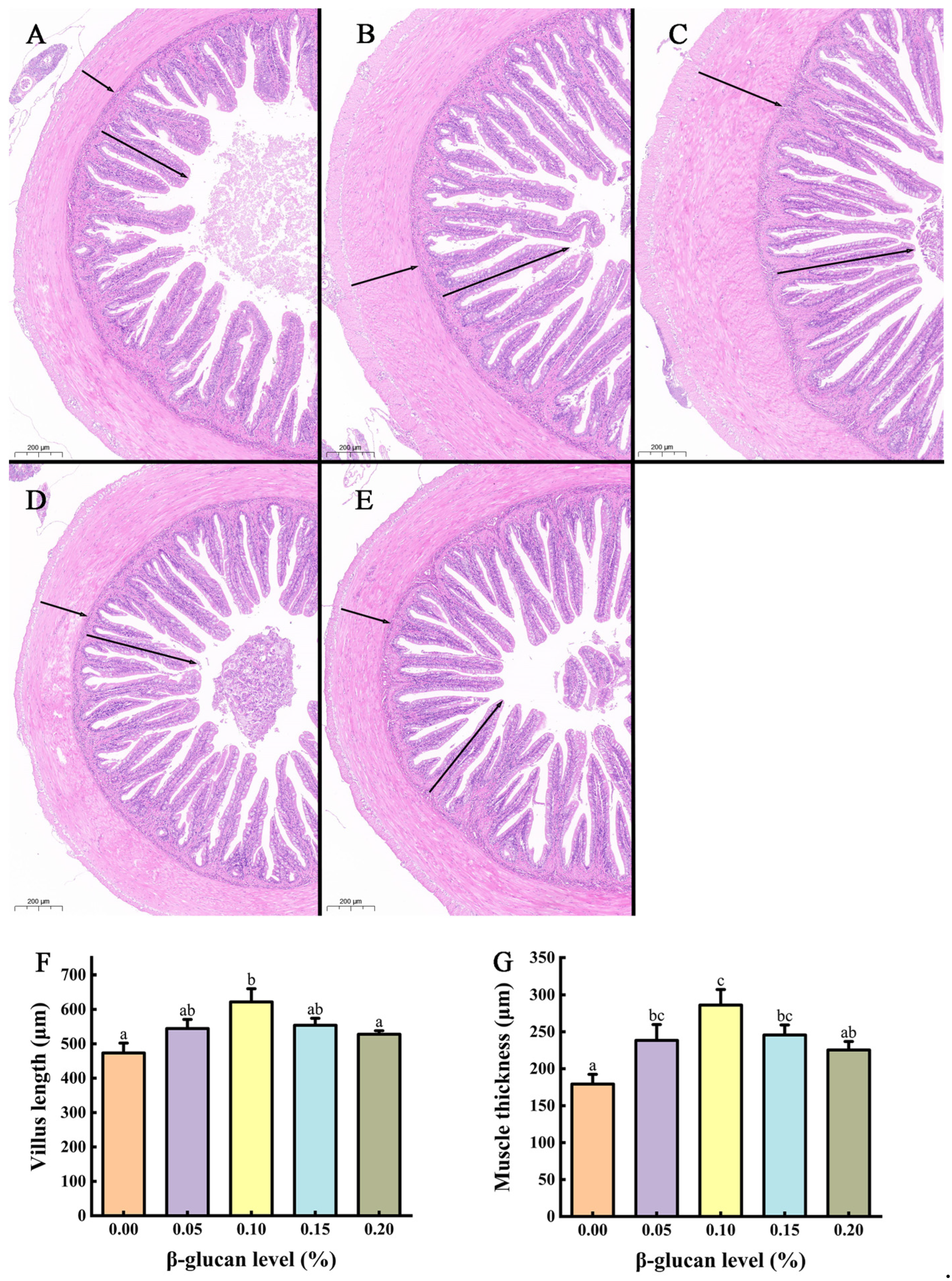
3.3. Mid-Gut Morphology

The intestinal morphological indexes of coral trout in different β-glucan groups are shown in Figure 2. The findings show that the mid-gut villus length and muscle thickness were markedly enhanced in the group treated with 0.10% β-glucan (p < 0.05) compared with the control group. Furthermore, the intestinal sections stained with H&E revealed that the morphology of the mid-gut did not change significantly under varying concentrations of β-glucan.

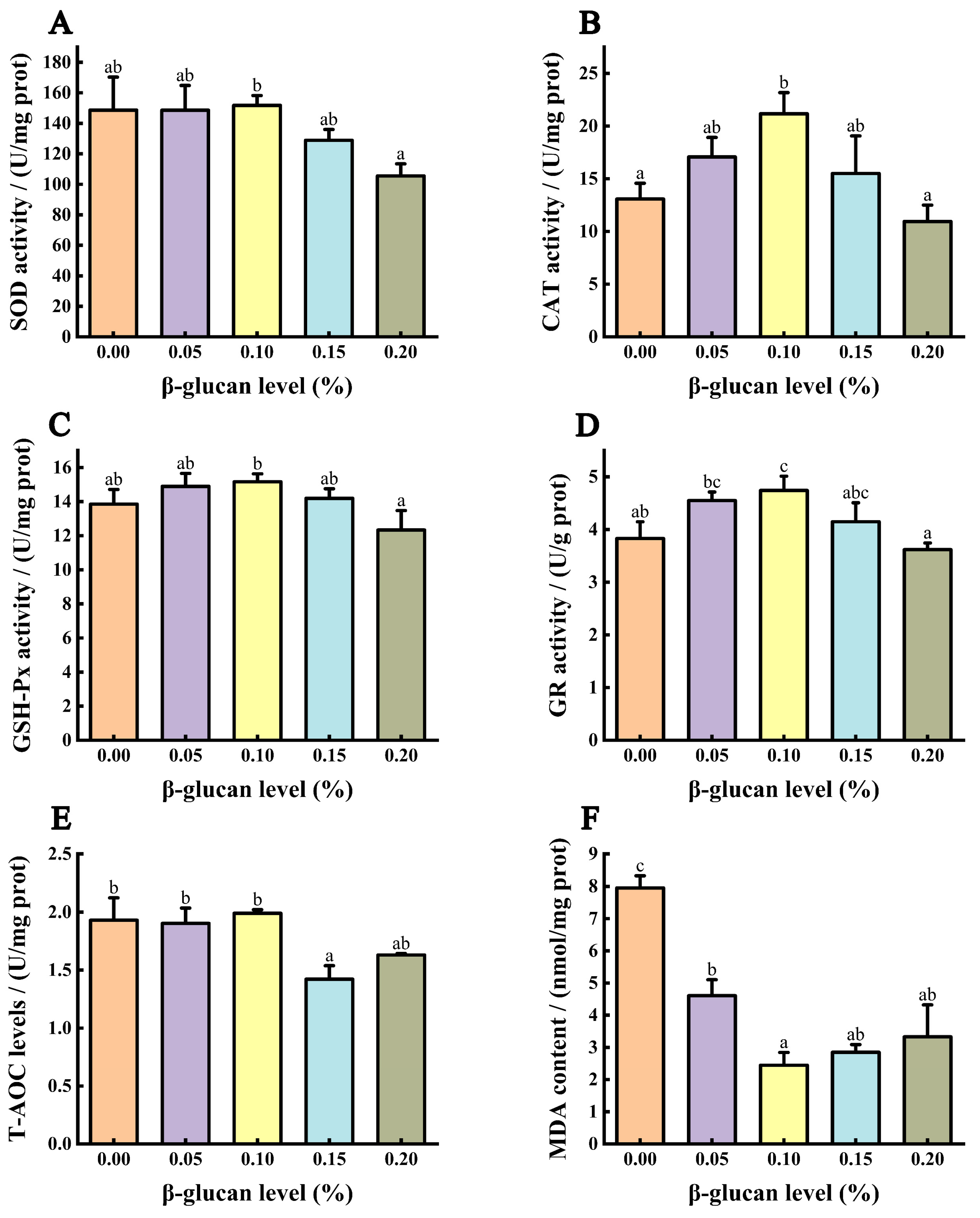
3.4. Antioxidant Capacity of the Liver

As shown in Figure 3, the catalase (CAT) and glutathione reductase (GR) activities in the 0.10% β-glucan group were significantly higher than those in the control and 0.20% β-glucan groups (p < 0.05). The 0.10% β-glucan group exhibited higher superoxide dismutase (SOD) and glutathione peroxidase (GSH-Px) activities compared to the 0.20% β-glucan group (p < 0.05). As the β-glucan content increased, the liver malondialdehyde (MDA) contents progressively decreased, with the lowest value reached in the 0.10% group (p < 0.05). However, with further increases in β-glucan content, the MDA content significantly increased (p < 0.05).

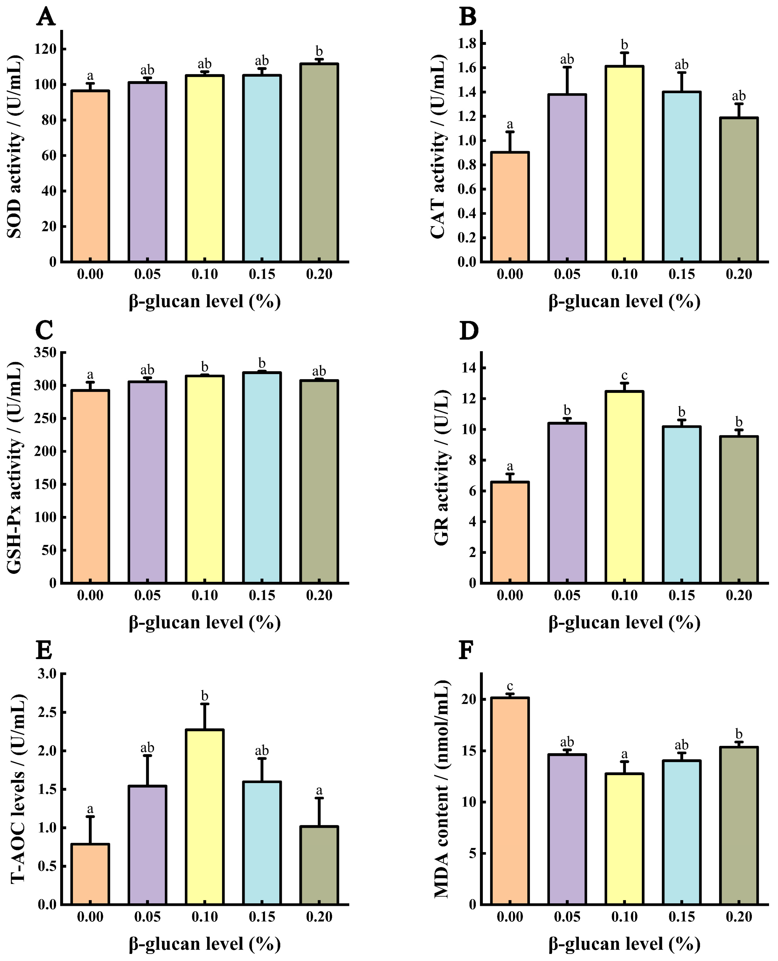
3.5. Antioxidant Capacity of Serum

As depicted in Figure 4, the CAT, GSH-Px, and GR activities and total antioxidant capacity (T-AOC) contents were significantly elevated in the 0.10% β-glucan group compared with the control group (p < 0.05). The SOD activity in the 0.20% β-glucan group and GSH-Px activity in the 0.10% to 0.15% β-glucan groups were notably higher than those in the control group (p < 0.05). In coral trout fed diets enriched with β-glucan, the GR activity was greater than that of the control group (p < 0.05), while the serum MDA content was significantly lower (p < 0.05).

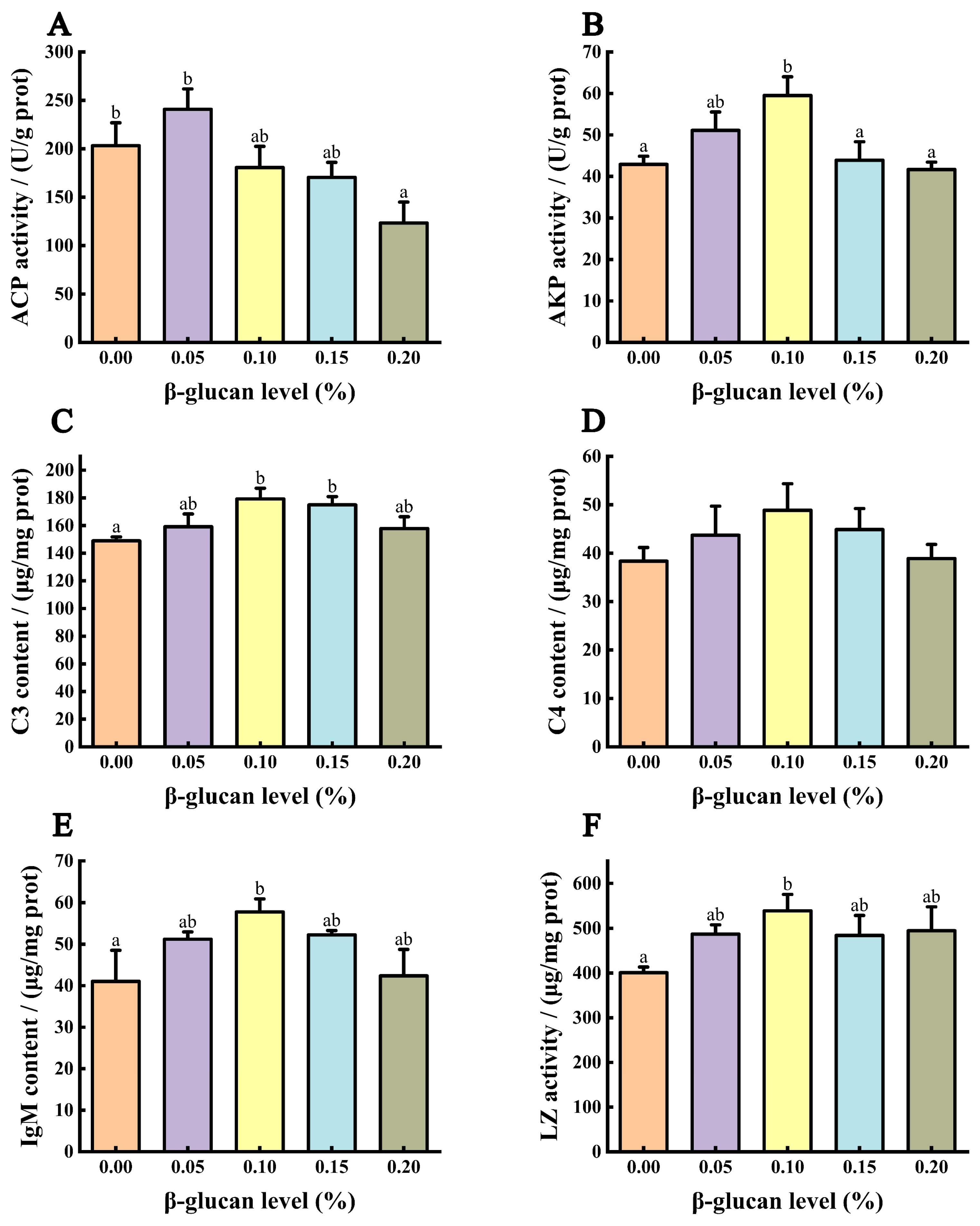
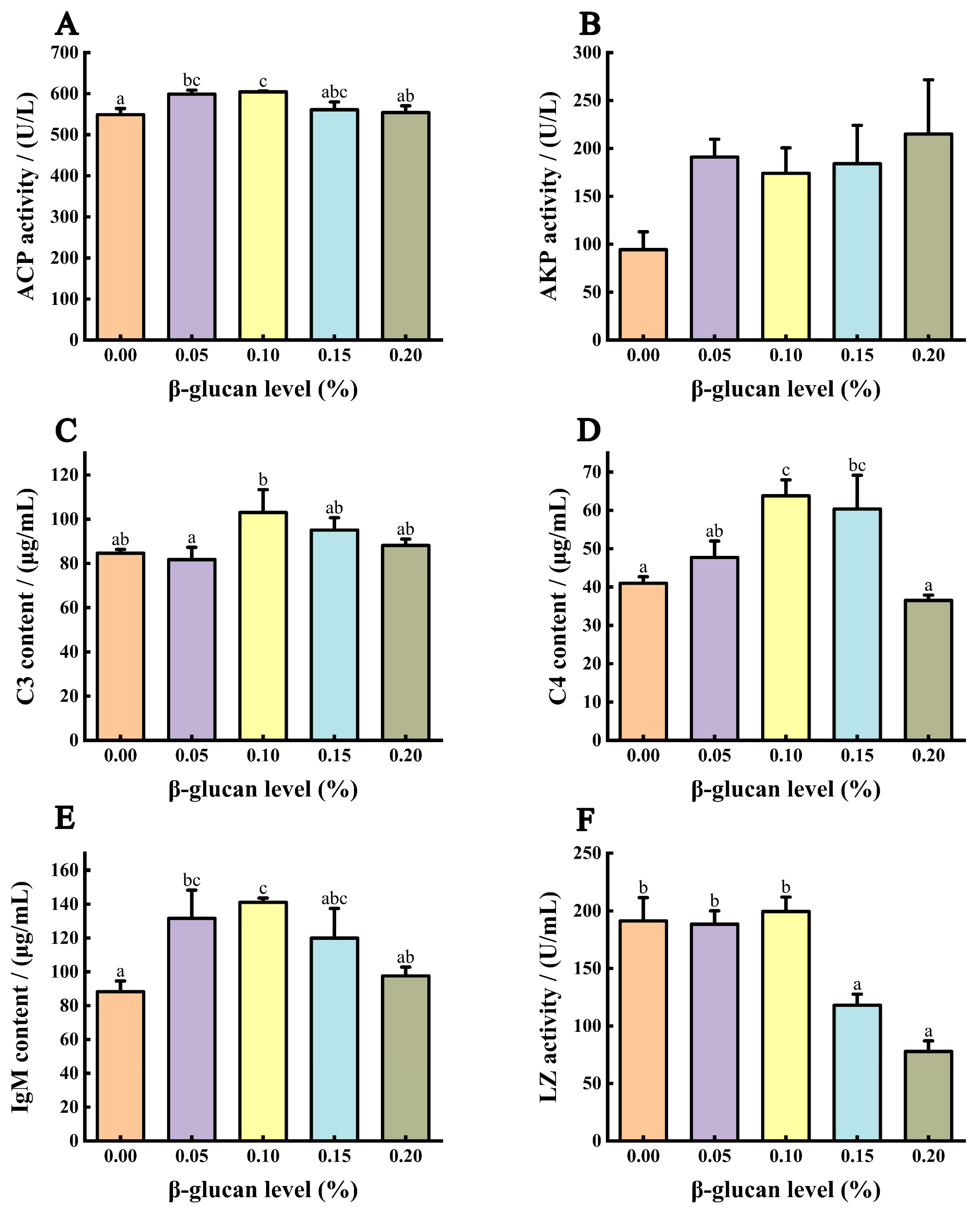
3.6. Immune Ability of Liver

As illustrated in Figure 5, with increasing β-glucan content in the diet, the activities of hepatic alkaline phosphatase (AKP) and lysozyme (LZ) and contents of complement 3 (C3) and immunoglobulin M (IgM) progressively increased, reaching their peak in the 0.10% group. Moreover, the C3 content significantly increased in the 0.10% to 0.15% β-glucan diet groups compared with the control group (p < 0.05); however, the acid phosphatase (ACP) activity significantly decreased in the 0.20% β-glucan group (p < 0.05). No significant difference in the liver complement 4 (C4) content was observed among the groups (p > 0.05).

3.7. Immune Ability of Serum

As illustrated in Figure 6, the highest average levels of ACP activity and IgM content were observed in the 0.05% to 0.10% diet treatments, which significantly differed from the levels in the control group (p < 0.05). The serum C4 content in the 0.10% to 0.15% treatments was considerably higher than that in the control group (p < 0.05). When 0.20% β-glucan was added, the serum ACP and LZ activities and C4 and IgM contents significantly decreased compared with that of the 0.10% β-glucan group (p < 0.05). No significant differences in serum AKP activity were observed among the treatments (p > 0.05).

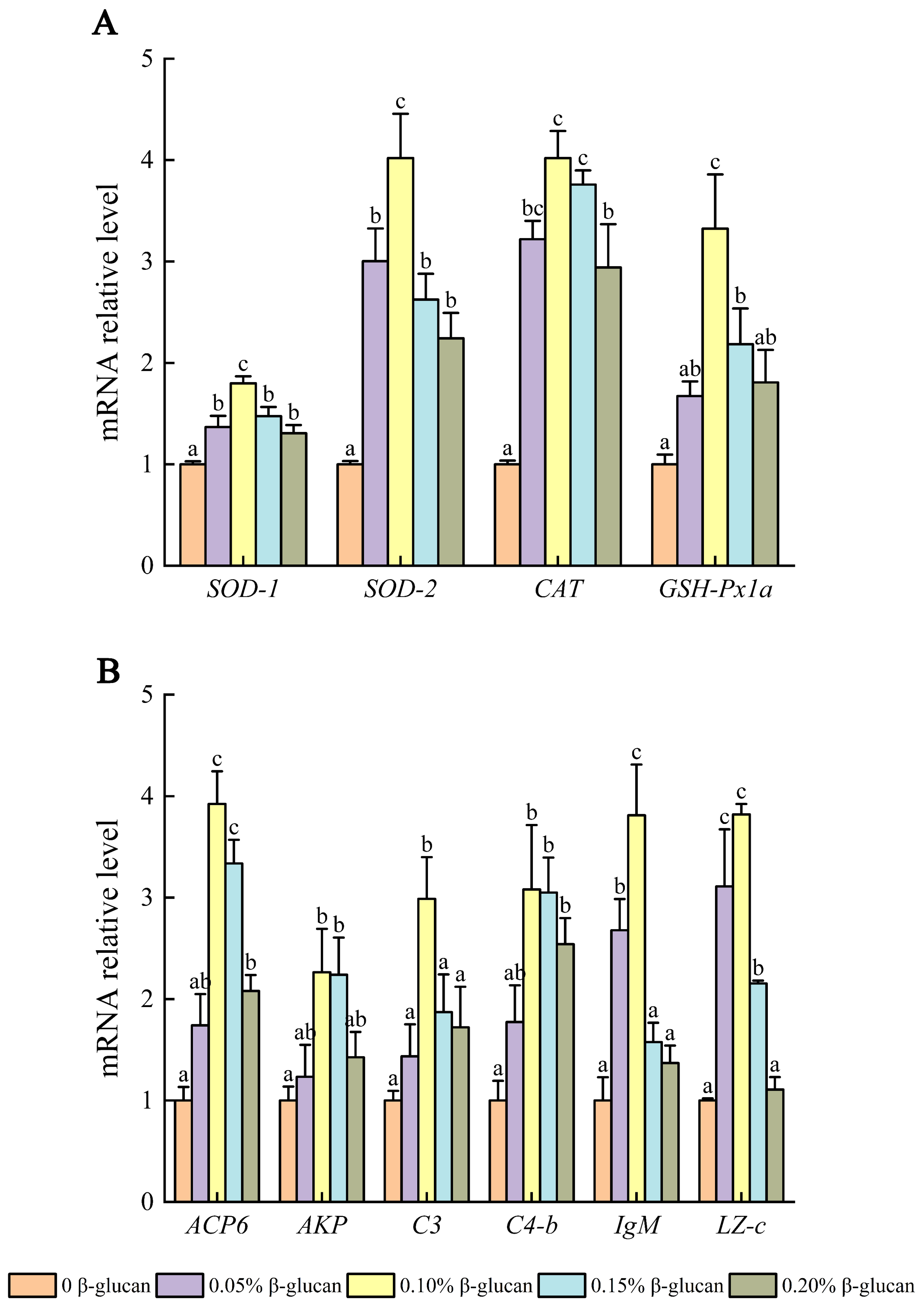
3.8. Relative mRNA Expression in Liver

The intestinal morphology indicators of coral trout in the different β-glucan groups are shown in Figure 7. The highest relative expression levels of copper-zinc superoxide dismutase (SOD-1), manganese superoxide dismutase (SOD-2), and CAT in the liver were observed in coral trout treated with β-glucan, and differences were significant relative to the control (p < 0.05). In the 0.10% diet group, the relative expression levels of SOD-1, SOD-2, CAT, GSH-Px1a, ACP6, C3, IgM, and LZ were markedly higher than those in the control and 0.20% diet groups (p < 0.05). In the 0.10% and 0.15% diet group, the relative expression levels of SOD-1, SOD-2, CAT, GSH-Px1a, ACP6, AKP, C4-b, and LZ-c were significantly increased relative to those of control (p < 0.05).

4. Discussion

Our research demonstrated that supplementation with 0.10% β-glucan led to the highest growth performance and thus was beneficial for coral trout growth. Our results are consistent with those of studies on pompano (Trachinotus ovatus) [35], Pacific red snapper (Lutjanus peru) [36], spotted rose snapper (Lutjanus guttatus) [37], Nile tilapia (O. niloticus) [38], and red sea bream (Pagrus major) [39]. In the present study, the optimal dosage of β-glucan (0.082%) was determined based on the SGR of coral trout. The mechanisms underlying the promoting effects of β-glucan on coral trout include increases in α-amylase and chymotrypsin activities under the suitable dose, which likely aided in improving their growth performance. Studies on tropical gar (Atractosteus tropicus) [40], Pacific red snapper [36], and white shrimp [23] showed that dietary β-glucan supplementation significantly enhanced digestive enzyme activities. Additionally, the villus length and muscle thickness in the intestines were increased under supplementation, which could also explain the growth-promoting effects of β-glucan. Similar results have been found in previous research, which showed that dietary β-glucan supplementation can improve villus length in pompano (T. ovatus) [41] and Chinese soft-shell turtle (Pelodiscus sinensis) [42] and increase muscle thickness in pearl gentian grouper (Epinephelus lanceolatus♂ × Epinephelus fuscoguttatus♀) [43]. Earlier studies indicated that β-glucan promoted the production of short-chain fatty acids (SCFAs), which can enhance gut health by promoting the differentiation and proliferation of intestinal epithelial cells [42]. Improvements in intestinal morphology can enhance the immune system of fish, activate pathogen defense mechanisms, promote health, and increase growth rates [44]. However, our study showed that when the β-glucan supplementation exceeded 0.10%, the growth of coral trout was suppressed. Similar studies have reported that the growth performance of aquatic animals showed initial improvements under increasing β-glucan supplementation but then stabilized and eventually declined [35,45]. We speculate that excessive β-glucan may lead to immunosuppression and inhibit growth performance [28,29,30]. However, whether β-glucan improves the growth performance of aquatic animals remains controversial. Studies have also shown that β-glucan does not significantly enhance the growth of tilapia [3], largemouth bass (Micropterus salmoides) [46], and tropical gar [40]. Current research suggests that the impact of β-glucan on the growth of aquatic animals depends on the species studied, the dosage and source of β-glucan, and the experimental conditions. Thus, tailored cultivation strategies should be developed for different fish species to achieve better growth performance.
Antioxidant enzymes are a crucial component in the defense system against oxidative stress in fish [47]. β-glucan can eliminate free radicals by regulating antioxidant enzyme activity, thereby preventing damage from hydrogen peroxide (H2O2) and reactive oxygen species (ROS) and enhancing the antioxidant capacity of aquatic animals [3,23,48]. MDA is a major product of lipid peroxidation that indicates the extent of peroxidation and cellular damage in organisms [49]. Our study demonstrated that supplementation with 0.10% β-glucan significantly increased enzyme activities in the liver (CAT and GR) and serum (CAT, GSH-Px, GR, and T-AOC) of coral trout and reduced the MDA contents. Consistent with our study, Dou et al. [3] reported that incorporating β-glucan into the diet of tilapia could significantly boost SOD, CAT, and GSH-Px activities and T-AOC contents and reduce MDA contents. Cao et al. [24] demonstrated that a suitable amount of β-glucan significantly improved the T-SOD, CAT, and GSH-Px activities and substantially diminished the MDA content in Pengze crucian carp. However, when the β-glucan content is too high, such as in the 0.2% group, the SOD, CAT, GSH-Px, and GR activities in the liver, and GR activity and T-AOC contents in the serum were significantly reduced compared to that in the 0.1% group. Similarly, Ren et al. found that a low dose of lentinan (a type of β-glucan) was more effective at enhancing the intestinal antioxidant capacity of juvenile taimen (Hucho taimen, Pallas) [50]. These findings demonstrate that an excessive level of β-glucan is not required for coral trout.
C3, C4, IgM, and LZ are important indicators of the immune status of fish [26]. Khanjani et al. [17] reported that an appropriate dose of β-glucan significantly increased the LZ activity and IgM, C3, and C4 contents in rainbow trout. Dou et al. [3] also demonstrated that β-glucan supplementation markedly increased the LZ activity and IgM, C3, and C4 contents in tilapia. ACP and AKP serve crucial roles in the infection of microbial pathogens, enhancing disease resistance in fish [51]. Supplementary β-glucan improved the immune status of white shrimp in low-salinity environments by activating ACP and AKP activities [23]. Additionally, Cao et al. [24] discovered that β-glucan enhanced the immune response in Pengze crucian carp by increasing the ACP and AKP activity. Consistent with the aforementioned studies, our study demonstrated that β-glucan enhanced nonspecific immunity in both the liver and serum. The 0.1% β-glucan group demonstrated significantly higher immune enzyme activities in the liver (IgM and C3 contents, AKP and LZ activities) and serum (IgM and C4 contents, ACP activity) compared to the control group. However, the beneficial effects of β-glucan on these immune indicators were not linear. As the β-glucan content increased, the 0.2% group showed significantly reduced liver ACP and serum LZ activity. The dietary inclusion of 0.1% β-glucan exhibited the most pronounced immunomodulatory effects. Similarly, studies on large yellow croaker (Pseudosciaena crocea) [45] and juvenile taimen [50] showed that lower amounts of β-glucan were more effective in enhancing immune responses compared to higher doses. Elevated levels of β-glucan may lead to immunosuppression [28,30], indicating that excessive β-glucan is not necessary for improving nonspecific immune responses in fish.
Furthermore, we explored the expression of genes related to antioxidant enzymes and immune indicators in the liver of coral trout and found that adding 0.10% β-glucan to the diet significantly increased the relative expression levels of SOD-1, SOD-2, CAT, GSH-Px1a, ACP6, AKP, LZ-c, IgM, C3, and C4-b in the liver. The increase in mRNA levels enhanced enzyme activity to some extent, although changes in enzyme activity do not necessarily correspond to changes in mRNA expression [52]. According to our results, only the activities of CAT, GSH-Px, and AKP and contents of C3, C4, and IgM in the liver aligned with the changes in their mRNA expression. However, the fish supplemented with 0.2% β-glucan showed significantly lower relative expression levels of SOD-1, SOD-2, CAT, GSH-Px1a, ACP6, C3, IgM, and LZ-c compared to those supplemented with 0.10% β-glucan. This trend was consistent with the changes in liver enzyme activities, indicating that excessive β-glucan diminished the beneficial effects.

5. Conclusions

An appropriate dose of β-glucan significantly promoted the growth performance of coral trout enhanced the activities of antioxidant and immune enzymes in the liver and serum, improved the expression of liver antioxidant and immune-related genes, and promoted intestinal health. However, an excessive level of 0.20% β-glucan showed reduced beneficial effects on coral trout compared with 0.10% β-glucan. Under the conditions of this experiment, the recommended dietary supplementation level of β-glucan for coral trout is 0.10%. Based on the cubic regression analysis, the optimal level of β-glucan for maximal growth of coral trout is 0.082%. These findings provide new insights and a comprehensive understanding of the positive effects of β-glucan on coral trout.

Supplementary Materials

The following supporting information can be downloaded at: https://www.mdpi.com/article/10.3390/fishes9080298/s1, Table S1: Composition and proximate chemical analysis of the experimental diets (expressed as % dry matter); Table S2: Primers and amplification conditions used for quantitative real-time PCR in this study; Method S1: Diet preparation; Method S2: Diet composition analysis; Method S3: Enzyme activity analysis; Method S4: Gene expression analysis.

Author Contributions

Conceptualization, X.H. and H.L.; methodology, X.H. and W.Y.; software, Z.L.; validation, H.L. and W.Y.; formal analysis, Z.M. and Y.Y.; resources, C.Z. and J.H.; data curation, X.H.; writing—original draft preparation, X.H.; writing—review and editing, W.Y.; visualization, Z.L. and Z.M.; supervision, Y.Y., C.Z. and J.H.; funding acquisition, W.Y. and H.L. All authors have read and agreed to the published version of the manuscript.

Funding

This research was funded by Hainan Province Science and Technology Special Fund (ZDYF2022XDNY349), Hainan Provincial Natural Science Foundation of China (321QN0942), Central Public-interest Scientific Institution Basal Research Fund, South China Sea Fisheries Research Institute, CAFS (2024RC15), Guangdong Provincial Rural Revitalization Strategy Special Funds for Seed Industry Revitalization Project (2022-SPY-02-003), the earmarked fund for HNARS (HNARS-03-Z02) and Central Public—interest Scientific Institution Basal Research Fund, CAFS (2023TD58).

Institutional Review Board Statement

The study was approved by the Animal Care and Use Committee of South China Sea Fisheries Research Institute, Chinese Academy of Fishery Sciences (Approval number SCSFRI2023-0731).

Informed Consent Statement

Not applicable.

Data Availability Statement

The original contributions presented in the study are included in the article/Supplementary Material. Further inquiries can be directed to the corresponding author.

Conflicts of Interest

The authors declare no conflicts of interest.

References

  1. Yang, Y.; Wu, L.N.; Chen, J.F.; Wu, X.; Xia, J.H.; Meng, Z.N.; Liu, X.C.; Lin, H.R. Whole-genome sequencing of leopard coral grouper (Plectropomus leopardus) and exploration of regulation mechanism of skin color and adaptive evolution. Zool. Res. 2020, 41, 328–340. [Google Scholar] [CrossRef]
  2. Hao, R.; Zhu, X.; Tian, C.; Jiang, M.; Huang, Y.; Li, G.; Zhu, C. LncRNA–miRNA–mRNA ceRNA network of different body colors in Plectropomus leopardus. Front. Mar. Sci. 2023, 10, 1170762. [Google Scholar] [CrossRef]
  3. Dou, X.; Huang, H.; Li, Y.; Deng, J.; Tan, B. Effects of dietary β-glucan on growth rate, antioxidant status, immune response, and resistance against Aeromonas hydrophila in genetic improvement of farmed tilapia (GIFT, Oreochromis niloticus). Aquac. Rep. 2023, 29, 101480. [Google Scholar] [CrossRef]
  4. Zhu, X. Regulation Mechanism of Astaxanthin and Vitamin C Improve the Growth and Immune Stress Incoral Trout (Plectropomus Leopardus). Doctoral Thesis, Guangdong Ocean University, Zhanjiang, China, 2022. [Google Scholar] [CrossRef]
  5. Cao, Q. Effect of Lactobacillus Salivarius GZPH2 on Gut Microbiota and Metabolome of Leopard Coral Grouper (Plectropomus Leopardus). Master’s Thesis, South China University of Technology, Guangzhou, China, 2020. [Google Scholar] [CrossRef]
  6. Boti, V.; Toli, V.; Efthymiou, C.; Albanis, T. Screening of commonly used antibiotics in fresh and saltwater samples impacted by aquacultures: Analytical methodology, occurrence and environmental risk assessment. Sustainability 2023, 15, 9199. [Google Scholar] [CrossRef]
  7. Hossain, A.; Habibullah-Al-Mamun, M.; Nagano, I.; Masunaga, S.; Kitazawa, D.; Matsuda, H. Antibiotics, antibiotic-resistant bacteria, and resistance genes in aquaculture: Risks, current concern, and future thinking. Environ. Sci. Pollut. Res. Int. 2022, 29, 11054–11075. [Google Scholar] [CrossRef] [PubMed]
  8. Meena, D.K.; Das, P.; Kumar, S.; Mandal, S.C.; Prusty, A.K.; Singh, S.K.; Akhtar, M.S.; Behera, B.K.; Kumar, K.; Pal, A.K.; et al. Beta-glucan: An ideal immunostimulant in aquaculture (a review). Fish Physiol. Biochem. 2013, 39, 431–457. [Google Scholar] [CrossRef] [PubMed]
  9. El-Boshy, M.E.; El-Ashram, A.M.; Abdelhamid, F.M.; Gadalla, H.A. Immunomodulatory effect of dietary Saccharomyces cerevisiae, β-glucan and laminaran in mercuric chloride treated Nile tilapia (Oreochromis niloticus) and experimentally infected with Aeromonas hydrophila. Fish Shellfish Immunol. 2010, 28, 802–808. [Google Scholar] [CrossRef]
  10. Elder, M.J.; Webster, S.J.; Chee, R.; Williams, D.L.; Hill Gaston, J.S.; Goodall, J.C. β-glucan size controls dectin-1-mediated immune responses in human dendritic cells by regulating IL-1β production. Front. Immunol. 2017, 8, 791. [Google Scholar] [CrossRef]
  11. Mironczuk-Chodakowska, I.; Kujawowicz, K.; Witkowska, A.M. Beta-glucans from fungi: Biological and health-promoting potential in the COVID-19 pandemic era. Nutrients 2021, 13, 3960. [Google Scholar] [CrossRef]
  12. Atta-Allah, A.A.; Ahmed, R.F.; Shahin, A.A.M.; Hassan, E.A.; El-Bialy, H.A.; El-Fouly, M.Z. Optimizing the synthesis of yeast Beta-glucan via response surface methodology for nanotechnology application. BMC Microbiol. 2023, 23, 110. [Google Scholar] [CrossRef]
  13. Singla, A.; Gupta, O.P.; Sagwal, V.; Kumar, A.; Patwa, N.; Mohan, N.; Ankush; Kumar, D.; Vir, O.; Singh, J.; et al. Beta-glucan as a soluble dietary fiber source: Origins, biosynthesis, extraction, purification, structural characteristics, bioavailability, biofunctional attributes, industrial utilization, and global trade. Nutrients 2024, 16, 900. [Google Scholar] [CrossRef] [PubMed]
  14. Zhu, F.; Du, B.; Xu, B. A critical review on production and industrial applications of beta-glucans. Food Hydrocoll. 2016, 52, 275–288. [Google Scholar] [CrossRef]
  15. Dalmo, R.A.; Bogwald, J. β-glucans as conductors of immune symphonies. Fish Shellfish Immunol. 2008, 25, 384–396. [Google Scholar] [CrossRef] [PubMed]
  16. Khanjani, M.H.; Sharifinia, M.; Ghaedi, G. β-glucan as a promising food additive and immunostimulant in aquaculture industry. Ann. Anim. Sci. 2022, 22, 817–827. [Google Scholar] [CrossRef]
  17. Khanjani, M.H.; Ghaedi, G.; Sharifinia, M. Effects of diets containing β-glucan on survival, growth performance, haematological, immunity and biochemical parameters of rainbow trout (Oncorhynchus mykiss ) fingerlings. Aquac. Res. 2021, 53, 1842–1850. [Google Scholar] [CrossRef]
  18. Huang, Q.; Zhu, Y.; Yu, J.; Fang, L.; Li, Y.; Wang, M.; Liu, J.; Yan, P.; Xia, J.; Liu, G.; et al. Effects of sulfated β-glucan from Saccharomyces cerevisiae on growth performance, antioxidant ability, nonspecific immunity, and intestinal flora of the red swamp crayfish (Procambarus clarkii). Fish Shellfish Immunol. 2022, 127, 891–900. [Google Scholar] [CrossRef]
  19. Aramli, M.S.; Kamangar, B.; Nazari, R.M. Effects of dietary β-glucan on the growth and innate immune response of juvenile Persian sturgeon, Acipenser persicus. Fish Shellfish Immunol. 2015, 47, 606–610. [Google Scholar] [CrossRef]
  20. Talpur, A.D.; Munir, M.B.; Mary, A.; Hashim, R. Dietary probiotics and prebiotics improved food acceptability, growth performance, haematology and immunological parameters and disease resistance against Aeromonas hydrophila in snakehead (Channa striata) fingerlings. Aquaculture 2014, 426–427, 14–20. [Google Scholar] [CrossRef]
  21. Lin, S.; Pan, Y.; Luo, L.; Luo, L. Effects of dietary β-1,3-glucan, chitosan or raffinose on the growth, innate immunity and resistance of koi (Cyprinus carpio koi). Fish Shellfish Immunol. 2011, 31, 788–794. [Google Scholar] [CrossRef]
  22. Nguyen, N.D.; Van Dang, P.; Le, A.Q.; Nguyen, T.K.L.; Pham, D.H.; Van Nguyen, N.; Nguyen, Q.H. Effect of oligochitosan and oligo-β-glucan supplementation on growth, innate immunity, and disease resistance of striped catfish (Pangasianodon hypophthalmus). Biotechnol. Appl. Biochem. 2017, 64, 564–571. [Google Scholar] [CrossRef]
  23. Qiao, Y.; Zhou, L.; Qu, Y.; Lu, K.; Han, F.; Li, E. Effects of different dietary β-glucan levels on antioxidant capacity and immunity, gut microbiota and transcriptome responses of white shrimp (Litopenaeus vannamei) under low salinity. Antioxidants 2022, 11, 2282. [Google Scholar] [CrossRef]
  24. Cao, H.; Yu, R.; Zhang, Y.; Hu, B.; Jian, S.; Wen, C.; Kajbaf, K.; Kumar, V.; Yang, G. Effects of dietary supplementation with β-glucan and Bacillus subtilis on growth, fillet quality, immune capacity, and antioxidant status of Pengze crucian carp (Carassius auratus var. Pengze). Aquaculture 2019, 508, 106–112. [Google Scholar] [CrossRef]
  25. Fath El-Bab, A.F.; Majrashi, K.A.; Sheikh, H.M.; Shafi, M.E.; El-Ratel, I.T.; Neamat-Allah, A.N.F.; El-Raghi, A.A.; Elazem, A.Y.A.; Abd-Elghany, M.F.; Abdelnour, S.A.; et al. Dietary supplementation of Nile tilapia (Oreochromis niloticus) with β-glucan and/or Bacillus coagulans: Synergistic impacts on performance, immune responses, redox status and expression of some related genes. Front. Vet. Sci. 2022, 9, 1011715. [Google Scholar] [CrossRef] [PubMed]
  26. Yu, W.; Yang, Y.; Chen, H.; Zhou, Q.; Zhang, Y.; Huang, X.; Huang, Z.; Li, T.; Zhou, C.; Ma, Z.; et al. Effects of dietary chitosan on the growth, health status and disease resistance of golden pompano (Trachinotus ovatus). Carbohydr. Polym. 2023, 300, 120237. [Google Scholar] [CrossRef] [PubMed]
  27. Yu, W.; Yang, Y.; Zhou, Q.; Huang, X.; Huang, Z.; Li, Y.; Wu, W.; Zhou, C.; Ma, Z.; Lin, H. Effects of dietary Astragalus polysaccharides on growth, health and resistance to Vibrio harveyi of Lates calcarifer. Int. J. Biol. Macromol. 2022, 207, 850–858. [Google Scholar] [CrossRef] [PubMed]
  28. Bai, N.; Zhang, W.; Mai, K.; Wang, X.; Xu, W.; Ma, H. Effects of discontinuous administration of β-glucan and glycyrrhizin on the growth and immunity of white shrimp Litopenaeus vannamei. Aquaculture 2010, 306, 218–224. [Google Scholar] [CrossRef]
  29. Rodrigues, M.V.; Zanuzzo, F.S.; Koch, J.F.A.; de Oliveira, C.A.F.; Sima, P.; Vetvicka, V. Development of fish immunity and the role of β-glucan in immune responses. Molecules 2020, 25, 5378. [Google Scholar] [CrossRef]
  30. Douxfils, J.; Fierro-Castro, C.; Mandiki, S.N.; Emile, W.; Tort, L.; Kestemont, P. Dietary β-glucans differentially modulate immune and stress-related gene expression in lymphoid organs from healthy and Aeromonas hydrophila-infected rainbow trout (Oncorhynchus mykiss). Fish Shellfish Immunol. 2017, 63, 285–296. [Google Scholar] [CrossRef] [PubMed]
  31. Yu, W.; Lin, H.; Yang, Y.; Zhou, Q.; Chen, H.; Huang, X.; Zhou, C.; Huang, Z.; Li, T. Effects of supplemental dietary Haematococcus pluvialison growth performance, antioxidant capacity, immune responses and resistance to Vibrio harveyi challenge of spotted sea bass Lateolabrax maculatus. Aquac. Nutr. 2020, 27, 355–365. [Google Scholar] [CrossRef]
  32. AOAC. Official Methods for Analysis, 19th ed.; Association of Official Analytical Chemists: Washington, DC, USA, 2012. [Google Scholar]
  33. Livak, K.J.; Schmittgen, T.D. Analysis of relative gene expression data using real-time quantitative PCR and the 2−ΔΔCT method. Methods 2001, 25, 402–408. [Google Scholar] [CrossRef]
  34. Xie, J.; Niu, J. Evaluation of four macro-algae on growth performance, anti-oxidant capacity and non-specific immunity in golden pompano (Trachinotus ovatus). Aquaculture 2022, 548, 737690. [Google Scholar] [CrossRef]
  35. Do Huu, H.; Sang, H.M.; Thanh Thuy, N.T. Dietary β-glucan improved growth performance, Vibrio counts, haematological parameters and stress resistance of pompano fish, Trachinotus ovatus Linnaeus, 1758. Fish Shellfish Immunol. 2016, 54, 402–410. [Google Scholar] [CrossRef]
  36. Guzman-Villanueva, L.T.; Ascencio-Valle, F.; Macias-Rodriguez, M.E.; Tovar-Ramirez, D. Effects of dietary β-1,3/1,6-glucan on the antioxidant and digestive enzyme activities of Pacific red snapper (Lutjanus peru) after exposure to lipopolysaccharides. Fish Physiol. Biochem. 2013, 40, 827–837. [Google Scholar] [CrossRef]
  37. Del Rio-Zaragoza, O.B.; Fajer-Avila, E.J.; Almazan-Rueda, P. Influence of β-glucan on innate immunity and resistance of Lutjanus guttatus to an experimental infection of dactylogyrid monogeneans. Parasite Immunol. 2011, 33, 483–494. [Google Scholar] [CrossRef]
  38. Welker, T.L.; Lim, C.; Yildirim-Aksoy, M.; Klesius, P.H. Use of diet crossover to determine the effects of β-glucan supplementation on immunity and growth of Nile tilapia, Oreochromis niloticus. J. World Aquac. Soc. 2012, 43, 335–348. [Google Scholar] [CrossRef]
  39. Dawood, M.A.O.; Koshio, S.; Ishikawa, M.; Yokoyama, S.; El Basuini, M.F.; Hossain, M.S.; Nhu, T.H.; Moss, A.S.; Dossou, S.; Wei, H. Dietary supplementation of β-glucan improves growth performance, the innate immune response and stress resistance of red sea bream, Pagrus major. Aquac. Nutr. 2017, 23, 148–159. [Google Scholar] [CrossRef]
  40. Nieves-Rodríguez, K.; Álvarez-González, C.; Peña-Marín, E.; Vega-Villasante, F.; Martínez-García, R.; Camarillo-Coop, S.; Tovar-Ramírez, D.; Guzmán-Villanueva, L.; Andree, K.; Gisbert, E. Effect of β-glucans in diets on growth, survival, digestive enzyme activity, and immune system and intestinal barrier gene expression for tropical gar (Atractosteus tropicus) juveniles. Fishes 2018, 3, 27. [Google Scholar] [CrossRef]
  41. Hoang, D.-H.; Thi Thanh Thuy, N.; Ky, P.X. A synergistic effect of dietary β-glucan and mannan oligosaccharide on growth performance, haematology, body composition, nutrient utilisation, and intestinal morphology in pompano, Trachinotus ovatus. Reg. Stud. Mar. Sci. 2024, 73, 103494. [Google Scholar] [CrossRef]
  42. Fu, H.; Qi, M.; Yang, Q.; Li, M.; Yao, G.; Bu, W.; Zheng, T.; Pi, X. Effects of dietary chito-oligosaccharide and β-glucan on the water quality and gut microbiota, intestinal morphology, immune response, and meat quality of Chinese soft-shell turtle (Pelodiscus sinensis). Front. Immunol. 2023, 14, 1266997. [Google Scholar] [CrossRef]
  43. Wang, F.; Wang, Z.; Cao, J.; Lu, Y. Long-and short-term dietary beta-glucan improves intestinal health and disease resistance in pearl gentian grouper (Epinephelus lanceolatus♂ x Epinephelus fuscoguttatus♀). Fish Physiol. Biochem. 2024, 50, 973–988. [Google Scholar] [CrossRef]
  44. Talpur, A.D.; Ikhwanuddin, M. Dietary effects of garlic (Allium sativum) on haemato-immunological parameters, survival, growth, and disease resistance against Vibrio harveyi infection in Asian sea bass, Lates calcarifer (Bloch). Aquaculture 2012, 364–365, 6–12. [Google Scholar] [CrossRef]
  45. Ai, Q.; Mai, K.; Zhang, L.; Tan, B.; Zhang, W.; Xu, W.; Li, H. Effects of dietary beta-1, 3 glucan on innate immune response of large yellow croaker, Pseudosciaena crocea. Fish Shellfish Immunol. 2007, 22, 394–402. [Google Scholar] [CrossRef] [PubMed]
  46. Zhang, Y.; Guo, M.; Li, N.; Dong, Z.; Cai, L.; Wu, B.; Xie, J.; Liu, L.; Ren, L.; Shi, B. New insights into β-glucan-enhanced immunity in largemouth bass Micropterus salmoides by transcriptome and intestinal microbial composition. Front. Immunol. 2022, 13, 1086103. [Google Scholar] [CrossRef]
  47. Deng, J.; Mai, K.; Chen, L.; Mi, H.; Zhang, L. Effects of replacing soybean meal with rubber seed meal on growth, antioxidant capacity, non-specific immune response, and resistance to Aeromonas hydrophila in tilapia (Oreochromis niloticus x O. Aureus). Fish Shellfish Immunol. 2015, 44, 436–444. [Google Scholar] [CrossRef]
  48. Pogue, R.; Murphy, E.J.; Fehrenbach, G.W.; Rezoagli, E.; Rowan, N.J. Exploiting immunomodulatory properties of β-glucans derived from natural products for improving health and sustainability in aquaculture-farmed organisms: Concise review of existing knowledge, innovation and future opportunities. Curr. Opin. Environ. Sci. Health 2021, 21, 100248. [Google Scholar] [CrossRef]
  49. Qiu, J.; Wang, W.N.; Wang, L.J.; Liu, Y.F.; Wang, A.L. Oxidative stress, DNA damage and osmolality in the Pacific white shrimp, Litopenaeus vannamei exposed to acute low temperature stress. Comp. Biochem. Physiol. C Toxicol. Pharmacol. 2011, 154, 36–41. [Google Scholar] [CrossRef] [PubMed]
  50. Ren, G.; Xu, L.; Lu, T.; Zhang, Y.; Wang, Y.; Yin, J. Protective effects of lentinan on lipopolysaccharide induced inflammatory response in intestine of juvenile taimen (Hucho taimen, Pallas). Int. J. Biol. Macromol. 2019, 121, 317–325. [Google Scholar] [CrossRef] [PubMed]
  51. Li, L.; Wei, X.F.; Yang, Z.Y.; Zhu, R.; Li, D.L.; Shang, G.J.; Wang, H.T.; Meng, S.T.; Wang, Y.T.; Liu, S.Y.; et al. Alleviative effect of poly-β-hydroxybutyrate on lipopolysaccharide-induced oxidative stress, inflammation and cell apoptosis in Cyprinus carpio. Int. J. Biol. Macromol. 2023, 253, 126784. [Google Scholar] [CrossRef]
  52. Zhu, X.; Hao, R.; Zhang, J.; Tian, C.; Hong, Y.; Zhu, C.; Li, G. Improved growth performance, digestive ability, antioxidant capacity, immunity and Vibrio harveyi resistance in coral trout (Plectropomus leopardus) with dietary vitamin C. Aquac. Rep. 2022, 24, 101111. [Google Scholar] [CrossRef]
Figure 1. Relationship between the SGR and β-glucan supplementation in coral trout diet. Values represent the mean ± SE (n = 3). Different letters (a, b, c) indicate significant differences (p < 0.05).
Fishes 09 00298 g001
Figure 2. Intestinal morphology of coral trout fed different experimental diets, with the long arrow indicating the villus length and the short arrow indicating the muscle thickness. (A) 0% β-glucan; (B) 0.05% β-glucan; (C) 0.10% β-glucan; (D) 0.15% β-glucan; (E) 0.20% β-glucan; (F) villus length; (G) muscle thickness. Scale bar: 200 μm. Values represent the mean ± SE (n = 3). Different letters (a, b, c) indicate significant differences (p < 0.05).
Fishes 09 00298 g002
Figure 3. Liver antioxidant ability of coral trout fed different experimental diets. Values represent the mean ± SE (n = 3). (A) SOD, superoxide dismutase; (B) CAT, catalase; (C) GSH-Px, glutathione peroxidase; (D) GR, glutathione reductase; (E) T-AOC, total antioxidant capacity; (F) MDA, malondialdehyde. Different letters (a, b, c) indicate significant differences (p < 0.05).
Fishes 09 00298 g003
Figure 4. Serum antioxidant ability of coral trout fed different experimental diets. Values represent the mean ± SE (n = 3). (A) SOD; (B) CAT; (C) GSH-Px; (D) GR; (E) T-AOC; (F) MDA. Different letters (a, b, c) indicate significant differences (p < 0.05).
Fishes 09 00298 g004
Figure 5. Liver immune ability of coral trout fed different experimental diets. Values represent the mean ± SE (n = 3). (A) ACP, acid phosphatase; (B) AKP, alkaline phosphatase; (C) C3, complement 3; (D) C4, complement 4; (E) IgM, immunoglobulin M; (F) LZ, lysozyme. Different letters (a, b) indicate significant differences (p < 0.05).
Fishes 09 00298 g005
Figure 6. Serum immune ability of coral trout fed different experimental diets. Values represent the mean ± SE (n = 3). (A) ACP; (B) AKP; (C) C3; (D) C4; (E) IgM; (F) LZ. Different letters (a, b, c) indicate significant differences (p < 0.05).
Fishes 09 00298 g006
Figure 7. Expression levels of genes related to liver antioxidant enzymes (A) and immune indicators (B) in coral trout fed different experimental diets. Values represent the mean ± SE (n = 3). SOD-1: copper-zinc superoxide dismutase; SOD-2: manganese superoxide dismutase; CAT: catalase; GSH-Px1a: glutathione peroxidase 1a; ACP6: acid phosphatase 6; AKP: alkaline phosphatase; LZ-c: lysozyme c; IgM: immunoglobulin M; C3: complement 3; C4-b: complement 4b; GAPDH: glyceraldehyde-3-phosphate dehydrogenase. Different letters (a, b, c) indicate significant differences (p < 0.05).
Fishes 09 00298 g007
Table 1. Growth performance of coral trout fed different experimental diets.
IndexDiets (β-Glucan %)
00.050.100.150.20
IBW (g)75.93 ± 0.2775.75 ± 0.0875.86 ± 0.1075.80 ± 0.3275.70 ± 0.42
FBW (g)106.65 ± 1.13 a111.93 ± 1.11 bc114.00 ± 0.99 c110.45 ± 1.28 abc108.50 ± 2.27 ab
WGR (%)40.48 ± 1.80 a47.76 ± 1.33 bc50.27 ± 1.21 c45.72 ± 1.54 abc43.35 ± 3.34 ab
SGR (%)0.61 ± 0.02 a0.70 ± 0.02 bc0.73 ± 0.01 c0.67 ± 0.02 abc0.64 ± 0.04 ab
FCR1.53 ± 0.07 c1.37 ± 0.04 ab1.24 ± 0.02 a1.47 ± 0.02 bc1.50 ± 0.04 bc
SR (%)90.00 ± 2.89 a100.00 ± 0.00 b100.00 ± 0.00 b96.67 ± 3.33 b98.33 ± 1.67 b
VSI (%)4.74 ± 0.294.29 ± 0.344.18 ± 0.124.49 ± 0.234.12 ± 0.16
HSI (%)1.16 ± 0.131.03 ± 0.061.09 ± 0.061.31 ± 0.041.03 ± 0.09
CF (%)2.20 ± 0.022.18 ± 0.052.03 ± 0.092.06 ± 0.082.08 ± 0.04
IBW, initial body weight; FBW, final body weight; WGR, weight gain rate; SGR, specific growth rate; FCR, feed conversion ratio; SR: survival rate, VSI, viscera somatic index; HSI, hepatic somatic indices, CF, condition factor. Values represent the mean ± SE (n = 3). Different superscript letters in the same row indicate a significant difference at p < 0.05.
Table 2. Digestive enzyme activities of coral trout fed different experimental diets.
IndexDiets (β-Glucan %)
00.050.100.150.20
α-Amylase (U/mg prot)1.17 ± 0.05 a1.39 ± 0.07 b1.47 ± 0.03 b1.40 ± 0.05 b1.27 ± 0.09 ab
Lipase (U/g prot)0.82 ± 0.070.97 ± 0.061.12 ± 0.121.09 ± 0.140.95 ± 0.08
Chymotrypsin (U/mg prot)1.19 ± 0.07 a1.60 ± 0.09 b1.73 ± 0.09 b1.67 ± 0.10 b1.47 ± 0.13 ab
Values represent the mean ± SE (n = 3). Different superscript letters in the same row indicate a significant difference at p < 0.05.
Disclaimer/Publisher’s Note: The statements, opinions and data contained in all publications are solely those of the individual author(s) and contributor(s) and not of MDPI and/or the editor(s). MDPI and/or the editor(s) disclaim responsibility for any injury to people or property resulting from any ideas, methods, instructions or products referred to in the content.

