Article

Acute Hypercapnia at South African Abalone Farms and Its Physiological and Commercial Consequences

1
Institute of Metabolic Physiology/Ecophysiology, Heinrich-Heine University, D-40225 Düsseldorf, Germany
2
HIK Abalone Farm (Pty) Ltd., 1 Whale Close, Hermanus 7200, South Africa
3
Department of Ichthyology and Fisheries Science, Rhodes University, P.O. Box 94, Grahamstown 6140, South Africa
4
Branch: Fisheries Management, Department of Forestry, Fisheries and the Environment (DFFE), Roggebaai, Cape Town 8012, South Africa
5
Department of Animal Sciences, Stellenbosch University, Stellenbosch 7602, South Africa
*
Author to whom correspondence should be addressed.
Fishes 2024, 9(8), 313; https://doi.org/10.3390/fishes9080313
Submission received: 17 July 2024 / Revised: 3 August 2024 / Accepted: 6 August 2024 / Published: 8 August 2024
(This article belongs to the Section Physiology and Biochemistry)

Abstract

:
Abalone Haliotis midae are distributed from the cold, hypercapnic waters of the dynamic Benguela Current Large Marine Ecosystem to the relatively warm, normocapnic waters of the Agulhas Current. The species supports an important fishery as well as a thriving aquaculture industry. Due to the relatively low capacity to regulate their acid–base balance and their need to calcify shell and radula, abalone are especially vulnerable to increasing ocean acidification. Exposure to acidified seawater, i.e., hypercapnia, also occurs during the farming operation and can originate from (a) changes in influent seawater, (b) pH decrease by accumulation of waste products, and (c) intentional hypercapnia for anaesthesia using CO2-saturated seawater for size grading. Currently, these are acute exposures to hypercapnia, but increasing ocean acidification can cause chronic exposure, if not mitigated. Wild South African abalone are already exposed to periodic hypercapnia during ocean upwelling events and will be more so in the future due to progressive ocean acidification. This study investigated the acute pH effects in isolation as an initial step in studying the acute physiological response of H. midae to provide a mechanistic basis for the design of complex multifactorial studies, imitating more closely what occurs on farms and in the natural habitat. The major findings relevant to the above conditions are as follows: 1. Acute exposure to hypercapnia induces a reversible, unbuffered respiratory acidosis. 2. The impact of acute hypercapnia is size-dependent and potentially fatal. 3. Exposure to extreme, short hypercapnia during anaesthesia causes a rapid imbalance in the acid–base state but a rapid subsequent recovery. LC50 for small, medium and large abalone range from pH 6.27 to 6.03, respectively, and sub-lethal levels from pH 6.8 to 6.2. These results can be used by abalone aquaculture farms to mitigate/avoid the impact of acute (and chronic) hypercapnia but also to standardise their anaesthesia method. They are also a proxy to estimate the effects on wild populations.
Key Contribution: Acute exposure to hypercapnia induces a reversible, unbuffered respiratory acidosis. The impact thereof is size-dependent and potentially fatal. Extreme short hypercapnia during “dipping” causes rapid imbalance of the acid–base state but subsequent recovery is rapid.

1. Introduction

The South African abalone, Haliotis midae, locally called “perlemoen”, is the largest of five South African abalone species [1,2]. Its distribution ranges from Saldanha Bay on the West Coast to Riet Point on the East Coast [3], where its habitat consists of rocky shores from the subtidal to depths of more than 30 m [1,4]. Perlemoen once supported a thriving fishery, but this is now much reduced and poaching dwarfs the legal harvest [5]. Most recently, the abalone fishery harvested about 50 t, whereas poaching was estimated to be at 3350 t [5]. Since the beginning of abalone cultivation in the early 1980s, aquaculture has replaced the fishery step by step as the main source of legal South African abalone and, in 2019, aquaculture produced more than 1650 t [6].
The distribution range of wild H. midae extends from the cold, hypercapnic waters of the dynamic Benguela Current Large Marine Ecosystem (BCLME) on the West coast of South Africa to the relatively warm, normocapnic waters of the Agulhas Current on the East coast of South Africa with mixing in between them. Currently, fourteen farms cultivate abalone in land-based flow-through tank systems close to the shoreline [7]. They are mainly concentrated in the Western Cape province of South Africa [6]. In the BCLME, wild abalone are exposed to acute hypercapnia during periodic episodes of upwelling events, mostly in summer, and could be exposed to chronic hypercapnia in the future according to general ocean acidification scenarios [8]. Abalone farms, due to their location, are partially or completely exposed to the conditions of the BCLME. They constantly pump large volumes of coastal seawater ashore, limiting meaningful pre-treatment, to avoid unfavourable ocean conditions [7]. During the five years that abalone remain on the farms from spawning to harvesting, they are potentially exposed to unfavourable ocean conditions on a regular basis [7].
The BCLME is one of the largest Eastern Boundary (upwelling) systems, characterised by frequent upwelling events accompanied by hypercapnia (pH levels 7.4–7.6) in 3–10-day cycles in austral spring and summer [9]. In addition, some of the farms are located in areas where low oxygen events have become common after the upwelling season when phytoplankton blooms collapse [10]. During such events, the pH can reach levels of 6.6 for several days [11]. Ongoing climate change is projected to exacerbate some of these unfavourable conditions: upwelling in the Southern BCLME, as in other poleward boundaries of upwelling systems, is predicted to increase by 10–20% by the end of the century [8]. This will be accompanied by a pH decrease by 0.3 units in the same period [8]. Upwelling events, and in turn hypercapnic episodes, are predicted to become longer, more frequent and severe in the near future [12,13,14,15].
Exposure of aquaculture-farmed abalone is not limited to spells of hypercapnia from the adjacent ocean but also to sources on the farms themselves. One such source is the accumulation of ammonia along the raceways and a concomitant decrease in pH [16,17]. Both ammonia and hypercapnia reduce growth, although pH seems to be the major factor [17]. In this regard, a lowered pH somewhat mitigates the toxicity of the accumulated ammonia by lowering the conversion of total ammonia nitrogen (TAN) to the more toxic free un-ionised ammonia (FAN) [18]. Another, intentional, exposure to hypercapnia happens during the handling process of grading (weight determination and sorting according to size). Grading is necessary due to heterogenous individual growth rates within size cohorts [19,20]. South African abalone farms use submerging (“dipping”) plates with abalone in CO2-saturated seawater to dislodge abalone gently. During this process, abalone from a certain size are exposed to hypercapnic seawater of a pH of about 4.9–5.2 for several minutes. This reduces handling stress and injury associated with mechanical removal of abalone from their substratum. During their lengthy life at HIK Abalone Farm (approximately 4–5 years), the average abalone experience this process four to five times. This can increase to eight or nine years for individuals kept for further on-growing for specific products where abalone of 200 g or more are required.
Hypercapnia has profound effects on the physiology of marine animals [21,22,23,24]. In addition, calcifying species like molluscs are more sensitive to acidification than non-calcifying species due to the additional energetic cost of calcification [8]. Abalone species have been shown to have lowered calcification and growth rates under chronic hypercapnia [7,25,26]. One reason for this may be due to their low capacity to regulate their extracellular acid–base balance [25], that impacts general metabolism but also calcification [21,22,27,28,29]. Species-specific knowledge of impacts on all aspects of abalone biology is relevant for abalone farming but also for the management of wild stocks [30]. Due to the short exposure and recovery thereafter, acute hypercapnia differs from chronic effects, depending on the severity of the hypercapnia. Acute exposure experiments are therefore required to uncover mechanisms of response, especially acid–base regulation capabilities, and the limitations of response. Climate change will cause the combined effects of a declining seawater pH, increasing temperature and a reduction in oxygen levels (see above).
In the present study, however, the acute pH effect was investigated in isolation as an initial step in studying the acute physiological response of H. midae. The present study was initiated to answer the questions: (1) How do H. midae respond to acute hypercapnic exposure? (2) What are safe acute pH levels for farmed abalone? (3) What is the effect of extreme hypercapnia during “dipping”? The results will provide important information for local abalone farms and will also be used as a proxy to estimate the effects on wild populations.

2. Materials and Methods

2.1. Experimental Animals

Abalone were provided by HIK Abalone Farm (Pty) Ltd. in Hermanus (South Africa). For acute hypercapnic exposure, so-called cocktail-size abalone (88.5 ± 10.3 g) were used, whereas abalone of three different sizes (2.2 ± 0.7 g, 6.8 ± 1.6 g and 14.7 ± 3.8 g) were used for the LC50 experiment. Abalone that were used in the “dipping” simulation were 92.1 ± 9.2 g. Abalone were transported from Hermanus within two hours to flow-through holding tanks (1000 L, flow rate approximately 400 L h−1) at the Marine Research Aquarium of the Department of Forestry, Fisheries and the Environment of South Africa (DFFE) in Cape Town. The abalone were held, separated by size class in baskets in normocapnic conditions for several weeks (ambient temperature TA ranged from 12 to 21 °C, salinity 34.5–35.0‰). Abalone were placed in plastic baskets with vertical plastic sheets inside tanks and covered by black sheets to provide shading. Abalone were fed ABFEED® daily (late afternoon) with leftovers removed the next morning. Feeding was discontinued two days prior to experimentation. All experiments were conducted at the Marine Research Aquarium.

2.2. Acute Response to Hypercapnia

For the acute response trial, cocktail-size abalone (88.5 ± 10.3 g) were selected from the holding tanks and transferred into separate labelled gauze bags in which they were placed in an acclimation tank (1000 L) and kept for 24 h prior to experimentation for adjusting to experimental temperature (water conditions given in Table 1). Subsequently, a haemolymph sample was taken from each abalone (see below), and five of the abalone in their nets transferred to experimental tanks (cross-section Ø = 1.2 m, depth = 1 m, containing approximately 940 L) under normocapnic conditions. The remaining five were transferred into another tank with the same dimensions, but filled with hypercapnic seawater (pH~7.3). The normocapnic pH represented that of the incoming seawater on the experimental day and was close to the level during non-upwelling periods in the subtidal zone. The experimental hypercapnia of ~pH 7.3 was selected because similar levels have been reported from abalone farms and, in the wild, can be expected in the near future during upwelling events in the BCLME, according to IPCC scenarios (see Introduction). In order to minimize the time difference in exposure to the set seawater parameters of the respective treatments, consecutive abalone were placed in alternating treatments after the initial of serial haemolymph samples (0 h) was withdrawn (i.e., first normocapnia, second hypercapnia). After 1.5, 3, 5, 8, 24 and 32 h (24 h exposure +8 h recovery), a haemolymph (50 µL) sample was withdrawn from the area of the pedal sinus between head and foot by syringe with hypodermic needle (Neomedic 1 mL, 29 G). Another set of five abalone were exposed to pH 5 for 5 min and, after haemolymph sampling, transferred into the normocapnic tank for recovery for 24 h. During this recovery, samples were taken after 1.5 h, 5 h and 24 h. After the 24 h exposure period, all abalone from the normocapnic and hypercapnic treatments were transferred into a normocapnic recovery tank. The seawater conditions are given in Table 1. Continuous circulation in the tanks was achieved by JVP-202 12,000 l h−1 propellers (Ningbo JT Pump Co., Ltd., Ningbo, China) and normal air was provided from the aquarium’s compressed air system. The pH of the hypercapnic tank was set by the use of a pH controller connected to a solenoid valve and a pH electrode (TUNZE, Penzberg, Germany) whereby CO2 was bubbled into the seawater as described previously [31]. Seawater pCO2 and [HCO3] were thus calculated using measured pH, salinity, TA and AT [32] as constants in CO2SYSv_2.1 software [33]. Oxygen concentration was determined using a Multi 350i meter set (WTW, Weilheim, Germany). Water quality was monitored by measuring NH3 concentration (Ammonia test kit, Sera, Heinsberg, Germany) which was always below the detection limit. Haemolymph pH was measured within 20 s after sampling at the temperature of the treatment tanks and total CO2 (cCO2) was determined immediately from a subsample (described in detail in [31] Knapp et al., 2015). From these measured parameters (pH and cCO2), pCO2, and [HCO3 + CO32−] were calculated using derivatives of the Henderson–Hasselbalch equation, using constants derived from Truchot [34]. Possible changes in haemocyanin oxygen affinity modulator concentrations in the haemolymph, namely Ca2+ and Mg2+ were determined by commercial kits (Diaglobal, Germany) on small subsamples from each abalone. Haemocyanin concentration was measured spectrophotometrically (335 nm) in diluted 1:50 haemolymph vs. a physiological abalone buffer (10 mM HEPES in 2.5% NaCl, pH 7.2) samples.

2.3. Determination of Seawater Acidification Toxicity Levels

For determination of LC50 for pH, abalone (2.2 ± 0.7 g, 6.8 ± 1.6 g and 14.7 ± 3.8 g) were placed on vertical plastic sheets and allowed to attach. Sheets were then placed vertically into plastic baskets which were submerged in a tank for acclimation (1000 L) to temperature 24 h prior to experimentation (water conditions given in Table 2). Baskets were covered with Styrofoam sheets for shading. To determine the LC50, sets of two experimental tanks each were used with the following targeted seawater pH values: 5.2, 5.6, 6.0, 6.4, 6.8 and 7.2 (detailed water parameters in Table 2). In each treatment, 2 × 10 animals of each size class were kept for a maximum of 48 h. The abalone were examined after 2, 4, 6, 8, 12, 24, 36 and 48 h exposure. Animals were considered to have died when they had fallen off their sheet and did not show any muscle movement in response to repeated mechanical stimulation. Seawater parameters were measured as described above.

2.4. Statistical Analysis

Acute response: to account for the non-independence of the multiple measurement, experimental results were analysed using repeated measure ANOVA; specifically, these were fitted using linear mixed effect models (LMM). Experimental animals were treated as a random effect. These yielded results for the study on the effect of treatment (whether there was treatment effect). These were followed by Tukey post-hoc analysis both to determine the magnitude and direction of the difference between pairs compared and its statistical significance among treatments (p < 0.05).
Seawater acidification toxicity levels: To model toxicity of pH to abalone at different size classes, a logistic generalized linear model (GLM) was fitted to the proportion of animals that died at different pH levels. Abalone from respective replicates were pooled for this analysis. Multiple models were considered starting from the simplest size invariant model, a model with main effect of pH and size, and a model with interaction effect of pH and size. From the best model, the model with main effect of pH and size, lethal doses for different probabilities of mortality, e.g., LC5, LC50 and LC95, were determined.
All analyses, visualisation and report generation were carried out in R [35] (R Core Team, 2024). Multiple R packages were utilised for data processing, visualisation, analysis and summary of results, including [36,37,38,39,40,41,42,43,44].

3. Results

3.1. Acute Response to Hypercapnia

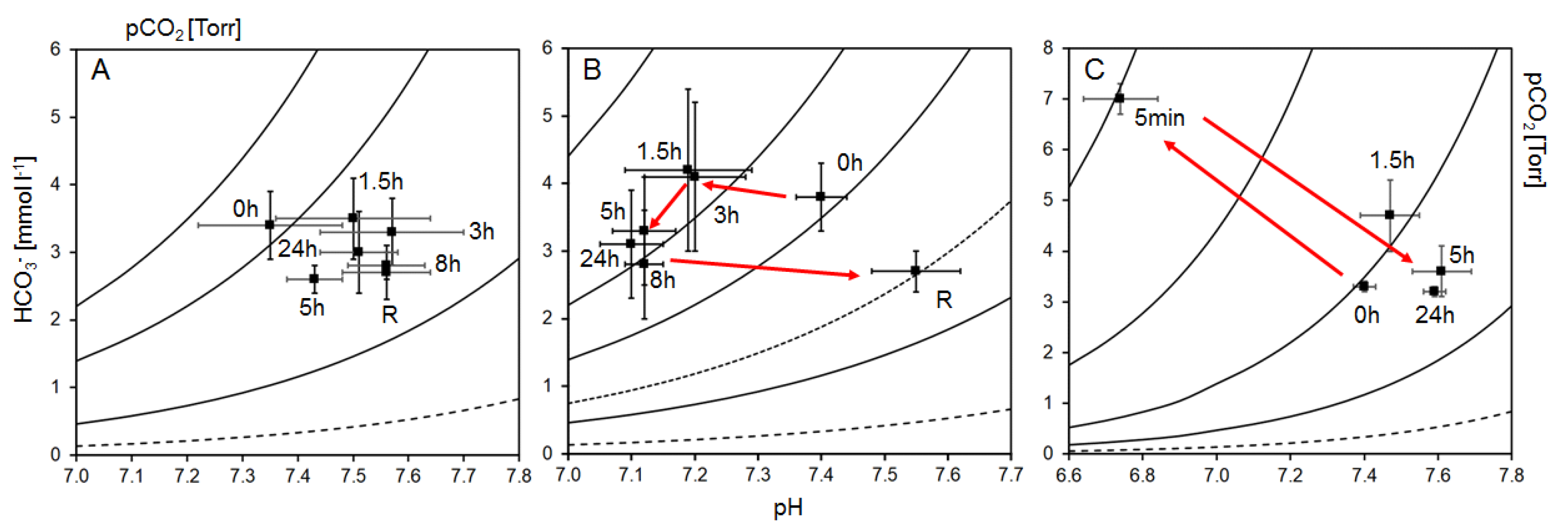
The acute exposure experiment showed a marked difference between the responses to normocapnic and hypercapnic conditions, respectively. In the normocapnic group, haemolymph pH increased by 0.22 units during the first 3 h from an initial pH of 7.35 and, except for the 5 h mark, in another 1.5 h, remained at this elevated level for the rest of the trial (Table 3, Figure 1A). Extracellular total CO2 (cCO2) levels remained relatively stable for the first 3 h of the trial and declined to a level approximately 20% lower thereafter where they remained until the 32 h time point (i.e., including 8 h recovery). Levels of pCO2 decreased from the initial value until the 8 h mark and subsequently remained at this level (Table 3). There was also a modest decline in [HCO3 + CO32−] throughout the exposure period (Table 3). The data were used to construct a Henderson–Hasselbalch diagram, depicting the extracellular pH, calculated [HCO3 + CO32−] and pCO2 values. It showed very little change in [HCO3 + CO32−], pCO2 and pH (Figure 2A).
Abalone in the hypercapnic treatment, due to the increase in seawater pCO2 and subsequently haemolymph pCO2 (Table 3), showed an extracellular pH decrease of 0.30 units from an initial value of 7.40 until the 24 h after exposure. During the subsequent recovery period in normocapnic seawater, pH increased by 0.45 units (Figure 1A, Table 3). This is an over-compensation of pH by approximately 0.15 pH units or a reduction in [H+] by 12 nM (30%) compared with initial levels. The level of extracellular total CO2 (cCO2) increased slightly by about 10% during the first 3 h of hypercapnic exposure from an initial 4.0 mmol L−1 (Table 3). Subsequently, cCO2 declined gradually to a level of just above 3 mmol L−1 and slightly further during subsequent recovery in normocapnic seawater. Values for [HCO3 + CO32−] followed a similar trend (Table 3, Figure 1B). The pCO2 increased by approximately 74% from its initial level, peaking at around 1.5–3 h after exposure, after which it decreased marginally but remained raised above the initial value. During the subsequent recovery in normocapnic seawater, pCO2 dropped to about half of the pre-incubation level. From the Henderson–Hasselbalch diagram, a substantial decrease in pH and an elevated pCO2 were apparent, leading to respiratory acidosis (shift to the left). However, there was no considerable elevation of bicarbonate (HCO3) levels, i.e., no or little bicarbonate buffering, indicating non-compensated respiratory acidosis. During the subsequent recovery in normocapnic seawater, the pH increased above pre-incubation levels, indicated by a shift to the right, leading to an alkalosis when compared to the initial pH measured in normocapnia.
Between treatments, haemolymph pHs differed substantially from the 1.5 h time interval onwards. They reached similar levels again after an 8 h recovery in normocapnic seawater (Table 3, Figure 1A). Total CO2 and calculated [HCO3 + CO32−], however, were similar in both treatments (Table 3, Figure 1B). The change in pCO2 in the hypercapnic group was more than double that of the normocapnic group from the 1.5 h mark and was only similar again after 8 h of subsequent recovery (Table 3). Levels of haemocyanin and the molecular oxygen affinity modulators Ca2+ and Mg2+ did not differ between treatments throughout the experiment (Table 3), probably indicating that serial sampling did not have a deleterious or measurable dilution effect on the acid–base parameters.
Alongside the above acute treatments, a separate group of abalone was exposed to an extremely low pH of 5.02 for 5 min after initial haemolymph sampling. This was to simulate “dipping” of abalone in CO2-gassed seawater at abalone farms for grading (weight determination and sorting according to size). The haemolymph pH decreased sharply by 0.66 units during these 5 min from an initial pH of 7.40 but subsequently increased above pre-incubation levels during 1.5 h of recovery in normocapnic seawater (Table 3, Figure 1C). Extracellular total CO2 (cCO2) and [HCO3 +CO32−] levels increased sharply by more than 100% during the 5 min of “dipping” (Table 3, Figure 1C). After transfer to normocapnic seawater for recovery, both decreased rapidly to initial levels and remained there for the remainder of the recovery period (Table 3, Figure 1C). Levels of pCO2 increased 10-fold from the initial value during “dipping”; thereafter, pCO2 returned rapidly to initial levels during recovery (Table 3). The Henderson–Hasselbalch diagram illustrates these extreme changes. Although there is some buffering by bicarbonate, this is not sufficient to prevent a respiratory acidosis (Figure 2C).

3.2. Seawater Acidification Toxicity Levels

For all three sizes of abalone, the exposure time of 48 h yielded the most substantial amount of data and was therefore chosen for probit analysis. Small abalone (2.2 g) in the present study were most sensitive to a lowered pH. The LC50 value was 6.27 and a pH of 6.4 was the highest (i.e., lowest [H+]) that caused mortality (after 48 h). The next highest experimental pH level (pH 6.8) was therefore regarded as the sub-lethal level (Table 4). The LC5 of 6.53 and LC100 of 5.89 were estimated from probit analysis for the exposure time of 48 h. The LC50 for medium abalone (6.8 g) was pH 6.18 and abalone began dying at a pH level of 6.4 (after 24 h), the sub-lethal level was therefore pH 6.8 (Table 5). An LC5 of 6.46 and LC100 of 5.78 were estimated. For the largest size class (14.7 g), an LC50 of 6.03 was calculated, whereas the sub-lethal pH was 6.2 after 48 h (Table 6). LC5 and LC100 were at pH 6.24 and pH 5.74, respectively. The shape (or the slope) of the toxicity curves (Figure 3) were not significantly different between the size classes, but it appears to suggest differences in the intercept. There was strong overlap in the confidence interval of LC50 between small and medium but less so between the large and the two other size classes. Although the point estimate of the LC50 for the three sizes appears to be different, it was associated with a higher standard error and hence larger confidence intervals that overlap between size classes. The relatively high standard error could potentially be the result of the sample size.

4. Discussion

Our research demonstrates that H. midae have the ability to survive episodes of acute hypercapnia at various levels and reveals the limits of this capability.
Compared with other invertebrates from the same habitat, the normocapnic extracellular pH of H. midae is relatively low. In normocapnic seawater (pH 8.05–8.28), pedal haemocoel pH was approximately 7.4 throughout the experiments. For comparison, two other invertebrate species had higher values despite lower normocapnic pH of 8.0: The coelomic fluid of the Cape urchin, Parechinus angulosus, had a pH of 7.6 (unpublished observations) and the extracellular pH of West Coast rock lobster, Jasus lalandii, was approximately 7.8 [31]. In their common kelp bed habitat, these three species form an ecological relationship [45] which may be impacted by their differences in acid–base regulation. However, values were similar to other abalone species under normocapnic conditions such as the Taiwan abalone H. diversicolor supertexta at pH 7.25 [27] and the European abalone H. tuberculata at pH 7.3–7.4 [25,46].
Following exposure to hypercapnia (pH 7.3) for 24 h, the abalone’s extracellular pH dropped even lower and reached a minimum of 7.10, effectively more than doubling the haemolymph’s acidity (Table 3). This acidification was not compensated by bicarbonate buffering: only a slight elevation by approximately 10% of [HCO3] was observed after 1.5–3 h. However, even this small increase seemed unsustainable as the bicarbonate levels declined to below initial values during the remaining course of the exposure (24 h) and subsequent normocapnic recovery. This short elevation of [HCO3] was also not sufficient to prevent a decline in haemolymph pH. At the same time, the haemolymph pCO2 was elevated by a net 74% in the first 3 h of exposure, after which a slight decline took place. This increased the outward pCO2 gradient to about 4 Torr above environmental levels, despite elevation of ambient pCO2 by about 1.4 Torr. The ambient pCO2 of 1.6 Torr would have been close to the range of resting haemolymph pCO2 and would have made removal of extracellular CO2 almost impossible. In the normocapnic group, the gradient was much lower at around 1.2 Torr after the same incubation period. The Henderson–Hasselbalch diagram (Figure 2) illustrates the interaction of plasma pH, calculated plasma bicarbonate and pCO2. While values were concentrated in a very restricted area throughout the entire experimentation in the normocapnic group (Figure 2A), the situation was different in the hypercapnic group. It revealed a non-compensated respiratory acidosis, i.e., no substantial elevation of bicarbonate (HCO3) levels to retain internal pH at pre-incubation/normocapnic levels (Figure 2B). This seems to be similar in another abalone species [46]; after 5 days of exposure to pH 7.7, H. tuberculata extracellular acidity was moderately lower by about 30% compared with normocapnia and seemed to increase again as the exposure progressed to the final 15 days. At pH 7.4, extracellular acidity dropped by approximately 70% to pH 7.15. Like in H. midae, no bicarbonate increase was observed and rather moderate buffering by haemolymph proteins is assumed [46]. The situation is different in the rock lobster J. lalandii from a similar habitat, which displays a transitional acidosis until bicarbonate fully compensates acid–base changes [47].
Although the above-mentioned short increase in bicarbonate levels does not keep pH within the physiological range, the oxygen affinity of haemocyanin should not be negatively affected due to the specific properties of abalone, and generally shelled-gastropod haemocyanin; a declining haemolymph pH elevates both oxygen affinity (positive/reverse Bohr effect) and non-cooperative oxygen binding [48,49]. Previous authors assumed that this is to maintain high oxygen saturation in the haemolymph during metabolic or respiratory acidosis in hypercapnic environments [50,51,52] and during functional hypoxia in muscle tissue [53,54].
While an outward gradient of pCO2 is ensured during acute (present study) and chronic hypercapnia [26], the extracellular pH is 0.3 pH units lower during hypercapnia in the present study and 0.25 units permanently under the same hypercapnic conditions [26]; this disturbance of the acid–base balance is most likely causing respiratory stress. Responses to this will require energy; however, the cost of acid–base regulation to the respiratory acidosis, are unknown so far. In general, research results on the impact of hypercapnia on the abalone metabolism are still scarce. However, the general stress response of abalone (to temperature, hypoxia/anoxia, handling, air exposure etc.) is to upregulate the anaerobic glycolytic flux [55,56] of their carbohydrate-centred metabolism [20,57,58]. Similar to H. midae, a disturbance of the acid–base balance was also reported for H. diversicolor supertexta during various levels of hypoxia [27]. We hypothesise, therefore, that the metabolic response to hypercapnia is similar to that for hypoxia, i.e., an increased anaerobic glycolytic flux. This speculation is supported by a report of a metabolic shift under hypercapnic stress conditions in H. midae that upregulates glycolysis for ATP synthesis in haemocytes [59].
Due to the increased energy consumption, stressed abalone have a lower growth rate [60]. The costly upregulation of glycolysis is indicative of such a scenario. A reduction in growth at a lowered pH was reported previously [7,16,26]. Acute physiological responses often differ distinctly from those to chronic exposure. When much larger H. midae (~180 g), however, were exposed to chronic hypercapnia (pH 7.3) for 18 months, it was evident that their extracellular pH was even lower at 7.05 and no bicarbonate buffering occurred [26].
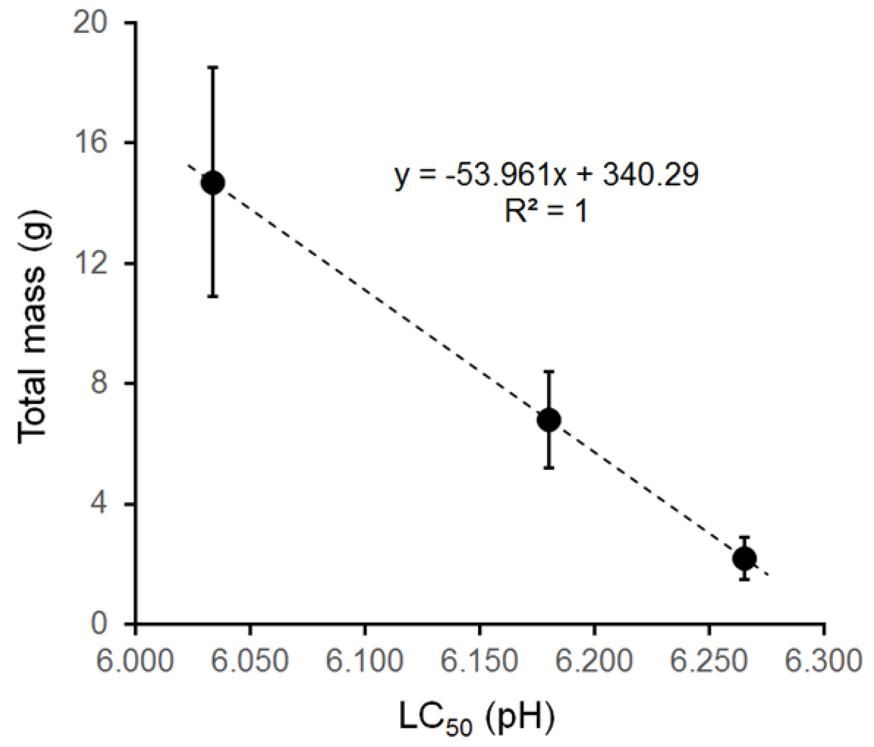
The capability of abalone to withstand stress generally increases with size, i.e., age [60]. The results of the seawater acidification toxicity test with farmed South African abalone are consistent with this statement. Although modelled toxicity curves for the size classes used (Figure 3) are not significantly different, there is an overall decrease in the LC values (i.e., increasing [H+]) with the increasing size of the abalone. For the smallest abalone (2.2 g), an LC50 of pH = 6.27 ([H+] = 543 nM) was determined, whereas for the medium (6.8 g) and large (14.7 g) abalone, these values were 6.18 ([H+] = 661 nM) and 6.02 ([H+] = 925 nM), respectively. The largest abalone in our study can therefore resist a 70% higher pH toxicity than the smallest one. This mass–response relationship is displayed in Figure 4. These results indicate that reported minimum in situ pH levels at farms of between 7.74 and 7.02 [61] and 7.5 [16] may not be of immediate danger to the survival of the abalone. They may become problematic, however, if such pH levels persist for extended periods: Table 4, Table 5 and Table 6 not only indicate which pH levels are toxic to different size abalone but they also reveal the time-dependence of exposure. During exposure times of up to 8 h, mortality has hardly commenced at any pH level tested, whereas mortalities subsequently (i.e., after longer exposure periods) occur at successively higher pH levels. At the 48 h time point, abalone start dying at pH levels of 6.0 to 6.4. Although many of the pH levels tested here are still too high to cause mortalities, they can limit growth and therefore make farms less economical [7,16,62]. Most South African aquaculture farms rely on large water influx from the nearby coastal waters and natural temperature fluctuations (and summer mortality) already cause concern for a number of abalone farms [7]. So far, imported hypercapnia does not seem to be a problem yet (own observation), although pH levels fluctuate, too. However, they can become a problem under most of the projected future scenarios [8]. Although currently of an acute nature, regular exposure to hypercapnic events may have negative effects and resemble chronic hypercapnia. This has been shown for this species as acidosis becomes more severe, reaching close to pH 7.0 after 12 [7] and 15 months [26], respectively. As a result, growth and shell formation was negatively affected [7,26]. This may affect both abalone ecology and aquaculture [25].
South African abalone farms primarily control pH levels through providing appropriate flow rates to the grow-out tanks. This is possible since they are land-based and mainly pump natural seawater through their systems extracted from the ocean. Strategies used on South African abalone farms to mitigate low pH include dosing with alkali chemicals [62] or co-culture with Ulva [7]. Ulva was shown to remove CO2 from an aquacultural mitigation system by photosynthesis and thereby lifted the pH compared with unmitigated levels [7]. Although both strategies revealed some economic benefits for farms due to an increase in productivity, further research is required to use and optimise such methods on a large-scale.
Another exposure to acute hypercapnia during aquaculture of the South African abalone is during removal from their tanks. This is necessary during handling activities such as tank maintenance, grading for size and harvesting. Abalone are attached to their substratum tightly and removal with mechanical force causes a slow-healing injury that can lead to mortality [63]. Grading (weight determination and sorting according to size) cannot be avoided because of heterogenous individual growth rates within a size cohort [19,20]. Large abalone would therefore outcompete smaller ones due to intraspecific competition [64]. Including the final harvest, removal is therefore necessary several times during an abalone’s life at a farm. If animals were to be removed mechanically, injuries and mortalities would accumulate, resulting in financial losses. As an alternative to mechanical removal, farms have turned to anaesthetic substances to reduce the handling stress and damage. The effect of several anaesthetics has been tested on abalone such as ethanol and clove oil [65,66], MgSO4, 2-phenoxyethanol, EDTA and procaine hydrochloride [63] and phenoxyethanol, benzocaine, eugenol (clove oil component) and CO2 [67]. At South African abalone farms, the use of MgSO4 for small abalone and dosing with CO2 (“dipping” in CO2-saturated seawater) for larger ones for the purpose of grading is now common practice (own observation). To evaluate the physiological impact of the latter on abalone, acid–base regulation was therefore analysed in a mimicked “dipping” period of 5 min in CO2-saturated seawater. During this “dipping”, the haemolymph pH drops sharply to 6.74 and pCO2 increases to almost 30 Torr. The haemolymph is therefore 455% more acidic than at the initial sampling. The [HCO3] more than doubles within the same period of time. Despite this bicarbonate buffering, the treatment leads to a strong respiratory acidosis as the Henderson–Hasselbalch diagram shows (Figure 2C). The ambient seawater pCO2 is almost 350 Torr, creating a strong inwardly directed gradient for carbon dioxide. Both pH and [HCO3] return to initial levels very quickly during subsequent recovery in normocapnic seawater. This recovery is also evident from observation of the abalone’s behaviour and indicates the reversibility of this mechanism. The rapid recovery is an important aspect when considering “dipping” in CO2-saturated seawater without compromising the animals’ growth potential permanently.
In a recent study, similar-sized (100 g) red abalone H. rufescens, were immersed in CO2-saturated seawater for 45 min for the purpose of pearl culture-related surgery [67]. The study tested a number of agents at different concentrations, such as Eugenol, Phenoxyethanol and Benzocaine, but also CO2 saturation. It revealed CO2 treatment to be the best method of anaesthesia: it was safe (no mortalities), was most effective in achieving full anaesthesia and did not lead to additional mucus secretion. This was despite the long exposure time of 30 min, compared with the 5 min exposure in the present study. The study found that glycogen reserves were used in various organs to meet the energetic cost of acute hypercapnic stress. Moreover, it showed that, during post-treatment recovery, depleted glycogen reserves were quickly replenished, especially in the digestive gland. This may be different in the present study due to the short dipping period of 5 min vs. 45 min in the case of the red abalone. In such a short period, it is possible that organ reserves of phosphagens (phosphoarginine) are sufficient to provide instantaneous energy [68,69]. Considering the inward pCO2 gradient in both cases, it is possible that gas exchange is severely interrupted. Anaerobic glycolysis is therefore likely to provide vital energy for some time. However, this was not investigated in the present study or in Rojas-Figueroa et al. [67].
The abalone used in the “dipping” simulation were much larger (91 g) than those in the standard toxicity test (2.2–14.7 g). The mass relationship with pH toxicity (Figure 3) would suggest that these large abalone were not exposed to lethal levels at the chosen pH of ~5. However, they dislodged from the surface very quickly, indicating that the linear mass–response relationship in Figure 3 does not extend to larger size abalone and that this pH level is potentially fatal even for this large size class. The combined results from our LC50 experiment and the “dipping” experiment indicate that this procedure is potentially fatal because of the reverse pCO2 gradient at such low pH levels and the size and time dependence thereof. CO2-saturated seawater seems to level out at a minimum pH range of 4.8 to 5.2 under temperature and salinity conditions at South African aquaculture farms. Unlike other chemical anaesthetics (such as MgSO4), this provides a certain safety net from accidental over-dosing and makes exposure time the critical factor to control. Despite the successful use of this procedure at South African farms, it would therefore be beneficial to standardise the method for consistent and safe use. This can ensure that overexposing the abalone to these potentially fatal conditions is avoided. Depending on size, there could be, for example, be a defined time period of “dipping” and a “best practice” protocol for recovery. In addition, it is recommended to investigate the exact impact of “dipping” on energy cost and subsequently the long-term effects on the growth of H. midae. Such knowledge will help to evaluate the energy cost of this method on growth parameters and if there is a need to search for better techniques. The need for such research is indicated from the response of related species; after transfer to normocapnia following short-term exposure to extreme hypercapnia (longer than in the present study), oysters do not recover, and growth remains retarded [70]. Furthermore, despite rapid recovery, the low pH levels achieved during the use of this method could potentially damage the exposed tissues of the abalone, and this may also be size or age dependent. Studies of the histology of tissues such as gills and epithelium after one or serial “dipping” should therefore be considered for different size classes.

5. Conclusions

The major findings of the present study with physiological and commercial consequences are as follows: 1. Acute exposure to hypercapnia induces a reversible, unbuffered respiratory acidosis. 2. The impact of acute hypercapnia is size-dependent and potentially fatal. 3. Exposure to extreme but short hypercapnia during “dipping” causes a rapid imbalance of the acid–base state but a speedy subsequent recovery. Details from the present study can be used by abalone aquaculture farms to not only mitigate/avoid the impact of acute (and chronic) hypercapnia but also to take the length of time and size dependence of the exposure into account. They also revealed the need to standardise the anaesthesia method for grading for consistent and safe use within aquaculture. The results provide a proxy to estimate the effects on wild populations, too, aiding the management of the fishing resources and conservation efforts in future.

Author Contributions

T.N.: conceptualisation, methodology, investigation, data collection, formal analysis, writing—original draft. C.R.B.: conceptualization, methodology, supervision, funding acquisition, writing—review and editing. M.N.: funding acquisition, methodology, writing—review and editing. D.Y.: formal analysis, writing—review and editing. L.A.: conceptualization, methodology, supervision, funding acquisition, writing—review and editing. All authors have read and agreed to the published version of the manuscript.

Funding

This research received no external funding.

Institutional Review Board Statement

Handling, transport and experiments were conducted in accordance with the Standard Operating Procedures established by the Aquaculture Animal Ethics Committee of the Department of Forestry, Fisheries and the Environment of South Africa (Code: 160330_ab_01_Auerswald; Date: 17 October 2017).

Data Availability Statement

Relevant information is included in the article. Raw data supporting the conclusions are available from the author, L.A., upon request.

Acknowledgments

We are grateful to A. Busby and team from the Seapoint research aquarium and staff from the Rock Lobster group for their support.

Conflicts of Interest

Author Matt Naylor was employed by the company HIK Abalone Farm (Pty) Ltd.. The remaining authors declare that the research was conducted in the absence of any commercial or financial relationships that could be construed as a potential conflict of interest.

References

  1. Newman, G.G. Distribution of the abalone (Haliotis midae) and the effect of temperature on productivity. Investig. Rep. Div. Sea Fish. 1969, 74, 1–7. [Google Scholar]
  2. Geiger, D.L. Distribution and biogeography of the recent Haliotidae (Gastropoda: Vetigastropoda) world-wide. Boll. Malacol. 1999, 35, 57–120. [Google Scholar]
  3. Rhode, C.; Bester-van der Merwe, A.E.; Roodt-Wilding, R. An assessment of spatio-temporal genetic variation in the South African abalone (Haliotis midae), using SNPs: Implications for conservation management. Conserv. Genet. 2017, 18, 17–31. [Google Scholar] [CrossRef]
  4. Barkai, A.; Griffiths, C.L. Diet of the South African abalone, Haliotis midae. S. Afr. J. Mar. Sci. 1986, 4, 37–44. [Google Scholar] [CrossRef]
  5. DFFE (Department of Forestry, Fisheries and the Environment). Status of the South African Marine Fishery Resources 2023; DEFF: Cape Town, South Africa, 2023.
  6. DEFF (Department of Environment, Forestry and Fisheries). Aquaculture Yearbook; DEFF: Cape Town, South Africa, 2020.
  7. Lester, N.C. The interaction of acidification and warming on the South African abalone, Haliotis midae, and the potential for mitigation in aquaculture. Ph.D. Thesis, University of Cape Town, Cape Town, South Africa, 2021. [Google Scholar]
  8. Cooley, S.; Schoeman, D.; Bopp, L.; Boyd, P.; Donner, S.; Ghebrehiwet, D.Y.; Ito, S.-I.; Kiessling, W.; Martinetto, P.; Ojea, E.; et al. Oceans and Coastal Ecosystems and Their Services. In Climate Change 2022: Impacts, Adaptation and Vulnerability; Contribution of Working Group II to the Sixth Assessment Report of the Intergovernmental Panel on Climate Change; Pörtner, H.-O., Roberts, D.C., Tignor, M., Poloczanska, E.S., Mintenbeck, K., Alegría, A., Craig, M., Langsdorf, S., Löschke, S., Möller, V., Eds.; Cambridge University Press: Cambridge, UK; New York, NY, USA, 2022; pp. 379–550. [Google Scholar] [CrossRef]
  9. Hill, A.E.; Hickey, B.M.; Shillington, F.A.; Strub, P.T.; Brink, K.H.; Barton, E.D.; Thomas, A.C. Eastern ocean boundaries coastal segment. In The Global Coastal Ocean, Regional Studies and Syntheses; Robinson, A.R., Brink, K.H., Eds.; John Wiley & Sons, Inc.: New York, NY, USA, 1998; pp. 29–68. [Google Scholar]
  10. Probyn, T.A.; Pitcher, G.C.; Monteiro, P.M.S.; Boyd, A.J.; Nelson, G. Physical processes contributing to harmful algal blooms in Saldanha Bay, South Africa. S. Afr. J. Mar. Sci. 2000, 22, 285–297. [Google Scholar] [CrossRef]
  11. Pitcher, G.C.; Figueiras, F.G.; Hickey, B.M.; Moita, M.T. The physical oceanography of upwelling systems and the development of harmful algal blooms. Prog. Oceanogr. 2010, 85, 5–32. [Google Scholar] [CrossRef] [PubMed]
  12. Bakun, A. Global climate change and intensification of coastal ocean upwelling. Science 1990, 247, 198–201. [Google Scholar] [CrossRef]
  13. Diaz, R.J.; Rosenberg, R. Spreading dead zones and consequences for marine ecosystems. Science 2008, 321, 926–929. [Google Scholar] [CrossRef]
  14. Pitcher, G.C.; Probyn, T.A. Anoxia in southern Benguela during the autumn of 2009 and its linkage to a bloom of the dinoflagellate Ceratium balechii. Harmful Algae 2011, 11, 23–32. [Google Scholar] [CrossRef]
  15. Sydeman, W.J.; García-Reyes, M.; Schoeman, D.S.; Rykaczewski, R.R.; Thompson, S.A.; Black, B.A.; Bograd, S.J. Climate change. Climate change and wind intensification in coastal upwelling ecosystems. Science 2014, 345, 77–80. [Google Scholar] [CrossRef]
  16. Naylor, M.A.; Kaiser, H.; Jones, C.L.W. Water quality in a serial-use raceway and its effect on the growth of South African abalone, Haliotis midae Linneaeus, 1785. Aquac. Res. 2011, 42, 918–930. [Google Scholar] [CrossRef]
  17. Naylor, M.A.; Kaiser, H.; Jones, C.L.W. The effect of free ammonia nitrogen, pH and supplementation with oxygen on the growth of South African abalone, Haliotis midae L. in an abalone serial-use raceway with three passes. Aquac. Res. 2014, 45, 213–224. [Google Scholar] [CrossRef]
  18. Reddy-Lopata, K.; Auerswald, L.; Cook, P.A. Ammonia toxicity and its effect on the growth of the South African abalone Haliotis midae Linnaeus. Aquaculture 2006, 261, 678–687. [Google Scholar] [CrossRef]
  19. Heath, P.; Moss, G. Is size grading important for farming the abalone Haliotis iris? Aquaculture 2009, 290, 80–86. [Google Scholar] [CrossRef]
  20. Venter, L.; Loots, D.T.; Vosloo, A.; Jansen van Rensburg, P.; Lindeque, J.Z. Abalone growth and associated aspects: Now from a metabolic perspective. Rev. Aquac. 2018, 10, 451–473. [Google Scholar] [CrossRef]
  21. Pörtner, H.O.; Langenbuch, M.; Reipschlager, A. Biological impact of elevated ocean CO2 concentrations: Lessons from animal physiology and earth history. J. Oceanogr. 2004, 60, 705–718. [Google Scholar] [CrossRef]
  22. Fabry, V.J.; Seibel, B.A.; Feely, R.A.; Orr, J.C. Impacts of Ocean acidification on marine fauna and ecosystem processes. ICES J. Mar. Sci. 2008, 65, 414–432. [Google Scholar] [CrossRef]
  23. Harvey, B.P.; Gwynn-Jones, D.; Moore, P.J. Meta-analysis reveals complex marine biological responses to the interactive effects of ocean acidification and warming. Ecol. Evol. 2013, 3, 1016–1030. [Google Scholar] [CrossRef] [PubMed]
  24. Hall-Spencer, J.M.; Harvey, B.P. Ocean acidification impacts on coastal ecosystem services due to habitat degradation. Emerg. Top. Life Sci. 2019, 3, 197–206. [Google Scholar]
  25. Avignon, S.; Auzoux-Bordenave, S.; Martin, S.; Dubois, P.; Badou, A.; Coheleach, M.; Richard, N.; Di Giglio, S.; Malet, L.; Servili, A.; et al. An integrated investigation of the effects of ocean acidification on adult abalone (Haliotis tuberculata). ICES J. Mar. Sci. 2020, 77, 757–772. [Google Scholar] [CrossRef]
  26. Haupt, T.M.; Novak, T.; Naylor, M.; Auerswald, L. The thermal response of adult abalone, Haliotis midae, following exposure to chronic hypercapnia. 2024; in preparation. [Google Scholar]
  27. Cheng, W.; Liub, C.-H.; Cheng, S.-Y.; Chen, J.-C. Effect of dissolved oxygen on the acid–base balance and ion concentration of Taiwan abalone Haliotis diversicolor supertexta. Aquaculture 2004, 231, 573–586. [Google Scholar] [CrossRef]
  28. Michaelidis, B.; Ouzounis, C.; Paleras, A.; Pörtner, H.O. Effects of long-term moderate hypercapnia on acid–base balance and growth rate in marine mussels Mytilus galloprovincialis. Mar. Ecol. Progr. Ser. 2005, 293, 109–118. [Google Scholar] [CrossRef]
  29. Melzner, F.; Gutowska, M.A.; Langenbuch, M.; Dupont, S.; Lucassen, M.; Thorndyke, M.C.; Bleich, M.; Pörtner, H.O. Physiological basis for high CO2 tolerance in marine ectothermic animals: Pre-adaptation through lifestyle and ontogeny? Biogeosciences 2009, 6, 2313–2331. [Google Scholar] [CrossRef]
  30. Morash, A.J.; Alter, K. Effects of environmental and farm stress on abalone physiology: Perspectives for abalone aquaculture in the face of global climate change. Rev. Aquac. 2015, 7, 1–27. Available online: https://www.researchgate.net/publication/276150674_Effects_of_environmental_and_farm_stress_on_abalone_physiology_Perspectives_for_abalone_aquaculture_in_the_face_of_global_climate_change (accessed on 1 February 2024).
  31. Knapp, J.L.; Bridges, C.R.; Krohn, J.; Hoffman, L.C.; Auerswald, L. Acid–base balance and changes in haemolymph properties of the South African rock lobsters, Jasus lalandii, a palinurid decapod, during chronic hypercapnia. Biochem. Biophys. Res. Commun. 2015, 461, 475–480. [Google Scholar] [CrossRef]
  32. Sarazin, G.; Michard, G.; Prevot, F. A rapid and accurate spectroscopic method for alkalinity measurements in sea water samples. Water Res. 1999, 33, 290–294. [Google Scholar] [CrossRef]
  33. Pierrot, D.E.; Lewis, E.; Wallace, D.W.R. MS excel program developed for CO2 system calculations. In ORNL/CDIAC-105a; Carbon Dioxide Information Analysis Center, Oak Ridge National Laroratory, US Department of Energy: Oak Ridge, TN, USA, 2006. [Google Scholar]
  34. Truchot, J.P. Carbon dioxide combining properties of the blood of the shore crab Carcinus maenas (L): Carbon dioxide solubility coefficient and carbonic acid dissociation constants. J. Exp. Biol. 1976, 64, 45–57. [Google Scholar] [CrossRef]
  35. R Core Team. R: A Language and Environment for Statistical Computing. R Foundation for Statistical Computing: Vienna, Austria, 2024. Available online: https://www.R-project.org/ (accessed on 1 February 2024).
  36. Allaire, J.; Xie, Y.; Dervieux, C.; McPherson, J.; Luraschi, J.; Ushey, K.; Atkins, A.; Wickham, H.; Cheng, J.; Chang, W.; et al. Rmarkdown: Dynamic Documents for R. 2024. Available online: https://github.com/rstudio/rmarkdown (accessed on 1 February 2024).
  37. Letaw, A. Captioner: Numbers Figures and Creates Simple Captions. 2015. Available online: https://github.com/adletaw/captioner (accessed on 1 February 2024).
  38. Wickham, H.; François, R.; Henry, L.; Müller, K.; Vaughan, D. Dplyr: A Grammar of Data Manipulation. 2023. Available online: https://dplyr.tidyverse.org (accessed on 1 February 2024).
  39. Wickham, H.; Chang, W.; Henry, L.; Pedersen, T.L.; Takahashi, K.; Wilke, C.; Woo, K.; Yutani, H.; Dunnington, D.; van den Brand, T. ggplot2: Create Elegant Data Visualisations Using the Grammar of Graphics. 2024. Available online: https://ggplot2.tidyverse.org (accessed on 1 February 2024).
  40. Wickham, H.; Henry, L. Purrr: Functional Programming Tools. 2023. Available online: https://purrr.tidyverse.org/ (accessed on 1 February 2024).
  41. Xie, Y. Knitr: A General-Purpose Package for Dynamic Report Generation in R. 2024. Available online: https://yihui.org/knitr/ (accessed on 1 February 2024).
  42. Bates, D.; Maechler, M.; Bolker, B.; Walker, S. Lme4: Linear Mixed-Effects Models Using Eigen and S4. 2024. Available online: https://github.com/lme4/lme4/ (accessed on 1 February 2024).
  43. Lenth, R.V. Emmeans: Estimated Marginal Means, Aka Least-Squares Means. 2024. Available online: https://github.com/rvlenth/emmeans (accessed on 1 February 2024).
  44. Pinheiro, J.; Bates, D.; R Core Team. Nlme: Linear and Nonlinear Mixed Effects Models. 2023. Available online: https://svn.r-project.org/R-packages/trunk/nlme/ (accessed on 1 February 2024).
  45. Tarr, R.J.Q.; Williams, P.V.G.; Mackenzie, A.J. Abalone, sea urchins and rock lobster: A possible ecological shift that may affect traditional fisheries. S. Afr. J. Mar. Sci. 1996, 17, 319–323. [Google Scholar] [CrossRef]
  46. Auzoux-Bordenave, S.; Chevret, S.; Badou, A.; Martin, S.; Di Giglio, S.; Dubois, P. Acid–base balance in the haemolymph of European abalone (Haliotis tuberculata) exposed to CO2-induced ocean acidification. Comp. Biochem. Physiol. Part A 2021, 259, 110996. [Google Scholar] [CrossRef]
  47. Knapp, J.L.; Bridges, C.R.; Krohn, J.; Hoffman, L.C.; Auerswald, L. The effects of hypercapnia on the West Coast rock lobster (Jasus lalandii) through acute exposure to decreased seawater pH-Physiological and biochemical responses. J. Exp. Mar. Biol. Ecol. 2016, 476, 58–64. [Google Scholar] [CrossRef]
  48. Brix, O. The adaptive significance of the reversed Bohr and Root shifts in blood from the marine gastropod, Buccinum undatum. J. Exp. Zool. 1982, 221, 27–36. [Google Scholar] [CrossRef]
  49. Wells, R.M.G.; Baldwin, J.; Speed, S.R.; Weber, R.E. Haemocyanin function in the New Zealand abalones Haliotis iris and H. australis: Relationships between oxygen-binding properties, muscle metabolism and habitat. Mar. Freshw. Res. 1998, 49, 143–149. [Google Scholar] [CrossRef]
  50. Brix, O.; Lykkeboe, G.; Johansen, K. Reversed Bohr and Root shifts in hemocyanin of the marine prosobranch, Buccinum undatum: Adaptations to a periodically hypoxic habitat. J. Comp. Physiol. 1979, 129, 97–103. [Google Scholar] [CrossRef]
  51. Mangum, C.P.; Burnett, L.E., Jr. The CO2 Sensitivity of the Hemocyanins and its relationship to Cl sensitivity. Biol. Bull. 1986, 171, 248–263. [Google Scholar] [CrossRef]
  52. Bridges, C.R.; Morris, S. Respiratory pigments: Interactions between oxygen and carbon dioxide transport. Can. J. Zool. 1989, 67, 2971–2985. [Google Scholar] [CrossRef]
  53. Donovan, D.; Baldwin, J.; Carefoot, T. The contribution of anaerobic energy to gastropod crawling and a re-estimation of minimum cost of transport in the abalone, Haliotis kamtschatkana (Jonas). J. Exp. Mar. Biol. Ecol. 1999, 235, 273–284. [Google Scholar] [CrossRef]
  54. Hickey, A.J.; Wells, R.M. Thermal constraints on glycolytic metabolism in the New Zealand abalone, Haliotis iris: The role of tauropine dehydrogenase. N. Z. J. Mar. Freshw. Res. 2003, 37, 723–731. [Google Scholar] [CrossRef]
  55. O’Omolo, S.; Gäde, G.; Cook, P.A.; Brown, A.C. Can the end products of anaerobic metabolism, tauropine and D-lactate, be used as metabolic stress indicators during transport of live South African abalone Haliotis midae? Afr. J. Mar. Sci. 2003, 25, 301–309. [Google Scholar] [CrossRef]
  56. Venter, L.; Loots, D.T.; Mienie, L.J.; van Rensburg, P.J.J.; Mason, S.; Vosloo, A.; Lindeque, J.Z. Uncovering the metabolic response of abalone (Haliotis midae) to environmental hypoxia through metabolomics. Metabolomics 2018, 14, 49. [Google Scholar] [CrossRef]
  57. Lee, S.-M. Utilization of dietary protein, lipid, and carbohydrate by abalone Haliotis discus hannai: A review. J. Shellfish Res. 2004, 23, 1027–1030. [Google Scholar]
  58. Durazo, E.; Viana, M.T. Fatty acid profile of cultured green abalone (Haliotis fulgens) exposed to lipid restriction and long-term starvation. Cienc. Mar. 2013, 39, 363–370. [Google Scholar] [CrossRef]
  59. Carroll, S.L.; Coyne, V.E. A proteomic analysis of the effect of ocean acidification on the haemocyte proteome of the South African abalone Haliotis midae. Fish Shellfish Immunol. 2021, 117, 274–290. [Google Scholar] [CrossRef] [PubMed]
  60. Fallu, R. Abalone Farming. In Fishing News Books; Blackwell Scient. Publisher: Oxford, UK, 1991; pp. 37–43. [Google Scholar]
  61. Naylor, M.A.; Kaiser, H.; Jones, C.L.W. The effect of dosing with sodium hydroxide (NaOH) on water pH and growth of Haliotis midae in an abalone serial-use raceway. Aquac. Int. 2013, 21, 467–479. [Google Scholar] [CrossRef]
  62. De Prisco, J.A. An Investigation of Some Key Physico-Chemical Water Quality Parameters of an Integrated Multi-Trophic Aquaculture (IMTA) System Operating Recirculation Methodology in the Western Cape of South Africa. Master’s Thesis, University of Cape Town, Cape Town, South Africa, 2020. [Google Scholar]
  63. White, H.; Hecht, T.; Potgieter, B. The effect of four anesthetics on Haliotis midae and their suitability for application in commercial abalone culture. Aquaculture 1996, 140, 145–151. [Google Scholar] [CrossRef]
  64. Van der Merwe, E. Toward best management practices for the growth of the abalone Haliotis midae Linnaeus on a commercial South African abalone farm. Master’s Thesis, University of the Western Cape, Cape Town, South Africa, 2009. [Google Scholar]
  65. Fanni, N.A.; Soeprijanto, F.R. Abalone (Haliotis sqaumata) anesthesia with ethanol on grading process. Russ. J. Agric. Socio-Econ. Sci. 2018, 2, 239–242. [Google Scholar]
  66. Fanni, N.A.; Shaleh, F.R.; Santanumurti, M.B. The role of clove (Sygnium aromaticum) oil as anaesthetics compound for abalone (Haliotis squamata). Iraqi J. Vet. Sci. 2021, 35, 335–342. [Google Scholar] [CrossRef]
  67. Rojas-Figueroa, A.; Angulo, C.; Araya, R.; Granados-Amores, A.; Guardiola, F.A.; Saucedo, P.E. Comparative analysis of anesthetic agents used in pre-operative therapy for pearl culture in the red abalone Haliotis rufescens (Swainson, 1822). Aquaculture 2023, 574, 739623. [Google Scholar] [CrossRef]
  68. Grieshaber, M.; Hardewig, I.; Kreutzer, U.; Pörtner, H.-O. Physiological and metabolic responses to hypoxia in invertebrates. In Reviews of Physiology, Biochemistry and Pharmacology; Springer: Berlin/Heidelberg, Germany, 1993; Volume 125, pp. 43–147. [Google Scholar]
  69. Venter, L.; Loots, D.T.; Mienie, L.J.; van Rensburg, P.J.J.; Mason, S.; Vosloo, A.; Lindeque, J.Z. The cross-tissue metabolic response of abalone (Haliotis midae) to functional hypoxia. Biol. Open 2018, 7, bio031070. [Google Scholar] [CrossRef]
  70. Lutier, M.; Pernet, F.; Di Poi, C. Pacific oysters do not compensate growth retardation following extreme acidification events. Biol. Lett. 2023, 19, 20230185. [Google Scholar] [CrossRef]
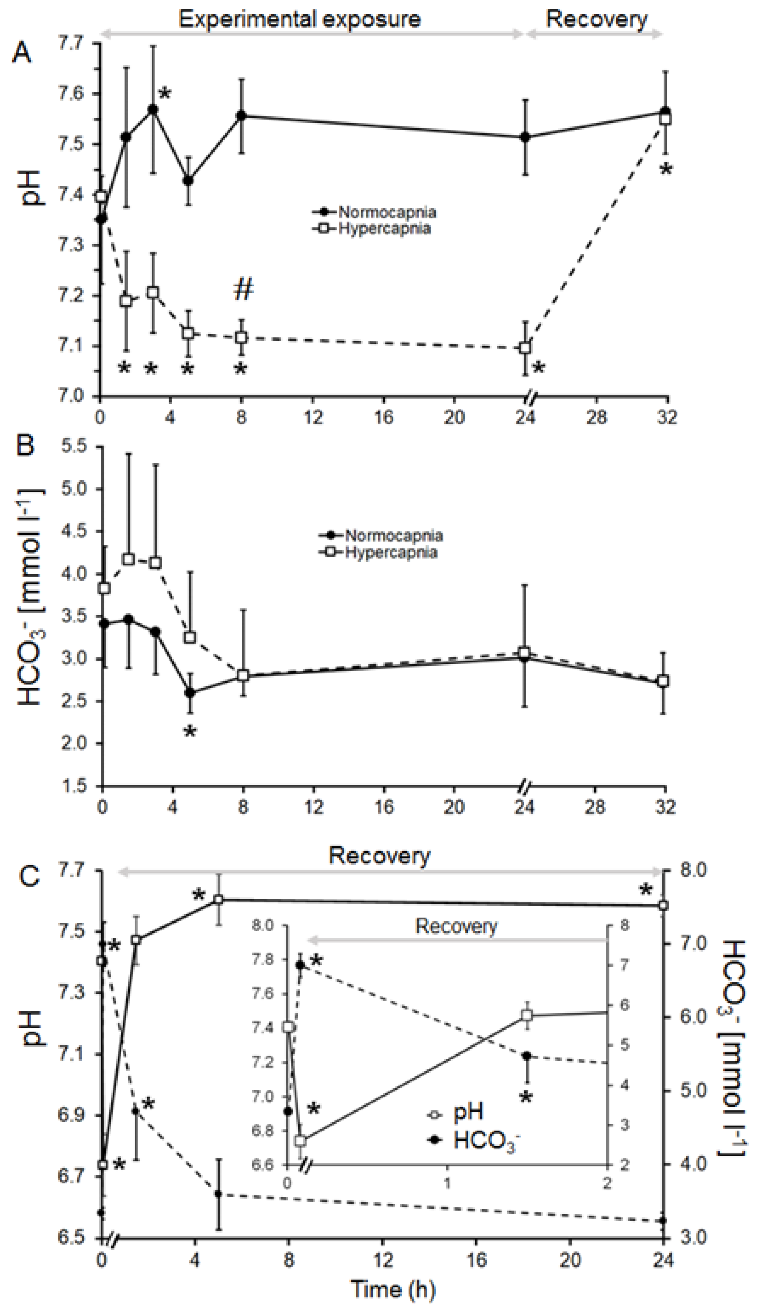
Figure 1. Time course of acid–base changes in H. midae haemolymph (A) measured pH and (B) calculated [HCO3 + CO32−] during acute exposure to normocapnic seawater (solid line) and hypercapnia (dashed line) for 24 h followed by 8 h recovery in normocapnic seawater. Panel (C) depicts pH and [HCO3 + CO32−] during dipping in low pH (5.02) seawater for 5 min and subsequent recovery in normocapnic seawater. Values are means ± S.D (n = 5). * Significantly different from initial value (t0) within treatment (p < 0.05). # Significantly different from the respective sampling time of the normocapnic treatment (p < 0.05).
Fishes 09 00313 g001
Figure 2. Henderson–Hasselbalch (pH-bicarbonate) diagrams for haemolymph of adult H. midae constructed from the time course of values during acute exposure presented in Table 3. (A) During 24 h normocapnia and subsequent 8 h recovery, (B) during 24 h hypercapnia followed by 8 h normocapnic recovery and (C) during 5 min “dipping” in pH 5.02 hypercapnia and subsequent recovery in normocapnic seawater. The pCO2 isopleths (full black lines) were derived from the Henderson–Hasselbalch equation. Note: isopleth for dipping pH was far outside the range of panel (C) and was therefore omitted. Appropriate values for the first dissociation constant (pK1) and solubility coefficient (α) were derived from Truchot [34]. Dashed line = normocapnic/recovery seawater isopleth, dotted line = hypercapnic seawater isopleth. Values are means ± S.D (n = 5). Arrows indicate course of pH and bicarbonate buffering during experimental exposure.
Fishes 09 00313 g002
Figure 3. Toxicity of pH to three size classes of abalone. Proportion surviving at the end of the experiment points (observations) and lines (model prediction). Shaded regions represent 95% confidence intervals.
Fishes 09 00313 g003
Figure 4. Relationship of LC50 (pH) and abalone mass for three sizes of abalone. Values for mass are means ± S.D (n = 5).
Fishes 09 00313 g004
Table 1. Physicochemical seawater conditions recorded during acclimation, acute exposure of H. midae to normocapnic and hypercapnic conditions and subsequent recovery and also to “dipping” treatment.
TreatmentTA
°C
pHAT
µmol kg −1
O2
%
Salinity
Ca2+
mmol L−1
Mg2+
mmol L−1
pCO2
Torr (µatm)
HCO3
mmol L−1
CO32−
mmol L−1
Acclimation19.3 ± 0.28.22 ± 0.082039 ± 698.7 ± 0.135.0 ± 0.010.3 ± 0.452.0 ± 1.10.2 ± 0.0 (209 ± 0)1.5 ± 0.00.2 ± 0.0
Normocapnia19.4 ± 0.78.28 ± 0.042048 ± 1297.8 ± 0.334.9 ± 0.010.3 ± 0.552.5 ± 1.10.2 ± 0.0 (176 ± 0)1.4 ± 0.00.2 ± 0.0
Hypercapnia19.5 ± 0.87.33 ± 0.052044 ± 2194.9 ± 0.034.9 ± 0.010.4 ± 0.351.5 ± 1.61.6 ± 0.2 (2142 ± 2)1.9 ± 0.00.0 ± 0.0
5 min dip19.4 ± 0.15.02 ± 0.062019 ± 589.4 ± 6.734.9 ± 0.010.2 ± 0.152.5 ± 0.9346 ± 46
(457,568 ± 60,582)
2.0 ± 0.00.0 ± 0.0
Recovery19.6 ± 0.28.27 ± 0.012031 ± 892.3 ± 0.234.9 ± 0.010.3 ± 0.852.4 ± 1.60.2 ± 0.0 (180 ± 0)1.4 ± 0.00.2 ± 0.0
Table 2. Physicochemical seawater conditions recorded during acclimation and determination of LC50 of three size classes of H. midae.
Treatment
(pH)
TA
°C
pHAT
µmol kg −1
O2
%
Salinity
Ca2+
mmol L−1
Mg2+
mmol L−1
pCO2
Torr (µatm)
HCO3
mmol L−1
CO32−
mmol L−1
Acclimation20.7 ± 0.48.05 ± 0.092011 ± 1197.6 ± 1.134.9 ± 0.110.6 ± 0.552.1 ± 1.00.2 ± 0.1 (338 ± 81)1.6 ± 0.10.2 ± 0.0
7.220.3 ± 0.07.20 ± 0.022051 ± 497.4 ± 0.134.2 ± 0.010.1 ± 0.453.1 ± 2.32.3 ± 0.0 (2960 ± 3)2.0 ± 0.00.0 ± 0.0
6.820.8 ± 1.06.79 ± 0.022036 ± 894.5 ± 0.234.2 ± 0.011.0 ± 0.452.5 ± 1.15.9 ± 0.0 (7732 ± 9)2.0 ± 0.00.0 ± 0.0
6.421.2 ± 1.16.39 ± 0.012046 ± 2289.0 ± 0.134.2 ± 0.010.6 ± 0.151.3 ± 1.015.0 ± 0.2 (19,677 ± 193)2.0 ± 0.00.0 ± 0.0
6.021.3 ± 0.26.00 ± 0.002031 ± 1688.6 ± 0.334.3 ± 0.010.7 ± 1.051.0 ± 1.336.6 ± 0.3 (48,182 ± 381)2.0 ± 0.00.0 ± 0.0
5.620.2 ± 0.35.61 ± 0.012028 ± 1383.7 ± 0.434.2 ± 0.010.9 ± 0.952.5 ± 1.689.7 ± 0.7 (117,984 ± 879)2.0 ± 0.00.0 ± 0.0
Table 3. Time course of in vivo haemolymph parameters of adult H. midae during acute exposure to normocapnic (pH 8.3) and hypercapnic (pH 7.3, 5.0) conditions.
Exposure TimepHcCO2
mmol L−1
pCO2
Torr
[HCO3 + CO32−]
mmol L−1
Ca2+
mmol L−1
Mg2+
mmol L−1
Haemocyanin
mg mL−1
Normocapnia (h)
07.35 ± 0.133.6 ± 0.43.5 ± 0.13.4 ± 0.511.9 ± 0.921.9 ± 0.512.4 ± 2.5
1.57.51 ± 0.14 *3.6 ± 0.42.4 ± 0.2 #3.5 ± 0.612.0 ± 0.822.0 ± 0.712.5 ± 1.9
37.57 ± 0.13 *3.4 ± 0.42.0 ± 0.2 #3.3 ± 0.511.7 ± 1.421.9 ± 0.712.8 ± 3.0
57.43 ± 0.052.7 ± 0.12.2 ± 0.0 *#2.6 ± 0.211.9 ± 0.721.8 ± 0.612.8 ± 2.7
87.56 ± 0.07 *2.9 ± 0.11.7 ± 0.1 *#2.8 ± 0.211.6 ± 0.921.8 ± 1.612.6 ± 2.2
247.51 ± 0.07 *3.1 ± 0.52.1 ± 0.2 #3.0 ± 0.611.1 ± 1.521.4 ± 1.812.5 ± 1.8
32 (Recovery)7.56 ± 0.08 *2.8 ± 0.21.7 ± 0.1 *2.7 ± 0.411.7 ± 1.121.7 ± 1.612.8 ± 1.6
Hypercapnia (h)
07.40 ± 0.044.0 ± 0.43.5 ± 0.13.8 ± 0.511.5 ± 1.221.7 ± 0.412.3 ± 1.9
1.57.19 ± 0.10 *#4.4 ± 1.16.1 ± 0.2 *#4.2 ± 1.211.0 ± 0.721.2 ± 1.212.5 ± 1.9
37.20 ± 0.08 *#4.4 ± 1.15.9 ± 0.2 *#4.1 ± 1.111.2 ± 0.121.3 ± 1.212.6 ± 1.7
57.12 ± 0.05 *#3.5 ± 0.75.6 ± 0.1 *#3.3± 0.811.0 ± 0.121.1 ± 0.712.7 ± 1.8
87.12 ± 0.03 *#3.0 ± 0.74.8 ± 0.1 #2.8 ± 0.811.3 ± 1.221.0 ± 1.612.9 ± 2.0
247.10 ± 0.05 *#3.3 ± 0.75.5 ± 0.1 *#3.1 ± 0.811.2 ± 2.320.8 ± 1.613.0 ± 1.7
32 (Recovery)7.55 ± 0.07 *2.8 ± 0.21.8 ± 0.1 *2.7 ± 0.312.0 ± 0.620.9 ± 1.312.7 ± 1.6
Anaesthesia
07.40 ± 0.033.5 ± 0.13.0 ± 0.23.3 ± 0.111.8 ± 1.222.5 ± 1.012.7 ± 2.2
5 min6.74 ± 0.10 *8.3 ± 0.5 *29.6 ± 6.5 *7.0 ± 0.3 *10.8 ± 1.122.1 ± 1.712.7 ± 1.9
1.5 (Recovery)7.47 ± 0.084.9 ± 0.7 *3.7 ± 0.74.7 ± 0.7 *11.1 ± 0.621.8 ± 0.913.4 ± 1.8
5 (Recovery)7.61 ± 0.08 *3.7 ± 0.52.1 ± 0.53.6 ± 0.511.2 ± 0.722.2 ± 0.812.8 ± 1.6
24 (Recovery)7.59 ± 0.03 *3.3 ± 0.11.9 ± 0.23.2 ± 0.111.6 ± 0.522.4 ± 0.813.0 ± 3.3
Values are means ± S.D. (n = 5). * Significantly different from initial value (t0) within treatment (p < 0.05). # Significantly different from the respective sampling time of the normocapnic treatment (p < 0.05).
Table 4. Toxicity of different pH levels to small abalone (2.2 ± 0.7 g).
pH Number of Dead Abalone after
2 h4 h6 h8 h10 h12 h24 h36 h48 h
ababababababababab
7.20 ± 0.02 (7.19–7.22)000000000000000000
6.80 ± 0.02 (6.77–6.80)000000000000000000
6.39 ± 0.01 (6.39–6.40)000000000000000022
6.00 ± 0.0 (6.00–6.01)0000001010102166910
5.61 ± 0.01 (5.60–5.61)0000000032458810101010
5.2 (5.18–5.21)0000000033668910101010
LC50 pH: 6.27
95% confidence limits: 6.18–6.4
Lower case letters a and b indicate parallel experiments (n = 10 each) under same conditions.
Table 5. Toxicity of different pH levels to medium abalone (6.8 ± 1.6 g).
pH Number of Dead Abalone after
2 h4 h6 h8 h10 h12 h24 h36 h48 h
ababababababababab
7.20 ± 0.02 (7.19–7.22)000000000000000000
6.80 ± 0.02 (6.77–6.80)000000000000000000
6.39 ± 0.01 (6.39–6.40)000000000000101011
6.00 ± 0.0 (6.00–6.01)000000000101016898
5.61 ± 0.01 (5.60–5.61)000000001233101010101010
5.2 (5.18–5.21)0000000133558910101010
LC50 pH: 6.18
95% confidence limits: 6.03–6.32
Lower case letters a and b indicate parallel experiments (n = 10 each) under same conditions.
Table 6. Toxicity of different pH levels to large abalone (14.7 ± 3.8 g).
pH Number of Dead Abalone after
2 h4 h6 h8 h10 h12 h24 h36 h48 h
ababababababababab
7.20 ± 0.02 (7.19–7.22)000000000000000000
6.80 ± 0.02 (6.77–6.80)000000000000000000
6.39 ± 0.01 (6.39–6.40)000000000000000000
6.00 ± 0.0 (6.00–6.01)000000000000003366
5.61 ± 0.01 (5.60–5.61)0000000010113210101010
5.2 ± 0.01 (5.18–5.21)0000001035668710101010
LC50 pH: 6.02
95% confidence limits: 5.98–6.02
Lower case letters a and b indicate parallel experiments (n = 10 each) under same conditions.
Disclaimer/Publisher’s Note: The statements, opinions and data contained in all publications are solely those of the individual author(s) and contributor(s) and not of MDPI and/or the editor(s). MDPI and/or the editor(s) disclaim responsibility for any injury to people or property resulting from any ideas, methods, instructions or products referred to in the content.