Article

Intermittent Detections of ISAV-HPR0 in a Salmon Recirculating Aquaculture System, and Implications for Sampling

1
USDA Agricultural Research Service, National Cold Water Marine Aquaculture Center, Franklin, ME 04634, USA
2
USDA Animal and Plant Health Inspection Service, Veterinary Services, National Veterinary Services Laboratories, Ames, IA 50010, USA
3
USDA Animal and Plant Health Inspection Service, Veterinary Services, Center for Epidemiology and Animal Health, Fort Collins, CO 80521, USA
*
Author to whom correspondence should be addressed.
Fishes 2024, 9(8), 325; https://doi.org/10.3390/fishes9080325
Submission received: 1 July 2024 / Revised: 5 August 2024 / Accepted: 16 August 2024 / Published: 17 August 2024
(This article belongs to the Section Welfare, Health and Disease)

Abstract

:
Infectious salmon anemia virus (ISAV) is an important pathogen in global Atlantic salmon (Salmo salar L.) aquaculture. The existence of both non-pathogenic (ISAV-HPR0) and pathogenic (HPR-deleted ISAV) forms of the virus impacts hatchery management. In November 2016, fish tested positive for ISAV-HPR0 at the National Cold Water Marine Aquaculture Center in Maine. A cohort exposed to the fish testing positive for ISAV were lethally sampled over a 7-month period (February–August 2017). No positive samples were detected during this time. Additional testing aimed to determine the extent of the ISAV infections in the facility’s fish and to investigate the water sources as potential virus entry points. Fish testing was designed to detect 2% pathogen prevalence with 95% confidence (assuming diagnostic sensitivity of 85%). Over a three-year period, ISAV-HPR0 was detected in spawning fish annually and once in smolts. Repeat testing of smolts from the affected tank three weeks later failed to detect ISAV-HPR0. Over a one-year period of weekly or biweekly evaluation of the incoming water sources, ISAV was never detected. These findings suggest that ISAV-HPR0 infections in monitored hatchery populations can evade detection and that episodes of high prevalence of ISAV-HPR0 associated with spawning can be highly transient. In both cases, conventional surveillance based on recurrent testing of healthy populations may provide only a very limited indication of the HPR0 status. Instead, targeting surveillance to periods of physiological stress, such as spawning and smoltification, and adjusting the sample sizes to account for a related surge in prevalence, should enhance the detection capacity in hatchery settings while also reducing testing costs.
Key Contribution: ISAV-HPR0 was initially detected in a recirculating aquaculture facility and then monitored through extra surveillance over a three-year period, with only intermittent detections. The results provide insights into improving surveillance to enhance detection in hatchery and recirculating aquaculture facilities.

1. Introduction

Infectious salmon anemia virus (ISAV) is a consequential pathogen for the Atlantic salmon (Salmo salar L.) global aquaculture industry. Initially identified in Norway in 1984 [1], it has since manifested in disease outbreaks as well as non-clinical occurrences in Scotland, Canada, the United States, Chile, the Faroe Islands and Iceland [2,3,4,5,6,7,8,9,10]. Clinical and pathological signs of the disease may include cumulative high mortality, severe anemia, and hemorrhages [1,11]. ISAV has caused significant economic costs to the industry, both from direct mortalities and culling of infected cages and also indirectly through regulatory and management practices designed to limit the spread of the pathogen.
The primary strategy for managing ISAV in farmed populations is to prevent the introduction of the virus. Strict fish health laws exist in most salmon-farming regions that limit or ban the movement of infected fish, especially across jurisdictional lines. However, management of ISAV is complicated by the existence of two forms of the virus, a culturable form and a non-culturable form. Culturable forms associated with clinical disease have in common the presence of an insertion or substitution in the fusion gene located on genomic segment 5 [12] as well as deletions within the highly polymorphic region (HPR) of the haemagglutinin-esterase (HE) gene located on genomic segment 6 [13,14]. The culturable forms of ISAV are referred to as HPR-deleted ISAV. A full length HPR is purported to be 105 nucleotides in length, which translates to 35 amino acids in the HE protein. To date, ISAV with a purported full length HPR region has not been isolated in cell culture and is referred to as the non-culturable form or ISAV-HPR0. The first detection of the non-culturable form with a full length HPR (ISAV-HPR0) occurred in wild salmon from the east coast of Scotland [15].
It has long been suggested that deletions from the full length HPR version virus give rise to the pathogenic and culturable forms of the virus [16,17], although evidence of the potential for insertions [18] and the existence of viral populations as quasi-species [19] complicate the picture. Further, despite the near ubiquity of ISAV-HPR0 in wild and farmed salmonid populations globally, there are only a few instances in which ISAV- HPR0 is linked phylogenetically to subsequent HPR-deleted infections in the same populations [20,21,22]. Consequently, a better understanding of the need for and potential benefits of efforts to detect and control ISAV-HPR0 infection is greatly needed.
Epidemiologic understanding of the spread and dynamics of ISAV-HPR0 to date has been primarily derived from surveillance and monitoring programs in net pen fish [4,23,24,25]. Laboratory studies have shown that ISAV-HPR0 has a tissue tropism for epithelial tissues [4,26], while HPR-deleted ISAV has a tropism for erythrocytes and endothelial cells that line blood vessels throughout the body [27,28,29,30]. Previous studies suggest that infections with ISAV-HPR0 are transitory in infected marine sites; some showing one or two detection peaks [4]. Elucidating the infection dynamics in non-clinical fish infected with a nonculturable transient ISAV-HPR0 has been challenged by the dependence on opportunistic samples collected during surveillance of marine net pens or hatchery screenings at spawning. The lack of a good challenge model, the number of samples needed, and the expense associated with testing further impede the understanding necessary to achieve control of a listed pathogen. Potential reservoirs and mechanisms of spread of ISAV-HPR0 within and between populations, the existence of chronic carriers intermittently shedding virus or chronic recirculation within population, and the best strategies for detection within affected populations need further study.
In November 2016, health verification conducted routinely at spawning revealed the presence of ISAV-HPR0 in Atlantic salmon broodstock at the U.S. Department of Agriculture (USDA) National Cold Water Marine Aquaculture Center (NCWMAC) in Maine. In collaboration with the USDA National Veterinary Service Laboratories (NVSL), a series of three studies were established to explore the occurrence of ISAV-HPR0 in the facility. These included an exposed cohort study, a spawning fish survey, and a facility-wide (full-facility) survey. The initial goal of the collaboration was to better understand the extent of the distribution of the virus, and the possible mechanisms for its persistence, in the facility. A secondary goal was to determine whether any evidence existed to support the introduction of the virus through a water pathway.

2. Materials and Methods

2.1. Water and Fish Sources

All the fish described in this study are part of the NCWMAC’s Atlantic salmon broodstock program. Fish in the broodstock program are only exposed to well water sources on the NCWMAC property, which are not treated prior to use: either a freshwater well (0 ppt), a brackish well (2–4 ppt) or a salty well (15–20 ppt). Each water source has a main line that runs through the facility, with distribution lines that branch off to each of the recirculation systems (RSs). At each RS, the various distribution lines meet at a manifold that allows for the desired water source to be selected and fed to the system make-up line. A sea water intake line that is sand-filtered and UV sterilized is also used on the property, but never in the RSs holding Atlantic salmon broodstock or their younger life stages. At the time of the study, the well water sources were not UV sterilized prior to use in the fish culture systems.
All the fish in the breeding program at the NCWMAC are maintained and cared for under standard operating procedures (SOPs) that are reviewed annually by the Institutional Animal Care and Use Committee (IACUC). All the year classes (YCs) of fish are health tested semi-annually (typically, in March and September) by lethally sampling 60 fish per YC for pathogens of regulatory concern, including ISAV [31]. Additional testing occurs once they reach spawning age, and they are lethally sampled in November (at the time of spawning). This routine health screening samples kidney, spleen, and heart tissues. Gill tissue, the most sensitive target for ISAV-HPR0 detection, was included in the November 2016 broodstock testing event and in the subsequent research (described here) that followed. Gill testing was not a component of the routine health screenings prior to November 2016.
Wolters et al. [32] provide a through description of the facility systems. The fish move through a series of five rooms (Figure 1) that roughly correspond to the age of the fish (i.e., egg incubation, parr, smolt, on-grow, and brood). Each room has a separate entry with foot baths and handwashing stations. The number of RSs in each room varies by tank numbers and tank sizes. Fish move through the facility in YC cohorts, and the systems are disinfected between cohorts using bleach (prior to July of 2019) or peracetic acid (Aquades, AquaTactics) (after July 2019). The egg incubation room contains a single RS, where eggs are separated by family in heath stack trays. Eggs were not routinely disinfected during the period of this study. The parr room contains 234 tanks on a single RS. Fish are organized by families but considered one cohort population. Fish from hatch to approximately one year old are raised in the parr room. The fish are then individually tagged and transferred to the smolt room for holding until approximately two years old. The smolt room contains two RSs with three tanks in each system for a total of six smolt tanks. Fish are next transferred to the on-grow room at around two years of age. The on-grow room has one RS with five tanks. Fish are held in the on-grow room until transfer into the broodstock room at approximately three years old. The broodstock room contains two identical RSs with four tanks each (brood systems 1 and 2) and a third RS with a single tank (brood system 3). The water from each tank goes through a swirl separator to remove large solids before entering the drain line for the RS and passing through a 60 µm drum filter. The filtered water goes to a sump that feeds a fluidized sand bio-filter and de-gassing/oxygenating tower before the water is cycled back to the fish tanks on the given RS. Each RS has its own sump, biofilter and degassing tower. The systems did not utilize UV or ozone sterilization of the water at the time of the study.
In late August or early September, approximately two months prior to spawning, brood fish are sorted and moved from brood system 1 to brood system 2, wherein one tank contains maturing males, one tank has maturing females, and one to two tanks have fish of mixed sex that do not appear to be maturing. Starting in late October, mature females are checked weekly to determine which fish are ready to spawn. Males and females are lethally spawned between late October and early December as they ripen. Near the peak of spawning, fish health inspectors collect samples from a subpopulation of the spawned females for pathogen testing. Fish that do not mature and spawn are combined into a single tank and held within the same system until after spawning and are typically slaughtered for donation to local food banks in January.

2.2. First Detection of ISAV-HPR0 in the Facility

On 8 November 2016, kidney, heart, and gill samples were lethally collected from 30 mature fish (out of a total of 417 fish spawned in the YC) for routine screening of the broodstock by an American Fisheries Society-Fish Health Section (AFS-FHS)-certified fish health inspector. Tissues collected for molecular testing were stored in RNAlater, while tissues collected for virus isolation were stored in a viral transport medium. Both were transported to a contracted aquatic pathogen diagnostic laboratory for testing. Additionally, at the time of spawning, samples of ovarian fluid were collected from 100% of the spawned females, frozen at −20 °C, and sent to the same contracted aquatic pathogen diagnostic laboratory.
Diagnostic testing was performed as a fee-for-service process by a single aquatic pathogen diagnostic laboratory following the USDA-APHIS guidelines as described below.

2.3. Molecular Testing

All the molecular testing for ISAV at both the USDA NVSL and the commercial laboratory utilized the established protocols validated by the NVSL [33]. Briefly, tissues were placed in 2 mL tubes with 4–6 1.5 mm steel beads and L-15 media with gentamicin (10% tissue suspension by mass) and homogenized (Fisherbrand Beadmill 24) in two runs of 60 s at a speed of 2.6 and stored in frozen tube racks for 2 min between runs. The tubes were then spun at 3000× g for 5 min at room temperature and the supernatant removed to a clean labeled tube. RNA was extracted using a Kingfisher Flex (ThermoFisher Scientific, Waltham, MA, USA) with Applied Biosystems MagMAX™-96 Viral RNA Isolation Kit following the USDA APHIS NVSL protocol. Extracted RNA was then used for real-time RT PCR using the segment 8-specific primers [34] (Table 1) following the USDA APHIS protocol. The reactions were 25 µL total with 5 µL of RNA template and 20 µL of Master Mix preparation with Xeno RNA amplification. The reactions were run in duplicate on either a QuantStudio 3 Real-Time PCR System (Applied Biosystems) or 7500 Fast Real-Time PCR System (Applied Biosystems, Waltham, MA, USA). The PCR results were called according to the guidelines in the USDA APHIS protocol, which are discussed in detail in Gustafson, Creekmore, Snekvik, Ferguson, Warg, Blair, Meyers, Stewart, Warheit, Kerwin, Goodwin, Rhodes, Whaley, Purcell, Bentz, Shasa, Bader and Winton [33].
Samples that were selected for sequencing based on the results of the segment 8 real-time RT PCR results were tested a second time by RT PCR using primers, 872EU and 1118EU specific to the HPR of the hemagglutinin gene on segment 6 (Table 1). The hemagglutinin PCR products were then sequenced to verify the presence/absence of a full length HPR. Sequence information from the segment 8 PCR amplicon and segment 6 HPR PCR amplicon were utilized for genotyping and further validated by NGS. We used segment 8 to assign/call the genotype. If we only had the HPR sequence, we just report if it is HPR-0 or deleted.

2.4. Exposed Cohort Study

Subsequent to the November 2016 detection of ISAV-HPR0 during routine health screening at spawning, a program was established to sample this exposed cohort over time to monitor for ISAV-HPR0. Fish in this exposed cohort were lethally sampled. The first sampling (n = 10) occurred in February 2017. Subsequently, samplings occurred monthly between April and August 2017 (n = 5; Table 2). Blood, gill, heart, kidney, liver, and spleen tissues were collected, preserved in RNAlater®, and stored at −80 °C for later analysis by PCR. All the samples were shipped to the NVSL for testing as described above.

2.5. ISAV-HPR0 Monitoring Program: Spawning

Starting in the fall of 2018 and continuing through the fall of 2021, all the fish of both sexes were lethally sampled at the time of spawning. Unique animal IDs allowed the tracing and linking of these samples to previous non-lethal gill testing from earlier life stages by individual, as described below in Section 2.6. Gill clips (all years), kidney (2019–2021) and ovarian fluid (2018, 2020, and 2021) were collected just after the fish were spawned and euthanized. Fish spawned on the same day, from the same tank, shared anesthesia baths prior to sampling. All the samples were placed in liquid nitrogen or frozen on dry ice immediately upon collection and then transferred to storage at −80 °C, except for kidney samples in 2019 and 2020, which were placed in RNAlater and then stored at 4 °C. The samples were then transferred to a commercial fish health lab on dry ice for RNA extraction and testing.

2.6. ISAV-HPR0 Monitoring Program: Full Facility

In July of 2020, a comprehensive monitoring program of all the life stages of fish and the incoming water sources at the facility was started. The program aimed to demonstrate the presence or absence of ISAV in each year class of fish on the premises using the following data streams: (1) gill samples that had been collected in 2019–2020 and stored at −80; (2) gill samples from a minimum of 180 fish from each year class in the facility (Table 3); and (3) water samples from the three well water sources and the sea water line collected at the manifold at least once every 2 weeks over the course of a year, with weekly sampling in the fall and spring. For all the fish larger than 100 g (older than parr), except those sampled at spawning, non-lethal gill clips were taken by anesthetizing the fish in MS-222 (Syndel) and then taking a gill clip of approximately 25–30 mg in size. Spawning fish were sampled as described above (Section 2.5). The tissue was placed directly in tubes and then frozen in liquid nitrogen before being stored at −80 °C until tested. Fish in the parr room were euthanized by overdose of MS-222 and either the whole animal (<1 g in size) or the head (fish ~ 2 g in size) was placed in the tube and frozen in liquid nitrogen before being stored at −80 °C until tested. Frozen samples were shipped to the commercial laboratory and tested using the methods described in Section 2.3. The fish sample sizes aimed to provide at least 95% probability of detecting ISAV-HPR0 should it occur in 2% or more of the year class in question, assuming 85% and 100% test sensitivity and specificity, respectively.
Biweekly water sampling commenced on 27 July 2020 and ended on 12 July 2021. Increased sampling occurred during the fall and spring, weekly from 21 September 2020 to 7 December 2020 and 5 April 2021 to 14 June 2021. The collections occurred at a single manifold where water from any of the four sources, freshwater well, brackish well, salty well, and sea water, could be accessed via a hose. The lines were flushed for a minimum of 20 min between water sources before samples were collected in a 2 L glass Nalgene bottle that had been baked to removed RNAases. Water was filtered and extracted based on methods from Vike et al. [35]. Water from each sample was measured in a separate baked, glass 1 L graduated cylinders and 2 L of each sample was filtered through 47 mm Zeta Plus 1MDS (3M) filters. The filters were immediately placed in cryotubes and stored at −80 °C until extraction with RNeasy spin column kits (Qiagen). The filters were extracted by briefly thawing on ice and placing the filter filtrate side down in a 60 mm petri dish with 1.2 mL of RLT buffer (Qiagen), ethanol and β-Mercaptoethanol added, as per the manufacturer’s instructions, and gently swirling at 150 RPM for 10 min at room temperature. The RLT buffer was removed from the petri dish and mixed with an equal volume of 70% ethanol before adding 700 µL of sample to the spin column and following the manufacturer’s instructions for RNA extraction with the optional DNase treatment. The spin columns were eluted with 50 µL of RNA-free water and then stored in the −80 until shipped to a commercial lab for real-time RT PCR testing following the methods in Section 2.3.

3. Results

3.1. Initial Detection and Exposed Cohort Study

The initial detection of ISAV-HPR0 was made from 30 fish screened on 8 November 2016 for routine health assessment during spawning. ISAV was detected in 8 of the 30 gill samples (27%) by real-time RT-PCR targeting segment 8 (Supplementary Table S1). Two of the eight samples where ISAV was detected were utilized for further PCR amplification of segment 6 amplicons and sequencing (NCIB Accession #s: PP830735 and PP830742). Sequencing revealed both samples were ISAV-HPR0. The remaining tissue and homogenates from these two samples were sent to the NVSL for confirmation. The NVSL sequencing efforts confirmed European ISAV-HPR0. The remaining six positive gill samples were not sequenced due to the high Ct values.
In the fall of 2017, a subsequent year class of spawning broodstock was tested for ISAV through the standard routine health screening for broodstock using only kidney samples. Of the 30 fish sampled, 2 were positive (7%) by RT-PCR for ISAV. Sequencing determined the infections to be European ISAV-HPR0 (Supplementary Table S2).

3.2. ISAV-HPR0 Monitoring Program: Spawning

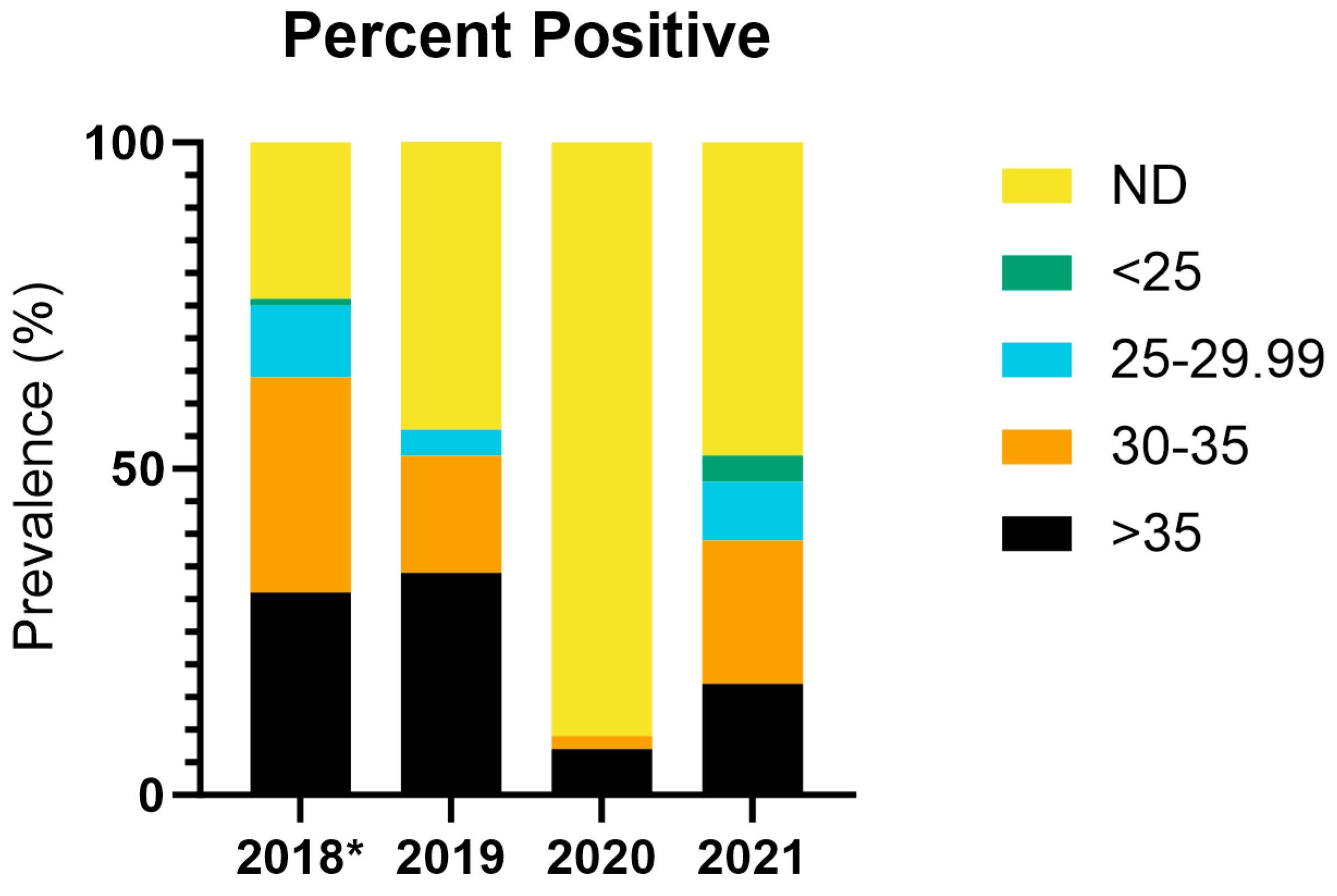
In 2018, 2019, and 2021, the proportion of gill clips positive for HPR0 at spawning ranged from 52.3% to 71.2% (Figure 2, Table 3). In 2020, the positive proportion was only 8.5% (Figure 2, Table 3). In comparison, in 2018, only one of the ovarian fluid samples (0.7%, n = 141) was positive for ISAV-HPR0. In 2019, one of the kidney samples (1.5%, n = 67) was positive and four were suspect (6%, n = 67). There were no positive kidney or ovarian samples from 2020, despite the sampling of 217 fish.

3.3. ISAV-HPR0 Monitoring Program: Full Facility

Between July of 2020 and September of 2021, a total of over 1250 non-spawning fish from various systems in the facility were sampled for ISAV (25 different samplings, Table 3). A minimum of 180 fish were sampled from each of the five year classes in the facility over that period. In addition, another 225 archived gill samples collected in 2019 and 2020 were tested. Detection of ISAV occurred in only one sampling period in early May 2021.
At that time, non-negative results were found in two of the YCs sampled. The sampling with the highest positive proportion involved fish from the 2019–2020 YC in the smolt room, while the other involved a single fish from the 2018–2019 YC in the on-grow room.
During the 6 May 2021 sampling event, 120 fish from year class 18–19 were sampled from the on-grow room. All the samples were negative except one. This sample was suspect, defined as having discordant results between two replicates for ISAV in the initial screening. Upon retesting the sample, ISAV was not detected. For the sampling event on 4 May 2021, 120 fish from YC 19–20 were sampled from the smolt room, with 57 fish being positive for ISAV-HPR0 and 15 being suspect. The fish in this tank were part of the selective breeding program and all were tagged with passive integrated transponders (PIT tags), allowing for the samples to be matched to individual fish. This same tank of fish was resampled twice, on 25 May 2021 (120 fish) and again on 10 August 2021 (115 fish). All the resampling results were negative. This included two of the fish with suspect results on May 4 that were indiscriminately resampled on both 25 May and 10 August, and a third that was resampled on 10 August only. From the 57 fish that tested positive on 4 May, six were resampled and tested negative on both subsequent dates. A further 13 fish that tested positive on the initial testing were resampled once on 10 August and also tested negative.
All the incoming water sources to the facility were tested once every two weeks over the course of a year and weekly during the fall and spring. In total, 144 water samples were tested, and ISAV was never detected in any of the samples. However, the method has been able to detect ISAV in water samples collected during spawning from a tank holding fish testing positive for ISAV, thereby demonstrating the method’s ability to detect ISAV in water.

4. Discussion

In 2016, the NCWMAC added RT-PCR screening of gill and heart tissue to meet new Canadian import requirements [36]. Following these new protocols, spawning fish at the NCWMAC facility were found to be positive for ISAV-HPR0 for the first time, despite routine screening of kidneys by virus isolation on chinook salmon embryo (CHSE-214) and Atlantic salmon kidney (ASK) cells lines and by RT-PCR on kidney tissue prior to 2016. ISAV-HPR0 in NCWMAC brood fish samples collected at the time of spawning has occurred annually since the initial 2016 ISAV detection in gill tissue. To date, there has only been a single occurrence event outside this window of sampling.
The ISAV-HPR0 occurrence window at the NCWMAC is of a short duration. The 2016 gill detection offered the opportunity to follow an exposed cohort over time. This group of fish, remaining in the same RS as those testing non-negative, repeatedly tested negative between three and nine months following the initial detection. It is possible that the 68 gill arch samples from the exposed cohort had never been infected with the virus. The high proportion of positives (26.7%) from the original group, however, counters this hypothesis. There was a 3-month window between when the positive fish were sampled on 8 November 2016, and the first sampling of the exposed cohort sampling on 15 February 2017. Consequently, a more likely explanation is that the exposed cohort had cleared the infection prior to sampling. Indeed, separate samplings in smolt detected ISAV in May of 2021 but were unable to detect the virus in the same cohort three weeks later.
ISAV-HPR0 has a known tissue tropism for gills [26], which was also seen in this study. However, as the gill is a surface tissue, the potential for environmental contamination for fish sharing a tank or anesthetic bath with another fish that is shedding virus can complicate the interpretation of the findings. We did not estimate prevalence values from the proportion of positives for this reason.
Smoltification is a period of physiological change for salmon that can be associated with suppression of the immune system [37]. Consequently, it was suspected this age class might be more susceptible to ISAV-HPR0 infection should this nonpathogenic virus occur within the hatchery as a chronic infection. Sampling in early May 2021 detected ISAV in 60% (Table 3) of the tested smolt population. However, as noted earlier, repeat sampling three weeks later failed to detect any signs of ISAV, even in individual fish that had tested positive in the earlier sampling. This population had also previously tested negative in February of 2021. Similarly, the initial detection in spawning fish in November of 2016 was not detected in the exposed cohort testing three months later, in February of 2017. These two findings suggest a short duration infection, which fits with the durations observed in experimental challenges using HPR-deleted ISAV [38] and with the short duration of ISAV-HPR0 infections observed in marine sites in the Faroes [4].
Our findings suggest that ISAV-HPR0 infections are either highly transient or are only detectable during short periods, in the order of weeks rather than multiple months (>4 months). This suggestion agrees with the hypothesis proposed by Nylund, Devold, Plarre, Isdal and Aarseth [14] that ISAV-HPR0 could spread horizontally across wild salmonid populations within a river system as a series of short-term infections, in much the same manner as influenza spreads within somewhat clustered human populations [39,40]. Alternatively, infections with ISAV-HPR0 may persist below our detection threshold for long periods of time and only rise above the threshold during brief outbreaks when environmental, host or pathogen conditions are favorable. This would be in congruence with the hypothesis that ISAV-HPR0 is maintained in broodstock facilities and spread to net pen farms at stocking [25,41,42]. Vertical transmission is a possibility and would be congruent with the consistent detection at spawning. There is evidence to support the potential of vertical transmission in ISAV [25,42,43,44,45]. However, there is evidence that vertical transmission does not occur as well, particularly in ISAV-HPR0 [20,46].
The recurrent detections of ISAV-HPR0 in spawning fish over a 5-year period, and the lack of a clear source for the repeated introduction, supports the possibility that the pathogen subsisted below the detection limits within the facility during this period. Over the three years of sampling, ISAV was only confirmed in another age class, smolting fish, once during the spring of 2021. A single non-negative finding in the on-grow system on the same sampling date as the smolt detections was never confirmed. The failure to detect the virus in other stages of fish or the incoming water sources suggests that the virus may have alternative pathways to remain in the environment. It is also possible the virus is capable of subsisting at extremely low infection rates, below normal monitoring detection limits, until the host and environmental condition are suitable for an outbreak, such as at smoltification or spawning.
These findings suggest (1) that ISAV-HPR0 infections can exist in hatchery populations without detections, and (2) that episodes of high prevalence of ISAV-HPR0 associated with spawning can be highly transient. In either case, conventional surveillance based on recurrent testing of healthy populations likely provides only a poor indication of the HPR0 status in hatchery settings. Targeting surveillance to periods of physiological change such as spawning and smoltification (which also precede common movement decisions) and adjusting the sample sizes to account for the likely surge in prevalence in those contexts should enhance the detection capacity while also reducing costs.
It is important to note the absence of HPR-deleted ISAV, despite recurrent findings of HPR0, in these populations. Consequently, the costs and benefits of enhanced sampling programs should be carefully assessed prior to their recommendation. It may be that targeting sampling to periods of physiologic change and/or implementing mitigations such as culling eggs from positive parents and disinfecting egg surfaces before incubation may provide sufficient protection against a form of the virus with otherwise rare consequence despite the potential for the virus to mutate to ISAV Δ [23,47].
As the salmonid industry expands to more land-based production and broodstock facilities, better understanding the surveillance capacity to detect and the consequence of occurrence of low-level ISAV-HPR0 infections is critical. Routine sampling to detect a pathogen that is highly transient may generate extensive costs without substantive benefits. The results of recurrent sampling for ISAV-HPR0 in the NCWMAC hatchery system suggest that the virus may evade routine sampling, even at high sample sizes. Consequently, targeting surveillance to life stages and periods of physiologic change may provide the greatest ability to detect and the greatest assurance of absence. Targeted sampling may also help determine how ISAV-HPR0 can evade elimination or circumvent established biosecurity and management practices. Not only will an improved understanding potentially lead to the eradication of ISAV-HPR0 from infected facilities but it will also help provide a better understanding of how to improve facility design, biosecurity, and management practices to minimize the risk of other potential pathogens in the growing recirculating aquaculture sector.

Supplementary Materials

The following supporting information can be downloaded at https://www.mdpi.com/article/10.3390/fishes9080325/s1, Table S1: Test results from the original detection and Table S2: GenBank accession numbers for the sequences.

Author Contributions

Conceptualization, M.P., J.W., L.G. and B.C.P.; methodology, M.P., J.W. and L.G.; formal analysis, M.P.; investigation, M.P. and J.W.; resources, M.P.; data curation, M.P.; writing—original draft preparation, M.P.; writing—review and editing, M.P, J.W., L.G. and B.C.P.; supervision, B.C.P.; project administration, J.W. and B.C.P.; funding acquisition, J.W. and B.C.P. All authors have read and agreed to the published version of the manuscript.

Funding

This project was funded by the USDA-ARS NCWMAC with funds from CRIS project number: 8030-31000-005-000D and funding from USDA-APHIS under interagency agreement 20-9419-0554IA.

Institutional Review Board Statement

The animal study protocol was approved by the Institutional Animal Care and Use Committee of the National Cold Water Marine Aquaculture Center (protocol #: 2020-01, approved on 22 June 2020).

Data Availability Statement

The data presented in this study are available on request from the corresponding author.

Acknowledgments

The authors would like to acknowledge Mark Polinski for his efforts in reviewing the manuscript and providing suggestions for improvement. The findings and conclusions in this publication are those of the authors and should not be construed to represent any official USDA or U.S. government determination or policy. Mention of trade names or commercial products in this publication is solely for the purpose of providing specific information and does not imply recommendation or endorsement by the U.S. Department of Agriculture. The USDA is an equal opportunity provider and employer.

Conflicts of Interest

The authors declare no conflicts of interest. The funders had no role in the design of the study; in the collection, analyses, or interpretation of data; in the writing of the manuscript; or in the decision to publish the results.

References

  1. Thorud, K.E.; Djupvik, H.O. Infectious anaemia in Atlantic salmon (Salmo salar L.). Bull. Eur. Assoc. Fish Pathol. 1988, 8, 109–111. [Google Scholar]
  2. Bouchard, D.; Keleher, W.; Opitz, H.M.; Blake, S.; Edwards, K.C.; Nicholson, B.L. Isolation of infectious salmon anemia virus (ISAV) from Atlantic salmon in New Brunswick, Canada. Dis. Aquat. Organ. 1999, 35, 131–137. [Google Scholar] [CrossRef]
  3. Bouchard, D.A.; Brockway, K.; Giray, C.; Keleher, W.; Merrill, P.L. First report of Infectious Salmon Anemia (ISA) in the United States. Bull. Eur. Assoc. Fish Pathol. 2001, 21, 86–88. [Google Scholar]
  4. Christiansen, D.H.; Østergaard, P.S.; Snow, M.; Dale, O.B.; Falk, K. A low-pathogenic variant of infectious salmon anemia virus (ISAV-HPR0) is highly prevalent and causes a non-clinical transient infection in farmed Atlantic salmon (Salmo salar L.) in the Faroe Islands. J. Gen. Virol. 2011, 92, 909–918. [Google Scholar] [CrossRef] [PubMed]
  5. Kibenge, F.S.; Garate, O.N.; Johnson, G.; Arriagada, R.; Kibenge, M.J.; Wadowska, D. Isolation and identification of infectious salmon anaemia virus (ISAV) from Coho salmon in Chile. Dis. Aquat. Organ. 2001, 45, 9–18. [Google Scholar] [CrossRef]
  6. Lovely, J.E.; Dannevig, B.H.; Falk, K.; Hutchin, L.; MacKinnon, A.M.; Melville, K.J.; Rimstad, E.; Griffiths, S.G. First identification of infectious salmon anaemia virus in North America with haemorrhagic kidney syndrome. Dis. Aquat. Organ. 1999, 35, 145–148. [Google Scholar] [CrossRef]
  7. Mullins, J.E.; Groman, D.; Wadowska, D. Infectious salmon anaemia in salt water Atlantic salmon (Salmo salar L.) in New Brunswick, Canada. Bull. Eur. Assoc. Fish Pathol. 1998, 18, 110–114. [Google Scholar]
  8. Rodger, H.D.; Turnbull, T.; Muir, F.; Millar, S.; Richards, R.H. Infectious salmon anaemia (ISA) in the United Kingdom. Bull. Eur. Assoc. Fish Pathol. 1998, 18, 115–116. [Google Scholar]
  9. Rowley, H.M.; Campbell, S.J.; Curran, W.L.; Turnbull, T.; Bryson, D.G. Isolation of infectious salmon anaemia virus (ISAV) from Scottish farmed Atlantic salmon, Salmo salar L. J. Fish Dis. 1999, 22, 483–487. [Google Scholar] [CrossRef]
  10. Anonymous. ISA hits the Faroes. Fish Farm. Int. 2000, 27, 47. [Google Scholar]
  11. Evensen, Ø.; Thorud, K.E.; Olsen, Y.A. A morphological study of the gross and light microscopic lesions of infectious anaemia in Atlantic salmon (Salmo salar). Res. Vet. Sci. 1991, 51, 215–222. [Google Scholar] [CrossRef] [PubMed]
  12. Fourrier, M.; Lester, K.; Markussen, T.; Falk, K.; Secombes, C.J.; McBeath, A.; Collet, B. Dual Mutation Events in the Haemagglutinin-Esterase and Fusion Protein from an Infectious Salmon Anaemia Virus HPR0 Genotype Promote Viral Fusion and Activation by an Ubiquitous Host Protease. PLoS ONE 2015, 10, e0142020. [Google Scholar] [CrossRef]
  13. Devold, M.; Falk, K.; Dale, B.; Krossoy, B.; Biering, E.; Aspehaug, V.; Nilsen, F.; Nylund, A. Strain variation, based on the hemagglutinin gene, in Norwegian ISA virus isolates collected from 1987 to 2001: Indications of recombination. Dis. Aquat. Org. 2001, 47, 119–128. [Google Scholar] [CrossRef]
  14. Nylund, A.; Devold, M.; Plarre, H.; Isdal, E.; Aarseth, M. Emergence and maintenance of infectious salmon anaemia virus (ISAV) in Europe: A new hypothesis. Dis. Aquat. Org. 2003, 56, 11–24. [Google Scholar] [CrossRef]
  15. Cunningham, C.O.; Gregory, A.; Black, J.; Simpson, I.; Raynard, R.S. A novel variant of the infectious salmon anaemia virus (ISAV) haemagglutinin gene suggests mechanisms for virus diversity. Bull. Eur. Assoc. Fish Pathol. 2002, 22, 366–374. [Google Scholar]
  16. Markussen, T.; Sindre, H.; Jonassen, C.M.; Tengs, T.; Kristoffersen, A.B.; Ramsell, J.; Numanovic, S.; Hjortaas, M.J.; Christiansen, D.H.; Dale, O.B.; et al. Ultra-deep pyrosequencing of partial surface protein genes from infectious Salmon Anaemia virus (ISAV) suggest novel mechanisms involved in transition to virulence. PLoS ONE 2013, 8, e81571. [Google Scholar] [CrossRef] [PubMed]
  17. Mjaaland, S.; Hungnes, O.; Teig, A.; Dannevig, B.H.; Thorud, K.; Rimstad, E. Polymorphism in the infectious salmon anemia virus hemagglutinin gene: Importance and possible implications for evolution and ecology of infectious salmon anemia disease. Virology 2002, 304, 379–391. [Google Scholar] [CrossRef]
  18. Cardenas, M.; Galleguillos, C.; Acevedo, K.; Ananias, C.; Alarcon, J.; Michelson, S.; Toledo, J.; Montoya, M.; Meneses, C.; Castro-Nallar, E.; et al. Rapid sequence modification in the highly polymorphic region (HPR) of the hemagglutinin gene of the infectious salmon anaemia virus (ISAV) suggests intra-segmental template switching recombination. J. Fish Dis. 2020, 43, 1483–1496. [Google Scholar] [CrossRef]
  19. Kulshreshtha, V.; Kibenge, M.; Salonius, K.; Simard, N.; Riveroll, A.; Kibenge, F. Identification of the 3′ and 5′ terminal sequences of the 8 rna genome segments of European and North American genotypes of infectious salmon anemia virus (an orthomyxovirus) and evidence for quasispecies based on the non-coding sequences of transcripts. Virol. J. 2010, 7, 338. [Google Scholar] [CrossRef]
  20. Christiansen, D.H.; Petersen, P.E.; Dahl, M.M.; Vest, N.; Aamelfot, M.; Kristoffersen, A.B.; Jansen, M.D.; Matejusova, I.; Gallagher, M.D.; Jonsson, G.; et al. No Evidence of the Vertical Transmission of Non-Virulent Infectious Salmon Anaemia Virus (ISAV-HPR0) in Farmed Atlantic Salmon. Viruses 2021, 13, 2428. [Google Scholar] [CrossRef] [PubMed]
  21. Lyngstad, T.M.; Kristoffersen, A.B.; Hjortaas, M.J.; Devold, M.; Aspehaug, V.; Larssen, R.B.; Jansen, P.A. Low virulent infectious salmon anaemia virus (ISAV-HPR0) is prevalent and geographically structured in Norwegian salmon farming. Dis. Aquat. Org. 2012, 101, 197–206. [Google Scholar] [CrossRef]
  22. Madhun, A.S.; Maehle, S.; Wennevik, V.; Karlsbakk, E. Prevalence and genotypes of infectious salmon anaemia virus (ISAV) in returning wild Atlantic salmon (Salmo salar L.) in northern Norway. J. Fish Dis. 2019, 42, 1217–1221. [Google Scholar] [CrossRef]
  23. Christiansen, D.H.; McBeath, A.J.A.; Aamelfot, M.; Matejusova, I.; Fourrier, M.; White, P.; Petersen, P.E.; Falk, K. First field evidence of the evolution from a non-virulent HPR0 to a virulent HPR-deleted infectious salmon anaemia virus. J. Gen. Virol. 2017, 98, 595–606. [Google Scholar] [CrossRef]
  24. Gustafson, L.; Remmenga, M.; Sandoval Del Valle, O.; Ibarra, R.; Antognoli, M.; Gallardo, A.; Rosenfeld, C.; Doddis, J.; Enriquez Sais, R.; Bell, E.; et al. Area contact networks and the spatio-temporal spread of infectious salmon anemia virus (ISAV) in Chile. Prev. Vet. Med. 2016, 125, 135–146. [Google Scholar] [CrossRef] [PubMed]
  25. Nylund, A.; Brattespe, J.; Plarre, H.; Kambestad, M.; Karlsen, M. Wild and farmed salmon (Salmo salar) as reservoirs for infectious salmon anaemia virus, and the importance of horizontal- and vertical transmission. PLoS ONE 2019, 14, e0215478. [Google Scholar] [CrossRef]
  26. Aamelfot, M.; Christiansen, D.H.; Dale, O.B.; McBeath, A.; Benestad, S.L.; Falk, K. Localised Infection of Atlantic Salmon Epithelial Cells by HPR0 Infectious Salmon Anaemia Virus. PLoS ONE 2016, 11, e0151723. [Google Scholar] [CrossRef]
  27. Aamelfot, M.; Dale, O.B.; Falk, K. Infectious salmon anaemia—Pathogenesis and tropism. J. Fish Dis. 2014, 37, 291–307. [Google Scholar] [CrossRef] [PubMed]
  28. Aamelfot, M.; Dale, O.B.; McBeath, A.; Falk, K. Host tropism of infectious salmon anaemia virus in marine and freshwater fish species. J. Fish Dis. 2015, 38, 687–694. [Google Scholar] [CrossRef] [PubMed]
  29. Aamelfot, M.; Dale, O.B.; Weli, S.C.; Koppang, E.O.; Falk, K. Expression of the Infectious Salmon Anemia Virus Receptor on Atlantic Salmon Endothelial Cells Correlates with the Cell Tropism of the Virus. J. Virol. 2012, 86, 10571–10578. [Google Scholar] [CrossRef]
  30. Fosse, J.H.; Aamelfot, M.; Sønstevold, T.; Weli, S.C.; Vendramin, N.; Petersen, P.E.; Solhaug, A.; Amundsen, M.M.; Heffernan, I.A.; Cuenca, A.; et al. Salmon Erythrocytes Sequester Active Virus Particles in Infectious Salmon Anaemia. Viruses 2022, 14, 310. [Google Scholar] [CrossRef] [PubMed]
  31. Maine, S.o. Importation of Live Marine Organisms. Chapter 24. Available online: https://www.maine.gov/dmr/rules-enforcement/regulations-rules (accessed on 1 February 2022).
  32. Wolters, W.; Masters, A.; Vinci, B.; Summerfelt, S. Design, loading, and water quality in recirculating systems for Atlantic Salmon (Salmo salar) at the USDA ARS National Cold Water Marine Aquaculture Center (Franklin, Maine). Aquacult. Eng. 2009, 41, 60–70. [Google Scholar] [CrossRef]
  33. Gustafson, L.L.; Creekmore, L.H.; Snekvik, K.R.; Ferguson, J.A.; Warg, J.V.; Blair, M.; Meyers, T.R.; Stewart, B.; Warheit, K.I.; Kerwin, J.; et al. A systematic surveillance programme for infectious salmon anaemia virus supports its absence in the Pacific Northwest of the United States. J. Fish Dis. 2018, 41, 337–346. [Google Scholar] [CrossRef]
  34. Snow, M.; McKay, P.; McBeath, A.J.; Black, J.; Doig, F.; Kerr, R.; Cunningham, C.O.; Nylund, A.; Devold, M. Development, application and validation of a Taqman real-time RT-PCR assay for the detection of infectious salmon anaemia virus (ISAV) in Atlantic salmon (Salmo salar). Dev. Biol. 2006, 126, 133–145; discussion 325–326. [Google Scholar]
  35. Vike, S.; Oelckers, K.; Duesund, H.; Erga, S.R.; Gonzalez, J.; Hamre, B.; Frette, O.; Nylund, A. Infectious salmon anemia (ISA) virus: Infectivity in seawater under different physical conditions. J. Aquat. Anim. Health 2014, 26, 33–42. [Google Scholar] [CrossRef] [PubMed]
  36. Government of Canada. Test Selection and Sampling Requirements for Aquatic Animal Imports. 2021. Available online: https://inspection.canada.ca/animal-health/aquatic-animals/imports/test-selection-and-sampling-requirements/eng/1548710570282/1548710570578 (accessed on 1 February 2022).
  37. Johansson, L.-H.; Timmerhaus, G.; Afanasyev, S.; Jørgensen, S.M.; Krasnov, A. Smoltification and seawater transfer of Atlantic salmon (Salmo salar L.) is associated with systemic repression of the immune transcriptome. Fish Shellfish. Immunol. 2016, 58, 33–41. [Google Scholar] [CrossRef] [PubMed]
  38. Raynard, R.S.; Snow, M.; Bruno, D.W. Experimental infection models and susceptibility of Atlantic salmon Salmo salar to a Scottish isolate of infectious salmon anaemia virus. Dis. Aquat. Org. 2001, 47, 169–174. [Google Scholar] [CrossRef] [PubMed]
  39. Charu, V.; Zeger, S.; Gog, J.; Bjornstad, O.N.; Kissler, S.; Simonsen, L.; Grenfell, B.T.; Viboud, C. Human mobility and the spatial transmission of influenza in the United States. PLoS Comput. Biol. 2017, 13, e1005382. [Google Scholar] [CrossRef] [PubMed]
  40. Viboud, C.; Bjornstad, O.N.; Smith, D.L.; Simonsen, L.; Miller, M.A.; Grenfell, B.T. Synchrony, waves, and spatial hierarchies in the spread of influenza. Science 2006, 312, 447–451. [Google Scholar] [CrossRef]
  41. Nylund, A.; Plarre, H.; Karlsen, M.; Fridell, F.; Ottem, K.F.; Bratland, A.; Saether, P.A. Transmission of infectious salmon anaemia virus (ISAV) in farmed populations of Atlantic salmon (Salmo salar). Arch. Virol. 2007, 152, 151–179. [Google Scholar] [CrossRef] [PubMed]
  42. Plarre, H.; Nylund, A.; Karlsen, M.; Brevik, O.; Saether, P.A.; Vike, S. Evolution of infectious salmon anaemia virus (ISA virus). Arch. Virol. 2012, 157, 2309–2326. [Google Scholar] [CrossRef]
  43. Skotheim, S.A. Co-infection with Norwegian Salmonid Alphavirus (NSAV) and Infectious Pancreatic Necrosis Virus (IPNV) in Chinook Salmon Embryo Cells (CHSE-214). Master Thesis, University of Bergen, Bergen, Norway, 2009. [Google Scholar]
  44. Marshall, S.H.; Ramírez, R.; Labra, A.; Carmona, M.; Muñoz, C. Bona Fide Evidence for Natural Vertical Transmission of Infectious Salmon Anemia Virus in Freshwater Brood Stocks of Farmed Atlantic Salmon (Salmo salar) in Southern Chile. J. Virol. 2014, 88, 6012–6018. [Google Scholar] [CrossRef]
  45. Vike, S.; Nylund, S.; Nylund, A. ISA virus in Chile: Evidence of vertical transmission. Arch. Virol. 2009, 154, 1–8. [Google Scholar] [CrossRef]
  46. Melville, K.J.; Griffiths, S.G. Absence of vertical transmission of infectious salmon anemia virus (ISAV) from individually infected Atlantic salmon Salmo salar. Dis. Aquat. Org. 1999, 38, 231–234. [Google Scholar] [CrossRef]
  47. Sommerset, I.; Wiik-Nielsen, J.; Moldal, T.; Oliveira, V.H.S.; Svendsen, J.C.; Haukaas, A.; Brun, E. Norwegian Fish Health Report 2023; Norwegian Veterinary Institute: Oslo, Norway, 2024. [Google Scholar]
Figure 1. Overview of the NCWMAC breeding program. Fertilized eggs start in incubation and then move to the parr system for year 1. In February of year 2, fish are moved to the smolt systems until Jan of year 3. They are then moved to on-grow until October of year 3, when they are finally moved to one of the three systems in the brood room until spawning in November–December of year 4. Fish leave the facility at two points indicated by dashed lines. The first is early summer of year 2, when a group goes for grow-out in marine net pens, and in November/December of year 4, when a portion of the eggs are transferred to industry. Fish do not re-enter the facility once they have left.
Fishes 09 00325 g001
Figure 2. Percent of gill samples testing positive for ISAV at spawning in 2018 through 2021. Results are grouped by CT value (legend). The * indicates that the reported CT values for 2018 do not include all the samples tested. A total of 27 samples from 2018 were only reported as not detected (n = 17), suspect (n = 1), or positive (n = 9) with no associated CT values from the contracted lab. These 27 samples were not included in the data reported in the figure.
Fishes 09 00325 g002
Table 1. Primers used for the detection of ISAV.
TargetPrimer IDSequenceReference
ISAV Seg8-F5′ CTACACAGCAGGATGCAGATGT 3′[34]
Segment 8Seg8-R5′ CAGGATGCCGGAAGTCGAT 3′
Seg8-P5′ (6-FAM) CATCGTCGCTGCAGTTC (MGB-NFG) 3′
European 872 EU5′ GCT GCT TCG TGT GAA TAT GAC 3′Warg
Segment 61118EU5′ TTC CAA CCT GCT AGG AAC 3′Unpub.
Table 2. The number of fish and the sample type collected for the exposed cohort study in 2017. On the final date, all the sample types were collected from 5 fish. From the remaining 33 fish in the population, only frozen gill arches were collected. ISAV was not detected in any of the samples listed below.
FixativeTissue15 Feb12 Apr26 May29 Jun19 Jul14 Aug
RNA LaterBlood1055555
Gill1055555
Heart1055555
Kidney1055555
Liver 55555
Spleen 55555
FrozenGill Arch10555538
Heart 55555
Kidney 55555
Liver 55555
Spleen 55555
Table 3. Timeline of the sample collection. The data listed are the total # of fish sampled/# of non-negative fish/room the fish were in when sampled. Cells with a light gray background are samples taken at spawning. Water indicates the number of weekly sampling events that occurred that month.
2018201920202021
YCNov.Dec.Apr.MayJul.Sep.Nov.Dec.MayJul.Aug.Sep.Oct.Nov.Dec.Jan.Feb.Mar.Apr.May-earlyMay-lateJun.Jul.Aug.Sep.Nov.
14–15174/133/B47/10/B
15–16 133/75/B4/1/B
16–17 30/0/G 30/0/B 180/0/B 218/23/B33/0/B
17–18 30/0/S60/0/S 90/0/B 90/0/B308/151/B
18–19 30/0/P30/0/P 15/0/S 60/0/G 120/1/G
19–20 60/0/P–S 120/72/S120/0/S 115/0/S
115/0/B2
20–21 180/0/P
Water 12345322244 41
B = brood, B2 = building 2, G = on-grow, P = parr, P-S = parr to smolt transfer, S = smolt.
Disclaimer/Publisher’s Note: The statements, opinions and data contained in all publications are solely those of the individual author(s) and contributor(s) and not of MDPI and/or the editor(s). MDPI and/or the editor(s) disclaim responsibility for any injury to people or property resulting from any ideas, methods, instructions or products referred to in the content.