Article

Laser Fabrication of Gold–sp-Carbon Films

1
School of Science, Westlake University, 18 Shilongshan Road, Hangzhou 310024, China
2
Institute of Natural Sciences, Westlake Institute for Advanced Study, 18 Shilongshan Road, Hangzhou 310024, China
3
Moscow Institute for Physics and Technology, Universitetskii Avenue 9, 141701 Dolgoprudny, Russia
4
Department of Physics and Applied Mathematics, Vladimir State University Named after AG and NG Stoletovs, Gor’kogo Street 87, 600000 Vladimir, Russia
5
Karelian Research Centre of RAS, Pushkinskaya Street 11, 185910 Petrozavodsk, Russia
6
Natural Sciences Center of General Physics Institute, Prokhorov General Physics Institute RAS, Leninskii Avenue 53, Building 19, 119991 Moscow, Russia
*
Authors to whom correspondence should be addressed.
Condens. Matter 2023, 8(4), 96; https://doi.org/10.3390/condmat8040096
Submission received: 16 August 2023 / Revised: 3 November 2023 / Accepted: 7 November 2023 / Published: 14 November 2023

Abstract

:
We develop a method for the laser synthesis and deposition of carbon–gold films formed by a net of linear sp-carbon chains and stabilized by gold nanoparticles. The originality of the method is in the simultaneous production of carbon chains and gold nanoparticles due to the laser fragmentation of the amorphous carbon and hydrogen tetrachloroaurate (III) or chloroauric acid. We study how surface resistivity alters the effect of the obtained films via the illumination in the visible spectral range.

1. Introduction

Carbon has established itself as one of the most promising materials for a number of nanotechnology applications. The variety of allotropic forms of carbon allows the creation of a wide range of nanostructures that possess unique optical and electronic properties [1]. Over the past decade, many research papers have been dedicated to the synthesis and fabrication of sp-hybridized carbon structures [2]. The nanoscience community pays special attention to one-dimensional sp-carbon due to its unusual band structure, leading to its ability to emit and absorb visible light. Moreover, carbyne is often considered as the thinnest possible conducting wire [3,4,5].
The main focus of this work is on the synthesis of stable macroscopically long linear carbon chains [6].
We are aware of a number of previous attempts to achieve this goal. Among the methods employed are the stabilization of linear chains due to their confinement in double-wall carbon nanotubes [7], the low temperature of pyrolise [8], PLAL (Pulsed Laser-Ablation in Liquids) [9] solid phase polymerization [10], etc. Still, the technology and synthesis of free-standing macroscopically long linear sp-carbon chains is far from being developed.
In this context, the experience of the synthesis of amorphous carbon (a-C) or tetrahedral amorphous-carbon (ta-C) stable films may be instructive. The control of the chain length and types of interatomic bonds in linear chains is essential for controlling the electric resistance of fabricated films [11]. Their electronic properties can also be optimized using optical irradiation [12]. In previous works, we developed the method of the synthesis and fabrication of linear carbon chains via the laser fragmentation of colloidal systems [13,14], resulting in the stabilization of linear sp-chains by gold anchors attached to their ends [15].
In the present study, we report an advanced method for the creation of linear carbon chains via the irradiation of a mixture of amorphous carbon and hydrogen tetrachloroaurate (III) or chloroauric acid. We use short laser pulses with a frequency corresponding to the absorption band of gold. We show that because of the simultaneous synthesis of gold nanoparticles and linear carbon chains due to laser irradiation, three-dimensional carbon nets with embedded gold nanoparticles can be formed. The deposition of these nets realized by the sputtering of a colloid allows for the formation of stable sp-carbon films formed by randomly oriented carbon chains and stabilized by gold nanoparticles [16]. The surface resistivity of deposited gold-carbon films is strongly sensitive to the wavelength and intensity of the external optical excitation. This method enables the optically controlled synthesis of conducting sp-carbon films that are suitable for optical sensor applications.

2. Experimental Section

2.1. Materials and Reagents

We used shungite carbon nanoparticles (ShC) distributed in the volume of distilled water (DI) at 4 mL (0.1 mg/mL) as a carbon precursor for linear chain formation. The ShC sample was provided by the NR team (Karelian Research Centre of RAS, Petrozavodsk, Russia) [17]. Water solutions of hydrogen tetrachloroaurate (III) or chloroauric acid (which was produced at the MOSMET (Moscow, Russia) in the form of yellow crystalline powder (with ~75% water content) for the synthesis of Au nanoparticles (AuNP) of 1 mL were obtained. Finally, we used shungite suspensions in aqueous solutions of hydrochloroauric acid (HAuCl4) as the starting samples. Ultrapure deionized water (resistivity of 18.2 MΩ/cm at 25 °C) was used as a solvent taken from a Milli-Q water purification system produced by Merck Millipore (Burlington, VT, USA). The initial water solution (before laser irradiation) of hydrochloroauric acid (HAuCl4) at a concentration of 5 × 10⁻⁴ mol/L was colorless and transparent. Glass coverslips of 50 × 50 × 0.5 mm, thoroughly cleaned in ethanol and acetone, were used as substrates for the deposition of carbon–Au complexes for optical study while a dielectric textolite was used for the electroconductivity study. We also used a 400-mesh cupper grid 01885-F produced by TED PELLA Inc. (Redding, CA, USA) as a substrate for high-resolution transmission electron microscopy (TEM) measurements. We sourced TEM with Titan3 model made by FEI company (Hillsboro, OR, USA).

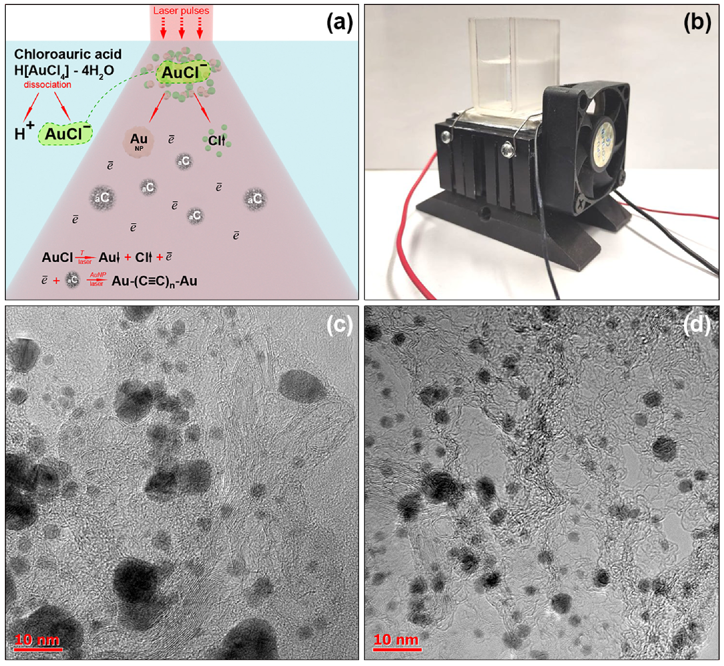
2.2. Linear Chain Formation Method

For the laser processing of colloidal systems, we designed and developed an experimental setup containing a Peltier element produced by Cryotherm (Moscow, Russia) that allowed us to maintain the temperature of a cuvette at the level of 50–55 °C. The temperature was measured using a thermocouple. As we used the Ytterbium (Yb) fiber laser source made by (Zhejiang, China) with a nanosecond pulse duration and irradiation size area of about 50 μm (the volume scanning regime with the speed of 50 μm/s), we were confident that significant temperature variations affecting the entire volume of liquid would not occur. This was essential for the disintegration and avoidance of synthesized linear carbon chains due to overheating during the irradiation of the cuvette containing a colloid using laser pulses [18]. For the initialization of chemical transformations, we used the laser source characterized by a wavelength of 532 nm, a pulse duration of 20 ns, and an average power of up to 10 W. The scheme of the employed laser fabrication process and its results are summarized in Figure 1.
The control time of laser processing is an important tool for the optimization and fragmentation of the amorphous carbon and chloroauric acid in a colloid. We found that the highest ratio of linear carbon chains fraction to the rest of the solution was achieved after 60 min of laser irradiation. At shorter times, the colloidal solution can be characterized by a significant fraction of the amorphous carbon and a great dispersion in the sizes of gold nanoparticles. Furthermore, long time laser irradiation leads to the total dissociation of initial components. The variation in the laser pulse energy also affects the speed and formation of the linear chains; however, the range of such a variation is limited. At its lower limit of about 0.1 mJ, the formation of gold nanoparticles is suppressed, while at its upper limit of about 1 mJ, the graphene flakes dissociate into an atomic plasma. That is why we had to use the laser source in the range of 0.1–1 mJ, which corresponds to the average power raging from 12.7 to 127.4 kW/cm2.
Hydrogen tetrachloroaurate (III) is a strong electrolyte that completely dissociates in a water solution (see scheme in Figure 1a). The periodic laser pulse action leads to the transfer of energy to the dispersive phase of the colloidal solution. On the other hand, the Peltier element provides cooling for the transparent cuvette bottom. Altogether, it provides a fluid circulation due to the temperature gradient inside the cuvette. Under a laser field action, the condition is achieved for Au reduction in the peripheral zone. The energy parameters of the laser pulses form a medium for metal sedimentation in the shape of quasi-spherical nanoparticles of an average diameter of about 10 nm (see TEM images in Figure 1c,d).
In the conditions of our experiments, the maximum temperature Tmax was achieved due to the irradiation of the colloid by a single laser pulse that does not exceed the carbon sublimation temperature (~5000 K). In this case, the energy that was absorbed by shungite particles was sufficient to provide their partial fragmentation without destroying the linear interatomic bonds. The synthesis of sp-chains is a challenging process because the linear carbon allotrope is unstable, and it undergoes transitions into other allotropic forms once the critical sizes of the corresponding seeds are achieved. Starting with 5 atoms, the linear chain is unstable, and it needs to be artificially stabilized. Au nanoparticles here play a role in the catalyst for carbon growth. They form mechanical anchors at the chains’ end (see Figure 1c,d). It is energetically advantageous for sp-carbon to form a single-electron bond with AuNp that prevents the folding and bending of the chains.

2.3. Apparatus

We studied the microstructure of the films formed by carbyne chains and stabilized with gold nanoparticles (Au NPs) by high-resolution transmission electron microscopy and scanning electron microscopy SEM using FEI Titan3 model made by FEI company (Hillsboro, OR, USA) with a spatial resolution of up to 2 Å and Quanta 200 3D model made by FEI company (Hillsboro, OR, USA). To investigate the optical properties of the carbon–Au complexes, we deposited droplets of the colloid on a substrate and dried them at room temperature.
The carbon phase configuration was investigated with the use of the Bruker Raman Senterra spectrometer (Billerica, MA, USA) at the excitation wavelength of 532 nm. The accumulation time was 10 s, and each spectrum was a result of averaging over 9 measurements. All the studies of liquid solutions were realized with the use of fused quartz cuvettes. The absorption spectra were measured using a spectrophotometer PerkinElmer (Shelton, CT, USA) Lambda 1050 equipped with the 150 mm integrating sphere.

3. Results and Discussion

3.1. Characterization of Raman Spectra

Comparing the results of Refs. [13,14] obtained with the use of a 1.06 µm laser with the results of the present study, we conclude that the formation mechanism of linear chains is different. The present approach to the formation of Au NPs implies that the irradiation of the solution is conducted via laser light, and the formation of gold NPs is simultaneous with the fragmentation of ShC. In the first stage, the disintegration of chloroauric acid resulted in the separation of gold atoms, forming clusters over time. In the second stage, these clusters interact with the carbon amorphous matrix and trigger its splitting into linear chains. This technique does not allow the formation of individual freestanding carbon chains; however, it is very efficient for the formation of thin films composed of a multitude of linear chains. An important advantage of the present method compared to the previous one is the very efficient fragmentation of amorphous carbon that leads to a dramatic decrease in the amorphous fraction due to the laser fragmentation, as confirmed by the Raman spectra (Figure 2).
The initial system characterized by the spectrum and shown by a red curve contains only ShC carbon peculiarities. It features two pronounced peaks, namely, D (n = 1335 cm−1) [19,20] and the second one (n = 1632 cm−1), respectively, which, in carbon, is a superposition of two main peaks as follows: 1615 and 1635 cm−1. Commonly, an additional shoulder at ~1620 cm−1 was observed in disordered graphite materials. This was attributed to the splitting of the degenerate E2g peak and to the breakdown of Raman selection rules, allowing a contribution from non-zone-center phonons. [15,16]. The broadening of the peaks at these frequencies is typical for the breathing modes C=C that are usually present in the amorphous carbon in the frequency range of 1330–1350 cm−1 [21,22] and are also characteristic of the effect of stretching C=C modes in a disordered graphite [23]. It is well known that amorphous sp2 carbon exhibits vibrational features resulting in the overlap of the two main bands, G and D [24,25], and the formation of C=O bonds [8], which is typical of colloidal solutions of ShC. Here, we clearly see the degradation of the D-peak with the exposure time, indicating the phase transformation of the amorphous carbon [17].
Furthermore, during the first 30 min of laser action, we observed the formation of the superposition of D and G peaks known in the literature as the D+G peak [17], with its maximum at 1632 nm, as shown by the blue line in Figure 2. This peak is typical of transpolyacetilens [26], and it reflects the vibration of a single bond in a polyyne chain in the absence of any variation in triple bonds [27]. We assumed that the scissor deformation vibrations of water played a role here. The shift in the diapason from the frequency of 1585 cm−1 up to 1627–1635 cm−1 is associated with free water molecules and indicates the hydrated state of water. The hydroxyl group can attach to chain defects. However, according to the TEM data in Figure 1, there are plenty of carbon threads that have conclusive traces in Raman spectra, and based on previous studies, the hydroxyl group does not lead to a loss in the stability of linear chains.
In the resulting spectra, we attribute the presence of a bordered peak at about 815 cm−1 to the formation of single electron bonds between Au NPs and the carbon matrix. It is noticeable that the lengths of this type of bond exceed those of single C-C pairs.
The second broad peak is centered at 2110 cm−1 and corresponds to the formation of short polyyne chains with vibrating triple bonds [28]. The frequency range of 1900–2300 cm−1 is usually ascribed to the formation of alternating –C≡C– bonds in chains of 2 to 10 atoms [29]. It is important to note that this Raman signal range is formed due to the multiphonon interaction that also leads to the oscillating intensity of the signal [11]. The enhancement of the laser action time to 60 min (black curve in Figure 2) leads to the formation of the material, which is much more homogeneous. We believe that the resonant excitation of plasmons in Au NPs via laser pulses heats these nanoparticles so that they easily release free electrons. These photoelectrons produce an additional impact on the amorphous carbon matrix and help its further disintegration. The newly formed linear sp-carbon chains attract each other by means of the van der Waals interaction and form stable one-dimensional crystals.

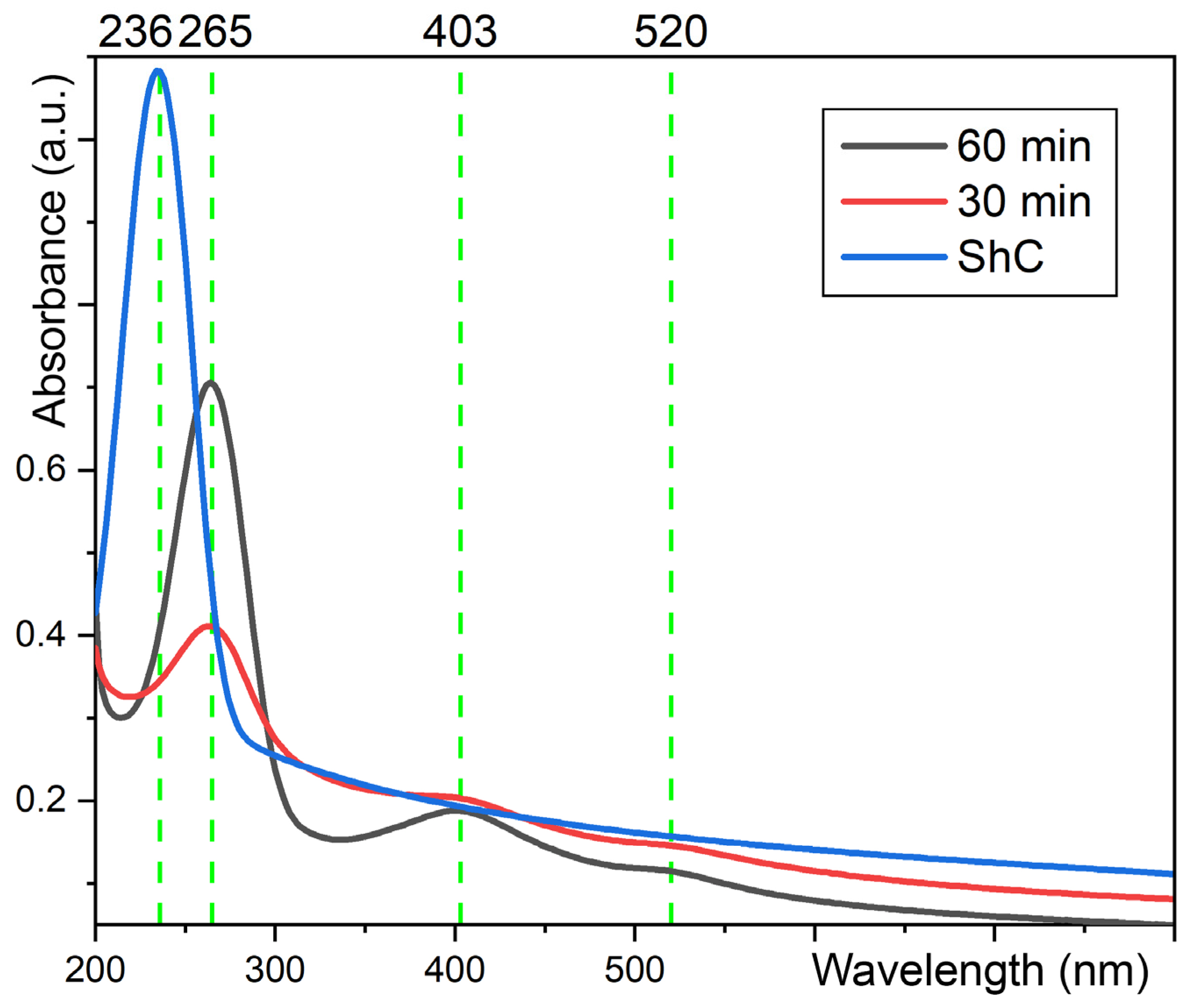
3.2. The Optical Absorption of the Carbyne Bundles Stabilized by Au NPs

One of the main problems of the deposition of the sp-carbon fraction process is a loss of stability. This typically leads the allotropic form to transition containing sp2-sp bonds [18]. To prevent this, we used the method of sputtering colloidal systems in the presence of a constant electric field [30].
The resulting absorption spectra (please see a black curve in Figure 3) contain the following three important bands: a series of pronounced peaks of about 230–265 nm and two broadened bands at 403 nm and 520 nm. To associate them with the different vibrational modes of the multielement system under study, we compared the spectra of deposited films at different stages of laser processing.
Namely, we compared the spectra of an initial shungite precursor inscribed with a blue line in Figure 3 with the spectra of the solution after 30 and 60 min of laser processing, indicated by red and blue curves in Figure 3, accordingly. We attribute the presence of singularities of peaks at 265 and 403 nm in the resulting spectra (shown by black lines) to the formation of carbon chains. The deposited sp-chains of a 10–12 atom length dominate the absorption band of 265 nm [31], and the presence of carbon chains with a length of more than 40 atoms is documented by the local maximum of 403 nm. The initial absorption spectra of amorphous carbon particles are responsible for the peak at 236 nm, which degrades rapidly after laser exposure starts. The evolution of the absorption spectrum with respect to the initial system shows that the number of linear chains in the solution increases as the exposure time increases from 30 to 60 min. From the slope of the curve in the range of 400–550 nm, we can deduce that the film contains longer carbon chains [32]. In addition, the presence of gold nanoparticles can be determined most reliably due to enhanced absorption at the plasmon resonance wavelength of 520 nm. The irradiation of a laser for 30 min results in the weak separation of absorption maxima at the wavelengths of 265 and 403 nm. This increase in the exposure time results in an increase in the intensity of the peak of 265 nm, which corresponds to the absorbance of short carbon chains (up to 12 atoms). This peak dramatically increases with the exposure time becoming twice as high. At the same time, the peak of 403 nm becomes more pronounced. These peaks were not observed during the irradiation of the water solution of chloroauric acid and shungite carbon nanoparticles (ShC). For these reasons, we conclusively determined on the key impact of the synthesized linear carbon structures on the resulting absorption spectra.

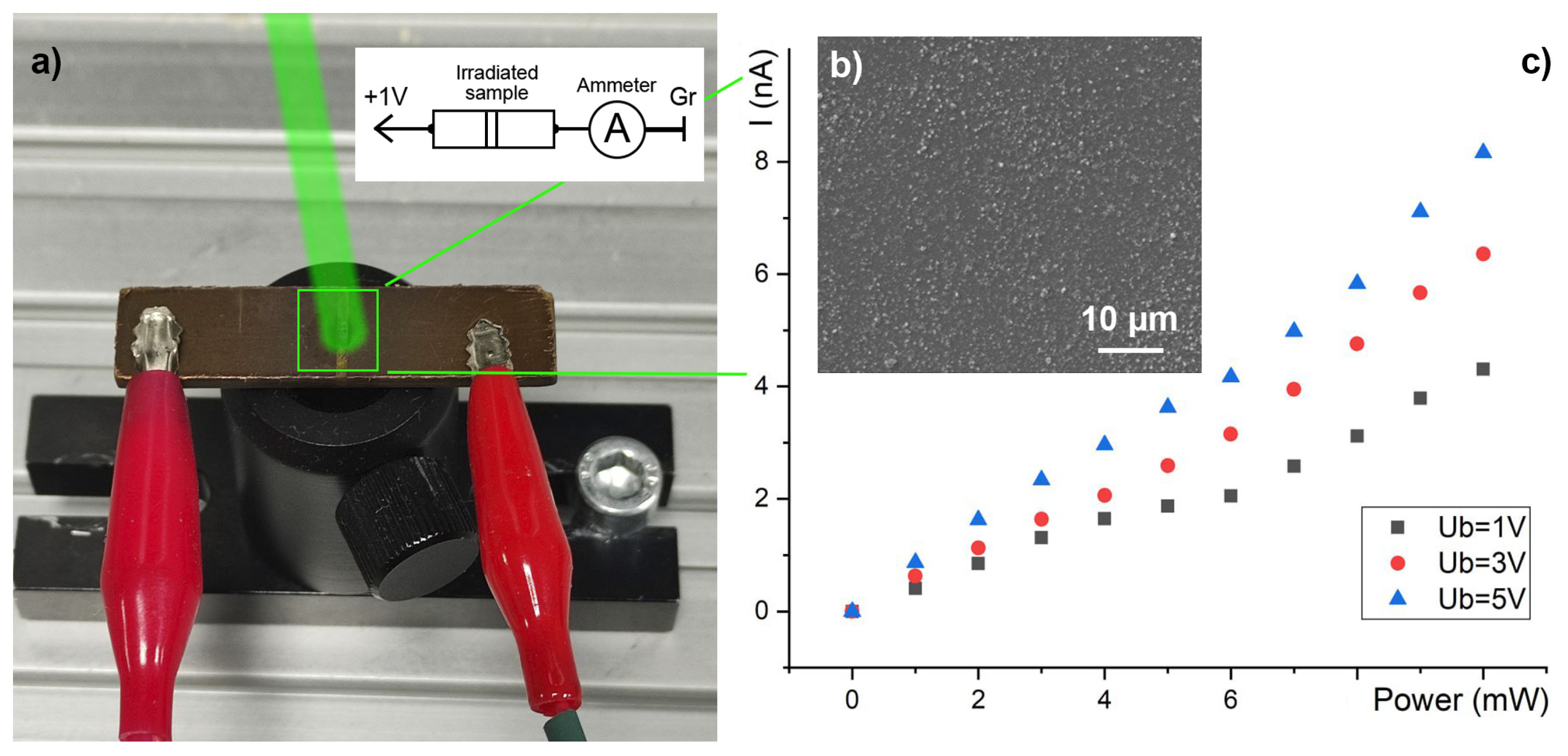
3.3. The Electroconductivity Measurements

To confirm the effect of resonant laser irradiation at the wavelength of 532 nm on the structural properties of the synthesized films, we performed photocurrent measurements. As a sample for this study, we deposited a carbon–gold solution obtained after 60 min of laser processing on a textolite substrate with the use of the sputtering method [30] (see Figure 4a). One can see that in the presence of the applied constant voltage of 1 V, the dark current in the deposited film was absent (see Figure 4b). The irradiation of the film via a CW laser produced by company (Moscow, Russia) light with power ranging from 0 to 10 mW focused on a surface of 1 cm2 and led to a linear increase in the detected current. The slope of this functional dependence changes when the input power exceeds 6 mW, indicating the generation of high concentrations of hot photoelectrons at 6 mW and higher powers.

4. Conclusions

We developed an experimental technique for the synthesis of thin metal–carbon films composed of linear sp-carbon chains stabilized with gold nanoparticles attached to their edges. The originality of this method is in the simultaneous laser-induced disintegration of the amorphous carbon and the chloroauric acid. The thermal stabilization of the irradiated solution was essential for the extension of the laser processing duration for up to 60 min. This time was long enough to achieve the disintegration of the full amount of amorphous carbon. The formation of linear carbon chains in our system was possible because of the effect of the resonant excitation of photoelectrons due to the laser light. These photoelectrons were then transferred from the surfaces of gold nanoparticles to the carbon matrix. The excitation of such photoelectrons was independently verified in the photocurrent experiment, where the deposited thin carbon–gold film was irradiated using laser light of the wavelength 532 nm and different intensities.

Author Contributions

Conceptualization, S.K. and A.K.; formal analysis and investigation, V.S., A.O. and A.A.; writing—original draft preparation, S.K. and A.O.; writing—review and editing, N.R., V.K. (Vitali Kononenko) and V.K. (Vitali Konov); supervision, A.K. and S.K.; project supervision, A.K. All authors have read and agreed to the published version of the manuscript.

Funding

This research was funded by Westlake University: Start-up funding for the International Center for Polaritonics, Project 1012516021801. This work was also supported by RSF grant 23-12-20004 in terms of the linear carbon chain synthesis conducted by the scientific group of Vladimir State University and 19-12-00255-П in terms of the laser synthesis of gold particles from acid solutions conducted by the scientific group of Prokhorov General Physics Institute.

Data Availability Statement

Not applicable.

Acknowledgments

TEM images were conducted in collaboration with the SMA LTD company for microscopy and analysis. This study was also carried out using the equipment of the interregional multispecialty and interdisciplinary center for the collective use of promising and competitive technologies in the areas of development and application in industry/mechanical engineering of domestic achievements in the field of nanotechnology (Agreement No. 075-15-2021-692 of 5 August 2021).

Conflicts of Interest

The authors declare no conflict of interest.

References

  1. Yang, G. Synthesis, properties, and applications of carbyne nanocrystals. Mater. Sci. Eng. R 2022, 151, 100692. [Google Scholar]
  2. Lv, C.F.; Yang, X.G.; Shan, C.X. One-dimensional sp carbon: Synthesis, properties, and modifications. Chin. Phys. B 2022, 31, 128103. [Google Scholar]
  3. Pan, B.; Xiao, J.; Li, J.; Liu, P.; Wang, C.; Yang, G. Carbyne with finite length: The one-dimensional sp carbon. Sci. Adv. 2015, 1, e1500857. [Google Scholar] [PubMed]
  4. Orekhov, N.; Logunov, M. Atomistic structure and anomalous heat capacity of low-density liquid carbon: Molecular dynamics study with machine learning potential. Carbon 2022, 192, 179–186. [Google Scholar]
  5. Ramberger, B.; Kresse, D. New insights into the 1D carbon chain through the RPA. Phys. Chem. Chem. Phys. 2021, 23, 5254. [Google Scholar]
  6. Chalifoux, W.A.; Tykwinski, R.R. Synthesis of polyynes to model the sp-carbon allotrope carbyne. Nat. Chem. 2010, 2, 967. [Google Scholar] [PubMed]
  7. Shi, L.; Rohringer, P.; Suenaga, K.; Niimi, Y.; Kotakoski, J.; Meyer, J.C.; Peterlik, H.; Wanko, M.; Cahangirov, S.; Rubio, A.; et al. Conned linear carbon chains as a route to bulk carbyne. Nat. Mater. 2016, 15, 634–639. [Google Scholar]
  8. Khanna, R.; Ikram-Ul-Haq, M.; Rawal, A.; Rajarao, R.; Sahajalla, V.; Cayumil, R.; Mikherjee, P.S. Formation of carbyne-like materials during low temperature pyrolysis of lignocellulosic biomass: A natural resource of linear sp carbons. Sci. Rep. 2017, 7, 16832. [Google Scholar] [CrossRef]
  9. Marabotti, P.; Peggiani, S.; Vidale, A.; Casari, C.S. Pulsed laser ablation in liquid of sp-carbon chains: Status and recent advances. arXiv 2022, arXiv:2206.01238. [Google Scholar]
  10. Sun, A.; Lauher, J.W.; Goroff, N.S. Preparation of poly(diiododiacetylene), an ordered conjugated polymer of carbon and iodine. Science 2006, 312, 1030–1033. [Google Scholar]
  11. Zavidovskii, I.A.; Streletskii, O.A.; Nishchak, O.Y.; Khaidarov, A.A.; Pavlikov, A.V. Resistivity of Thin Carbon Films with Different sp-Bonds Fractions. Tech. Phys. 2020, 65, 139–144. [Google Scholar] [CrossRef]
  12. Samyshkin, V.; Lelekova, A.; Osipov, A.; Bukharov, D.; Skryabin, I.; Arakelian, S.; Kucherik, A.; Kutrovskaya, S. Photosensitive free-standing ultra-thin carbyne-gold films. Opt. Quantum Electron. 2019, 51, 394. [Google Scholar] [CrossRef]
  13. Kucherik, A.O.; Arakelian, S.M.; Garnov, S.V.; Kutrovskaya, S.V.; Nogtev, D.S.; Osipov, A.V.; Khor’kov, K.S. Two-stage laser-induced synthesis of linear carbon chains. Quantum Electron. 2016, 46, 627. [Google Scholar] [CrossRef]
  14. Kutrovskaya, S.; Osipov, A.; Baryshev, S.; Zasedatelev, A.; Samyshkin, V.; Demirchyan, S.; Pulci, O.; Grassano, D.; Gontrani, L.; Hartmann, R.; et al. Excitonic Fine Structure in Emission of Linear Carbon Chains. Nano Lett. 2020, 20, 6502–6509. [Google Scholar] [CrossRef] [PubMed]
  15. Al-Jishi, R.; Dresselhaus, G. Lattice-dynamical model for graphite. Phys. Rev. B 1982, 26, 4514. [Google Scholar]
  16. Nemanich, R.J.; Solin, S.A. First-and second-order Raman scattering from finite-size crystals of graphite. Phys. Rev. B 1979, 20, 392. [Google Scholar] [CrossRef]
  17. Ravagnan, L.; Siviero, F.; Lenardi, C.; Piseri, P.; Barborini, E.; Milani, P.; Casari, C.S.; Bassi, A.L.; Bottani, C.E. Cluster-Beam Deposition and in situ Characterization of Carbyne-Rich Carbon Films. Phys. Rev. Lett. 2002, 89, 285506. [Google Scholar] [CrossRef]
  18. Kucherik, A.; Arakelian, S.; Vartanyan, T.; Kutrovskaya, S.; Osipov, A.; Povolotskaya, A.; Povolotskii, A.; Man’shina, A. Laser-induced synthesis of metalcarbon materials for implementing surface enhanced Raman scattering. Opt. Spectrosc. 2016, 121, 263–270. [Google Scholar] [CrossRef]
  19. Rozhkova, N.; Kovalchuk, A.; Goryunov, A.; Borisova, A.; Osipov, A.; Kucherik, A.; Rozhkov, S. Thin film coatings from aqueous dispersion of graphene-based nanocarbon and its hybrids with metal nanoparticles. Coatings 2022, 12, 600. [Google Scholar] [CrossRef]
  20. Cui, W.; Saito, T.; Ayala, P.; Pichler, T.; Shi, L. Oxidation stability of confined linear carbon chains, carbon nanotubes, and graphene nanoribbons as 1D nanocarbons. Nanoscale 2019, 11, 15253–15258. [Google Scholar] [PubMed]
  21. Sadezky, A.; Muchkenhuber, H.; Grothe, H.; Niessner, R.; Poschl, U. Raman microspectroscopy of soot and related carbonaceous materials: Spectral analysis and structural information. Carbon 2005, 43, 1731. [Google Scholar] [CrossRef]
  22. Karekin, S.; Esmeryan, D.; Castano, E.C.; Bressler, H.A.; Abolghasemibizaki, M.; Fergusson, P.C.; Roberts, A.; Mohammadi, R. Kinetically driven graphite-like to diamond-like carbon transformation in low temperature laminar diffusion flames. Diam. Relat. Mater 2017, 75, 58. [Google Scholar]
  23. Sidorov, A.; Leks, E.; Podsvirov, O.; Vinogradov, A. Crystallization and silicon carbide formation in two-layer amorphous silicon-carbon films during electron irradiation. Tech. Phys. 2022, 11, 1475. [Google Scholar] [CrossRef]
  24. Ferrari, A.; Robertson, J. Resonant Raman spectroscopy of disordered, amorphous, and diamondlike carbon. Phys. Rev. B Condens. Matter Mater. Phys. 2001, 64, 075414. [Google Scholar] [CrossRef]
  25. Condorelli, M.; D’Urso, L.; Compagnini, G.; Fazio, E.; Neri, F.; Litvinenko, V.; Kurmaev, E.; Zhidkov, I. The catalytic role of platinum nanoparticles in laser generated nanocarbons. Appl. Surf. Sci. 2021, 558, 149890. [Google Scholar] [CrossRef]
  26. Merlen, A.; Buijnsters, J.; Pardanaud, C. A guide to and review of the use of multiwavelength Raman spectroscopy for characterizing defective aromatic carbon solids: From graphene to amorphous carbons. Coatings 2017, 7, 153. [Google Scholar] [CrossRef]
  27. Moura, T.A.; Neves, W.Q.; Alencar, R.S.; Kim, Y.; Endo, M.; Vasconcelos, T.L.; Costa, D.G.; Candiotto, G.; Capaz, R.B.; Araujo, P.T.; et al. Resonance Raman spectroscopy characterization of linear carbon chains encapsulated by multi-walled carbon nanotubes. Carbon 2023, 212, 118123. [Google Scholar] [CrossRef]
  28. Wang, J.; Zhang, S.; Zhou, J.; Liu, R.; Du, R.; Xu, H.; Liu, Z.; Zhang, J.; Liu, Z. Identifying sp–sp2 carbon materials by Raman and infrared spectroscopies. Phys. Chem. Chem. Phys. 2014, 16, 11303–11309. [Google Scholar] [CrossRef] [PubMed]
  29. Milani, A.; Barbieri, V.; Facibeni, A.; Russo, V.; Bassi, A.; Lucotti, A.; Tommasini, M.; Tzirakis, M.; Diederich, F.; Casari, C.S. Structure modulated charge transfer in carbon atomic wires. Sci. Rep. 2019, 9, 1648. [Google Scholar] [CrossRef]
  30. Kutrovskaya, S.; Chestnov, I.; Osipov, A.; Samyshkin, V.; Sapegina, I.; Kavokin, A.; Kucherik, A. Electric field assisted alignment of monoatomic carbon chains. Sci. Rep. 2020, 10, 9709. [Google Scholar] [CrossRef]
  31. Eisler, S.; Slepkov, A.D.; Elliott, E.; Luu, T.; McDonald, R.; Hegmann, F.A.; Tykwinski, R.R. Polyynes as a Model for Carbyne: Synthesis, Physical Properties, and Nonlinear Optical Response. J. Am. Chem. Soc. 2005, 9, 127. [Google Scholar] [CrossRef] [PubMed]
  32. Arutyunyan, N.; Kononenko, V.; Gololobov, V.; Obraztsova, E. Resonant Effects in SERS Spectra of Linear Carbon Chains. Phys. Status Solidi B 2017, 255, 1700254. [Google Scholar] [CrossRef]
Figure 1. (a) The schematic process of chloroauric acid dissociation, AuNP recovery in the zone of a presence of free electrons under laser pulses action and ShC transformation into linear chains dopped with AuNP; (b) A cuvette with a DI solution of ShC nanoparticles and hydrogen tetrachloroaurate (III): A bottom cuvette side is a subject to forced cooling that initiates a flow circulation in a solution due to the temperature difference between a cooled bottom and the top of the solution heated by laser pulses; (c,d) TEM images of carbyne bundles with AuNPs appear as dark spheres (c) after 30 min and (d) 60 min of laser pulse actions on the studied solution. The scale bars are 10 nm.
Condensedmatter 08 00096 g001
Figure 2. Raman spectra of a multielement solution taken at different stages of laser processing: the red curve shows singularities characteristic of the initial solution; blue and black lines demonstrate the evolution of the studied solution after 30 and 60 min of laser action, respectively.
Condensedmatter 08 00096 g002
Figure 3. Absorption spectra of the investigated thin films recorded after 30 and 60 min of the laser action.
Condensedmatter 08 00096 g003
Figure 4. (a) The experimental setup allowing a photocurrent measurement to be performed in a small area of the green cube containing a deposited thin film of the carbyne bundles stabilized by AuNPs; (b) The TEM image of the deposited thin film under study; (c) The photocurrent induced in the irradiated area by the CW laser of the power, as indicated by the horizontal axis. Black, red, and blue points correspond to different voltages applied between two contacts seen in (a).
Condensedmatter 08 00096 g004
Disclaimer/Publisher’s Note: The statements, opinions and data contained in all publications are solely those of the individual author(s) and contributor(s) and not of MDPI and/or the editor(s). MDPI and/or the editor(s) disclaim responsibility for any injury to people or property resulting from any ideas, methods, instructions or products referred to in the content.

