
Clinical and Radiological Results after Fracture-Dislocations of the Ankle: A Medium- to Long-Term Followup Study


Abstract
:1. Introduction
2. Materials and Methods
Statistical Analysis
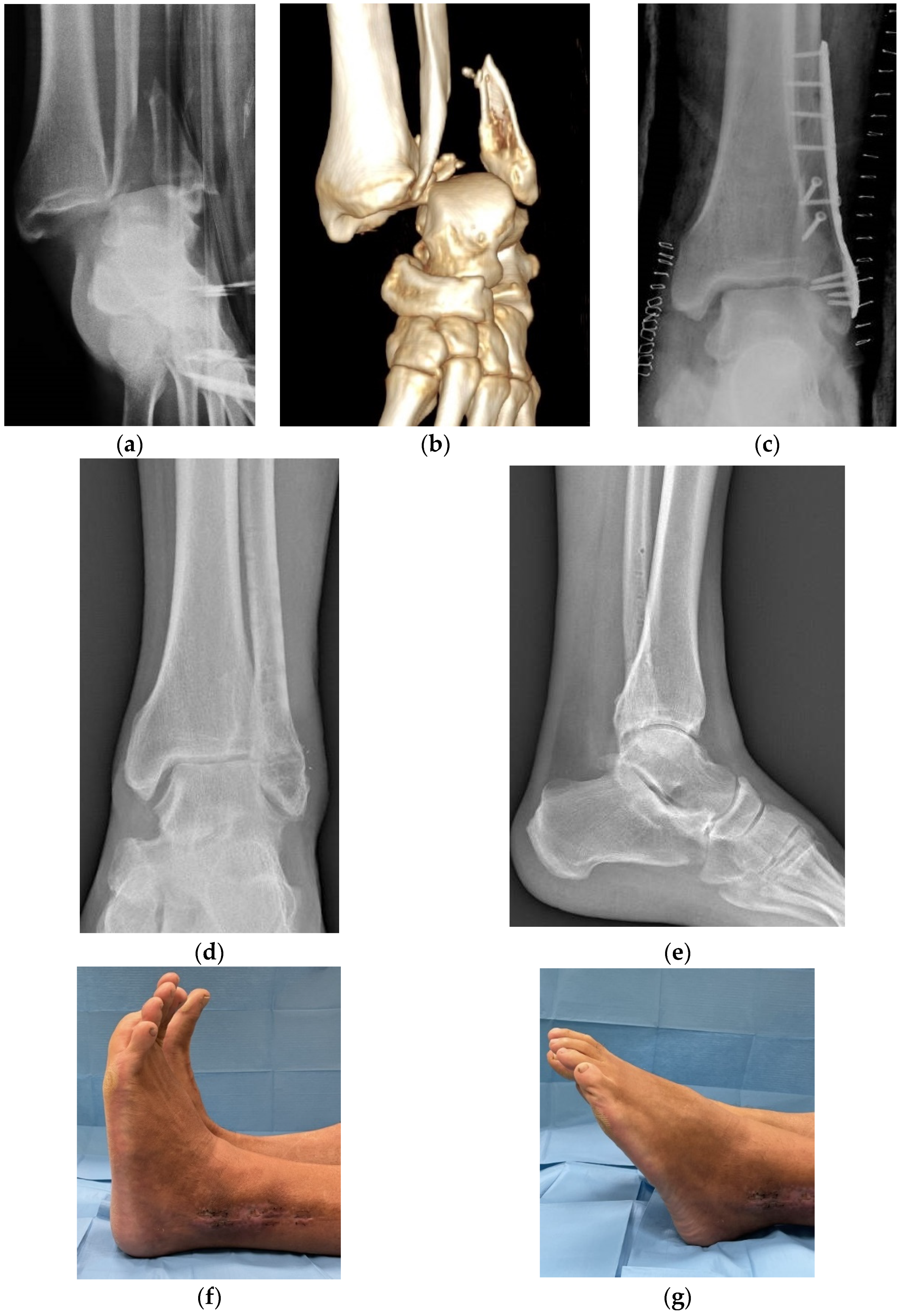
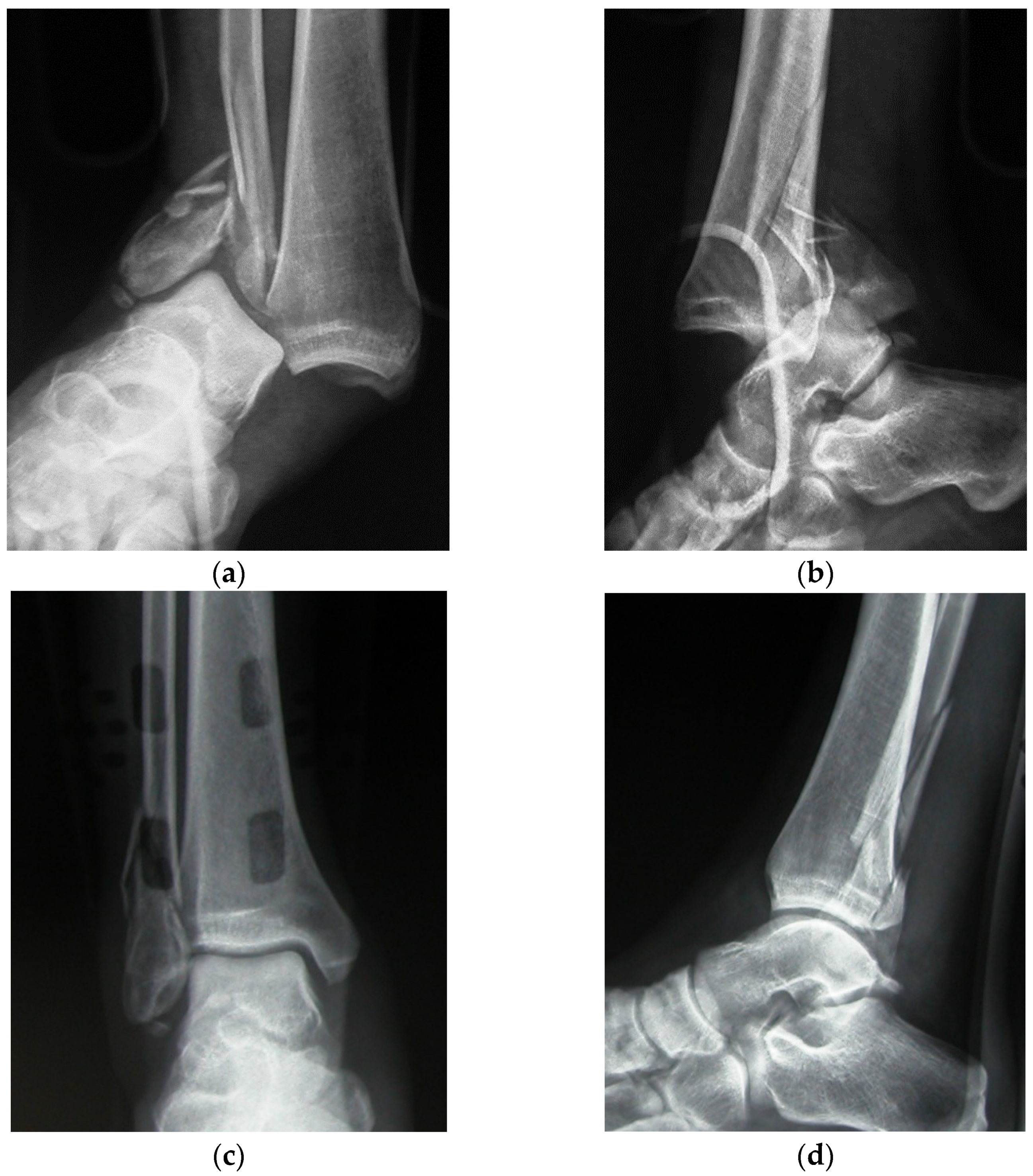
3. Results
4. Discussion
5. Conclusions
Author Contributions
Funding
Institutional Review Board Statement
Informed Consent Statement
Data Availability Statement
Conflicts of Interest
References
- Leeds, H.C.; Ehrlich, M.G. Instability of the distal tibiofibular syndesmosis after bimalleolar and trimalleolar ankle fracture. J. Bone J. Surg. Am. 1984, 66, 490–503. [Google Scholar] [CrossRef]
- Egol, K.A.; Tejwani, N.C.; Walsh, M.G.; Capla, E.L.; Koval, K.J. Predictors of short-term functional outcome following ankle fracture surgery. J. Bone J. Surg. Am. 2006, 88, 974–979. [Google Scholar] [CrossRef]
- Court-Brown, C.M.; Caesar, B. Epidemiology of adult fractures: A review. Injury 2006, 37, 691–697. [Google Scholar] [CrossRef] [PubMed]
- Schepers, T.; De Vries, M.R.; Van Lieshout, E.M.M.; Van der Elst, M. The timing of ankle fracture surgery and the effect on infections complications: A case series and systematic review of the literature. Int. Orthop. 2013, 37, 489–494. [Google Scholar] [CrossRef] [PubMed] [Green Version]
- Shibuyan, N.; Davis, M.L.; Jupiter, D.C. Epidemiology of foot and ankle fractures in the United States: An analysis of the National Trauma Data Bank (2007 to 2011). J. Foot Ankle Surg. 2014, 53, 606–608. [Google Scholar] [CrossRef] [PubMed]
- Verhage, S.M.; Schipper, I.B.; Hoogendoorn, J.M. Long-term functional and radiographic outcomes in 243 operated ankle fractures. J. Foot Ankle Res. 2015, 8, 45–50. [Google Scholar] [CrossRef] [PubMed] [Green Version]
- De Avila, V.R.; Bento, T.; Gomes, W.; Leitao, J.; De Sousa, N.J. Functional outcomes and quality of life after ankle fracture surgically treated: A systematic review. J. Sport Rehabil. 2018, 27, 274–283. [Google Scholar] [CrossRef] [PubMed] [Green Version]
- Stiehl, J.B.; Schwartz, H.S. Long-term results of pronation-external rotation ankle fracture-dislocations treated with anatomical open reduction, internal fixation. J. Orthop. Trauma 1990, 4, 339–345. [Google Scholar] [CrossRef] [PubMed]
- Warner, S.J.; Schttel, P.C.; Hinds, R.M.; Helfet, D.L.; Lorich, D.G. Fracture-dislocation demonstrate poorer postoperative functional outcomes among pronation external rotation IV ankle fractures. Foot Ankle Int. 2015, 36, 641–647. [Google Scholar] [CrossRef] [PubMed]
- Carragee, E.J.; Csongradi, J.S.; Blek, E.E. Early complications in the operative treatment of ankle fractures. Influence of delay before operation. J. Bone Jt. Surg. Br. 1991, 73, 79–82. [Google Scholar] [CrossRef] [PubMed] [Green Version]
- Sculco, P.K.; Lazaro, L.E.; Little, M.M.; Berkes, M.B.; Warner, S.J.; Helfet, D.L.; Lorich, D.G. Dislocation is a risk factor for poor outcome after supination external rotation type ankle fractures. Arch. Orthop. Trauma Surg. 2016, 136, 9–15. [Google Scholar] [CrossRef] [PubMed]
- Tantigate, D.; Ho, G.; Kirschenbaum, J.; Baker, H.C.; Asherman, B.; Freibott, C.; Greisberg, J.; Vosseller, J.T. Functional outcomes after fracture-dislocation of the ankle. Foot Ankle Spec. 2020, 13, 18–26. [Google Scholar] [CrossRef] [PubMed]
- Bosworth, D.M. Fracture-dislocation of the ankle with fixed displacement of the fibula behind the tibia. J. Bone Jt. Surg. Am. 1947, 29, 130–135. [Google Scholar]
- Kitaoka, H.B.; Alexander, I.J.; Adelaar, R.S.; Nunley, J.A.; Myerson, M.S.; Sanders, M. Clinical rating systems for the ankle-hindfoot, midfoot, hallux, and lesser toes. Foot Ankle Int. 1994, 15, 349–353. [Google Scholar] [CrossRef] [PubMed]
- Van Dijk, C.N.; Verhagen, R.A.; Tol, J.L. Arthroscopy for problems after ankle fracture. J. Bone Jt. Surg. Br. 1997, 79, 280–284. [Google Scholar] [CrossRef]
- Buyukkuscu, M.O.; Basilgan, S.; Mollaomeroglu, A.; Misir, A.; Basar, H. Splinting vs. temporary external fixation in the initial treatment of ankle-fracture-dislocation. Foot Ankle Surg. 2022, 28, 235–239. [Google Scholar] [CrossRef] [PubMed]
- Baker, J.R.; Patel, S.N.; Teichman, A.J.; Bochat, S.E.S.; Fleischer, A.E.; Knight, J.M. Bivalved fiberglass cast compared with plaster splint immobilization for initial management of ankle fracture-dislocation: A treatment algorithm. Foot Ankle Spec. 2012, 5, 160–167. [Google Scholar] [CrossRef] [PubMed]
- Friedman, J.; Ly, A.; Mauffrey, C.; Stahel, P.F. Temporary transarticular K-wire fixation of critical ankle injuries at risk: A neglected “damage control” strategy? Orhopaedics 2015, 38, 122–127. [Google Scholar] [CrossRef] [PubMed] [Green Version]
- Wawrose, R.A.; Grossman, L.S.; Tagliaferro, M.; Siska, P.A.; Moloney, G.B.; Tarkin, I.S. Temporizing external fixation VS splinting following ankle fracture dislocation. Foot Ankle Int. 2020, 41, 177–182. [Google Scholar] [CrossRef] [PubMed]
- Farsetti, P.; Caterini, R.; Potenza, V.; De Luna, V.; De Maio, F.; Ippolito, E. Immediate continous passive motion after internal fixation of an ankle fracture. Optim. Eng. 2009, 10, 63–69. [Google Scholar]


| Case | Sex—Age | Side | Energy | Type of Fracture | Weber Classif. | Quality of Reduction | Follow-Up Years | AOFAS | Radiograp. Results (Van Dijk) |
|---|---|---|---|---|---|---|---|---|---|
| 1 | M—20 | R | High | Unimal. | B | Good | 4.6 | 88 | 0 |
| 2 | F—51 | R | High | Unimal. | C | Excellent | 6.8 | 92 | 0 |
| 3 | F—39 | R | High | Bimal. | B | Good | 6.6 | 84 | 0 |
| 4 | F—77 | R | Low | Bimal. | C | Good | 4.2 | 92 | 1 |
| 5 | F—59 | R | High | Trimal. | B | Good | 4.6 | 96 | 1 |
| 6 | M—45 | R | High | Unimal. | A | Excellent | 4.2 | 84 | 0 |
| 7 | M—35 | L | High | Bimal. | B | Excellent | 5.3 | 88 | 0 |
| 8 | F—68 | R | Low | Bimal. | B | Excellent | 5.8 | 90 | 1 |
| 9 | F—26 | R | High | Bimal. | B | Excellent | 3.9 | 98 | 0 |
| 10 | F—79 | R | Low | Unimal. | C | Good | 4.8 | 85 | 1 |
| 11 | F—49 | L | High | Bimal. | B | Good | 5.2 | 75 | 1 |
| 12 | M—59 | R | High | Bimal. | C | Excellent | 5.5 | 92 | 0 |
| 13 | M—32 | R | High | Bimal. | B | Excellent | 5.3 | 82 | 0 |
| 14 | M—24 | R | High | Unimal. | B | Excellent | 6.8 | 80 | 0 |
| 15 | F—16 | R | High | Unimal. | C | Excellent | 3.7 | 96 | 0 |
| 16 | F—45 | R | High | Trimal. | B | Good | 5.9 | 88 | 0 |
| 17 | F—71 | R | Low | Trimal. | B | Good | 5.9 | 93 | 1 |
| 18 | M—45 | L | High | Unimal. | B | Excellent | 3.8 | 85 | 0 |
| 19 | F—24 | R | High | Bimal. | C | Excellent | 4.8 | 84 | 0 |
| 20 | F—66 | R | Low | Trimal. | B | Excellent | 5.2 | 92 | 0 |
| 21 | M—50 | R | High | Unimal. | C | Excellent | 6.1 | 94 | 0 |
| 22 | F—45 | L | High | Unimal. | B | Good | 4.9 | 86 | 1 |
| 23 | M—28 | R | High | Bimal. | C | Good | 5.6 | 76 | 1 |
| 24 | F—74 | R | Low | Trimal. | B | Good | 6.2 | 88 | 1 |
| 25 | F—28 | R | High | Unimal. | C | Excellent | 5.9 | 94 | 0 |
| 26 | F—62 | R | High | Bimal. | C | Good | 4.4 | 85 | 1 |
Publisher’s Note: MDPI stays neutral with regard to jurisdictional claims in published maps and institutional affiliations. |
© 2022 by the authors. Licensee MDPI, Basel, Switzerland. This article is an open access article distributed under the terms and conditions of the Creative Commons Attribution (CC BY) license (https://creativecommons.org/licenses/by/4.0/).
Share and Cite
De Luna, V.; Caterini, A.; Casci, C.; Marsiolo, M.; Efremov, K.; De Maio, F.; Farsetti, P. Clinical and Radiological Results after Fracture-Dislocations of the Ankle: A Medium- to Long-Term Followup Study. J. Funct. Morphol. Kinesiol. 2022, 7, 30. https://doi.org/10.3390/jfmk7020030
De Luna V, Caterini A, Casci C, Marsiolo M, Efremov K, De Maio F, Farsetti P. Clinical and Radiological Results after Fracture-Dislocations of the Ankle: A Medium- to Long-Term Followup Study. Journal of Functional Morphology and Kinesiology. 2022; 7(2):30. https://doi.org/10.3390/jfmk7020030
Chicago/Turabian StyleDe Luna, Vincenzo, Alessandro Caterini, Chiara Casci, Martina Marsiolo, Kristian Efremov, Fernando De Maio, and Pasquale Farsetti. 2022. "Clinical and Radiological Results after Fracture-Dislocations of the Ankle: A Medium- to Long-Term Followup Study" Journal of Functional Morphology and Kinesiology 7, no. 2: 30. https://doi.org/10.3390/jfmk7020030
APA StyleDe Luna, V., Caterini, A., Casci, C., Marsiolo, M., Efremov, K., De Maio, F., & Farsetti, P. (2022). Clinical and Radiological Results after Fracture-Dislocations of the Ankle: A Medium- to Long-Term Followup Study. Journal of Functional Morphology and Kinesiology, 7(2), 30. https://doi.org/10.3390/jfmk7020030