Article

Optimization of DD-110 Neutron Generator Output for Boron Neutron Capture Therapy Using Monte Carlo Simulation

1
Physics Department, Faculty of Science, King Abdulaziz University, Jeddah 21589, Saudi Arabia
2
Department of Physics, Faculty of Science, Menoufia University, Shebin El-Koom, Menoufia 32511, Egypt
*
Author to whom correspondence should be addressed.
Quantum Beam Sci. 2025, 9(2), 12; https://doi.org/10.3390/qubs9020012
Submission received: 25 September 2024 / Revised: 12 March 2025 / Accepted: 10 April 2025 / Published: 15 April 2025

Abstract

:
Boron neutron capture therapy (BNCT) is a specialized cancer treatment that leverages the high absorption cross-section of boron for thermal neutrons. When boron captures neutrons, it undergoes a nuclear reaction that produces alpha particles and lithium ions, which have high linear energy transfer (LET) and can effectively damage nearby cancer cells while minimizing harm to surrounding healthy tissues. This targeted approach makes BNCT particularly advantageous for treating tumors situated in sensitive areas where traditional radiation therapies may pose risks to critical structures. In this study, the deuterium–deuterium (DD) neutron generator, specifically the DD-110 model (neutron yield Y = 1 × 10¹⁰ n/s), served as the neutron source for BNCT. The fast neutrons produced by this generator were thermalized to the epithermal energy range using a beam-shaping assembly (BSA). The BSA was designed with a moderator composed of 32 cm of MgF2, a reflector made of 76 cm of Pb, and filters including 3 cm of Pb and 1.52 cm of Bi. A collimator, featuring a 10 cm high Pb cone frustum with a 12 cm aperture diameter, was also employed to optimize beam characteristics. The entire system’s performance was modeled and simulated using the MCNPX code, focusing on parameters both in-air and in-phantom to evaluate its efficacy. The findings indicated that the BSA configuration yielded an optimal thermal-to-epithermal flux ratio ( φ t h e r / φ e p t h ) of 0.19, a current-to-flux ratio of 0.87, and a gamma dose-to-epithermal flux ratio of 1.71 × 10⁻¹³ Gy/cm2, all aligning with IAEA recommendations. The simulated system showed acceptable ratios for φ t h e r / φ e p t h , gamma dose to epithermal flux, and beam collimation. Notably, the advantage depth was recorded at 5.5 cm, with an advantage ratio of 2.29 and an advantage depth dose rate of 4.1 × 10⁻⁴ Gy.Eq/min. The epithermal neutron flux of D110 exceeded D109, but D110’s fast neutron contamination increased ~6.6 times. On the other hand, D110’s gamma contamination decreased by 30%. Based on these findings, optimizing neutron source characteristics is crucial for BNCT efficacy. Future research should focus on developing advanced neutron generators that balance these factors, aiming to produce optimal neutron yields for enhanced treatment outcomes and broader applicability.

1. Introduction

In 2022, there were 20 million new cases of cancer and 9.7 million cancer-related deaths [1]. The primary brain tumor that is malignant and most frequently occurs is called glioblastoma (GBM). Of all primary malignant brain tumors, GBM makes up 48% [2]. An estimated yearly incidence rate of 0.59 to 5 per 100,000 people is found in worldwide studies. Patients with glioblastoma are thought to have an average survival time of only 12 to 18 months [3]. Head and neck cancers (HNCs) account for over 325,000 fatalities and 660,000 new cases annually, making them the seventh most prevalent cancer diagnosed globally [4]. With an expected 1.5 million new cases in 2022, skin malignancies are the most common type of tumors diagnosed globally. An estimated 330,000 new instances of melanoma were reported globally in 2022, and the disease claimed the lives of over 60,000 people [5]. Boron neutron capture therapy (BNCT) is a form of radiation therapy used mainly to treat certain types of cancer, which are GBM, HNCs, melanoma, and other sensitive organ tumors.
One kind of radiation treatment that makes use of the special qualities of boron-10, a stable isotope of boron, is boron neutron capture therapy (BNCT). Certain cancers appear to respond well to BNCT, especially those which are in the brain and other delicate regions where conventional radiation therapy may be difficult to administer [6,7,8]. The idea behind BNCT is that atoms of boron-10 absorb thermal neutrons and subsequently break down, producing particles with a high LET. According to radiation treatment energy classification, high-energy lithium and alpha ions are produced by this process and the near-cancer cells primarily absorb them [9], as presented in Figure 1. This differential effect results from the fact that alpha and lithium-ions have short route track lengths and that boron-10 preferentially accumulates in tumor cells at higher concentrations than in normal cells. Then, within a cell’s dimensions, they run out of energy.
1n + 10B → 11B*4He + 7Li (6.3%)
1n + 10B → 11B*4He + 7Li + γ (93.7%)
The energies of the lithium and alpha particles in the first kind of reaction are 1.01 MeV and 1.78 MeV, respectively [10]. The energy of the alpha particle in the second type of reaction is 1.47 MeV, while the energy of the lithium-particle is 0.84 MeV. The remaining energy, 0.48 MeV, is gamma-ray energy [10]. These particles have a total average kinetic energy of 2.33 MeV and a range of 12–13 µm in tissue, which is comparable to the 10–100 µm cellular dimensions of mammals [11]. In addition to this, these particles have higher relative biological effectiveness (RBE) than conventional radiation therapy particles, electrons, and gamma rays. These points make BNCT a promising radiation treatment for certain types of tumors, with recent and future applications [12].
An injectable substance containing boron-10 is first administered to the patient during BNCT administration. This chemical accumulates in the tumor due to the high metabolism rate of tumor cells compared to normal tissue cells. Once the patient has amassed enough of the substance within the tumor, high-energy particles that cause damage to the tumor cells are released when the patient is exposed to a neutron beam.
Compared to traditional radiation therapy, which uses an X-ray or electron beam, BNCT has the advantage of employing high LET particles, attaining a larger absorbed dosage in the tumor relative to normal tissue, preserving the latter [13].
In 1936, Gordon Locher [14] first proposed BNCT. He put forward the principles of BNCT following the insight of Goldhaber. In 1934, Taylor and Goldhaber [15] discovered that the stable 10B element has a large thermal neutron capture cross-section and then immediately breaks down into an energetic 4He2+ with a rebounding 7Li ion. These particles have a total average kinetic energy of 2.33 MeV and a range of 12–13 µm in tissue, which is comparable to the cellular dimensions of mammals. During 1959–1961, the first BNCT clinical examinations were carried out at Brookhaven National Laboratory (BNL) and Massachusetts General Hospital (MGH) to treat glioblastoma multiforme (GBM) [16]. Borax, or sodium tetraborate, was the boron delivery agent. Like most scientific discoveries, the initial outcomes were not satisfactory. Following decades of a lot of advancements, there are currently 33 BNCT facilities globally, spread across 13 countries [16].
The neutron source, neutron energy spectrum, and boron delivery agent have the greatest influence on the effectiveness of BNCT. Because of its strong affinity for tumor cells, 4-borono-L-phenylalanine (BPA) is now the most employed boron agent in BNCT [17]. Since the efficiency of the treatment is dependent on the ratio between tumor boron concentration to normal tissue boron concentration and this is constantly being improved, efforts in this area are still active, trying enhance this ratio and decrease the toxicity of the boron compound. Reactors and particle accelerators are the options that provide the right neutron flux to reduce treatment time when it comes to neutron sources for BNCT. Reactors are the original BNCT neutron sources and provide higher neutron fluxes than particle accelerators [18]; however, reactor-based BNCT (RB-BNCT) is very expensive to install, difficult to operate, and requires a license because it is not registered as a medical device [6]. Therefore, the current neutron sources for BNCT are particle accelerators. For neutron sources, there are various kinds of particle accelerators and reactions. These sources include deuterium accelerators for the reactions 7Li (2H,n)8Be, 9Be (2H,n)10B, and 13C(2H,n)14N, as well as proton accelerators with targets of Li–7Li(p,n)7Be or Be–9Be(p,n)9B. The other accelerator is a deuterium accelerator for the fusion reaction of d + d and d + t. In this work, deuterium accelerators for fusion reaction, also known as neutron generators, are selected as the source of neutron [19].
In d + t − 3H(d,n)4He and d + d − 2H(d,n)3He reaction neutron generators, the resulting energies are 14.1 MeV and 2.45 MeV, respectively. The tritium element is a beta emitter, and the d + t process produces far more energetic neutrons than epithermal neutrons; hence, it is unlikely to be selected as a BNCT neutron source [19]. In this work, a d + d neutron generator was utilized as a neutron source, particularly a DD-110 neutron generator, built by Adelphi tech. There is no previous work that assesses this neutron generator model as a neutron source for deep-seated BNCT. Beam-shaping assembly (BSA) is intended to attenuate the 2.45 MeV neutrons that this generator produces down to the epithermal region (0.5 eV to 10 keV). The reason why the BSA’s output port detects epithermal neutrons instead of thermal ones is that, as they approach deeply ingrained tumors, their energy drops, and they transform into thermal neutrons, which are then captured by the boron element. Different Monte Carlo simulation codes have been used by our group in different studies for calculating different dose parameters [20,21,22]. In this study, the MCNPX code was used. The optimization of the output neutrons through BSA was investigated. In addition to this, the in-air and clinical parameters of the overall design were also evaluated.

2. Materials and Methods

2.1. DD-110 Neutron Generator

Designing the DD-110 neutron generator (Adelphi Technology, Inc., Redwood City, CA, USA) was the initial step in this project. An ion beam from a microwave plasma ion source drove the generator. The ion source generated a high plasma density for a high current and high D+ content by utilizing the electron cyclotron resonance (ECR) effect. It accelerated to the deuterium target after the deuterium ion was created. The target was 50 mm in diameter. Neutrons were isotopically expelled from the collision when the deuterium target was bombarded by the accelerating deuterium. Monoenergetic 2.45 MeV 1 × 10¹⁰ neutrons per second were released in this system. The whole size of the neutron generator was 26.67 cm diameter and 63.5 cm length cylinder [23]. MCNPX code (Los Alamos National Laboratory, Los Alamos, NM, USA) was utilized for this purpose. The simulations were run until all tallies of interest achieved relative uncertainties below 0.1 (10%) at the 1σ confidence level.

2.2. Beam-Shaping Assembly

The design of the beam-shaping assembly that thermalized the fast neutrons to epithermal neutrons constituted the following stage. Table 1 lists the classification of neutrons according to their energy.
The goal of the BSA was not only to reduce neutron energy but also to direct the isotopic neutrons towards the BSA output port, reduce the energy of fast neutrons and, in turn, their number, filter out gamma rays produced by the collision of fast neutrons with hydrogen and nitrogen atoms, shield the surrounding environment, reduce out-of-field exposure, and collimate the final yield to the appropriate aperture dimension. Table 2 provides a numeric guideline for these elements.
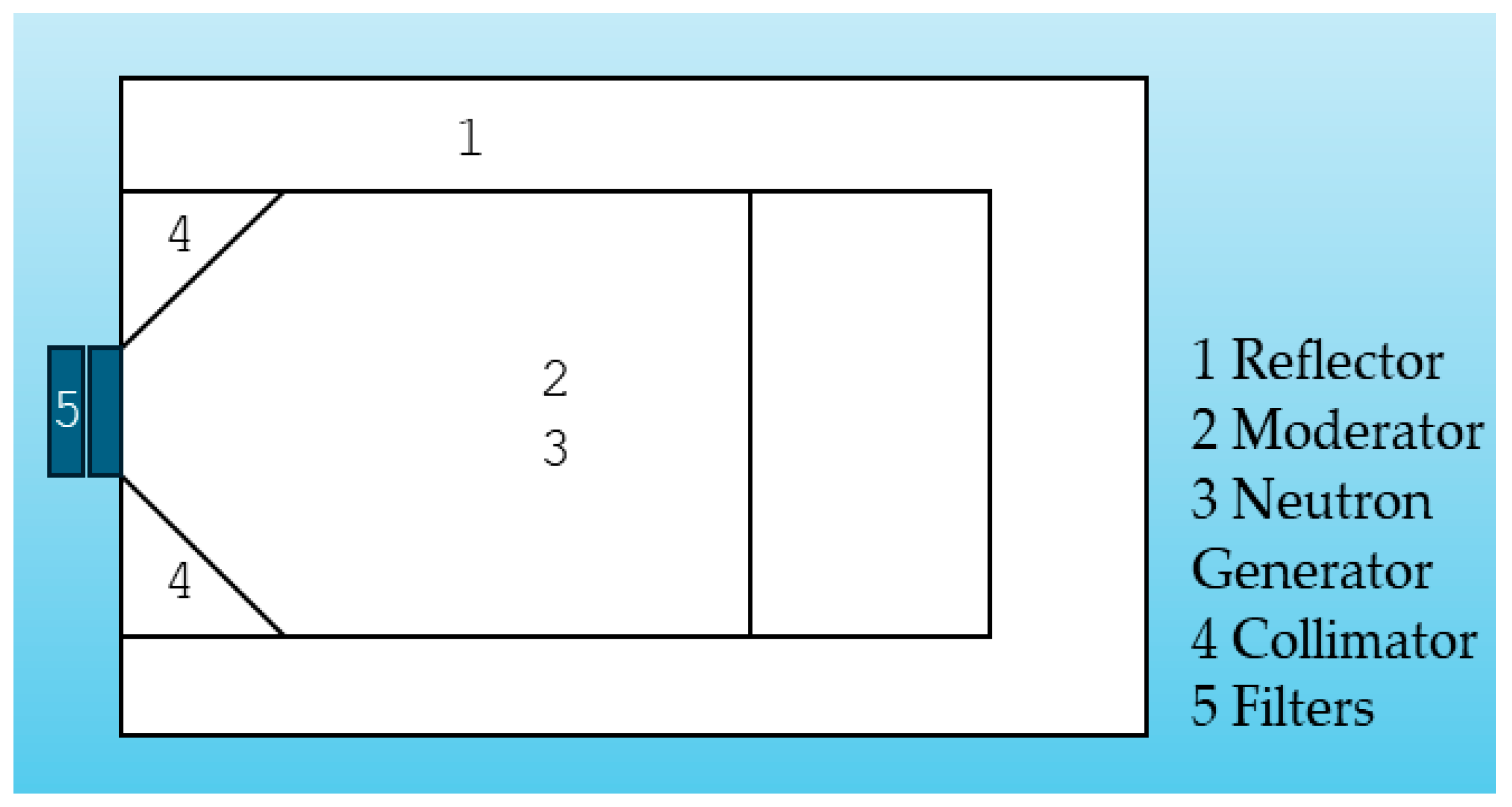
The IAEA has proposed these values as the beam spectrum standards for epithermal neutron-based treatments. The BSA was built with parts that could provide outcomes meeting these requirements. The key parts of the BSA were as follows: The moderator reduced the fast neutron energy to an epithermal neutron energy range. The reflector returned the neutrons that were outgoing from the collimator direction. The filter removed unwanted beam components. The collimator collimated the final output to the desired region. The general setup of the neutron generator and BSA is presented in Figure 2.
The first step of designing the BSA was to select the appropriate moderator material and its dimensions. The moderator material was selected based on the ability of elastic collision with neutrons. As such, these materials are mostly lightweight materials. Further consideration needed to be given to ensure that the moderator did not decrease the neutron flux much. The other parameter of the moderator was the thickness of the material. In this work, 10 materials were assessed as moderators for the given neutron generator using the MCNPX code.
Finding a suitable filter and reflector followed the selection of the right moderator. The unique property of reflectors is their heaviness. The last step in designing the BSA consisted of researching several collimator materials with various aperture sizes and select the best one. The choices of reflector and shielding for the neutron generator were made simultaneously. Subsequently, the aggregate impact of the several constituents of BSA was evaluated.

2.3. Clinical Parameters and Dose Calculation

After the overall design of the BSA, its clinical efficacy needed to be studied using phantoms. The most used in-phantom parameters for BNCT are advantage depth (AD), advantage ratio (AR), advantage depth dose rate (ADDR), and treatment time. Advantage depth is a depth in the irradiated body at which the total therapeutic tumor dose equals the maximum healthy tissue dose. It is a measure of beam penetration ability. The advantage ratio is the ratio of the total therapeutic dose to the total healthy tissue dose over the AD. The advantage depth dose rate, as the name indicates, is the dose rate at the AD. Treatment time is the maximum irradiation time until the healthy tissue dose limit is reached.
Neutrons interact with atoms in 3 main ways. The first one is the elastic scattering of fast and epithermal neutrons by low-atomic-mass atoms. In the BNCT modality, this type of reaction takes place when the neutron interacts with a hydrogen atom (1H(n,n’)1H). The second interaction is the inelastic scattering of neutrons. The last one is the neutron absorption reaction. This type of reaction is the main reaction that takes place in the BNCT. Neutron absorption reactions in the BNCT are 1H (n,Υ)2H, 14N(n,p)14C, and 10B(n, α)7Li. Hence, the absorbed doses in this treatment modality are these four types of reactions. These dose transfer mechanisms have different biological effectiveness. The overall calculation of the total absorbed dose is given in Equation (3) [24].
D t = C B E B D B + R B E H D H + R B E N D N + R B E γ D γ
C B E B is the compound biological effectiveness of the boron dose, while R B E H , R B E N , and R B E γ are the relative biological effectiveness of the hydrogen, nitrogen, and gamma doses, respectively. Their values are given in Table 3.
The boron dose analysis was dependent on the concentration of the boron compound in the tumor and healthy tissue. In this work, the boron concentrations in the tumor and the normal tissue were 33.3 ppm and 10 ppm, respectively. The ratio between the tumor boron concentration and the healthy tissue boron concentration was 3.33 [25].

3. Results and Discussion

For the moderator, there were nine candidate materials used in this work: MgF2, LiF, D2O, TiF3, Al, AlF3, Al2O3, Pb, and CaF2. Initially, these materials were examined using a standard reflector, devoid of a filter and a collimator. The setup of the simulation is presented in Figure 3.
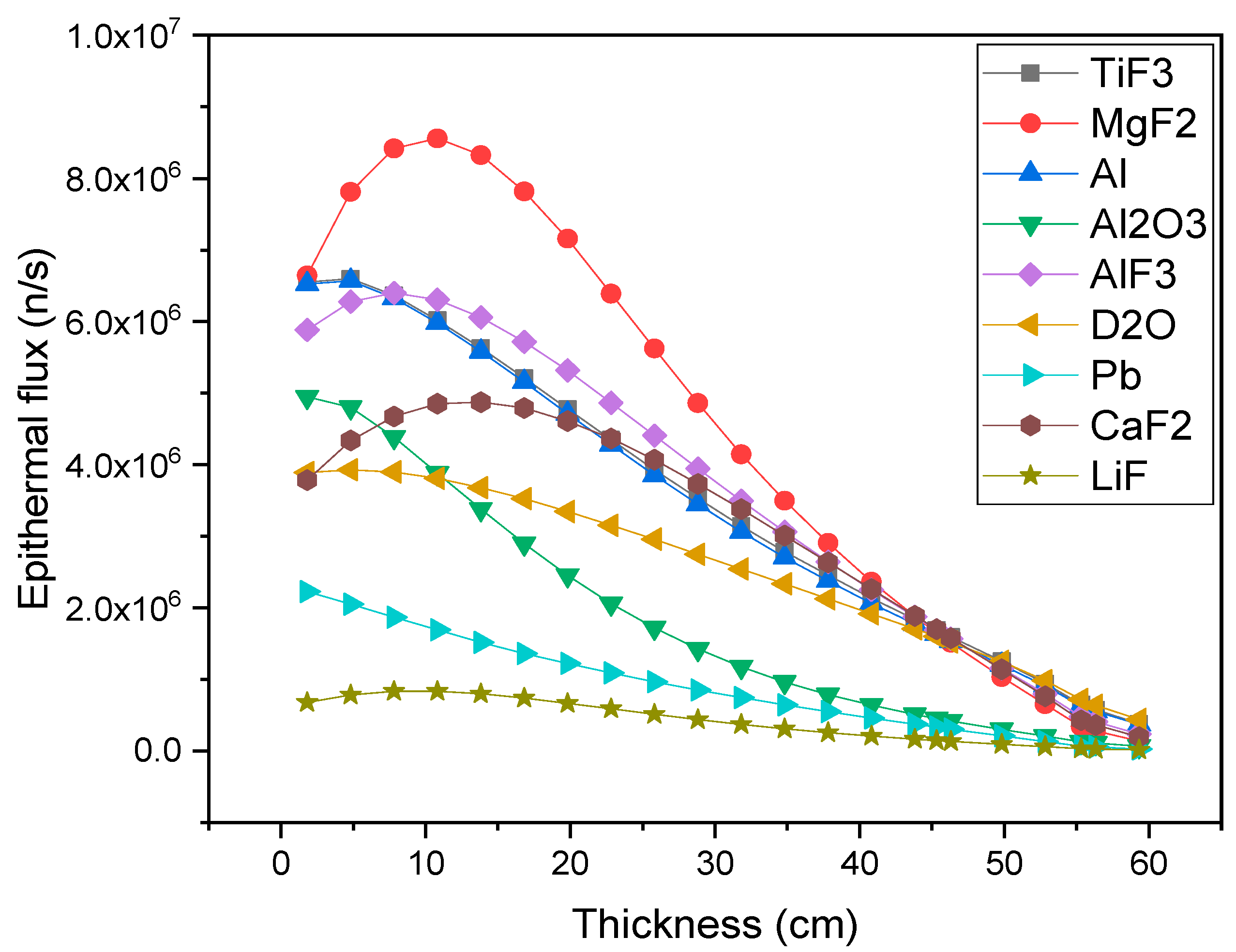
The small boxes in the moderator were inserted to calculate beam parameters at different thicknesses, and this helped to assess the change in these parameters according to the thickness. This simulation helped us reach a sensible selection of moderators and improve them further. The result of this simulation is given in Figure 4. It can be interpreted as the affinity of different materials to converting fast neutrons into epithermal neutrons at varying thicknesses.
With a step of 3 cm, the epithermal flux was counted from 1.81 cm to 59.31 cm. The 2.45 MeV neutrons were isotopically expelled from the target, causing some of them to travel in a different direction to the position of the moderator. The neutrons were then reflected by the reflector. They expended some energy on their travel. This explains why, for some materials, the epithermal flux at 1.81 cm was in the order of 106, which was an unexpected value. This graph makes it evident that the maximal epithermal flux and associated thickness of the various materials varied. Certain materials exhibited more epithermal flux for a given neutron spectrum at a certain thickness in comparison to another, but not across the entire thickness range. For example, MgF2 exhibited maximum epithermal neutron flux between 1.81 cm and 42 cm but not at thicknesses greater than 42 cm. The reason for this is that, in addition to the cross-section values for the materials being energy-dependent, the energy of the neutron changed as it passed through a material. As a result, the relative values of epithermal flux for various materials differed. At most thicknesses, MgF2, TiF3, Al, AlF3, and CaF2 achieved better results than the other materials, as advised by the IAEA report [19]. The epithermal flux was nearly constant for LiF and Pb. This was a positive result, since they effectively filtered out thermal, fast, and gamma rays without compromising the epithermal flux. Furthermore, materials with a high slope of degradation like Al2O3 were challenging to work with for optimizing other parameters.
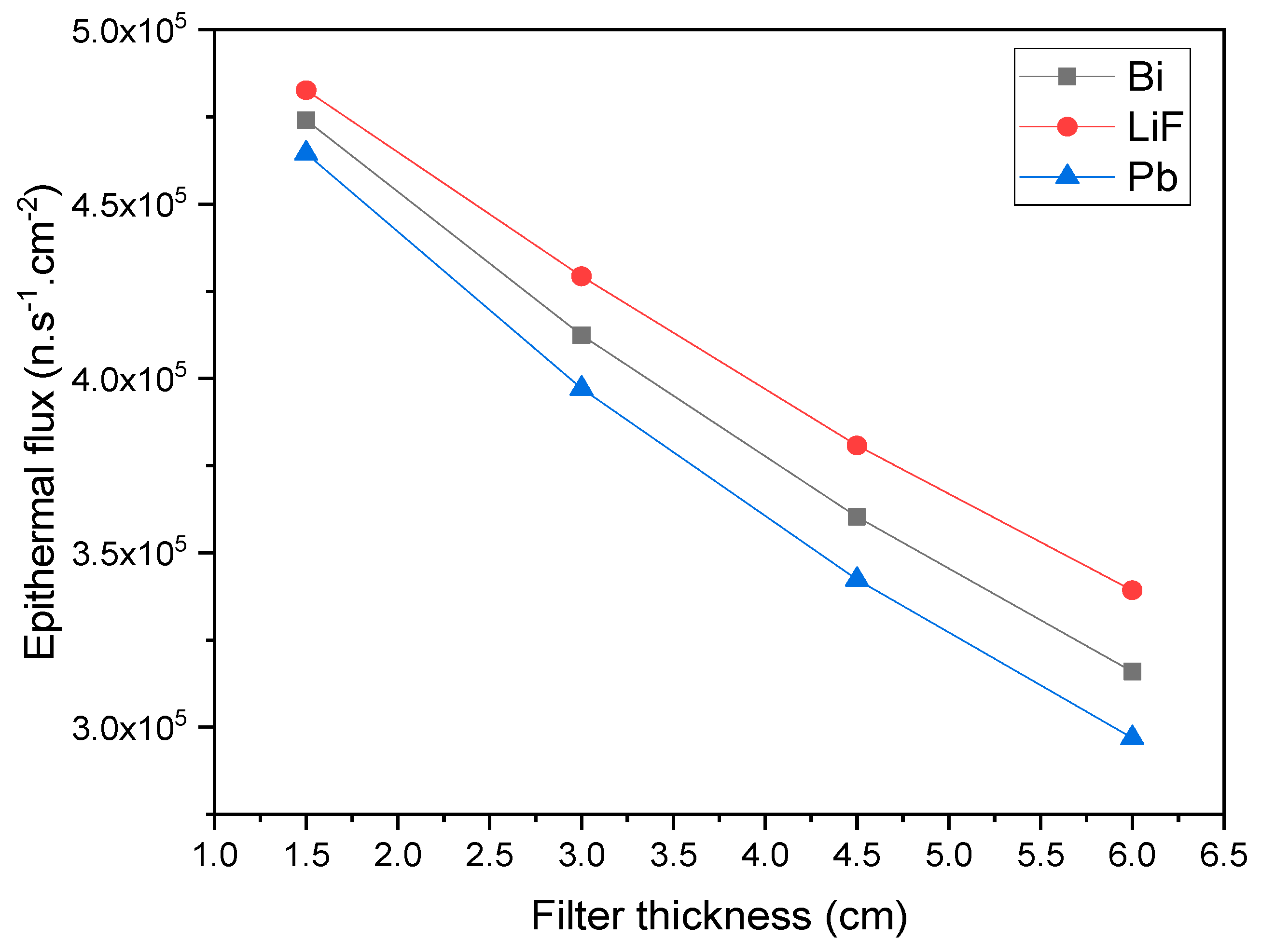
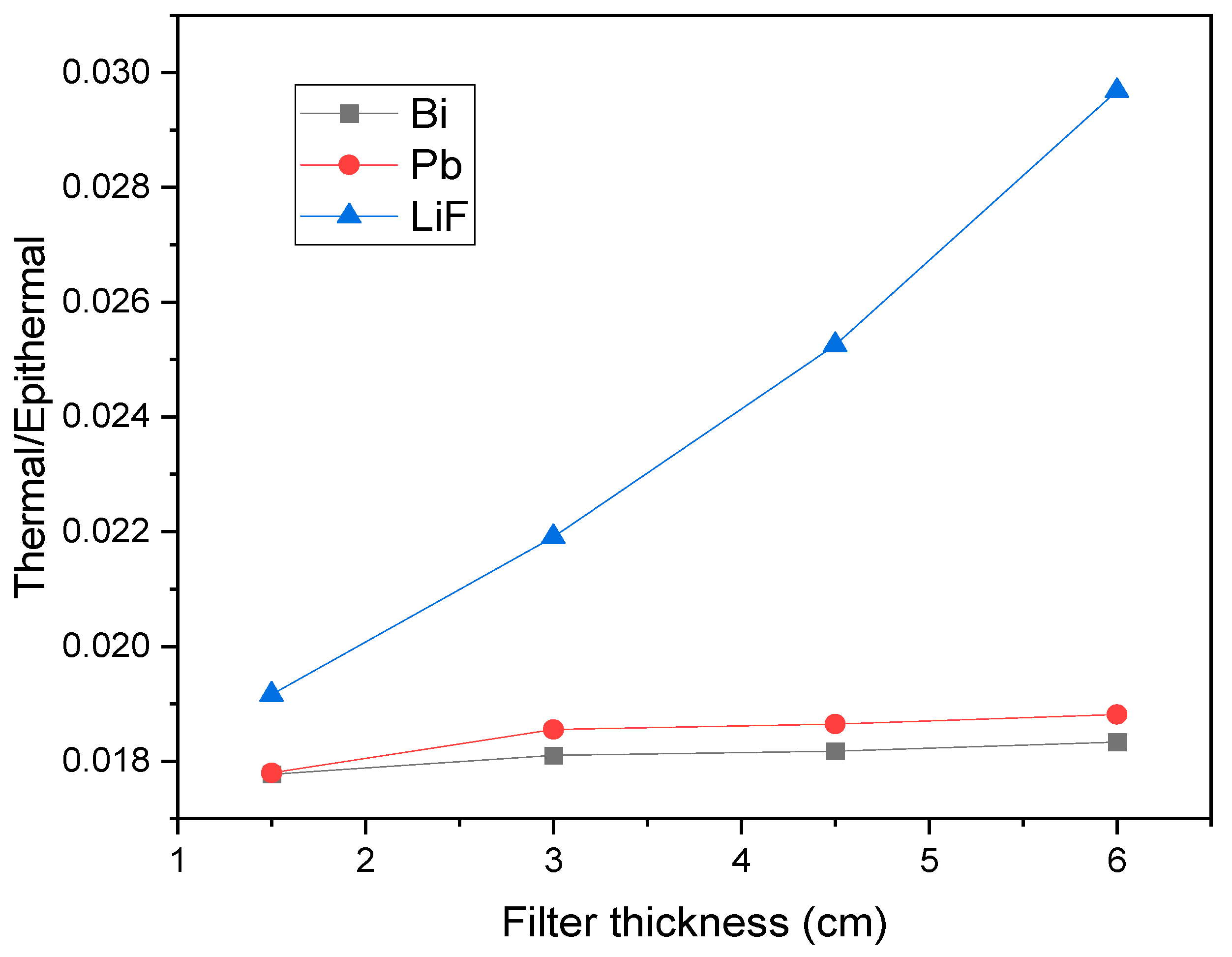
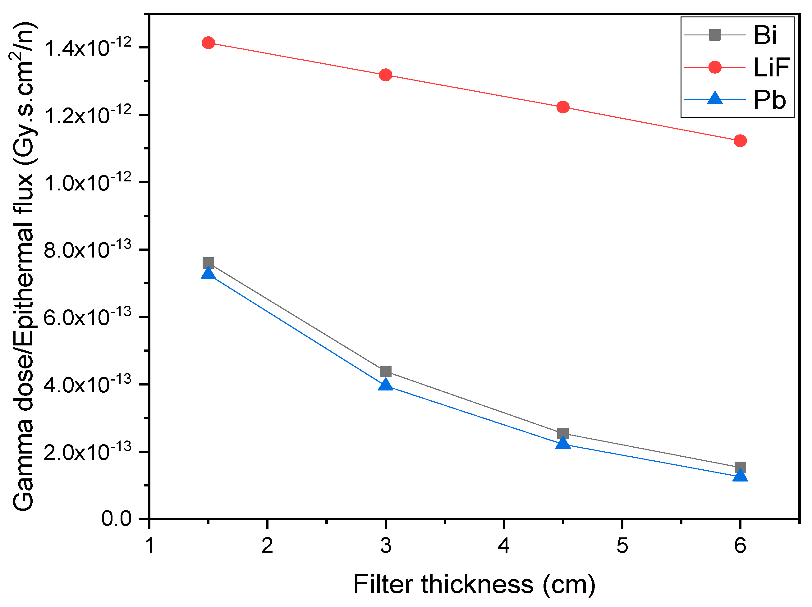
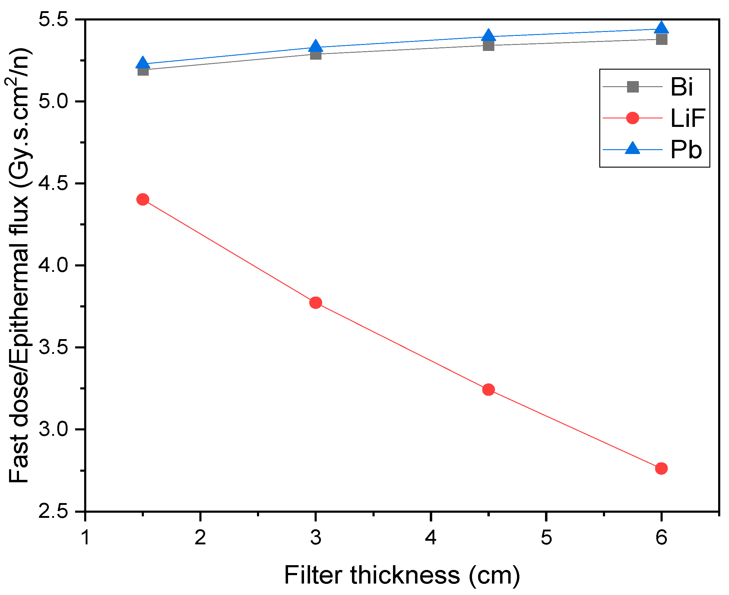
The next step after selecting the optimal moderator was to alter the thickness of the moderator and use filters to lower the dosages of gamma and fast neutrons as well as those of thermal neutrons in surface tissues, as the system was intended to target deep-seated tumors. In this project, the filter candidate materials were LiF, Pb, and Bi: LiF is good at absorbing thermal neutrons, Pb is good at filtering gamma rays and fast neutrons, and Bi is a gamma ray filter. The effect of these materials in the BSA is displayed in Figure 5, Figure 6, Figure 7 and Figure 8. The fact that the output epithermal flux dropped as the filter material’s thickness increase is something that requires attention, meaning that it is vital to balance lowering unneeded dosages and enhancing epithermal flow.
Using combinations of different filters, the output could be improved, as shown in Table 4.
In this work, hundreds of different trials were carried out for different components of BSA. The best result from these trials was obtained when the moderator was 32 cm of MgF2, the reflector was 76 cm of Pb, the filters were 3 cm Pb and 1.52 cm Bi, and the collimator was a cone frustum of Pb with height 10 cm and its aperture diameter 12 cm, as shown in row 4 of Table 4. The current-to-epithermal flux ratio of this BSA was 0.87, which is a good result. With further trials and different combinations, the output may be improved.
Figure 9 shows the configuration utilized to evaluate in-phantom parameters.
A rectangular parallelepiped phantom with dimensions of 14 cm × 17 cm × 14 cm was employed in the dosage calculation setup. A 1.5 cm radius tumor, the typical size of several tumor forms, was modeled inside the phantom. In order to examine the effectiveness of the system with tumors in varying places, the sampled tumor’s center was shifted from 1.92 cm to 13 cm.
Figure 10 provides a general evaluation of the clinical parameters. For this BSA output, the AD was 5.5 cm, the AR was 2.29, and the ADDR was 4.1 × 10⁻⁴ Gy.Eq/min. The ADDR decreased excessively because the neutron generator’s initial output was insufficient. In order to obtain the recommended epithermal flux, the generator’s initial neutron fluence needs to be 1.14 × 10¹³ n/s. Achieving 1.14 × 10¹³ n/s neutron fluence would result in an ADDR of 0.47 Gy.Eq/min, meaning that the therapy could be completed in 30 min or less.
Table 5 and Table 6 compare the proposed system to the DD-109 (Y = 2 × 10⁹ n/s), the preceding neutron generator in the DD neutron generator series. When compared with the DD-109 neutron generator-based BNCT, the DD-110 system presented several good enhancements, although not meeting all of the IAEA’s requirements.
In terms of beam quality, the DD-110 showed improvements in some areas while facing challenges in others. The DD-110 showed a significant improvement in epithermal neutron flux, showing a 4.36-fold increase compared to the DD-109. This enhancement is particularly beneficial for BNCT, as it provides a higher flux of neutrons in the desired energy range. However, it still limits its clinical utility, as these values would necessitate impractically long treatment times and restrict effectiveness to shallow tumors. While the system does not fulfill all IAEA requirements, its evaluation remains valuable for guiding iterative refinements in neutron source design and highlighting gaps in performance metrics.
Table 5 also shows a lower thermal-to-epithermal flux ratio for DD-110, which is advantageous and well within IAEA recommendations. The DD-110 also showed a reduction in gamma dose contamination, meeting IAEA standards, better than DD-109. These improvements suggest a better-quality neutron beam for treatment purposes. However, DD-110 showed an increase in fast neutron contamination compared to DD-109, with both generators exceeding IAEA recommendations in this aspect.
When considering in-phantom parameters (see Table 6), DD-110 presented a mixed profile. It offered a shallower advantage depth compared to DD-109, which may make it more suitable for treating tumors closer to the surface. However, this came at the cost of a lower advantage ratio. On the positive side, DD-110 demonstrated an improved ADDR and a shorter treatment time. These factors could lead to more efficient treatment sessions and potentially improved patient comfort.
Overall, the DD-110 neutron generator shows significant advancements in several key areas important for BNCT, particularly in terms of epithermal neutron flux, reduced gamma contamination, and treatment efficiency. However, the increased fast neutron contamination and shallower advantage depth present challenges that need to be considered. The choice between these generators would ultimately depend on the specific requirements of the BNCT application, including factors such as the typical depth of tumors being treated and the desired balance between treatment time and beam quality
Crucially, no current BNCT facility completely meets every IAEA criterion, underlining the need for investigating all newly developed neutron sources for their potential contributions, even modest progress in one domain such as neutron beam optimization or boron carrier development, as these could synergize with advancements in other areas. The interdisciplinary character of BNCT means that even minor advancements in one area can have significant impacts across the field, underscoring the importance of comprehensive documentation and open dissemination of all research outcomes. Ultimately, this collaborative approach fosters a dynamic research environment where every contribution, regardless of its immediate impact, has the potential to drive substantial progress in the complex landscape of BNCT research and development.

4. Conclusions

From the perspective of a medical physicists, BNCT entails a few steps: producing the necessary quantity of neutrons, adjusting them to the proper energy range, and figuring out the dosage deposition. This research leads to the conclusion that the DD-110 neutron generator emits less neutrons. Given that neutron generators are inexpensive, lightweight, and easy to construct and operate, more advancements in this field are encouraged. Further fast neutron dose filters should have a higher thickness in order to reduce the fast neutron dose. Moreover, as the epithermal flux drops with the addition of the fast neutron filter, the right fast neutron dosage and epithermal flux could be obtained with a neutron generator with an output of 2 × 10¹³ n/s. Such improvement cannot be achieved using a DD source because it is beyond current technological capabilities
Broadly speaking, BNCT is a promising radiation treatment. One of the inputs in this treatment is a selective neutron source. Compared to research reactors and accelerators, neutron generators are safer, more affordable, and have a more straightforward design. On the other hand, neutron generators produce fewer neutrons than advised. Therefore, neutron generators need to be improved even further.
Recently, BNCT has been put into practice; the next challenge facing this treatment modality is making it simple and easy. The major aspect making this treatment modality simple and uncomplicated is a good neutron source. Thus, a future research topic should be obtaining neutron sources, such as DD and DT neutron generators, that can create the right number of neutrons. Such neutron generator-based BNCT improvements offer valuable insights for neutron source developers, enabling them to effectively utilize neutrons in clinical fields.

Author Contributions

Conceptualization, H.D.; methodology, H.D. and M.U.; software, H.D.; validation, H.D. and M.U.; formal analysis, H.D.; investigation, H.D.; data curation, H.D.; writing—original draft preparation, H.D. and M.U.; writing—review and editing, H.D. and M.U.; visualization, H.D. and M.U.; supervision, H.D.; and project administration, H.D. All authors have read and agreed to the published version of the manuscript.

Funding

This project was funded by the Deanship of Scientific Research (DSR) at King Abdulaziz University (KAU), Jeddah, under grant no. (G: 1566-130-1440).

Data Availability Statement

The data that support the findings of this study are available from the corresponding author upon reasonable request.

Acknowledgments

This project was funded by the Deanship of Scientific Research (DSR) at King Abdulaziz University (KAU), Jeddah, under grant no. (G: 1566-130-1440). The authors, therefore, acknowledge with thanks DSR for technical and financial support.

Conflicts of Interest

The authors declare no conflicts of interest.

References

  1. Sathishkumar, K.; Chaturvedi, M.; Das, P.; Stephen, S.; Mathur, P. Cancer incidence estimates for 2022 & projection for 2025: Result from National Cancer Registry Programme, India. Indian J. Med. Res. 2022, 156, 598–607. [Google Scholar] [PubMed]
  2. Lapointe, S.; Perry, A.; Butowski, N.A. Primary brain tumours in adults. Lancet 2018, 392, 432–446. [Google Scholar] [CrossRef] [PubMed]
  3. Schaff, L.R.; Mellinghoff, I.K. Glioblastoma and other primary brain malignancies in adults: A review. Jama 2023, 329, 574–587. [Google Scholar] [CrossRef]
  4. Alsharif, A.; Alsharif, M.T.; Samman, M.; Binmadi, N.; Kassim, S.; Mourad, S.; Warnakulasuriya, S. Forecasting Head and Neck Cancer Trends in GCC Countries: Implications for Public Health Policy and Strategy. Risk Manag. Healthc. Policy 2023, 16, 2943–2952. [Google Scholar] [CrossRef]
  5. Meram, A.T.; Woo, B.M. Oral Cancer Management. In Peterson’s Principles of Oral and Maxillofacial Surgery; Springer: Cham, Switzerland, 2022; pp. 1009–1055. [Google Scholar]
  6. Matsumoto, Y.; Fukumitsu, N.; Ishikawa, H.; Nakai, K.; Sakurai, H. A critical review of radiation therapy: From particle beam therapy (proton, carbon, and BNCT) to beyond. J. Pers. Med. 2021, 11, 825. [Google Scholar] [CrossRef]
  7. Kanno, H.; Nagata, H.; Ishiguro, A.; Tsuzuranuki, S.; Nakano, S.; Nonaka, T.; Kiyohara, K.; Kimura, T.; Sugawara, A.; Okazaki, Y.; et al. Designation products: Boron neutron capture therapy for head and neck carcinoma. Oncologist 2021, 26, e1250–e1255. [Google Scholar] [CrossRef] [PubMed]
  8. Yong, Z.; Song, Z.; Zhou, Y.; Liu, T.; Zhang, Z.; Zhao, Y.; Chen, Y.; Jin, C.; Chen, X.; Lu, J.; et al. Boron neutron capture therapy for malignant melanoma: First clinical case report in China. Chin. J. Cancer Res. 2016, 28, 634. [Google Scholar] [CrossRef]
  9. Coderre, J.A.; Morris, G.M. The radiation biology of boron neutron capture therapy. Radiat. Res. 1999, 151, 1–18. [Google Scholar] [CrossRef]
  10. He, H.; Li, J.; Jiang, P.; Tian, S.; Wang, H.; Fan, R.; Liu, J.; Yang, Y.; Liu, Z.; Wang, J. The basis and advances in clinical application of boron neutron capture therapy. Radiat. Oncol. 2021, 16, 1–8. [Google Scholar] [CrossRef]
  11. Karaoglu, A.; Arce, P.; Obradors, D.; Lagares, J.I.; Unak, P. Calculation by GAMOS/Geant4 simulation of cellular energy distributions from alpha and lithium-7 particles created by BNCT. Appl. Radiat. Isot. 2018, 132, 206–211. [Google Scholar] [CrossRef]
  12. Valentin, J. Relative biological effectiveness (RBE), quality factor (Q), and radiation weighting factor (wR): ICRP Publication 92. Ann. ICRP 2003, 33, 1–121. [Google Scholar]
  13. Malouff, T.D.; Seneviratne, D.S.; Ebner, D.K.; Stross, W.C.; Waddle, M.R.; Trifiletti, D.M.; Krishnan, S. Boron neutron capture therapy: A review of clinical applications. Front. Oncol. 2021, 11, 351. [Google Scholar] [CrossRef] [PubMed]
  14. Locher, G.L. Biological effects and therapeutic possibilities of neutrons. Am. J. Roentgenol. 1936, 36, 1–13. [Google Scholar]
  15. Taylor, H.; Goldhaber, M. Detection of nuclear disintegration in a photographic emulsion. Nature 1935, 135, 341. [Google Scholar] [CrossRef]
  16. Jin, W.H.; Seldon, C.; Butkus, M.; Sauerwein, W.; Giap, H.B. A review of boron neutron capture therapy: Its history and current challenges. Int. J. Part. Ther. 2022, 9, 71–82. [Google Scholar] [CrossRef]
  17. Lamba, M.; Goswami, A.; Bandyopadhyay, A. A periodic development of BPA and BSH based derivatives in boron neutron capture therapy (BNCT). Chem. Commun. 2021, 57, 827–839. [Google Scholar] [CrossRef]
  18. Sakurai, Y.; Tanaka, H.; Takata, T.; Fujimoto, N.; Suzuki, M.; Masunaga, S.; Kinashi, Y.; Kondo, N.; Narabayashi, M.; Nakagawa, Y.; et al. Advances in boron neutron capture therapy (BNCT) at kyoto university-From reactor-based BNCT to accelerator-based BNCT. J. Korean Phys. Soc. 2015, 67, 76–81. [Google Scholar] [CrossRef]
  19. Ahmed, M.; Bedogni, R.; Chen, Y.; Dagrosa, A.; Green, S. Advances in Boron Neutron Capture Therapy; International Atomic Energy Agency: Vienna, Austria, 2023. [Google Scholar]
  20. Donya, H.; Seniwal, B.; Darwesh, R.; Fonseca, T.C.J.T. Prospective Monte Carlo simulation for choosing high efficient detectors for small-field dosimetry. In Theory, Application, and Implementation of Monte Carlo Method in Science and Technology; IntechOpen: Rijeka, Croatia, 2019; p. 39. [Google Scholar]
  21. Donya, H. Pencil-beam fluence evaluation based on Monte Carlo simulations algorithm of high energetic treatment photons. J. Med. Signals Sens. 2018, 8, 81–86. [Google Scholar] [CrossRef] [PubMed]
  22. Alem-Bezoubiri, A.; Bezoubiri, F.; Speiser, M.; Suleiman, S.A.; Donya, H.; Chami, A.C. Monte Carlo study of organ doses and related secondary cancer risk estimations for patients undergoing prostate radiotherapy: Algerian Population-Based Study. Appl. Radiat. Isot. 2024, 216, 111595. [Google Scholar] [CrossRef]
  23. Robalino, L.E.C.; Fernández, G.F.G.; Gallego, E.; Guzmán-García, K.A.; Vega-Carrillo, H.R. Study by Monte Carlo methods of an explosives detection system made up with a DD neutron generator and NaI (Tl) gamma detectors. Appl. Radiat. Isot. 2018, 141, 167–175. [Google Scholar]
  24. Rinard, P. Neutron interactions with matter. In Passiv. Nondestruct. Assay Nucl. Mater.; Reilly, D., Ensslin, N., Smith, H., Eds.; Los Alamos National Laboratory Report LA-UR-90-732; Los Alamos National Laboratory: Los Alamos, NM, USA, 1991; pp. 375–377. [Google Scholar]
  25. Obayashi, S.; Kato, I.; Ono, K.; Masunaga, S.-I.; Suzuki, M.; Nagata, K.; Sakurai, Y.; Yura, Y. Delivery of 10boron to oral squamous cell carcinoma using boronophenylalanine and borocaptate sodium for boron neutron capture therapy. Oral Oncol. 2004, 40, 474–482. [Google Scholar] [CrossRef] [PubMed]
  26. Hsieh, M.; Liu, Y.; Mostafaei, F.; Poulson, J.M.; Nie, L.H. A feasibility study of a deuterium–deuterium neutron generator-based boron neutron capture therapy system for treatment of brain tumors. Med. Phys. 2017, 44, 637–643. [Google Scholar] [CrossRef] [PubMed]
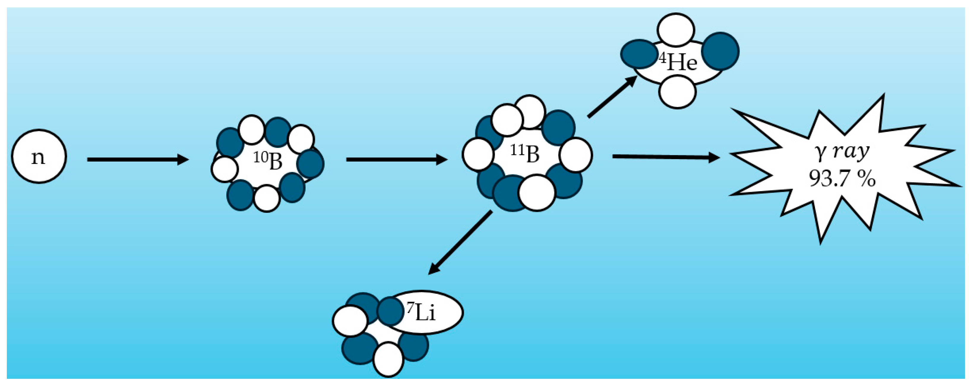
Figure 1. Boron neutron capture nuclear reaction (93.7%).
Qubs 09 00012 g001
Figure 2. General beam-shaping assembly (BSA).
Qubs 09 00012 g002
Figure 3. Examining epithermal flux at different moderator thicknesses ((left image) created in visedX_24E version (visedX_24E, Los Alamos National Laboratory, Los Alamos, NM, USA), where the blue color represents the environment of the system, the orange represents the reflector and collimator, the white and red represent the neutron generator and the moderator, respectively, and the faces of the small boxes are the areas in which epithermal fluxes at different thicknesses are calculated (right image) in the schematic diagram in the left image.
Qubs 09 00012 g003
Figure 4. Epithermal flux for different material moderators versus their thicknesses.
Qubs 09 00012 g004
Figure 5. Effect of different filter materials on epithermal flux of the BSA with a 32 cm MgF2 moderator.
Qubs 09 00012 g005
Figure 6. Effect of different filter materials on thermal-to-epithermal flux ratio of the BSA with a 32 cm MgF2 moderator (the recommended value is ≤0.05).
Qubs 09 00012 g006
Figure 7. Effect of different filter materials on gamma dose-to-epithermal flux ratio of the BSA with a 32 cm MgF2 moderator.
Qubs 09 00012 g007
Figure 8. Effect of different filter materials on fast dose to epithermal flux ratio of the BSA with a 32 cm MgF2 moderator.
Qubs 09 00012 g008
Figure 9. The setup for dose calculation using a rectangular phantom, the yellow region. The small red circle in the phantom is a tumor with a diameter of 3 cm created with the visedX_24E application.
Qubs 09 00012 g009
Figure 10. Dose rate of tumor and healthy tissue in parallelepiped phantom.
Qubs 09 00012 g010
Table 1. Energy-based neutron classification [19].
Neutron TypeEnergy Range
Thermal neutron<0.5 eV
Epithermal neutron1eV–10 keV
Fast neutron>10 keV
Table 2. IAEA recommendation of neutron beam parameters for BNCT [19].
ParameterRecommended Value
Thermal neutron<0.5 eV
Therapeutic   epithermal   flux   ( φ e p t h )≥5 × 10⁸ nepith.cm−2s−1
Thermal - to - epithermal   flux   ratio   ( φ t h e r / φ e p t h )≤0.05
Beam   collimation   ( J / φ e p t h )≥0.7
Fast   neutron   dose - to - epithermal   flux   ratio   ( D fast / φ e p t h )≤2 × 10⁻¹³ Gy.cm2
Gamma   dose - to - epithermal   flux   ratio   ( D γ / φ e p t h )≤2 × 10⁻¹³ Gy.cm2
Therapeutic   epithermal   flux   ( φ e p t h )≥5 × 10⁸ nepith.cm−2s−1
Thermal - to - epithermal   flux   ratio   ( φ t h e r / φ e p t h )≤0.05
Beam   collimation   ( J / φ e p t h )≥0.7
Fast   neutron   dose - to - epithermal   flux   ratio   ( D fast / φ e p t h )≤7 × 10⁻¹³ Gy.cm2
Gamma   dose - to - epithermal   flux   ratio   ( D γ / φ e p t h )≤2 × 10⁻¹³ Gy.cm2
Table 3. Radiation weight factors in BNCT [19].
C B E B (Normal Tissue) C B E B (Tumor) R B E H R B E N R B E γ H
1.353.8331
Table 4. Effect of different combinations of filters on different parameter values (32 cm MgF2 moderator).
Pb (cm)LiF (cm)Bi (cm) φ e p t h
nepith.cm−2s−1
φ t h e r / φ e p t h Dfast / φ e p t h
(Gy.cm2)
D γ / φ e p t h
(Gy.cm2)
0.5200.56.01 × 1051.79 × 10−24.17 × 10−126.71 × 10⁻¹³
1.5200.525.46 × 1051.86 × 10−24.19 × 10−124.46 × 10⁻¹³
301.524.36 × 1051.88 × 10−24.19 × 10−121.71 × 10⁻¹³
31.5204.72 × 1052.41 × 10−23.87 × 10−124.82 × 10⁻¹³
Table 5. In-air parameter comparison of DD-109 and DD-110 neutron generators.
Generator
Model
φ e p t h
(nepith.cm−2s−1)
φ t h e r / φ e p t h D fast / φ e p t h
(Gy.cm2)
D γ / φ e p t h (Gy.cm2)
DD-109 [26]1 × 1050.055.5 × 10−132.49 × 10⁻¹³
DD-110 (proposed generator)4.36 × 1050.024.19 × 10−121.71 × 10⁻¹³
IAEA recommendations≥5 × 10⁸≤0.05≤2 × 10⁻¹³≤2 × 10⁻¹³
Table 6. In-phantom parameter comparison of DD-109 and DD-110 neutron generators.
Generator
Model
AD
(cm)
ARADDR (Gy.Eq/min)TT
(min)
DD-109 [26]12.13.70.3140
DD-110 (proposed generator)5.52.290.4730
IAEA recommendationsNo recommended value≤60
Disclaimer/Publisher’s Note: The statements, opinions and data contained in all publications are solely those of the individual author(s) and contributor(s) and not of MDPI and/or the editor(s). MDPI and/or the editor(s) disclaim responsibility for any injury to people or property resulting from any ideas, methods, instructions or products referred to in the content.