Review

Sigma-Class Glutathione Transferases (GSTσ): A New Target with Potential for Helminth Control

by
Lluvia de Carolina Sánchez Pérez
1,
Rafael A. Zubillaga
1,*,
Ponciano García-Gutiérrez
1 and
Abraham Landa
2,*
1
Departamento de Química, Universidad Autónoma Metropolitana-Iztapalapa, Mexico City C.P. 09310, Mexico
2
Departamento de Microbiología y Parasitología, Facultad de Medicina, Universidad Nacional Autónoma de México, Ciudad Universitaria, Mexico City C.P. 04510, Mexico
*
Authors to whom correspondence should be addressed.
Trop. Med. Infect. Dis. 2024, 9(4), 85; https://doi.org/10.3390/tropicalmed9040085
Submission received: 23 February 2024 / Revised: 10 April 2024 / Accepted: 12 April 2024 / Published: 16 April 2024
(This article belongs to the Section Infectious Diseases)

Abstract

:
Glutathione transferases (GSTs EC 2.5.1.18) are critical components of phase II metabolism, instrumental in xenobiotics’ metabolism. Their primary function involves conjugating glutathione to both endogenous and exogenous toxic compounds, which increases their solubility and enables their ejection from cells. They also play a role in the transport of non-substrate compounds and immunomodulation, aiding in parasite establishment within its host. The cytosolic GST subfamily is the most abundant and diverse in helminths, and sigma-class GST (GSTσ) belongs to it. This review focuses on three key functions of GSTσ: serving as a detoxifying agent that provides drug resistance, functioning as an immune system modulator through its involvement in prostaglandins synthesis, and acting as a vaccine antigen.

1. Introduction

Detoxification is an important enzyme function, and organisms have evolved complex and efficient mechanisms to perform it. One such mechanism is xenobiotic metabolism, existing in three phases: bioactivation, conjugation, and transport [1]. Xenobiotics are chemical substances that are not nutrients and are not necessary for maintaining homeostasis or normal physiological and biochemical functions. Some can be toxic, such as drugs, food additives, and heavy metals [2].
Different families of enzymes perform this metabolic process through three phases. In phase I, cytochrome P450 (CPY) enzymes are predominantly used. Phase II involves glutathione transferases (GSTs), a crucial group responsible for conjugating reduced glutathione with toxic compounds to enhance their solubility and ease excretion. Lastly, in phase III, the compounds are expelled from cells through transporters such as ABC [3]. This review focuses on sigma-class GST (GSTσ), discussing studies on and contributions to this class of enzymes in various organisms to better understand its functions. The aim is to show its potential for controlling diseases caused by helminths, with a focus on developing environmentally friendly and safe products for humans.

2. Glutathione Transferases (GSTs)

GSTs (EC 2.5.1.18) are a multifunctional superfamily of enzymes found in all aerobic organisms [4]. They primarily catalyze the conjugation of the thiolate ion of reduced glutathione (GSH) to the electrophilic centers of hydrophobic organic molecules [3,5]. This process is essentially an evolutionary adaptation for the efficient elimination of numerous potentially toxic chemicals [6].
GSTs assist in the metabolism of harmful endogenous compounds, such as free radicals and reactive oxygen species. They also process xenobiotics, including aldehydes, quinones, epoxides, hydroperoxides, drugs, carcinogens, and environmental pollutants. Furthermore, GSTs play a role in the sequestration and transport of these compounds [7,8]. Within the GST family in helminths, there are three subfamilies: the cytosolic subfamily, which is abundant in cells; the mitochondrial subfamily; and the microsomal subfamily (MAPEG). The latter is associated with the membrane and is involved in the metabolism of eicosanoids and glutathione. To date, these subfamilies have been identified and described in mammals, reptiles, plants, mollusks, insects, helminths, and bacteria [9,10,11].
GSTs perform several functions, which include conjugation with glutathione (GSH), the biosynthesis of key biological mediators, the modulation of cellular signals, and the regulation of calcium channels. They also transport non-substrate molecules, including bilirubin, the heme group, steroids, hormones, and bile salts [10,12]. Moreover, they play an important role in the catabolism of tyrosine and catalyze the isomerization reactions essential for the synthesis of prostaglandins and steroids. GSTs also aid in the removal of reactive oxygen species and the regeneration of S-thiolated proteins, offering protection against oxidative cell damage [10,13]. Table 1 shows the core functions of the three main GST families, along with the types that each one possesses, based on primary structure similarities, substrate specificity, inhibition properties, and isoelectric point [14]. For a more in-depth understanding, we recommend reviewing [15].
The various classes of GSTs have multiple isoforms that serve the same function, with slight differences in their primary structure and subsequent electrical charge. This has been observed across a range of organisms, including rats, humans, plants, parasites, and insects. The presence of multiple isoforms provides biological and adaptative advantages, allowing organisms to biotransform and eliminate a broader of toxic com-pounds, which would be impossible with just one isoform [14]. The level of sequence identity for enzymes within the same class is typically high, around 50%, with in a species. However, this drops to 20–40% between different classes [24,25]. The most exten-sively studied GST family is the cytosolic family, largely due to its significant role in human pharmacology.
The structural features of most cytosolic GST classes—such as their compact folds, conserved structure, well-defined domains, and binding cavities at G, H, and L sites—make them easier to study [10].

2.1. Cytosolic GST (cGST)

Thirteen classes of GSTs in animals and plants are displayed in Table 1. Mammals boast the most classifications, with seven, while plants have six. These have been identified in insects, helminths, mollusks, and fungi [13,26]. The cGST subfamily, which contributes to approximately 3–4% of a cell’s total soluble protein, underlines its significance. This subfamily is further divided based on structural similarity and the type of catalytic residue at the GSH binding site forming the GS- thiolate anion. The alpha, mu, pi, and sigma classes, which seemingly evolved more recently, activate GSH using a tyrosine near the N-terminus, whereas the older theta, omega, and zeta classes utilize serine or cysteine for activation [27]. The quaternary structure of cGST is composed of two monomers, each weighing between 23 and 28 kDa, forming a roughly 50 kDa dimer. The dimer can feature identical (homodimer) or different (heterodimer) subunits from the same class. The dimeric configuration is crucial for enhancing protein stability and ensuring the proper active site structure for efficient catalysis [28,29].
Each monomeric unit of cGST integrates an N-terminal domain harboring a GSH binding site (G site) composed of four mixed β strands, of which the first, second, and fourth point in the same direction, while the third is antiparallel. This domain also includes three α helices and is arranged in a βαβαββα format, resembling that of thioredoxin [3]. The C-terminal domain contains a hydrophobic binding site for electrophilic substrates (H-site) with only α helices, the count of which varies between five and six according to the class. Notably, the H-site displays considerable structural variability among classes, along with differing specificity and substrate recognition attributes, given its plasticity and flexibility. This contributes to the enzyme’s catalytic promiscuity, enabling it to bind to various electrophilic substrates [8]. These structural differences aid cells in eliminating a more diverse range of toxic compounds, as the binding site (H) affinity differs across classes [30].

2.1.1. Glutathione (GSH)

Glutathione, a water-soluble tripeptide composed of glutamic acid, cysteine, and glycine (L-g-glutamyl-L-cysteinyl-glycine), is an essential factor in maintaining cellular REDOX balance and is found throughout the body [31]. This molecule exists in reduced (GSH) and oxidized (GSSG) states, as well as being bound to proteins. The free form predominantly exists within cells, serving as a reductant or substrate in protection against oxidative and xenobiotic stresses [2]. Meticulously evolved over time, glutathione performs many functions, with one of the most significant being the detoxification of foreign compounds (xenobiotics) through enzyme-regulated processes involving GST [31,32]. If these potentially hazardous xenobiotics were not neutralized by GSH, they could react directly with DNA, RNA, lipids, or proteins within cells, causing significant damage. Additionally, the introduction of GSH to reactive electrophiles tags the R group with the tripeptide, setting it up for recognition by ATP-dependent GSH export pumps, which expel such conjugates from the cell [33,34].

2.1.2. Catalytic Mechanism of the Conjugation Reaction

To catalyze the conjugation reaction, certain physicochemical alterations are needed. First, a structural rearrangement must take place, characterized by typical conformational changes induced by the substrate. Second, the ionization of reduced GSH, which produces the thiolate anion (GS-), is required. The binding of GSH to GST has been found to decrease the pKa value of the -SH group at the G site [35,36]. This reduction in the pKa of the thiol group improves its ionization capability, thereby generating a more potent nucleophile (RS- versus RSH). This enhanced nucleophile can then interact more effectively with the electrophilic substrate at the H-site at a neutral pH. This accounts for the high conservation of tyrosine and why its -OH group acts as the acceptor in the hydrogen bond formed between the G site and the GSH [4,18].

2.1.3. Substrates and Specificity

A variety of substrates are currently utilized, along with other parameters, to ascertain the specificity of a GST and its classification. Many GSTs demonstrate enzymatic activity toward 1-chloro-2,4-dinitrobenzene (CDNB), which serves as a standard substrate. However, various other substrates have been employed, including 1,2-dichloro-4-nitrobenzene (DCNB), ethacrynic acid, lipid hydroperoxides, and reactive carbonyls. Furthermore, inhibitors have been used as class markers, such as bromosulfophthalein for the alpha class, cibacron blue, and triphenyltin chloride for the mu class. Other substances employed are hematin-related compounds and bile acids, which help to define their class and specificity (Table 2) [37,38].
Although various substrates have been used to identify new GSTs, the specificity or inhibition degree of each enzyme for the same compound can differ, even if they belong to the same class. A comparative study of activities with 4-hydroxyalkenal (HNEs) and other activated alkenes has provided an understanding of certain GSTs’ active site properties. It has been proven that the specificity of 15 GSTs against 4-hydroxyalkenals (HNEs) is not related to their class membership; the higher the substrate’s hydrophobicity, the greater the binding free energy. The free-energy differences are estimated from their specificity constants (kcat/KM) [35]. For some enzymes, the active site’s steric limitations seem to offset the increased binding energy attributed to the increase in substrate chain length.
It is worth noting how glutathione (GSH) interacts with cytosolic GSTs. The tripeptide adopts an extended configuration upon binding to the enzyme at a β-strand end, with the cysteine sulfur pointing toward its monomer’s core, where it interacts with the hydrogen-bond donor. For example, for recombinant GSTσ from Taenia solium (TsMσGST), the formation of a hydrogen bond between the GS- sulfur and the Tyr14 hydroxyl has been observed [44]. In the case of theta- and zeta-class GSTs, the hydroxyl-group donor originates from a serine [19].
The most conserved region across all GSTs begins with a cis-proline residue before the β3 strand and continues through the α3 helix. This region plays a crucial role in binding the γ-glutamyl residue of GSH by providing hydrogen-bond interactions with glutamine or glutamate, followed by a serine or threonine located at the turn between the β4 strand and the α3 helix [5,55].

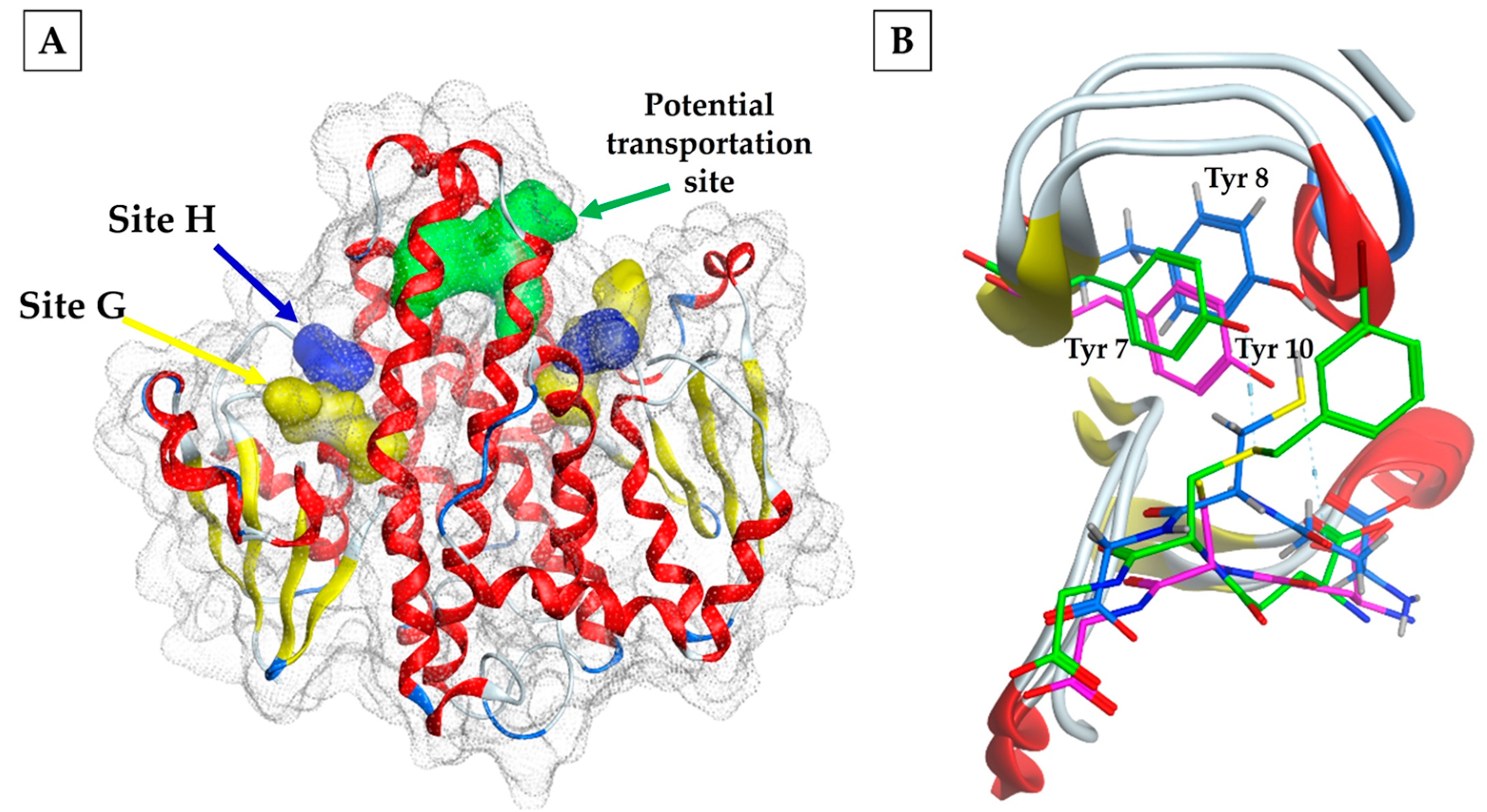
3. Structural and Functional Generalities of GSTσ

GSTσ was initially described in various species, including squid, humans, mice, rats, birds, insects, helminths, mollusks, and frogs [9]. According to Ji and colleagues [56], GSTσ evolutionarily diverged first from the same ancestral precursor as the alpha, mu, and pi classes. Its three-dimensional structure parallels that of other cytosolic GSTs, although a much shorter C-terminal domain gives it a more open active site. The N-terminal domain consists of α-helices and β-strands, which are smaller than those in the C-terminal domain. The latter, composed solely of α helices, contains most of the residues forming the H-site cavity [57].
The specific architecture of each GST’s catalytic site allows for unique functions besides detoxification. For example, the rat hematopoietic prostaglandin D synthase (H-PGDS) enzyme, which is a type of GSTσ, solely catalyzes the isomerization of PGH2 to PGD2. A similar function is demonstrated by the GSTσ of Schistosoma mansoni but not by the GSTσ of squid, which produces a mixture of PGD2, PGE2, and PGF from the same substrate [58,59,60]. In mammals, prostaglandins mediate various functions, including immune-response modulation, sleep promotion, hormone release, and vasodilation. The generation of diverse products by a single GST class could be attributed to an organism’s need to regulate its physiological activities or to the need to primarily evade the host’s immune response.
A noted variation occurs in the FhGST-S1 variant of Fasciola hepatica, which exhibits a disulfide bond between the cysteines at positions 26 and 196. This bond is crucial for the formation of the active site and is thought to help stabilize the enzyme when released into the host’s stomach. It is worth mentioning that FhGST-S1 is expressed in the Fasciola’s extracellular vesicles and can be released to modulate the host’s immune response [57].
Conversely, there is a third potential binding site named “L” (Figure 1A). This site is a hydrophobic cavity located approximately 14 Å from the hydroxyl group of Tyr-7 of the active site, that binds non-catalytic ligands, such as synthetic organic compounds, reactive oxylipins, phenols, flavonoids, and unstable intermediates. This binding process results in the sequestration or storage of these compounds or their transport to specified sites. Notably, this binding may inhibit the catalytic activity of the GSTs either competitively or non-competitively [61]. In the case of the squid enzyme, the L site transports glutathione with its conjugates, as revealed by the crystal structure of the protein complexed with S-(3-iodobenzyl) glutathione [56].
Electrostatic interactions near the active site are considered significant. As commonly observed, GSH binds consistently among all cytosolic GSTs, creating a hydrogen bond between the cysteine’s sulfhydryl group and the catalytic tyrosine; this residue is highly conserved in sigma-class GSTs, as well as in the alpha, mu, and pi classes. The preservation of Tyr as a hydrogen-bond donor has been identified in the GSTσ in several organisms, as demonstrated by bmGST1 in the insect Bombix mori, FhGST-S1 in Fasciola hepatica, and glutathione transferase in the squid Loligo vulgaris, where Tyr 8, Tyr 10, and Tyr 7 form the hydrogen bond, respectively [57,62,63] (Figure 1B).
In other cGSTs, a different catalytic residue is present, such as Ser in the theta and zeta classes or Cys in the omega class. Ser plays a role similar to that of Tyr in alpha-class GSTs, such as mu, pi, and sigma. Meanwhile, Cys, in the omega class, forms a mixed disulfide with GSH, resulting in variations in their catalysis mechanism. An interesting characteristic of GSTσ is its low activity against the universal substrate for GST, 1-chloro-2,4-dinitrobenzene (CDNB), but high activity against 4-hydroxynonenal (HNE). The latter is a cytotoxic peroxidation compound produced by oxidative stress [46]. For F. hepatica, two isoforms of GSTσ, FhGST-S1 and FhGST-S2, are reported. FhGST-S2 lacks total activity against the most common GST substrates, except for reactive aldehydes such as trans-2-Nonenal and trans-2,4-decadienal. This suggests a unique role for this isoform [42], as shown in Table 2.

3.1. Role of GSTσ in Helminths

Helminths have co-evolved with their host species, developing adaptive mechanisms for survival. For instance, they alter the genes that code for their actin isoforms and activate their detoxification enzymatic systems. These systems decrease the effects of drugs and, consequently, increase resistance [64]. Among helminths, GSTs are the predominant enzymes involved in the phase II metabolism of xenobiotics [12]. This prevalence is attributed to the parasites’ low expression of phase I detoxification enzymes, such as the cytochrome P450 family and glutathione peroxidase. Consequently, GST is the lead detoxification system in helminths [65,66].

GSTσ as Prostaglandin Synthase in Helminths

Recently, it was discovered that helminth GSTσ can also synthesize prostaglandins, which regulate cellular homeostasis and multiple physiological responses in humans. The main types of prostaglandins are PGD2, PGE2, PGF2, TXA2 (thromboxane), and PGI2 (prostacyclin). The first two are produced by GSTσ in helminths and have distinct functions. PGD2 triggers sleepiness during parasitic infection, restrains the migration of the host’s epidermal Langerhans cells (LC), and instigates programmed cell death. Conversely, PGE2 acts as a powerful immunomodulator, causing inflammation, fever, headaches, and bodily aches, reducing cytokine production, enabling signal coupling during phagocytosis, and enhancing the parasite’s ability to penetrate host cells [9,67]. It is recognized that prostaglandins are crucial for the initiation and progress of parasitosis.
In the context of helminth infections, their prostaglandins impact their growth and reproduction, as well as manipulate the host’s immune system to their advantage [39,68]. Notably, the prostaglandins PGD2 and PGE2 are found throughout various stages of the parasite’s development, synthesized by the helminth’s GSTσ and the microsomal GST (MAPEG), respectively. High concentrations of PGD2 have been found in non-embryonic eggs, indicating a potential role in egg development [40]. Prostaglandin production similarly modulates the host’s immune response in the sexually immature metacercariae stage.
The presence of GSTσ in tissues that interact directly with the host suggests that prostaglandin synthesis is vital for the parasite’s survival and reproduction, contributing to changes in the parasite’s pathophysiology and its ability to invade and establish itself. Specifically, PGE2 also impacts the fertilization and survival of eggs. Researchers have discovered that GSTσ from Schistosoma mansoni (Sm28GST) produces PGD2 upon penetrating the host’s skin. PGD2 inhibits the migration of LCs to the skin’s surface. Given LCs’ essential role in establishing skin immunity by triggering immune responses in the presence of antigens, this shows how S. mansoni has developed a strategy to neutralize the host’s immune response [69]. Moreover, PGD2 obstructs the LCs’ departure from the epidermis and the accumulation of dendritic cells in the lymph nodes, responsible for increasing the immune response [70]. This suppressive effect is mediated by soluble lipophilic factors released by the parasites, not host-derived anti-inflammatory cytokines, emphasizing PGD2’s role in stalling LC migration. These findings highlight the intriguing strategies deployed by S. mansoni to combat the host’s immune response, which may pave the way for developing new treatments.
The nematode Onchocerca volvulus produces GSTσ (Ov-GST1), which expresses PGD2 in the hypodermis, cuticle, and epicuticle upon host contact. GSTs, enzymes typically located within a cell’s cytosol, being found outside cells, as with Ov-GST1, may indicate an evolutionary adaptation. This pattern mirrors that of F. hepatica FhGST-S1’s GSTσ, which is released through extracellular vesicles [43]. Similar observations have been made in the cestodes Echinococcus granulosus (EgGST2) and Taenia solium (Ts24GST), where the location of GSTσ in the protoscolex and scolex’s cytosol has been confirmed. The biochemical properties and locations of these enzymes have led to suggestions of their role in drug resistance and immune-response modulation [38]. A glycosylated GSTσ, Onchocerca ochengi OoGST1, has been identified and characterized in the nematode, known to infect cattle and humans [71]. A glycan analysis of OoGST1 revealed the presence of α(1–3)fucose, a common carbohydrate in helminth glycoproteins, known to induce a TH2-type immune response [72]. OoGST1 can also synthesize the prostaglandins PGD2 and PGE2. Therefore, identifying GSTσ enzymes capable of producing PGD2 or PGE2 in helminths is of significant pharmacological interest because they play crucial roles in modifying the host’s immune response during parasitic infections. Thus, they are doubly strategic pharmacological targets; inhibiting them disrupts their detoxifying function and assists in negating the parasite’s immune evasion efforts.

3.2. GSTσ of Helminths as Vaccine Candidate

Vaccination holds numerous advantages over chemotherapy, prompting increased efforts to create vaccines against human parasites [73]. The theory is that immunizing an individual with a helminth antigen can confer protection against it and similar species. Despite varying levels of efficacy in existing research, the quest for effective vaccine antigens continues [74]. Studies have suggested that the full or specific epitopes of GSTσ can instigate an immune response, producing specific antibodies in the host, hence their consideration as vaccine candidates [75,76,77]. In particular, for T. solium, the immunization of mice with cytosolic GST fractions of 25 kDa and 26 kDa lessened the cysticerci burden by 90% [78]. Due to the cross-reaction that was observed with anti-Ts24GST antibodies with human GSTσ, the possibility of using specific epitopes can be explored (78).
The development of a novel purification method for F. hepatica’s native protein, nFhGST1, revealed increased conjugation activity with CDNB compared with its previously obtained recombinant counterpart, rFhGST1 [40,41]. This discrepancy suggests potential structural differences between the two proteins, likely stemming from post-translational modifications. Another study found that the GSTσ isolated from F. hepatica, nFhGST1, remained undetected by sera from goats and sheep immunized with the recombinant enzyme rFhGST-S1 [42]. However, the enzyme weakly registered after 4 weeks of infection, with a stronger detection in 12-week-old animals. This indicates an element present in the wild form of nFhGST-S that is not in the recombinant protein, likely triggering the immune-system responses of infected goats [79]. A post-translational analysis revealed that the wild protein nFhGST-S1 undergoes glycosylation and phosphorylation—processes that are absent in rFhGST-S1 [42]. Consequently, it is important to study how the production modes of proteins impact their final structure and antigenicity [80]. As of now, no suitable antigen has been found for an F. hepatica vaccine.
An example is Bilhvax®, a vaccine based on the Sh28GST antigen, which has completed both phase 1 and 2 clinical trials and is currently under phase 3 testing. Determining the crystallographic structure of this antigen has paved the way for identifying specific inhibitors [81]. An initial phase 1 clinical trial involving Sh28GST as the vaccine antigen for urinary schistosomiasis in humans revealed that it triggers a TH2-type immune response, with antibodies inhibiting Sh28GST’s enzymatic activity [82]. These findings facilitated further human research and led to phase 2 trials, which demonstrated that Bilhvax®, when combined with praziquantel, is safe for infected adults and children. Later, the same group reported phase 3 trial results, which showed that Bilhvax® effectively controls the parasite at both the egg and adult stages, reducing the worm population by 40–70% post-infection in various animal models, including in primates. Another significant result was the vaccine’s considerable impact on the fertility of S. haematobium, reducing the viability of the eggs expelled by the host and, thereby, significantly blocking disease transmission. These findings led to the concept of an “anti-fertility vaccine” [76].
Another, GST-1 from Necator americanus (Na-GST-1) belonging to the nu class, has also been identified as a vaccine candidate, and phase 1 clinical trials have been performed in Brazil, the United States, and Gabon [83,84,85].

3.3. GSTσ of Helminths as Drug Target

Recently, a compound known as Ha14 was discovered, and it effectively suppresses GSTσ activity in F. hepatica and rFgGST-S1 in F. gigantica [42]. In vitro, tests demonstrated that Ha14 competently manages the adult species and diminishes egg vitality, often outperforming Triclabendazole (TCBZ), the standard reference drug. This illustrates that Ha14 is a promising candidate for controlling Fasciola, indicating a potential leader in the quest for analogs with superior inhibitory characteristics.
Because mammalian hosts also possess GSTs, it is important that, in the search for inhibitors of helminth GSTs, the structures of the host’s homologous GSTs are also considered in the selection of drug binding sites in which there are few common residues, as this would achieve better selectivity toward the parasite enzyme [86].

4. Perspectives

Vaccination is a well-acknowledged solution for controlling pathogens. Still, there are no commercial vaccines available for humans despite the identification of numerous promising helminth antigens. Helminth GSTσ antigens, which trigger a host’s humoral response, may offer a novel treatment model for these parasitic infections. Their utility lies in GSTσ’s essential role in producing PGD2 and PGE2—crucial elements at various stages of a helminth’s life cycle.
Notably, PGD2 plays a critical role in modulating the host’s immune system, making parasitosis easier. Additionally, GSTσ’s detoxifying property enables it to resist drugs, thus contributing to parasite survivability. As such, inhibiting this enzyme could substantially increase the parasite’s susceptibility and suppress PGD2 production. Developing inhibitors for GSTσ, as well as integrating them into vaccines with other cGSTs, could enhance their efficacy. Additionally, the simultaneous use of vaccines and anti-GST drugs could drastically reduce the incidence of such infectious diseases. Thus, GSTσ presents a viable target and a fresh strategy for parasite control that could be beneficial in medical applications.

Author Contributions

L.d.C.S.P. and R.A.Z. researched the literature and wrote the first draft. L.d.C.S.P., R.A.Z., P.G.-G., and A.L. reviewed and edited the manuscript. R.A.Z. and A.L. drafted the final manuscript. All authors have read and agreed to the published version of the manuscript.

Funding

This research was funded by the Consejo Nacional de Ciencia y Tecnología (CONACYT-Frontier Science CF19-7397) and the Dirección de Cómputo y de Tecnologías de Información y Comunicación (Miztli, LANCAD-UNAM-DGTIC-344). The authors thank Consejo Nacional de Humanidades, Ciencias y Tecnologías (CONAHCYT) for the scholarship granted for the execution of the LlCSP postdoctoral stay (CVU 467703).

Conflicts of Interest

The authors declare no conflicts of interest.

References

  1. Stanley, L.A. Drug Metabolism. In Pharmacognosy Fundamentals, Applications and Strategies; Badal, S., Delgoda, R., Eds.; Academic Press: Cambridge, MA, USA, 2017; pp. 527–545. [Google Scholar]
  2. Tsuchida, S.; Yamada, T. Glutathione Transferases. In Reference Module in Biomedical Research; Elsevier: Amsterdam, The Netherlands, 2014; pp. 1–13. [Google Scholar]
  3. Wu, B.; Dong, D. Human cytosolic glutathione transferases: Structure, function, and proteins: A nexus of glutathione transferases and epoxide hydrolases in a metalloenzyme superfamily. Trends Pharmacol. Sci. 2012, 33, 656–668. [Google Scholar] [CrossRef] [PubMed]
  4. Fabrini, R.; Bocedi, A.; Dawood, K.F.; Turella, P.; Stella, L.; Parker, M.W.; Pedersen, J.Z.; Federici, G.; Antonini, G.; Ricci, G. The extended catalysis of glutathione transferase. FEBS Lett. 2010, 585, 341–345. [Google Scholar] [CrossRef] [PubMed]
  5. Wilce, M.C.J.; Parker, M.W. Structure and function of glutathione S-transferases. Biochim. Biophys. Acta 1994, 1205, 1–18. [Google Scholar] [CrossRef] [PubMed]
  6. Deponte, M. Glutathione catalysis and the reaction mechanisms of glutathione-dependent enzymes. Biochim. Biophys. Acta 2013, 1830, 3217–3266. [Google Scholar] [CrossRef]
  7. Board, P.G.; Menon, D. Glutathione transferases, regulators of cellular metabolism and physiology. Biochim. Biophys. Acta 2013, 1830, 3267–3288. [Google Scholar] [CrossRef]
  8. Dasaria, S.; Ganjayia, M.S.; Yellanurkondab, P.; Bashac, S.; Meriga, B. Role of glutathione S-transferases in detoxification of a polycyclic aromatic hydrocarbon, methylcholanthrene. Chem. Biol. Interac. 2018, 294, 81–90. [Google Scholar] [CrossRef] [PubMed]
  9. Flanagan, J.U.; Smythe, M.L. Sigma-class glutathione transferases. Drug Metab. Rev. 2011, 43, 194–214. [Google Scholar] [CrossRef]
  10. Oakley, A. Glutathione transferases: A structural perspective. Drug Metab. Rev. 2011, 43, 38–151. [Google Scholar] [CrossRef]
  11. Sun, H.D.; Ru, Y.W.; Zhang, D.J.; Yin, S.Y.; Yin, L.; Xie, Y.Y.; Guan, Y.F.; Liu, S.Q. Proteomic analysis of glutathione S-transferase isoforms in mouse liver mitochondria. World J. Gastroenterol. 2012, 18, 3435–3442. [Google Scholar] [CrossRef]
  12. Torres-Rivera, A.; Landa, A. Glutathione transferase from parasites: A biochemical view. Acta Trop. 2008, 105, 99–112. [Google Scholar] [CrossRef]
  13. Sheehan, D.; GMeade, G.; Foley, V.M.; Dowd, C.A. Structure, Function and Evolution of Glutathione Transferases: Implications for Classification of Non-Mammalian Members of An Ancient Enzyme Superfamily. Biochem. J. 2001, 15, 1–16. [Google Scholar] [CrossRef]
  14. Mannervik, B.; Alin, P.T.; Guthenberg, C.; Jensson, H.; Tahir, M.K.; Warholm, M.; Jornvall, H. Identification of three classes of cytosolic glutathione transferase common to several mammalian species: Correlation between structural data and enzymatic properties. Proc. Natl. Acad. Sci. USA 1985, 82, 7202–7206. [Google Scholar] [CrossRef] [PubMed]
  15. Allocati, N.; Luca, F.; Masulli, M.; Di Ilio, C. Glutathione transferases in bacteria. FEBS J. 2008, 276, 58–75. [Google Scholar] [CrossRef]
  16. Nava, G.; Robert, L.; Plancarte, A. Characterization of Taenia solium cysticerci microsomal glutathione S-transferase activity. Parasitol. Res. 2007, 101, 1373–1381. [Google Scholar] [CrossRef] [PubMed]
  17. Rigsby, R.E.; Fillgrove, K.L.; Beihoffer, L.A.; Armstrong, R.N. Fosfomycin resistance drug discovery. Methods Enzymol. 2005, 401, 367–379. [Google Scholar] [PubMed]
  18. Armstrong, R.N.; Morgenstern, R.; Board, P.G. 10.16 Glutathione Transferases. In Comprehensive Toxicology, 3rd ed.; McQueen, C.A., Ed.; Elsevier Ltd.: Amsterdam, The Netherlands, 2018; pp. 326–362. [Google Scholar]
  19. Armstrong, R.N. Mechanistic Diversity in a Metalloenzyme Superfamily. Biochemistry 2000, 39, 13625–13632. [Google Scholar] [CrossRef] [PubMed]
  20. Hayes, J.D.; Strange, R.C. Glutathione S-Transferase Polymorphisms and Their Biological Consequences. Pharmacology 2000, 61, 154–166. [Google Scholar] [CrossRef] [PubMed]
  21. Ladner, J.E.; Parsons, J.F.; Rife, C.L.; Gilliland, G.L.; Armstrong, R.N. Parallel evolutionary pathways for glutathione transferases: Structure and mechanism of the mitochondrial class kappa enzyme rGSTK1-1. Biochemistry 2004, 43, 352–361. [Google Scholar] [CrossRef] [PubMed]
  22. Ranson, H.; Hemingway, J. Mosquito glutathione transferases. Methods Enzymol. 2005, 401, 226–241. [Google Scholar]
  23. Song, X.-W.; Zhong, Q.-S.; Ji, Y.-H.; Zhang, Y.-M.; Tang, J.; Feng, F.; Bi, J.-X.; Xie, J.; Li, B. Characterization of a sigma class GST (GST6) required for cellular detoxification and embryogenesis in Tribolium Castaneum. Insect Sci 2021, 29, 215–229. [Google Scholar] [CrossRef]
  24. Pearson, W.R. Phylogenies of Glutathione Transferase Families. In: Phase ii: Glutathione transferases. Methods Enzymol. 2005, 401, 186–204. [Google Scholar]
  25. Mashiyama, S.T.; Malabanan, M.M.; Akiva, E.; Bhosle, R.; Branch, M.C.; Hillerich, B.; Jagessar, K.; Kim, J.; Patskovsky, Y.; Seidel, R.D.; et al. Large-scale determination of sequence, structure, and function relationships in cytosolic glutathione transferases across the biosphere. PLoS Biol. 2014, 12, e1001843. [Google Scholar] [CrossRef] [PubMed]
  26. Chelvanayagam, G.; Parker, M.W.; Board, P.G. Fly fishing for GST: A unified nomenclature for mammalian and insect glutathione transferases. Chem. Biol. Interact. 2001, 133, 256–260. [Google Scholar]
  27. Atkinson, H.J.; Babbitt, P.C. Glutathione Transferases Are Structural and Functional Outliers in the Thioredoxin Fold. Biochemistry 2009, 48, 11108–11116. [Google Scholar] [CrossRef] [PubMed]
  28. Buetler, T.M.; Eaton, D.L. Glutathione S-transferases: Amino acid sequence comparison, classification and phylogenetic relationship. J. Environ. Sci. Health C Toxicol. 1992, 10, 181–203. [Google Scholar] [CrossRef]
  29. Abdalla, A.M.; Bruns, C.M.; Tainer, J.A.; Mannervik, B.; Stenberg, G. Design of a monomeric human glutathione transferase GSTP1, a structurally stable but catalytically inactive protein. Protein Eng. 2002, 15, 827–834. [Google Scholar] [CrossRef] [PubMed]
  30. Oakley, A.J.; Lo Bello, M.; Mazzetti, A.P.; Federici, G.; Parker, M.W. The glutathione conjugate of ethacrynic acid can bind to human pi class glutathione transferase P1-1 in two different modes. FEBS Lett. 1997, 419, 32–36. [Google Scholar] [CrossRef] [PubMed]
  31. Denzoin, V.L.A.; Soraci, A.L.; Tapia, M.O. Homeostasis del glutatión. Acta Bioquim. Clin. Latinoam. 2013, 47, 529–539. [Google Scholar]
  32. Singhal, S.S.; Singh, P.S.; Singhal, P.; Horne, D.; Singhal, J.; Awasthi, S. Antioxidant Role of Glutathione S-Transferases: 4-Hydroxynonenal, a Key Molecule in Stress-Mediated Signaling. Toxicol. Appl. Pharmacol. 2015, 289, 361–370. [Google Scholar] [CrossRef]
  33. Board, P.G. Transport of Glutathione S-Conjugate from human erythrocytes. FEBS Lett. 1981, 124, 163–165. [Google Scholar] [CrossRef]
  34. Wheeler, J.B.; Stourman, N.V.; Their, R.; Dommermuth, A.; Vuilleumier, S.; Rose, J.A.; Armstrong, R.N.; Guengerich, F.P. Conjugation of haloalkanes by bacterial and mammalian glutathione transferases: Mono-and dihalomethanes. Chem. Res. Toxicol. 2001, 14, 1118–1127. [Google Scholar] [CrossRef] [PubMed]
  35. Danielson, U.H.; Esterbauer, H.; Mannervik, B. Structure activity relationships of 4-hydroxyalkenals in the conjugation catalyzed by mammalian glutathione transferases. Biochem. J. 1987, 247, 707–713. [Google Scholar] [CrossRef] [PubMed]
  36. Labrou, N.E.; Karavangeli, M.; Tsaftaris, A.; Clonis, Y.D. Kinetic analysis of maize glutathione S-transferase I catalyzing the detoxification from chloroacetanilide herbicides. Planta 2005, 222, 91–97. [Google Scholar] [CrossRef] [PubMed]
  37. Walker, J.; Crowley, P.; Moreman, A.D.; Barrett, J. Biochemical properties of cloned glutathione S-transferases from Schistosoma mansoni and Schistosoma japonicum. Mol. Biochem. Parasitol. 1993, 61, 255–264. [Google Scholar] [CrossRef] [PubMed]
  38. Arbildi, P.; La-Rocca, S.; Lopez, V.; Da-Costa, N.; Fernandez, V. Echinococcus granulosus: Evidence of a heterodimeric glutathione transferase built up by phylogenetically distant subunits. Mol. Biochem. Parasitol. 2017, 211, 26–30. [Google Scholar] [CrossRef] [PubMed]
  39. Meyer, D.J.; Muimo, R.; Thomas, M.; Coates, D.; Isaac, R.E. Purification and characterization of prostaglandin-H E-isomerase, a sigma-class glutathione S-transferase, from Ascaridia galli. Biochem. J. 1996, 313, 223–227. [Google Scholar] [CrossRef] [PubMed]
  40. LaCourse, E.J.; Perally, S.; Morphew, R.M.; Moxon, J.V.; Prescott, M.; Dowling, D.J.; O’Neill, S.M.; Kipar, A.; Hetzel, U.; Hoey, E.; et al. The Sigma Class Glutathione Transferase from the Liver Fluke Fasciola hepatica. PLoS Negl. Trop. Dis. 2012, 6, e1666. [Google Scholar] [CrossRef] [PubMed]
  41. Duncan, J.; Cutress, D.; Morphew, R.M.; Brophy, P.M. Purification of native sigma Class Glutathione Transferase from Fasciola hepatica. Mol. Biochem. Parasitol. 2018, 222, 45–50. [Google Scholar] [CrossRef]
  42. Cutress, D.J. Towards Validation of the Sigma Class GST from the Liver Fluke Fasciola hepatica as Chemotherapeutic Targets. Ph.D. Dissertation, Aberystwyth University, Department of Life Sciences, Aberystwyth, UK, 2019. [Google Scholar]
  43. Sommer, A.; Rickert, R.; Fischer, P.; Steinhart, H.; Walter, R.D.; Liebau, E. A dominant role for extracellular glutathione S-transferase from Onchocerca volvulus is the production of prostaglandin D2. Infect. Immun. 2003, 71, 3603–3606. [Google Scholar] [CrossRef]
  44. Nguyen, H.A.; Bae, Y.A.; Lee, E.G.; Kim, S.H.; Diaz, S.; Camacho, P.; Nawa, Y.; Kang, I.; Kong, Y. A novel Sigma-like glutathione transferase of Taenia solium metacestode. Int. J. Parasitol. 2010, 40, 1097–1106. [Google Scholar] [CrossRef]
  45. Hirowatari, A.; Nagaoka, S.; Yamada, N.; Yamamoto, K. Identifying a sigma class glutathione S-transferase 2 from the silkworm Bombyx mori. J. Insect Biotechnol. Sericol. 2017, 9, 1_001–1_007. [Google Scholar]
  46. Singh, S.P.; Coronella, J.A.; Benes, H.; Cochrane, B.J.; Zimniak, P. Catalytic function of Drosophila melanogaster glutathione S-transferase DmGST1-1 (GST-2) in conjugation of lipid peroxidation end products. Eur. J. Biochem. 2001, 268, 2912–2923. [Google Scholar] [CrossRef]
  47. Yamamoto, K.; Fujii, Y.H.; Aso, Y.; Banno, Y.; Koga, K. Expression and Characterization of a Sigma-Class Glutathione S-Transferase of the Fall Webworm, Hyphantria cunea. Biosci. Biotechnol. Biochem. 2007, 71, 553–560. [Google Scholar] [CrossRef]
  48. Qin, G.; Jia, M.; Liu, T.; Zhang, X.; Guo, Y.; Zhu, K.Y.; Ma, E.; Zhang, J. Characterization and Functional Analysis of four Glutathione S-Transferases from the Migratory Locust, Locusta migratoria. PLoS ONE 2013, 8, e58410. [Google Scholar] [CrossRef]
  49. Hassan, F.; Singh, K.P.; Ali, V.; Behera, S.; Shivam, P.; Das, P.; Dinesh, D.S. Detection and functional characterization of sigma class GST in Phlebotomus argentipes and its role in stress tolerance and DDT resistance. Sci. Rep. 2019, 9, 19636. [Google Scholar] [CrossRef] [PubMed]
  50. Valles, S.M.; Perer, P.O.; Strong, A.C. Purification, biochemical characterization, and cDNA cloning of a glutathione S-transferase from the red imported fire ant, Solenopsis invicta. Insect Biochem. Mol. Biol. 2003, 33, 981–988. [Google Scholar] [CrossRef] [PubMed]
  51. Abdalla, A.M.; Ghada, S.A.; Karim, S.A.A. Biochemical characterization and peptide mass fingerprinting of two glutathione transferases from Biomphalaria alexandrina snails (Gastropoda: Planorbidae). J. Genet. Eng. Biotechnol. 2022, 20, 99. [Google Scholar] [CrossRef]
  52. Li, H.; Yang, Z.; Huang, Q.; Li, Y. Molecular cloning and characterization of a sigma-class glutathione S-Transferase from the freshwater mussel Hyriopsis cumingii. Microbiol. Immunol. 2015, 59, 219–230. [Google Scholar] [CrossRef]
  53. Harris, J.; Coles, B.; Meyer, D.J.; Kettere, B. The Isolation and Characterization of The Major Glutathione S-Transferase from The Squid Loligo vulgaris. Comp. Biochem. Physiol. Part-B Biochem. Mol. Biol. 1991, 98, 511–515. [Google Scholar] [CrossRef] [PubMed]
  54. Umasuthan, N.; Revathy, K.S.; Lee, Y.; Whang, I.; Choi, C.Y.; Lee, J. A novel molluscan sigma-like glutathione S-transferase from Manila clam, Ruditapes philippinarum: Cloning, characterization and transcriptional profiling. Comp. Biochem. Physiol. C Toxicol. Pharmacol. 2012, 155, 539–550. [Google Scholar] [CrossRef]
  55. Polekhina, G.; Board, P.G.; Blackburn, A.C.; Parker, M.W. Crystal Structure of Maleylacetoacetate Isomerase/Glutathione Transferase Zeta Reveals the Molecular Basis for Its Remarkable Catalytic Promiscuity. Biochemistry 2001, 40, 1567–1576. [Google Scholar] [CrossRef] [PubMed]
  56. Ji, X.; von Rosenvinge, E.C.; Johnson, W.W.; Armstrongw, R.N.; Gilliland, G.L. Location of a potential transport binding site in a sigma class glutathione transferase by x-ray crystallography. Proc. Natl. Acad. Sci. USA 1996, 93, 8208–8213. [Google Scholar] [CrossRef] [PubMed]
  57. Line, K.; Isupov, M.N.; LaCourse, E.J.; Cutress, D.J.; Morphew, R.M.; Brophy, P.M.; Littlechild, J.A. X-ray structure of Fasciola hepatica sigma class glutathione transferase 1 reveals a disulfide bond to support stability in gastro-intestinal environment. Sci. Rep. 2019, 9, 902. [Google Scholar] [CrossRef] [PubMed]
  58. Kanaoka, Y.; Ago, H.; Inagaki, E.; Nanayama, T.; Miyano, M.; Kikuno, R.; Fujii, Y.; Eguchi, N.; Toh, H.; Urade, Y.; et al. Cloning and Crystal Structure of Hematopoietic Prostaglandin D Synthase. Cell 1997, 90, 1085–1095. [Google Scholar] [CrossRef] [PubMed]
  59. Kanaoka, Y.; Fujimori, K.; Kikuno, R.; Sakaguchi, Y.; Urade, Y.; Hayaishi, O. Structure and chromosomal localisation of human and mouse genes for haematopoietic prostaglandin D synthase. Eur. J. Biochem. 2000, 267, 3315–3322. [Google Scholar] [CrossRef] [PubMed]
  60. Hervé, M.; Angeli, V.; Pinzar, E.; Wintjens, R.; Faveeuw, C.; Narumiya, S.; Capron, A.; Urade, Y.; Capron, M.; Riveau, G.; et al. Pivotal roles of the parasite PGD2 synthase and of the host D prostanoid receptor 1 in schistosome immune evasión. Eur. J. Inmunol. 2003, 33, 2764–2772. [Google Scholar] [CrossRef]
  61. Axarli, I.; Dhavala, P.; Papageorgiou, A.C.; Labrou, N.E. Crystallographic and Functional Characterization of the Fluorodifen-inducible Glutathione Transferase from Glycine max Reveals an Active Site Topography Suited for Diphenylether Herbicides and a Novel L-site. J. Mol. Biol. 2009, 385, 984–1002. [Google Scholar] [CrossRef] [PubMed]
  62. Yamamoto, K.; Higashiura, A.; Suzuki, M.; Aritake, K.; Urade, Y.; Uodome, N.; Nakagawa, A. Crystal structure of a Bombyx mori sigma-class glutathione transferase exhibiting prostaglandin E synthase activity. Biochim. Biophys. Acta 2013, 1830, 3711–3718. [Google Scholar] [CrossRef] [PubMed]
  63. Ji, X.; von Rosenvinge, E.C.; Johnson, W.W.; Tomarev, S.I.; Piatigorsky, J.; Armstrong, R.N.; Gilliland, G.L. Three-dimensional structure, catalytic properties, and evolution of a sigma class glutathione transferase from squid, a progenitor of the lens S-crystallins of cephalopods. Biochemistry 1995, 34, 5317–5328. [Google Scholar] [CrossRef]
  64. Geerts, S.; Gryseels, B. Drug resistance in human helminths: Current situation and lessons from livestock. Clin. Microbiol. Rev. 2000, 13, 207–222. [Google Scholar] [CrossRef]
  65. Brophy, P.M.; Barrett, J. Glutathione transferase in helminths. Parasitology 1990, 100, 345–349. [Google Scholar] [CrossRef] [PubMed]
  66. Cwiklinski, K.; Dalton, J.P.; Dufresne, P.J.; LaCourse, J.; Williams, D.J.L.; Hodgkinson, J.; Paterson, S. The Fasciola hepatica genome: Gene duplication and polymorphism reveal adaptation to the host environment and the capacity for rapid evolution. Genome Biol. 2015, 16, 1–13. [Google Scholar] [CrossRef] [PubMed]
  67. Eida, A.M. Proteinoids and parasitic diseases. Parasitol. United J. 2015, 8, 38–51. [Google Scholar] [CrossRef]
  68. Miller, W.D.; Dill, K.D. Ligand binding to proteins: The binding landscape model. Protein Sci. 1997, 6, 2166–2179. [Google Scholar] [CrossRef] [PubMed]
  69. Angeli, V.; Faveeuw, C.; Roye, O.; Fontaine, J.; Teissier, E.; Capron, A.; Wolowczuk, I.; Capron, M.; Trottein, F. Role of the parasite-derived prostaglandin D2 in the inhibition of epidermal Langerhans cell migration during schistosomiasis infection. J. Exp. Med. 2001, 193, 1135–1147. [Google Scholar] [CrossRef] [PubMed]
  70. Cervonic, V.; Houston, S.A.; Scott, C.L.; Aumeunier, A.; Yrlid, U.; Mowat, A.M.; Milling SW, F. Intestinal CD103− dendritic cells migrate in lymph and prime effector T cells. Mucosal Immunol. 2013, 6, 104–113. [Google Scholar]
  71. Rose, C.; Praulins, G.; Armstrong, S.D.; Casas-Sanchez, A.; Davis, J.; Molyneux, G.; Yunta, C.; Stead, Z.; Prescott, M.; Perally, S.; et al. Characterization of a novel glycosylated glutathione transferase of Onchocerca ochengi, closest relative of the human river blindness parasite. Parasitology 2019, 146, 1773–1784. [Google Scholar] [CrossRef] [PubMed]
  72. Faveeuw, C.; Mallevaey, T.; Paschinger, K.; Fontaine, W.J.; Mollicone, R.; Oriol, R.; Altmann, F.; Lerouge, P.; Capron, M.; Trottei, F. Schistosome N-glycans containing core α3-fucose and core β2-xylose epitopes are strong inducers of Th2 responses in mice. Eur. J. Immunol. 2003, 33, 1271–1281. [Google Scholar] [CrossRef] [PubMed]
  73. Toet, H.; Piedrafita, D.M.; Spithill, T.W. Liver fluke vaccines in ruminants: Strategies, progress and future opportunities. Int. J. Parasitol. 2014, 44, 915–927. [Google Scholar] [CrossRef]
  74. Spithill, T.W.; Piedrafita, D.; Smooker, P.M. Immunological approaches for the control of fasciolosis. Int. J. Parasitol. 1997, 27, 1221–1235. [Google Scholar] [CrossRef]
  75. Diemert, D.J.; Bottazzi, M.E.; Plieskatt, J.; Hotez, P.J.; Bethony, J.M. Lessons along the Critical Path: Developing Vaccines against Human Helminths. Trends Parasitol. 2018, 34, 749–758. [Google Scholar] [CrossRef] [PubMed]
  76. Riveau, G.; Schacht, A.M.; Dompnier, J.P.; Deplanque, D.; Seck, M.; Waucquier, N.; Senghor, S.; Delcroix-Genete, D.; Hermann, I.E.; Idris-Khodja, I.N.; et al. Safety and efficacy of the rSh28GST urinary schistosomiasis vaccine: A phase 3 randomized, controlled trial in Senegalese children. PLoS Negl. Trop. Dis. 2018, 12, e0006968. [Google Scholar] [CrossRef] [PubMed]
  77. Miranda-Blancas, R.; Rodríguez-Lima, O.; García-Gutiérrez, P.; Flores-López, R.; Jiménez, L.; Zubillaga, R.A.; Rudiño-Piñera, E.; Landa, A. Biochemical characterization and gene structure analysis of the 24-kDa glutathione transferase sigma from Taenia solium. FEBS Open Bio 2024, 1–14. [Google Scholar] [CrossRef] [PubMed]
  78. Vibanco-Pérez, N.; Jimenez, L.; Mendoza-Hernandez, G.; Landa, A. Characterization of a recombinant mu-class glutathione S-transferase from Taenia solium. Parasitol. Res. 2002, 88, 398–404. [Google Scholar]
  79. Jensen, O.N. Interpreting the protein language using proteomics. Nat. Rev. Mol. Cell Biol. 2006, 7, 391–403. [Google Scholar] [CrossRef] [PubMed]
  80. Molina-Hernández, V.; Mulcahy, G.; Pérez, J.; Martínez-Moreno, A.; Donnelly, S.; O’Neill, S.M.; Dalton, J.P.; Cwiklinski, K. Fasciola hepatica vaccine: We may not be there yet but we’re on the right road. Vet. Parasitol. 2015, 208, 101–111. [Google Scholar] [CrossRef] [PubMed]
  81. Johnson, K.A.; Angelucci, F.; Bellelli, A.; Hervé, M.; Fontaine, J.; Tsernoglou, D.; Capron, A.; Trottein, F.; Brunori, M. Crystal Structure of the 28 kDa Glutathione S-Transferase from Schistosoma haematobium. Biochemistry 2003, 42, 10084–10094. [Google Scholar] [CrossRef] [PubMed]
  82. Riveau, G.; Deplanque, D.; Remoue, F.; Schacht, A.M.; Capron, H.V.M.; Thiry, M.; Martial, J.; Libersa, C.; Capron, A. Safety and Immunogenicity of rSh28GST Antigen in Humans: Phase 1 Randomized Clinical Study of a Vaccine Candidate against Urinary Schistosomiasis. PLoS Negl. Trop. Dis. 2012, 6, e1704. [Google Scholar] [CrossRef]
  83. Zhan, B.; Perally, S.; Brophy, P.M.; Xue, J.; Goud, G.; Liu, S.; Deumic, V.; de Oliveira, L.M.; Bethony, J.; Bottazzi, M.E.; et al. Molecular cloning, biochemical characterization, and partial protective immunity of the heme-binding glutathione S-transferases from the human hookworm Necator americanus. Infect. Immun. 2010, 78, 1552–1563. [Google Scholar] [CrossRef]
  84. Diemert, D.J.; Freire, J.; Valente, V.; Fraga, C.G.; Talles, F.; Grahek, S.; Campbell, D.; Jariwala, A.; Periago, M.V.; Enk, M.; et al. Safety and immunogenicity of the Na-GST-1 hookworm vaccine in Brazilian and American adults. PLoS Negl. Trop. Dis. 2017, 11, e0005574, Erratum in: PLoS Negl. Trop. Dis. 2020, 14, e0008670. [Google Scholar] [CrossRef]
  85. Zinsou, J.F.; Diemert, D.J.; Dejon-Agobé, J.C.; Adégbité, B.R.; Honkpehedji, Y.J.; Vodonou, K.G.; Bikangui, R.; Edoa, J.R.; Massinga Loembe, M.; Li, G.; et al. Safety and immunogenicity of the co-administered Na-APR-1 and Na-GST-1 hookworm vaccines in school-aged children in Gabon: A randomised, controlled, observer-blind, phase 1, dose-escalation trial. Lancet Infect. Dis. 2024. [Google Scholar] [CrossRef] [PubMed]
  86. García-Gutiérrez, P.; Zubillaga, R.A.; Téllez-Plancarte, A.; Flores-López, R.; Camarillo-Cadena, M.; Landa, A. Discovery of a new non-substrate inhibitor of the 26.5 kDa glutathione transferase from Taenia solium by virtual screening. J. Mol. Graph Model. 2020, 100, 107707. [Google Scholar] [CrossRef] [PubMed]
Figure 1. (A) Identification of the G (yellow) and H (blue) sites and a third site with the potential to transport non-substrate compounds (green). (B) Superposition of three crystallographic structures of GSTσ, where the conservation of Tyr is observed as the residue responsible for activating the thiol group of GSH (2GSQ.pdb (green), GSTσ in squid; 2WB9.pdb (magenta), FhGST-S1; 3VPQ.pdb (blue), and bmGST1.
Tropicalmed 09 00085 g001
Table 1. Classification of the three main GST superfamilies, functions, and classes. The subfamily with the greatest diversity is the cytosolic subfamily, with 13 different classes [10,16,17,18,19,20,21,22,23].
G S TCytosolicInvolved in metabolization of xenobiotics, drugs, and
Insecticides and in immunomodulation via the synthesis of prostaglandin D2
Mammals:
Classes: alpha, mu, pi, sigma,
theta, zeta, omega
Plants:
Classes: phi, tau, lambda, iota,
theta, sigma, zeta
Insects and Helminths
Classes: alfa-mu, epsilon, delta,
mu, sigma, omega, theta, zeta
MitochondrialThey participate in energy and lipid metabolism in the mitochondriaHumans
Class: Kappa
Helminths
Unclassified
MAPEG
(Membrane-associated)
Involved in the biosynthesis of eicosanoids, glutathione, and prostaglandin E2Humans
  • 5-Lipoxygenase activating protein (FLAP)
  • Leukotriene C4 synthase
  • Microsomal prostaglandin E2 synthase 1 (MPGES1)
Table 2. Enzymatic activity of GSTσ from helminths, insects, and mollusks on different substrates.
OrganismGSTσActivity on Different Substrates (µmol/min/mg)
CDNB aDCNB bEAC cHEN dCHP ePC fReference
Helminths
Ascaris gallinN.D.N.D.260 0.51Detected[39]
Fasciola hepaticarFhGST-S1474 ± 292N.D.898 ± 204645 ± 1297080 ± 1010 Detected[40]
rFhGSTs1a13,900 ± 907 [41]
nFhGSTs1b7360± 216N.D.1730 ± 212 [42]
Onchocerca volvulusnOv-GST1 100.649± 0.015 N.D.Detected[43]
Schistosoma mansonirSm28GST7270 ± 251580 ± 97158 ± 97287 ± 17167 ± 7Detected[37]
Taenia soliumrTsMσGST1.08N.D.N.D.8.4 ± 0.4 [44]
Insects
Bombyx morirbmGSTS20.006 0.04 5.43 Detected[45]
Drosophila melanogasternDmGSTS1-10.49 ± 0.02 0.44 ± 0.010.24 ± 0.029.4 ± 0.60.173 [46]
Hyphantria cuneanhcGST6.99 ± 0.78 0.90 ± 0.09 [47]
Migratory locustarLmGSTs53890 ± 9627.41 ± 0.96 [48]
Phlebotomus argentipesrPargGSTσ8.75 20492.5 [49]
Solenopsis invictanSiGSTS187.4 ± 5.1 [50]
Tribolium castaneumrTcGSTS63.8 17666.7 [23]
Mollusks
Biomphalaria alexandrinanBaGST230 ± 1.5 0.60 ± 0.02 [51]
Hyriopsis cumingiirHcGSTS4.54 ± 0.08 [52]
Loligo vulgarisnL. vulgarisGST2730.6N.D.17.71.2 [53]
Ruditapes philippinarumrRpGSTσ4.6 ± 0.170.28 ± 0.030.39 ± 0.02 N.D.[54]
a CDNB = 1,2-dichloro-4-nitrobenzene, b DCNB = 3,4-Dichloronitrobenzene, c EAC = ethacrynic acid, d HEN = 4-hydroxynonenal, e CHP = cumene hydroperoxide, f PC = prostaglandin. Origin of protein: n = native, r = recombinant.
Disclaimer/Publisher’s Note: The statements, opinions and data contained in all publications are solely those of the individual author(s) and contributor(s) and not of MDPI and/or the editor(s). MDPI and/or the editor(s) disclaim responsibility for any injury to people or property resulting from any ideas, methods, instructions or products referred to in the content.

