Abstract
Non-destructive evaluation techniques using infrared and terahertz waves were employed to examine an aged violin and an inlaid dish. The results suggest that active thermography can rapidly reveal the general features of deterioration, while optical coherence tomography and THz imaging visualise cross-sectional images by scanning. These techniques are complementary and provide useful information for conservation planning.
1. Introduction
Non-destructive techniques for observing internal structures using pulsed terahertz waves and infrared rays have been used in cultural heritage studies for more than two decades. This presentation discusses the complementary use of three transportable non-destructive evaluation (NDE) devices—active thermography, optical coherence tomography (OCT), and THz time-domain imaging (THz-TDI)—from a practical viewpoint.
2. Experiments
The objects used in this study are a top plate of a violin made by Eugène Charteux and an alabaster inlaid dish, shown in Figure 1a,b, respectively. The deteriorated top plate of the violin is examined using active thermography and THz-TDI. The inlaid dish, which was examined by THz-TDI and OCT, has no notable defects except for a small crack on the surface.
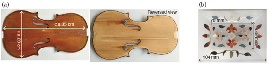
Figure 1.
Objects under examination: (a) aged violin, (b) inlaid dish.
Active thermography is a technology for observing the internal structure of an object in quasi real-time by irradiating it with an optical pulse containing infrared frequencies. The propagation of the thermal pulse is recorded by an infrared camera as the time dependence of the temperature change [1]. This technique has recently been applied to artwork examinations [2,3]. Unlike conventional passive thermography, the propagation of the thermal pulse can potentially provide information on the approximate location of the defect in the depth profile, based on the detected delay time. In this study, an active thermography system with two xenon lamps and an InSb infrared camera was used [4]. OCT is a technique used to observe the internal structure of an object. It irradiates the object with infrared light and detects the interferometric signal of the reflected waves generated at the reference mirror and the interface between materials with different refractive indices in the object under test [5,6]. The OCT system used in this study uses an optical source that sweeps a narrow pulse over the range at the centre wavelength of 1700 nm [7]. The Fourier transformation is used to obtain the depth profile from the interferogram. THz-TDI is a technique that uses a THz pulse to observe the internal structure of an object. When an object is irradiated with a THz pulse, reflection pulses are generated from the surface and internal interfaces with a delay depending on their differences in refractive index. The depth profile is obtained by detecting the reflection pulse sequence [8,9]. The THz-TDI system used in this study is equipped with a two-dimensional scanning system [10]. To verify the results of the violin, the interior of the wood panel was observed using X-ray CT [11].
3. Aged Violin Observation
The aged violin was examined by using active thermography, dividing it into three parts, shown as thin solid, thick solid, and dashed rectangles in Figure 2a. Figure 2b,c show examples of the phase images obtained by performing a Fourier transform on the time response to the temperature change during cooling after irradiation. Images acquired shortly after the start of the observation, i.e., analysed at higher frequencies, show near-surface conditions, while images at lower frequencies reveal more internal conditions. Black specks, which indicate areas of low heat conductivity, were observed in both images obtained at 1.079 Hz (Figure 2b) and at 0.196 Hz (Figure 2c), where no surface defects were recognised. In particular, there are cavities in a deeper position around the F-hole, as indicated by the dotted rectangles in Figure 2c.
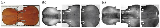
Figure 2.
Observation of a top plate of an aged violin by active thermography: (a) three observation areas; (b) phase images at 1.079 Hz; (c) phase images at 0.196 Hz.
The THz-TDI system used in this study could not maintain a constant incident angle of 90 degrees on curved surfaces, so the relatively flat area around the F-hole, indicated by the white dotted rectangle in Figure 3a, has been chosen as an example. The cross-sectional images along the lines a-a’ and b-b’ are shown in Figure 3b. Both images show numerous internal reflections, suggesting that this area is porous. In particular, along the line b-b’ in Figure 3b(i), a significant discontinuity is recognised 10 ps after reflection from the surface, as indicated by the yellow circle. This corresponds to a depth of 1.5 mm, calculated with a refractive index of 1. Figure 3c(i) shows a sliced image near the surface, obtained by integrating the THz pulses from 4 ps to 8 ps after the first reflection at the surface. Some cavities, estimated to be insect paths, appeared in the vicinity of the surface. Figure 3c(ii) shows the sliced image of the area between the two white dotted lines in Figure 3b(ii), i.e., between 10 ps and 20 ps from the reflection at the surface. The large discontinuity, which appeared in Figure 3b(ii), is visible on the slice plane, as indicated by the yellow circle.
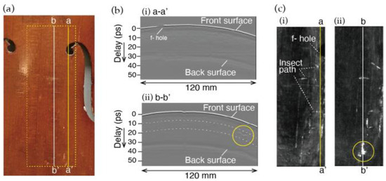
Figure 3.
Observation of a top plate of an aged violin by THz-TDI: (a) observation areas; (b) cross-sectional images along the lines a-a’ and b-b’ indicated in (a); (c) sliced images (i) near the surface and (ii) between two white dotted lines in (b).
The results obtained with active thermography and THz-TDI were compared with sliced images by X-Ray CT, one of the most well-established and widely used NDE methods for industrial and medical applications. The regions in Figure 2c where black specks are visible correspond to areas with cavities in the X-ray CT images shown in Figure 4a,b. Since active thermography can rapidly reveal the general features of deterioration, it has the potential to be used as an in situ assessment tool. THz-TDI, on the other hand, has a high enough spatial resolution to recognise insect paths. For example, the paths that appear in the white solid rectangle in Figure 4c can also be seen in Figure 3c. However, a device applicable to curved objects is likely to be a robot-mounted system, which makes in situ evaluation difficult [12].
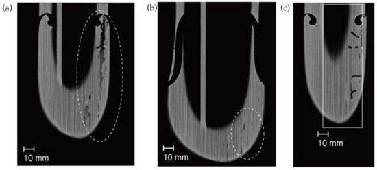
Figure 4.
Examples of X-ray CT images of a top plate of an aged violin: (a) near the f-hole; (b) bottom part; (c) near the f-hole at a different depth to (a).
4. Inlaid Dish Observation
Both THz-TDI and OCT techniques were used to observe an inlaid alabaster dish. To avoid the influence of variations in incident angles, only the results for the bottom flat part of the dish are discussed. Figure 5b shows a cross-sectional image obtained by THz-TDI, along the black dotted lines in Figure 5a. The THz pulse can pass through the entire dish, and shells and stones at different depths are recognised as internal interfaces. Figure 5c shows a cross-sectional image obtained by OCT along the yellow solid line in Figure 5a. The black area with no reflection seen around the shell on the right-hand side of the image is presumed to be a cavity. OCT revealed the gap of several tens of microns in the central part, but due to a lack of resolution in the depth direction, THz-TDI can only suggest the presence of an internal interface. The thickness of the inlaid shells and stones is approximately 2 mm and is consistent with the results of both techniques. These results demonstrate that THz-TDI is effective in understanding the internal structure of the dish as a whole, while OCT is particularly useful for observing the shape of embedded materials and their interfaces with alabaster base.
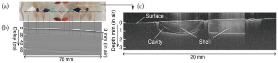
Figure 5.
Cross sectional observation of inlaid alabaster dish by THz-TDI and OCT: (a) observation area; (b) by THz-TDI; (c) by OCT.
5. Conclusions
Three commercial and transportable systems using electromagnetic waves were compared as practical examination tools in the field of cultural heritage research. Experimental results show that active thermography can reveal the general features of deterioration. OCT is suitable for depth profiling near the surface at a high resolution, and THz-TDI can be used to roughly examine the internal structure of objects. Since each NDE technique has its own advantages and limitations, combining and comparing data from various techniques is desirable for the diagnosis of cultural heritage.
Author Contributions
Conceptualization, K.F.; experiments and data analysis using OCT, T.T.; experiments and data analysis using X-ray CT, H.I. and S.M.; experiments and data analysis using active thermography, Y.U. and A.N.; writing—original draft preparation, K.F. All authors have read and agreed to the published version of the manuscript.
Funding
This research received no external funding.
Institutional Review Board Statement
Not applicable.
Informed Consent Statement
Not applicable.
Data Availability Statement
The data presented in this study are available upon request from the corresponding author.
Acknowledgments
The authors would like to express their sincere thanks to Koji Suzuki for providing the old violins and information on stringed instruments, and for helpful discussions on the experimental results.
Conflicts of Interest
The authors declare that the research was conducted in the absence of any commercial or financial relationships that could be construed as a potential conflict of interest.
References
- Maldague, X.P.V.; Marinetti, S. Pulse phase infrared thermography. J. Appl. Phys. 1996, 79, 2694–2698. [Google Scholar] [CrossRef]
- Deleu, N.; Hillen, M.; Steenackers, G.; Borms, G.; Janssens, K.; Van der Stighelen, K.; Van der Snickt, G. Combined macro X-ray fluorescence (MA-XRF) and pulse phase thermography (PPT) imaging for the technical study of panel paintings. Talanta 2024, 270, 125533. [Google Scholar] [CrossRef] [PubMed]
- Kunikata, S.; Tsuchiya, Y.; Fukunaga, K. Delamination of Wax-Resin Linings in Oil Paintings: Visualization and Analysis Using Infrared Active Thermography and Terahertz Time-Domain Imaging. J. Cult. Herit. 2025, 73, 295–304. [Google Scholar] [CrossRef]
- Laboratory NDT Systems for Pulse Thermography. Available online: https://www.edevis.com/en/products/laboratory-testing-systems/ptvis/ (accessed on 9 September 2025).
- Liang, H.; Cid, M.G.; Cucu, R.G.; Dobre, G.M.; Podoleanu, A.G.; Pedro, J.; Saunders, D. En-face Optical Coherence Tomography—A novel application of non-invasive imaging to art conservation. Opt. Express 2005, 13, 6133–6144. [Google Scholar] [CrossRef] [PubMed]
- Ford, T.; Iwanicka, M.; Platania, E.; Targowski, P.; Hendriks, E. Munch and optical coherence tomography: Unravelling historical and artist applied varnish layers in painting collections. Eur. Phys. J. Plus 2021, 136, 899. [Google Scholar] [CrossRef]
- Configurable OCT System. Available online: https://ois.santec.com/products/swept-source-oct-system (accessed on 9 September 2025).
- Mittleman, D.M. Twenty years of terahertz imaging. Opt. Express 2018, 26, 9417–9431. [Google Scholar] [CrossRef] [PubMed]
- Fukunaga, K. THz Technology Applied to Cultural Heritage in Practice; Springer: Tokyo, Japan, 2016. [Google Scholar]
- Terahertz Gauging and Imaging. Available online: https://lunainc.com/product/terahertz-control-unit-0 (accessed on 9 September 2025).
- X-Ray Nondestructive Inspection System. Available online: https://www.toshiba-itc.com/en/hihakai/txscanner/ (accessed on 9 September 2025).
- The Irys System by Das-Nano. Available online: https://das-nano.com/products/irys/ (accessed on 9 September 2025).


Disclaimer/Publisher’s Note: The statements, opinions and data contained in all publications are solely those of the individual author(s) and contributor(s) and not of MDPI and/or the editor(s). MDPI and/or the editor(s) disclaim responsibility for any injury to people or property resulting from any ideas, methods, instructions or products referred to in the content. |
© 2025 by the authors. Licensee MDPI, Basel, Switzerland. This article is an open access article distributed under the terms and conditions of the Creative Commons Attribution (CC BY) license (https://creativecommons.org/licenses/by/4.0/).