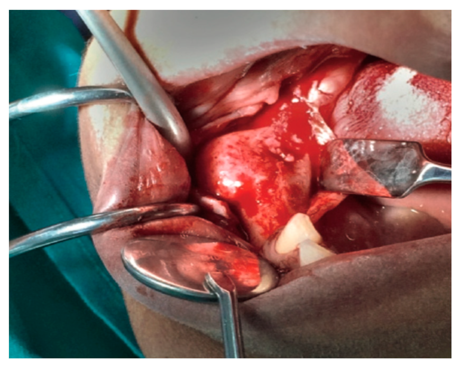
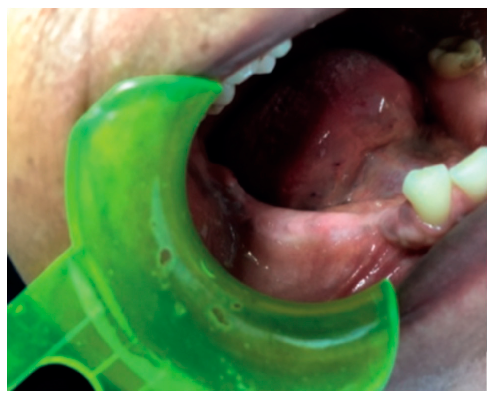
Craniofacial Fibrous Dysplasia: Diagnosis and Treatment Options †
Conflicts of Interest
References
- Riminucci, M.; Fisher, L.W.; Shenker, A.; Spiegel, A.M.; Bianco, P.; Gehron Robey, P. Fibrous dysplasia of bone in the McCune-Albright syndrome: Abnormalities in bone formation. Am. J. Pathol. 1997, 151, 1587–1600. [Google Scholar] [PubMed]
- Adetayo, O.A.; Salcedo, S.E.; Borad, V.; Richards, S.S.; Workman, A.D.; Ray, A.O. Fibrous dysplasia: An overview of disease process, indications for surgical management, and a case report. Eplasty 2015, 15, e6. [Google Scholar] [PubMed]
- Ricalde, P.; Magliocca, K.R.; Lee, J.S. Craniofacial fibrous dysplasia. Oral Maxillofac. Surg. Clin. N. Am. 2012, 24, 427–441. [Google Scholar] [CrossRef] [PubMed]


Publisher’s Note: MDPI stays neutral with regard to jurisdictional claims in published maps and institutional affiliations. |
© 2019 by the authors. Licensee MDPI, Basel, Switzerland. This article is an open access article distributed under the terms and conditions of the Creative Commons Attribution (CC BY) license (https://creativecommons.org/licenses/by/4.0/).
Share and Cite
Joseph, G.; Sara, G.; Matteo, P.; Alessandro, M.; Caterina, D.B. Craniofacial Fibrous Dysplasia: Diagnosis and Treatment Options. Proceedings 2019, 35, 59. https://doi.org/10.3390/proceedings2019035059
Joseph G, Sara G, Matteo P, Alessandro M, Caterina DB. Craniofacial Fibrous Dysplasia: Diagnosis and Treatment Options. Proceedings. 2019; 35(1):59. https://doi.org/10.3390/proceedings2019035059
Chicago/Turabian StyleJoseph, Garibaldi, Grasso Sara, Piazzai Matteo, Merlini Alessandro, and Del Buono Caterina. 2019. "Craniofacial Fibrous Dysplasia: Diagnosis and Treatment Options" Proceedings 35, no. 1: 59. https://doi.org/10.3390/proceedings2019035059
APA StyleJoseph, G., Sara, G., Matteo, P., Alessandro, M., & Caterina, D. B. (2019). Craniofacial Fibrous Dysplasia: Diagnosis and Treatment Options. Proceedings, 35(1), 59. https://doi.org/10.3390/proceedings2019035059


