Proceeding Paper

Development of Anti-TNFR Antibody-Conjugated Nanoparticles †

1
InoCure s.r.o, R&d Lab, Prumyslová 1960, 250 88 Celákovice, Czech Republic
2
Division of Molecular Internal Medicine, Department of Internal Medicine II, University Hospital Würzburg, Auverahaus, Grombühlstrasse 12, 97080 Würzburg, Germany
*
Author to whom correspondence should be addressed.
Presented at the 1st International Electronic Conference on Pharmaceutics, 1–15 December 2020; Available online: https://iecp2020.sciforum.net/.
Proceedings 2021, 78(1), 55; https://doi.org/10.3390/IECP2020-08684
Published: 1 December 2020
(This article belongs to the Proceedings of The 1st International Electronic Conference on Pharmaceutics)

Abstract

:
Immunotherapy is considered as a new pillar of cancer treatment. However, the application of some promising immunotherapeutic antibodies, such as antibodies against certain immune-stimulatory receptors of the TNF receptor superfamily (TNFRs), including CD40, 41BB, CD27 and anti-fibroblast growth factor-inducible 14 (anti-Fn14), are limited due to their low bioactivity. It has been previously shown that the bioactivity of such anti-TNFR antibodies could be improved by crosslinking or attachment to the plasma membrane by interaction with Fcγ receptors (FcγR). Both result in the proximity of multiple antibody-bound TNFR molecules, which allow for the activation of proinflammatory signaling pathways. In this work, we have grafted antibodies on gold nanoparticles to simulate the “activating” effect of FcγR-bound, and thus plasma membrane-presented anti-TNFR antibodies. We have developed and optimized the method for the preparation of gold nanoparticles, their functionalization with poly-ethylene glycol (PEG) linkers, and grafting of antibodies on the surface. We showed here that antibodies, including the anti-Fn14 antibody PDL192, can be successfully attached to nanoparticles without affecting antigen binding. We hypothesize that conjugation of monoclonal anti-TNFR antibodies to the inorganic nanoparticles is a promising technique to boost the efficacy of these immunotherapeutic antibodies.

1. Introduction

Cancer immunotherapy is a very attractive field with high potential to provide cures for difficult cancers. Immunotherapy relies on the stimulation or silencing of signaling pathways of relevance for tumour development. Some of the promising immunotherapeutics target tumor necrosis factor receptor superfamily (TNFRSF) receptors (TNFRs), which are naturally involved in the regulation or even the inhibition of tumor growth [1]. The interactions between the ligands of the TNF superfamily (TNFSF) and their TNFRs regulate innate and adaptive immune responses, including natural killer cell activation, T cell co-stimulation and control of B cell homeostasis [1]. However, there are many obstacles to the production of recombinant soluble TNFSF ligands, and they show poor pharmacokinetics (low serum half-life of only around 10–30 min) [2,3,4]. Thus, agonistic antibodies targeting TNFRs such as CD40-, 41BB, CD27 and Fn14 are considered as alternative TNFR agoists to soluble TNFSF ligand molecules. Unfortunately, anti-TNFR antibodies targeting a subgroup of TNFRs, including CD40, 41BB, CD27 and Fn14, typically lack agonistic activity as free molecules. Instead, FcγR binding is required for these antibodies in order to stimulate receptor signaling [5].
Medler et al. have shown that “activating” the FcγR-dependent cell surface anchoring of IgG antibodies can be replaced by anchoring domains which are genetically fused to antibodies that recognize cell-surface-exposed structures distinct from FcγRs [4]. We hypothesized that the activating effects of the plasma-membrane-associated presentation of antibodies can be simulated by grafting antibodies to a solid support. In this work, we have developed and optimized a method for attaching antibodies to gold nanoparticles with the expectation of increasing their activity.
Gold nanoparticles (AuNPs) are widely used in different biomedical applications and are used as a platform for nanobiological conjugates, such as oligonucleotides [6], antibodies [7] and proteins [8]. In addition, the physicochemical and optoelectronic properties of the spherical AuNPs, such as surface plasmon resonance, conductivity, large surface-to-volume ratio, excellent biocompatibility and low toxicity, extend the possibility of exploiting them as a new generation of drug-delivery systems [9]. All these properties, when combined, make gold nanoparticles a promising tool to deliver the therapeutic agents to the targeted cells. In this work, we hypothesize that gold nanoparticles can be exploited as a platform to immobilize antibodies against TNFRs, to enhance their agonistic activity. More specifically, we used a known gold nanoparticle synthesis protocol and optimized it for the grafting of antibodies, including the anti-Fn14 antibody PDL192, under preservation of their antigen-binding abilities.

2. Experiments

2.1. Materials

Gold (III) chloride acid trihydrate was obtained from VWR International. mPEG-SH/mPEG-Thiol (5 kDa) and SH-PEG-COOH/Thiol-PEG-Acetic Acid (5 kDa) were obtained from Biochempeg. The 38.8 mM, trisodium citrate, 1-ethyl-3-(3-dimethylaminopropyl)carbodiimide hydrochloride (EDC) were obtained from Thermo Scientific™, N-Hydroxysuccinimide(1-hydroxy-2,5 pyrrolidinedione) (NHS) was obtained from Sigma-Aldrich, β-(N-Morpholino)ethansulfonsaure (MES), water free ≥99% and was obtained from VWR chemicals. A total of 50 mM TRIS with 0.33 mg/mL mPEG-SH was used as a blocking buffer.
The following antibodies were used: Humira (anti TNF alpha), Cosentyx® (IL-17A monoclonal antibody) and anti-Fn14 PDL192. GpL-TNC-TNF has been described elsehere (Lang et al., 2016) [10] Fn14ed-GpL was generated by cloning the Gaussia princeps luciferase w/o leader to the C-terminus of the extracellular domain of Fn14.

2.2. Methods

2.2.1. AuNPs Synthesis

The protocol for AuNP synthesis was adapted from [11,12], with slight modifications. A total of 100 mL of 0.4 mM chloroauric acid solution was boiled in a clean 300-mL glass flask with stir bar. A reflux column was attached on top of the flask to prevent a decrease in the solution’ volume. The entire apparatus was placed on a hot plate and boiled while stirring. A total of 1 mL of 38.8 mM trisodium citrate solution was added to the solution to produce 60 nm spherical monodisperse gold nanoparticles. Other sizes were created based on the amount of added trisodium citrate and/or the concentration of auric salt within a range between 15 and 100 nm. Upon addition of the trisodium citrate, the colour of the solution changed to blue in about 30 s, and then to red in another 150 s. The colour change during synthesis is attributed to the increase in the size of gold nanoparticles as the citrate ions reduce the gold ions [13]. The boiling was continued for another 10 min and then the mixture was cooled to room temperature.

2.2.2. AuNPs Functionalization

To modify the surface of the produced gold nanoparticles with functional carboxyl groups, a volume of HS-PEG-COOH solution with a selected concentration was added to the whole amount of the produced colloidal of AuNPs to obtain 100 μg/mL of HS-PEG-COOH in the solution, and was left to mix for one hour. The whole amount of carboxyl-modified AuNPs was centrifuged in a big centrifuging device (10,000 RPM for 10 min), then the pellet was collected in an Eppendorf tube and washed twice with mPEG (0.33 mg/mL) (in order to fill the unmodified places on the surface of HOOC-PEG-AuNPs and to compensate for the washed stabilizer trisodium citrate).

2.2.3. AuNPs Grafting

The carboxyl-modified AuNPs were conjugated with the protein of interest according to the EDC-NHS covalent binding procedure adapted from [14,15,16,17,18]. In brief, a number of purified C-AuNPs were resuspended in a volume of the activation/coupling buffer (50 mM MES, pH 6.0) and washed with it three times. Then, 24 μL of EDC (200 mM) and 240 μL of NHS (200 mM) were added to 1 mL of the previous solution of AuNPs, and incubated for 30 min in RT. After washing the particles from the EDC and NHS reagents three times with the activation/coupling buffer, 500 μL of the activated C-AuNPs were incubated with 500 μL of a selected concentration of the protein of interest (anti-TNF, anti-Fn14, anti-IL17A). The antibody-grafted AuNPs were washed from the excess of the unconjugated protein three times with the blocking buffer (Tris 50 mM in mPEG-SH (0.33 mg/mL)) to block the free activated carboxyl sites on the surface of gold nanoparticles. Finally, the antibody-grafted AuNPs were resuspended in a volume of the blocking buffer to be ready for application later.

2.2.4. Characterization

UV-Vis: AuNPs samples were collected at each stage immediately after synthesis and their optical properties were evaluated by UV–vis spectrophotometry (SpectraMax—Molecular Devices, San Jose, CA, USA). The absorption spectra were acquired in the range of 450–650 nm with a step of 5/10 nm.
DLS: The sizes of the obtained AuNPs (unPEGylated, PEGylated and grafted particles) were analyzed using Nanophox 123 Dynamic Light Scattering (DLS) with photon cross-correlation spectroscopy from Sympatec (Sympatec GmbH —System | Partikel | Technik, Clausthal-Zellerfeld, Germany). The particles were purified by centrifugation at 22,000× g for 10 min, diluted 100 times in distilled water, then analyzed. All DLS experiments were carried out at a temperature of 25 °C.
Zetasizer: The effective surface charges on the gold nanoparticles were measured using zeta-potential (Malvern Instruments Zetasizer, Malvern Panalytical Ltd., Cambridge, UK). Reported zeta potential measurements were collected on aqueous solution in which AuNPs were diluted 10–100 times, depending on their concentration.

3. Results

3.1. Optimization of Gold Nanoparticles Synthesis Protocol

3.1.1. Controlling the Size and Concentration of Gold Nanoparticles

The size of AuNPs can be adjusted by controlling the concentration of the auric salt (HAuCL4·3H2O) [12]. To optimize the procedure for the preparation of the AuNPs needed for our work, i.e., to obtain AuNPs with a size below 200 nm, we set the trisodium citrate concentration to 38.8 mM and varied the concentration of gold chloride and boiling duration. UV-vis absorption was used to characterize the particle size (the wavelength of the maximum absorbance of the plasmon band of the spherical particles AuNPs is dependent on the particle size) [19]. As shown in Figure 1a, increasing the concentration of the gold chloride leads to a lambda max shift from 520 nm to 550 nm, indicating an increase in particle size from 15 nm to 80 nm. On the other hand, different results were noted for the influence of the boiling duration. In this case, the λmax absorbance of the colloidal solution was measured after 5, 20 and 40 min of boiling. As shown in Figure 1b, the optical density increased with fixed λmax absorbance, indicating that the total amount of the produced gold nanoparticles increased with continuous boiling [13].

3.1.2. Functionalization of the Gold Nanoparticles

Trisodium citrate plays a role as a reducing agent and a stabilizer of the produced gold nanoparticles [20]. The abundance of the negative charges of the citrate structure surrounding the surface of AuNPs is known to prevent their aggregation. However, the stabilization effect of citrate is not significant enough for storing the particles long-term, and can be lost after purification. To increase the long-term stability of the AuNPs, and to introduce chemical groups for the subsequent functionalization, particles were functionalized with a layer of SH- and carboxyl- (5 kDa HOOC-PEG-SH) or methoxy- (5 kDa H3C-O-PEG-SH) containg polymers. Grafted particles were purified by washing with distilled water to discard the excess of trisodium citrate and free polymer molecules. As indicated in Figure 2 the increase in particle size after the PEGylation grafted with two types of polymers is similar, most likely due to the identical molecular weight. To confirm the PEGylation, zeta potential measurements were performed. As shown in Table 1 the PEGylation leads to a change in ζ potential values. In comparison with the citrate stabilization, the dominant charge of the particles after PEGylation is even more negative after PEGylation with the carboxyl-terminated polymer (−20 mV), and less negative when methoxy-PEG was used (−7 mV).

3.2. Characterization the Conjugation of C-AuNPs with Different Thersputics Antibodies

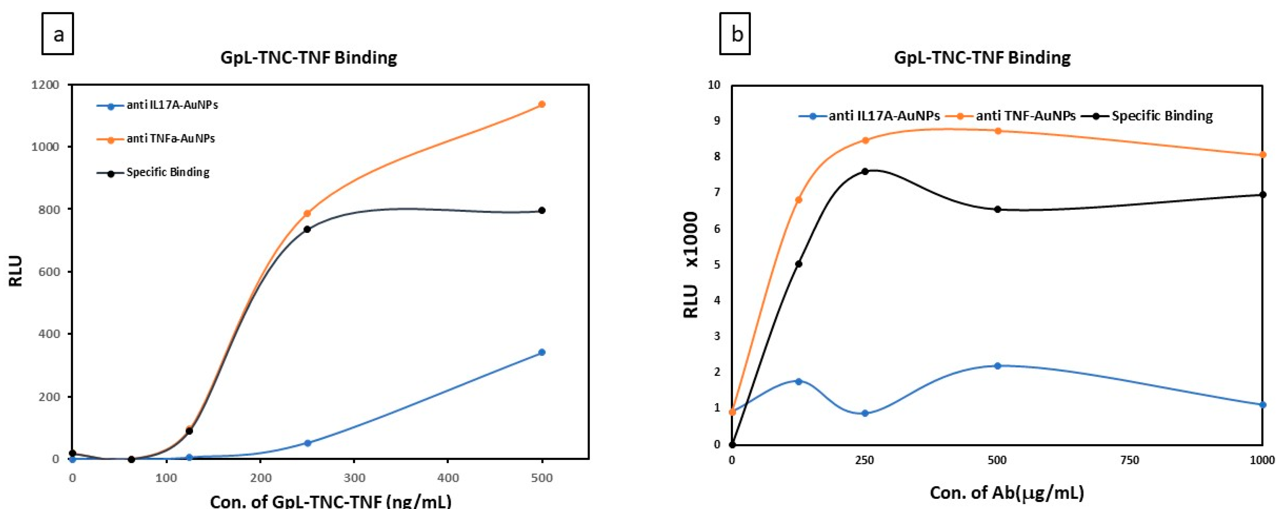
Carboxyl-modified gold nanoparticles C-AuNPs (ca. 60 nm, 25 mg/mL) were conjugated with the following proteins: anti-Fn14 antibody PDL192, anti-TNF or anti-IL17A. The conjugation process was performed by following the EDC/NHS covalent coupling procedure (described in the methods). To confirm not only the conjugation of C-AuNPs with the antibodies, but also their post-conjugation functionality, binding studies using the antigens TNF and Fn14 fused to the Gaussia princeps Luciferase (GpL-TNF-TNC, Fn14ed-GpL) [10]. The GpL domain allows for the quantification of the binding to the corresponding gold-nanoparticle-associated antibodies by measurement of luminescence upon removal of the free GpL fusion protein molecules. The binding curves between GpL-TNF-TNC and anti-TNF-AuNPs (total biding) or anti-IL17A-AuNPs (unspecific binding) are shown in Figure 1a and Figure 3. A serial dilution of the GpL-linked antigen (from 0 ng/mL to 500 ng/mL) was mixed with the fixed concentration (25 mg/mL) of the AuNPs conjugated with antibody (1 mg/mL conc in linking solution). As shown in Figure 3a, in all cases, the luminescence increased with the increasing antigen concentration. However, a notably higher binding was observed with anti-TNF than with anti IL17A AuNPs. This indicates the high specific binding of GpL-TNC-TNF to gold nanoparticle immobilized anti-TNF.
Thus, the conjugation of the antibody did not notably affect the interaction between the conjugated Ab and its antigen.
In order to evaluate the maximum amount of the antibody that can be conjugated to the nanoparticles, increasing concentrations of anti-TNF or anti-IL17A were used for conjugation. The binding of a GpL-TNF-TNC solution with constant concentration was then determined, as shown in Figure 3b. With a concentration of 250 μg/mL of anti-TNF in the coupling reaction, the maximum amount of antibody could be immobilized on the particles. Increasing the antibody concentration did not lead to a further increase in antigen binding. Similar binding studies with anti-Fn14 PDL192-AuNPs and their GpL-fused antigen (Fn14ed-GpL) were performed. As shown in Figure 4, maximum conjugation capacity was again reached at the antibody concentration of 250 μg/mL—similar to the results shown in Figure 3b.

4. Discussion

Since the early work of Turkevich and Frens, methods to produce gold nanoparticles in the scale from 9 to 120 nm, and with defined size distribution, have been optimized for various applications [13,21]. Gold NPs can be coated with a ligand shell, which provides colloidal stability, or conjugated with (biological) molecules via thiols ([22], Rivera-Gil et al.). However, attaching Abs directly to the surface of NPs has drawbacks; that is, the process may affect their activity by blocking the active side. To overcome this problem, a method depending on directional covalent binding to a functional polymer on the surface of NPs has been investigated in the literature [23]. One effective method is the use of 1-ethyl-3- (3-dimethylaminopropyl) carbodiimide (EDC) chemistry [24].
Antibodies bear amine groups on some of their amino-acid side chains, including residues in the antigen-binding site. To detect the probability of their binding to antigen-binding-irrelevant sites of the antibody, we functionalized AuNPs with a carboxyl-containing polymer and then conjugated them with the therapeutic Abs using EDC/NHS chemistry, and carried out binding studies. The binding studies revealed that, even though some antibodies might have coupled through their antigen-binding sites, others coupled through antigen-biding-irrelevant amino acids and remained active.
Recent studies found that specific antibodies for the TNF receptor Fn14 can mimic some effects of the soluble TWEAK, Fn14-specific TNF ligand, by triggering the related signaling pathways. However, the soluble anti-Fn14 antibody failed to activate all of the needed signaling pathways of the soluble TWEAK. This drawback of the soluble anti-Fn14 can be overcome by oligomerization using protein G, or by anchoring to Fcγ receptors of the effector cells, which provides the antibody with agonistic activity [4,25]. In future work, we will exploit the gold nanoparticles as a platform to immobilize the therapeutic antibody (PDL192) as a model. We hypothesize that the AuNPs-conjugated anti-Fn14 monoclonal IgG1 antibody will possess agonistic activity which resembles the Fcγ receptors anchoring-dependent efficiency. Such anchored antibodies should then be able to trigger the associated proinflammatory signaling pathways. If successful, this approach will enable promising applications of nanocomposites with antitumor antibodies. Furthermore, we will work on the nanoencapsulation of these nanoparticles to target tumor tissues specifically and prevent the systemic side effects associated with such antibodies.

5. Conclusions

Gold nanoparticles of diameter ca. 60 nm have been synthesized by a gold chloride reduction in sodium citrate, then functionalized with COOH-PEG-SH to stabilize the colloidal solution of the gold nanoparticles. We have also demonstrated that the carboxyl-modified gold nanoparticles can be coupled with the antibodies of interest using the EDC/NHS coupling procedure. The binding studies of the Ab-grafted AuNPs against their labeled specific antigen confirmed the conjugation of C-AuNPs. We showed that our coupling protocol allows for the conjugation of the antibody so that its activity is maintained. Our future work will focus on the in vitro assays needed to compare the activity of the conjugated antibodies and their soluble variants.

Author Contributions

A.A. designed the study, performed the experiments, data processing, analysis of the results and wrote the paper; A.S. and H.W. helped with method development, supervised the work and contributed with insights and discussions; A.S. and M.B. guided the design of work and experiments, helped with the writing of the manuscript and reviewed the paper. All authors have read and agreed to the published version of the manuscript.

Institutional Review Board Statement

Not applicable.

Informed Consent Statement

Not applicable.

Data Availability Statement

The data presented in this study are available on request from the corresponding author.

Acknowledgments

This is a part of project which has received funding from the European Union’s Horizon 2020 research and innovation programme under the Marie Sklodowska-Curie grant agreement No 813871.

Conflicts of Interest

The authors declare no conflict of interest. The founding sponsors had no role in the design of the study; in the collection, analyses, or interpretation of data; in the writing of the manuscript, and in the decision to publish the results.

Abbreviations

The following abbreviations are used in this manuscript:
Abs Antibodies
AuNPsGold nanoparticles
C-AuNPsCarboxyl-modified gold nanoparticles
DLSDynamic light scattering
GpLGaussia princeps luciferase
IgGImmunoglobulin G
PEGPoly-ethylene glycol
PDL192Anti Fn14 monoclonal IgG1 antibody
SPRSurface Plasmon Resonance
TNFTumor Necrosis Factor
TWEAKTNF-related weak inducer of apoptosis

References

  1. Bodmer, J.-L.; Schneider, P.; Tschopp, J. The molecular architecture of the TNF superfamily. Trends Biochem. Sci. 2002, 27, 19–26. [Google Scholar] [CrossRef]
  2. Kelley, S.K.; Harris, L.A.; Xie, D.; Deforge, L.; Totpal, K.; Bussiere, J.; Fox, J.A. Preclinical Studies to Predict the Disposition of Apo2L/Tumor Necrosis Factor-Related Apoptosis-Inducing Ligand in Humans: Characterization of in Vivo Efficacy, Pharmacokinetics, and Safety. J. Pharmacol. Exp. Ther. 2001, 299, 31–38. [Google Scholar] [PubMed]
  3. Alerts, E. Cachectin / tumor necrosis factor : production, distribution, and metabolic fate in vivo. J. Immunol. 2019, 135, 3972–3977. [Google Scholar]
  4. Medler, J.; Nelke, J.; Weisenberger, D.; Steinfatt, T.; Rothaug, M.; Berr, S.; Hünig, T.; Beilhack, A.; Wajant, H. TNFRSF receptor-specific antibody fusion proteins with targeting controlled FcγR-independent agonistic activity. Cell Death Dis. 2019, 10. [Google Scholar] [CrossRef]
  5. Wajant, H. Principles of antibody-mediated TNF receptor activation. Cell Death Differ. 2015, 22, 1727–1741. [Google Scholar] [CrossRef] [PubMed]
  6. Zhang, T.; Chen, P.; Sun, Y.; Xing, Y.; Yang, Y.; Dong, Y.; Xu, L.; Yang, Z.; Liu, D. A new strategy improves assembly efficiency of DNA mono-modified gold nanoparticles. Chem. Commun. (Camb.) 2011, 47, 5774–5776. [Google Scholar] [CrossRef]
  7. El-Sayed, I.H.; Huang, X.; El-Sayed, M.A. Surface Plasmon Resonance Scattering and Absorption of Anti-EGFR Antibody Conjugated Gold Nanoparticles in Cancer Diagnostics: Applications in Oral Cancer. Nano Letters 2005. Available online: https://kopernio.com/viewer?doi=10.1021/nl050074e&token=WzExNDE5NzUsIjEwLjEwMjEvbmwwNTAwNzRlIl0.-akvz5cNi2_mB0A37P4eWnqNTSM (accessed on 20 December 2019). [CrossRef]
  8. You, C.-C.; Arvizo, R.R.; Rotello, V.M. Regulation of alpha-chymotrypsin activity on the surface of substrate-functionalized gold nanoparticles. Chem. Commun. (Camb.) 2006, 2905–2907. [Google Scholar] [CrossRef]
  9. Yeh, Y.-C.; Creran, B.; Rotello, V.M. Gold nanoparticles: preparation, properties, and applications in bionanotechnology. Nanoscale 2012, 4, 1871–1880. [Google Scholar] [CrossRef]
  10. Lang, I.; Fu, S.; Wyzgol, A.; Fick, A.; Trebing, J.; Arana, A.C.; Weisenberger, D.; Wajant, H. Binding Studies of TNF Receptor Superfamily (TNFRSF) Receptors on Intact Cells. J. Biol. Chem. 2016, 291, 5022–5037. [Google Scholar] [CrossRef]
  11. Kimling, J.; Maier, M.; Okenve, B.; Kotaidis, V.; Ballot, H.; Plech, A. Turkevich method for gold nanoparticle synthesis revisited. J. Phys. Chem. B 2006, 110, 15700–15707. [Google Scholar] [CrossRef] [PubMed]
  12. Frens, G. Controlled Nucleation for the Regulation of the Particle Size in Monodisperse Gold Suspensions|Kopernio. Available online: https://kopernio.com/viewer?doi=10.1038/physci241020a0&token=WzExNDE5NzUsIjEwLjEwMzgvcGh5c2NpMjQxMDIwYTAiXQ.es09awC7jdG9BahFkjNV3cCqjpQ (accessed on 16 December 2019).
  13. Haiss, W.; Thanh, N.T.K.; Aveyard, J.; Fernig, D.G. Determination of size and concentration of gold nanoparticles from UV-Vis spectra. Anal. Chem. 2007, 79, 4215–4221. [Google Scholar] [CrossRef] [PubMed]
  14. Lomant, A.J.; Fairbanks, G. Chemical probes of extended biological structures: synthesis and properties of the cleavable protein cross-linking reagent [35S]dithiobis(succinimidyl propionate). J. Mol. Biol. 1976, 104, 243–261. [Google Scholar] [CrossRef]
  15. Grabarek, Z.; Gergely, J. Zero-length crosslinking procedure with the use of active esters. Anal. Biochem. 1990, 185, 131–135. [Google Scholar] [CrossRef]
  16. Nakajima, N.; Ikada, Y. Mechanism of Amide Formation by Carbodiimide for Bioconjugation in Aqueous Media. Bioconj. Chem. 1995, 6, 123–130. [Google Scholar] [CrossRef] [PubMed]
  17. Staros, J. V. Membrane-Impermeant Cross-Linking Reagents: Probes of the Structure and Dynamics of Membrane Proteins. Acc. Chem. Res. 1988, 21, 435–441. [Google Scholar] [CrossRef]
  18. Staros, J.V.; Wright, R.W.; Swingle, D.M. Enhancement by N-hydroxysulfosuccinimide of water-soluble carbodiimide-mediated coupling reactions. Anal. Biochem. 1986, 156, 220–222. [Google Scholar] [CrossRef]
  19. Ðorđević, L.; Arcudi, F.; Prato, M. Preparation, functionalization and characterization of engineered carbon nanodots. Nat. Protoc. 2019, 14, 2931–2953. [Google Scholar] [CrossRef]
  20. Oliveira, J.P.; Prado, A.R.; Keijok, W.J.; Ribeiro, M.R.N.; Pontes, M.J.; Nogueira, B.V.; Guimarães, M.C.C. A helpful method for controlled synthesis of monodisperse gold nanoparticles through response surface modeling. Arab. J. Chem. 2020. [Google Scholar] [CrossRef]
  21. Liz-marz, L.M. Nanometals: Formation and color. Mater. Today 2004, 7, 26–31. [Google Scholar] [CrossRef]
  22. Montenegro, J.; Grazu, V.; Sukhanova, A.; Agarwal, S.; De, J.M.; Nabiev, I.; Greiner, A.; Parak, W.J. Controlled antibody/(bio-) conjugation of inorganic nanoparticles for targeted delivery. Adv. Drug Deliv. Rev. 2013. [Google Scholar] [CrossRef] [PubMed]
  23. Hermanson, G. Bioconjugate Techniques, 2nd ed.; Academic Press: Cambridge, MA, USA, 2008; ISBN 9780123705013. [Google Scholar]
  24. Bartczak, D.; Kanaras, A.G. Preparation of peptide-functionalized gold nanoparticles using one pot EDC/Sulfo-NHS coupling. Langmuir 2011, 27, 10119–10123. [Google Scholar] [CrossRef] [PubMed]
  25. Salzmann, S.; Seher, A.; Trebing, J.; Weisenberger, D.; Rosenthal, A.; Siegmund, D.; Wajant, H. Fibroblast growth factor inducible (Fn14)-specific antibodies concomitantly display signaling pathway-specific agonistic and antagonistic activity. J. Biol. Chem. 2013, 288, 13455–13466. [Google Scholar] [CrossRef] [PubMed]
Figure 1. λmax absorbance of the (SPR) of AuNPs colloidal. Comparasion between spectrum of the absorbance of AuNPs colloidals resulting from (a) different concentration of HAuCL4·3H2O or (b) different boiling duration.
Proceedings 78 00055 g001
Figure 2. Diameter distribution graphs from dynamic light scattering (DLS) show the increase in particle size after PEGylation with carboxyl-PEG-SH and mPEG-SH.
Proceedings 78 00055 g002
Figure 3. Binding of GpL-TNC-TNF fusion protein to either anti-TNF-AuNPs or anti-IL17A-AuNPs: Carboxyl-modified gold nanoparticles C-AuNPs (ca. 60 nm, 25 mg/mL) were conjugated with either a fixed concentration of anti-TNF or anti-IL17A. Then, binding studies were carried out with serial dilutions of GpL-TNC-TNF (a). Alternatively, different concentrations of the two antibodies were used for gold nanoparticle conjugation, and GpL-TNF-TNC binding with a fixed concentration was analyzed (b). The curve of the specific binding was obtained by subtracting the binding to anti-IL17A from the values of the binding to anti-TNF.
Proceedings 78 00055 g003
Figure 4. Binding of Fn14ed-GpL fusion protein to either antiTNF-AuNPs or PDL192 -AuNPs: Carboxyl-modified gold nanoparticles C-AuNPs (ca. 60 nm, 25 mg/mL) were conjugated with different concentrations of both antibodies, and then the signals of the emitted light resulting from the binding with fixed concentration of Fn14ed-GpL were measured and plotted. The curve of the specific binding was obtained by subtraction of the binding to anti-TNF from the values of the binding to PDL192.
Proceedings 78 00055 g004
Table 1. Comparison of ζ potential values and particles size between the un-PEGylated and PEGylated gold nanoparticles.
Sample StructureParticles Sizeζ Potential
Trisodium citrate—AuNPs60.19 nm−14 mv
mPEG-AuNPs80.45 nm−7 mv
HOOC-PEG-AuNPs86.5 nm−20 mv
Publisher’s Note: MDPI stays neutral with regard to jurisdictional claims in published maps and institutional affiliations.